INTERNAL MEDICINE BOARDS - PART 2 doc
Bạn đang xem bản rút gọn của tài liệu. Xem và tải ngay bản đầy đủ của tài liệu tại đây (758.08 KB, 54 trang )
AMBULATORY MEDICINE
54
■
Empiric therapy is often indicated in the absence of a suspected organic
etiology. Oral phosphodiesterase inhibitors (sildenafil, vardenafil,
tadalafil) are first-line therapy but are contraindicated with nitrates or ac-
tive cardiac disease (can cause hypotension and sudden death).
■
Psychosexual counseling is first-line therapy for psychogenic ED.
■
Second-line therapies include intraurethral alprostadil suppositories, vac-
uum constrictive pumps, and penile prostheses.
Prostatitis
The differential includes acute bacterial prostatitis, chronic bacterial prostati-
tis, nonbacterial prostatitis, and prostatodynia. See Table 2.15 for key features
of each.
SYMPTOMS/EXAM
Presents with irritative voiding symptoms and perineal or suprapubic pain.
Acute bacterial prostatitis is notable for the presence of fever and an exqui-
sitely tender prostate.
TREATMENT
Table 2.15 outlines the treatment of prostatitis and prostatodynia.
Genital Lesions
Table 2.16 outlines the differential diagnosis and treatment of STIs that pre-
sent as genital lesions. Figures 2.23 through 2.26 illustrate genital HSV le-
Rapid onset of ED suggests
psychogenic causes or
medication side effects. More
gradual onset is associated
with medical conditions. Low
libido along with ED suggests
a psychogenic, medication-
related, or hormonal cause.
TABLE 2.14. Medical Conditions Associated with ED
CONDITION EXAMPLES/COMMENTS
Psychogenic disorders Performance anxiety, depression, mental stress.
Obesity, physical inactivity
Diabetes mellitus ED is seen in up to 50% of cases.
Peripheral vascular disease
Endocrine disorders Hypogonadism, hyperprolactinemia, thyroid abnormalities.
Pelvic surgery
Spinal cord injury
Drugs of abuse Amphetamines, cocaine, marijuana, alcohol, tobacco.
Medications Antihypertensives: Thiazides, β-blockers, clonidine, methyldopa.
Antiandrogens: Spironolactone, H
2
blockers, finasteride.
Antidepressants: TCAs, SSRIs.
Other: Antipsychotics, benzodiazepines, opiates.
All patients with genital
lesions should be screened for
syphilis (serology).
AMBULATORY MEDICINE
55
sions, genital warts, syphilitic chancre, and chancroid, respectively. Refer to
the Women’s Health chapter for a detailed discussion of gonorrheal and
chlamydial infections (cervicitis, PID). The diagnosis and treatment of ure-
thritis in men follow the same principles as those of cervicitis in women.
ORTHOPEDICS
Rotator Cuff Tendinitis or Tear
The spectrum of pathology ranges from subacromial bursitis and rotator cuff
tendinitis to partial or full rotator cuff tear. Due to excessive overhead motion
(e.g., baseball players).
SYMPTOMS
Presents with nonspecific pain in the shoulder with occasional radiation down
the lateral arm that worsens at night or with overhead movement. Motor
weakness with abduction is seen in the presence of a tear.
TABLE 2.15. Treatment of Prostatitis and Prostatodynia
ACUTE BACTERIAL CHRONIC BACTERIAL NONBACTERIAL
PROSTATITIS PROSTATITIS PROSTATITIS PROSTATODYNIA
Fever +− −−
UA +− −−
Expressed Contraindicated. ++−
prostatic
secretions
Bacterial culture ++ −−
Prostate exam Very tender. Normal, boggy, or Normal, boggy, or Usually normal.
indurated. indurated.
Etiology Gram-
ᮎ rods (E. coli); Gram-ᮎ rods; less Unknown; perhaps Varies; includes voiding
less commonly gram-
ᮍ commonly enterococcus. Ureaplasma, dysfunction and pelvic
organisms (enterococcus). Mycoplasma, Chlamydia. floor musculature
dysfunction.
Treatment IV ampicillin and TMP-SMX; Erythromycin × 3–6 α-blocking drugs (e.g.,
aminoglycosides until fluoroquinolones × weeks if response at two terazosin) for bladder
organism sensitivities 6–12 weeks. weeks. neck and urethral
are obtained; then spasms;
switch to benzodiazepine and
fluoroquinolones × 4–6 biofeedback for pelvic
weeks. floor dysfunction.
Adapted, with permission, from Tierney LM et al. Current Medical Diagnosis & Treatment, 43rd ed. New York: McGraw-Hill, 2003:
914.
AMBULATORY MEDICINE
56
TABLE 2.16. Differential Diagnosis of Genital Lesions
GENITAL WARTS
(CONDYLOMATA
HSV ACUMINATA)1° SYPHILIS CHANCROID
Cause HSV-2 > HSV-1. HPV. Treponema pallidum. Haemophilus ducreyi.
Incubation 1°: +/− asymptomatic; 1–6 months; triggers 2–6 weeks. 3–5 days.
period/ prodrome consists of include pregnancy and
triggers malaise, genital immunosuppression.
paresthesias, and fever.
Reactivation: Most
commonly occurs with
symptoms; triggers
include stress, fever,
and infection.
Symptoms Painful, grouped vesicles; Warty “cauliflower” Painless, clean-based Pustule or pustules
tingling, dysesthesia. growths or none. ulcer (“chancre”). erode to form a painful
Asymptomatic shedding ulcer with a necrotic base.
is common.
Exam Groups of multiple, small Warty growths or none. Ulcer on genitalia; Usually unilateral,
vesicles. nontender regional tender, fluctuant, matted
lymph nodes. nodes with overlying
erythema.
Diagnosis Mostly clinical; ᮍ viral Clinical if wartlike; 4% Serology: RPR
ᮍ 1–2 Culture of lesion on
culture or DFA or Tzanck acetic acid applied to the weeks after the special media.
smear with ᮍ intranuclear lesion turns tissue white 1° lesion is first
inclusions and with papillae. seen.
multinucleated giant cells. Immunofluorescence or
darkfield microscopy of
fluid with treponemes.
Treatment Acute episodes: Acyclovir Trichloroacetic acid; Benzathine penicillin Azithromycin 1 g PO × 1
400 mg TID, famciclovir podophyllin G IM × 1; in penicillin- or ceftriaxone 250 mg
250 mg TID, valacyclovir (contraindicated in allergic patients, IM × 1.
1000 mg BID × 10 days pregnancy); imiquimod. doxycycline or tetracycline
(first episode) or × 5 PO × 2 weeks.
days (recurrence).
Suppression: Acyclovir
400 mg BID or
famciclovir 250 mg BID
or valacyclovir 500 mg
BID or 1 g QD.
AMBULATORY MEDICINE
57
E
XAM
■
Exam reveals pain with abduction between 60 and 120 degrees. Tears lead
to weakness on abduction (“drop arm test”).
■
Pain elicited by 60–120 degrees of passive abduction (impingement sign)
suggests impingement or trapping of an inflamed rotator cuff on the over-
lying acromion.
DIFFERENTIAL
■
Bicipital tendinitis: Due to repetitive overhead motion (e.g., throwing,
swimming). Exam reveals tenderness along the biceps tendon or muscle.
■
Degenerative joint disease.
■
Systemic arthritis: RA, pseudogout.
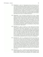
FIGURE 2.24.
Penile warts.
Note the multiple soft, filiform papules on the glans penis
and prepuce. (Reproduced, with permission, from Wolff K
et al. Fitzpatrick’s Color Atlas & Synopsis of Clinical Der-
matology, 5th ed. New York: McGraw-Hill, 2005: 888.)
FIGURE 2.23.
1° HSV infection in a female.
Note the multiple, painful, grouped vesicles. (Reproduced,
with permission, from Wendel GD, Cunningham FG: Sex-
ually transmitted diseases in pregnancy. In Williams Obstet-
rics, 18th ed. (Suppl 13). Norwalk, CT: Appleton & Lange,
August/September 1991.)
FIGURE 2.26.
Chancroid.
Note the multiple painful, punched-out ulcers with under-
mined borders on the labia. (Reproduced, with permission,
from Kasper DL et al. Harrison’s Principles of Internal Med-
icine, 16th ed. New York: McGraw-Hill, 2004.)
FIGURE 2.25.
Syphilitic chancre.
This dry-based, painless ulcer with indurated borders is typ-
ical for a 1° chancre in a male patient. (Reproduced, with
permission, from Bondi EE et al. Dermatology: Diagnosis &
Treatment. Stamford, CT: Appleton & Lange, 1991: 394.)
AMBULATORY MEDICINE
58
■
Referred pain: May be derived from a pulmonary process (e.g., pul-
monary embolism, pleural effusion), a subdiaphragmatic process, cervical
spine disease, or brachial plexopathy.
■
Adhesive capsulitis (frozen shoulder): Presents with progressive loss of
range of motion (ROM), usually more from stiffness than from pain. Can
follow rotator cuff tendinitis; more common in diabetics and older pa-
tients.
DIAGNOSIS
■
Diagnosis is made by the history and exam.
■
An MRI can be obtained if a complete tear is suspected or if no improve-
ment is seen despite conservative therapy and the patient is a surgical can-
didate.
TREATMENT
■
↓ exacerbating activities; NSAIDs.
■
Steroid injection is a common treatment but is no more effective than
NSAID therapy.
■
ROM exercises and rotator cuff strengthening can be initiated once acute
pain has resolved.
■
Refer to orthopedics for possible surgery if there is a complete tear or if no
improvement is seen with conservative therapy after several months.
Knee Pain
Table 2.17 outlines the etiologies and clinical characteristics of common knee
injuries.
DIAGNOSIS
■
In a patient who presents after acute trauma, the Ottawa Knee Rules
identify situations in which x-ray imaging is necessary to rule out a knee
fracture. These guidelines recommend that an x-ray be obtained if any of
the following is present:
■
Patient age ≥ 55 years.
■
Tenderness at the head of the fibula.
■
Isolated patellar tenderness.
■
Inability to bear weight both immediately after trauma and on exam.
■
Inability to flex the knee to 90 degrees.
■
MRI is most sensitive for soft tissue injuries (e.g., meniscal and ligament
tears).
Foot and Ankle Pain
A common reason for 1° care visits; may be acute or chronic.
DIFFERENTIAL
See Table 2.18 for common causes of foot pain.
DIAGNOSIS
In acute ankle or foot pain after trauma, use the Ottawa Ankle Rules to deter-
mine the need for x-ray imaging (see Figure 2.27).
Knee swelling immediately
post-trauma suggests a
ligamentous tear (with
hemarthrosis). Swelling
occurring hours to days after
trauma suggests meniscal
injury.
The thin female teenager who
is an “exercise nut” is
particularly prone to stress
fractures.
AMBULATORY MEDICINE
59
Lower Back Pain (LBP)
Extremely common, with up to 80% of the population affected at some time.
Three-quarters of LBP patients improve within one month. Most have self-
limited, nonspecific mechanical causes of LBP.
EXAM
■
A 1° goal of initial evaluation is to rule out serious conditions as indicated
by neurologic or systemic findings (see below).
■
A straight-leg raise test is ᮍ and indicates nerve root irritation if passively
straightening the leg in the supine or seated position causes radicular pain
at less than a 60-degree angle. Has poor specificity (40%) but excellent
sensitivity (80%) for lumbar disk herniation.
TABLE 2.17. Common Knee Injuries
ILIOTIBIAL PATELLOFEMORAL MEDIAL MENISCUS
BAND SYNDROME ANSERINE BURSITIS PAIN SYNDROME TEAR ACL TEAR
Those Runners; Runners, obese or Runners/ Twisting of the knee Twisting trauma,
affected/ deconditioned deconditioned deconditioned while the foot is often in noncontact
mechanism patients. patients, people patients, often with firmly planted on sports (e.g.,
who work on their chondromalacia of the ground (soccer, skiing).
knees. the patella. More football).
common in women.
Symptoms Lateral knee pain Pain medial and Anterior knee pain; Pop or tear at time Audible “pop” and
that is gradual; inferior to the knee often exacerbated of injury; severe giving way;
tightness after joint. by walking up and pain with “locking,” immediate
running. down stairs/hills. “catching,” and swelling.
swelling that peaks
the next day.
Exam Tenderness over Localized Pain on patellar Medial joint line
ᮍ anterior drawer
the lateral femoral tenderness. compression while tenderness; pain sign,
ᮍ Lachman’s
epicondyle. the patient contracts on hyperflexion and test, effusion.
the quadriceps. hyperextension;
Exam is often effusion;
ᮍ
nonspecific. McMurray’s test.
Treatment Rest and abstain Avoid exacerbating Quadriceps Treat conservatively: Conservative; ACL
from running until activities. Hamstring strengthening, avoid RICE (rest, ice, reconstruction if
symptoms subside. stretches and flexion loads, compression, the patient has a
Then resume gentle quadriceps bicycling may be elevation); high activity level.
stretching, strengthening. well tolerated. quadriceps
especially before strengthening with
running. physical therapy;
surgery only if
symptoms persist.
New-onset back pain in a
patient with a previous
diagnosis of cancer represents
metastasis until proven
otherwise. Spinal cord
compression is a
neurosurgical emergency.
AMBULATORY MEDICINE
60
TABLE 2.18. Common Causes of Foot and Ankle Pain
CAUSE SEEN IN/ETIOLOGY SYMPTOMS DIAGNOSIS TREATMENT
Plantar Obese patients, Plantar pain, especially Tenderness over insertion ↓ prolonged standing;
fasciitis prolonged standing, with first steps in of the plantar fascia at arch supports; NSAIDs;
runners. morning. the medial heel. Bone stretches. In 80% of
spurs on x-ray are cases, symptoms
neither sensitive nor resolve within one year.
specific for plantar
fasciitis.
Stress fracture Runners, especially Foot pain that worsens X-ray may miss early Hard-soled shoe or
women. with weight bearing. fractures. Obtain bone walking cast for 3–4
scan or MRI in the weeks. Avoid
presence of high exacerbating activities
suspicion and when x-ray until fully healed.
is ᮎ.
Metatarsalgia Seen in those with Pain in the area of the Clinical diagnosis; Avoid offending shoes;
prolonged pressure on metatarsal heads (one exclude other etiologies. NSAIDs.
the anterior feet, or multiple).
especially from high
heels.
Morton’s Entrapment of the Forefoot pain and Usually a clinical diagnosis Broad-toed shoes,
neuroma interdigital nerve. Affects paresthesias radiating (tenderness in affected orthotics, corticosteroid
women more than men. to toes; the third web web space); MRI can injections. Surgery should
space is classic. Patients confirm when surgery is be reserved for
feel pain while wearing a consideration. refractory cases.
shoes but not when
barefoot.
Bunions Those who use ill-fitting Foot pain in the area of Deformity of the first Pain control and well-
(hallux footwear. Women are the first metatarsal. MTP joint with valgus fitting shoes for early
valgus) affected more than deviation of the great toe. bunions; surgical
men. correction (osteotomy)
when pain/functional
impairment are severe.
Gout Those with risk factors Sudden onset of Inflammatory signs at NSAIDs, colchicine, oral
for gout. Men are exquisite pain in the the first MTP. Other joints or intra-articular
affected more than first MTP with redness/ or risk factors for gout corticosteroids.
women. swelling. Can also may be present.
present as midfoot or
Achilles tenosynovitis.
AMBULATORY MEDICINE
61
■
A wide-based gait and a ᮍ Romberg sign are specific signs of spinal steno-
sis.
■
Exam may also localize the origin of the nerve root syndrome (see Table
2.19).
DIFFERENTIAL
■
Serious causes of back pain can be distinguished as follows:
■
Cancer: Age > 50, a previous cancer history, unexplained weight loss.
■
Compression fracture: Age > 50, significant trauma, a history of osteo-
porosis, corticosteroid use.
■
Infection (epidural abscess, diskitis, osteomyelitis, or endocarditis):
Fever, recent skin or urinary infection, immunosuppression, IV drug
use.
■
Cauda equina syndrome: Bilateral leg weakness, bowel or bladder in-
continence, saddle anesthesia.
■
Less urgent causes of back pain include herniated disk; spinal stenosis;
sciatica; musculoskeletal strain; and referred pain from a kidney stone, an
intra-abdominal process, or herpes zoster. Table 2.20 outlines the distin-
guishing features of herniated disk and spinal stenosis.
DIAGNOSIS
■
The history and clinical exam are helpful in identifying the cause.
■
A plain x-ray is indicated only if fracture, osteomyelitis, or cancer is being
considered. Plain films are insensitive for metastasis, infection, and disk
disease.
■
MRI (or CT) is indicated urgently in cases of suspected cauda equina syn-
drome, cancer, or infection. For patients with suspected disk disease, imag-
ing is not indicated unless symptoms persist for > 6 weeks or significant
neurologic findings are present, particularly if surgery is being considered.
■
The specificity of MRI is low, and care should be taken to intervene only
when symptoms and physical findings can clearly be attributed to the ab-
normalities found on imaging.
TABLE 2.18. Common Causes of Foot and Ankle Pain (continued)
CAUSE SEEN IN/ETIOLOGY SYMPTOMS DIAGNOSIS TREATMENT
Achilles Athletes. Consider Pain with running or Tenderness at the Achilles NSAIDs, stretches,
tendinitis Achilles tendon tear and jumping that worsens insertion on the avoidance of offending
spondyloarthropathies in with dorsiflexion of the calcaneus. Consider an activity.
the differential. foot. MRI if Achilles tendon
tear is suspected.
Tarsal tunnel Entrapment of the Heel/plantar foot pain Tinel’s sign— NSAIDs, corticosteroid
syndrome posterior tibial nerve and paresthesias. Pain reproduction of injections, orthotics.
under the medial flexor at night and after symptoms by tapping the
retinaculum. Can be prolonged weight tibial nerve posterior and
post-traumatic or from bearing. inferior to the medial
chronic overuse. malleolus. X-ray is
indicated to rule out
associated bony
abnormalities.
Back pain causes—
DISC MASS
Degeneration (DJD,
osteoporosis,
spondylosis)
Infection/Injury
Spondylitis
Compression fracture
Multiple myeloma/Mets
(cancer of the breast,
kidney, lung, prostate,
or thyroid)
Abdominal
pain/Aneurysm
Skin (herpes zoster),
Strain, Scoliosis, and
lordosis
Slipped disk/
Spondylolisthesis
AMBULATORY MEDICINE
TREATMENT
■
For mechanical causes of acute LBP, conservative therapy with NSAIDs
and muscle relaxants, education, and early return to ordinary activity are
indicated in the absence of major neurologic deficits or other alarm symp-
toms, as most cases of LBP resolve within 1–3 months. Bed rest is ineffec-
tive.
■
Massage and manipulation by a chiropractor or physical therapist are safe
and effective for benign, mechanical causes of LBP.
■
Spinal stenosis can be treated with exercises to ↓ lumbar lordosis. Epidural
corticosteroid injections provide some relief. Decompressive laminectomy
may provide at least short-term symptom improvement for a majority of
patients. Surgery for lumbar disk herniation is reserved for refractory radic-
ular symptoms (duration > 6 weeks) or severe motor deficits.
FIGURE 2.27.
Ottawa Ankle Rules for x-rays in ankle/foot trauma.
(Reproduced, with permission, from Tintinalli JE et al. Tintinalli’s Emergency Medicine: A
Comprehensive Study Guide, 6th ed. New York: McGraw-Hill, 2004.)
TABLE 2.19. Nerve Root Syndromes (Sciatica)
NERVE ROOT STRENGTH SENSORY REFLEXES
S1 Ankle plantar flexion (toe walking). Lateral foot. Achilles.
L5 Great toe dorsiflexion. Medial forefoot. None.
L4 (less common) Ankle dorsiflexion (heel walking). Medial calf. Knee jerk.
62
“Red flags” in the history of a
patient with new-onset back
pain:
■
Age > 50
■
History of cancer
■
Fever
■
Weight loss
■
IV drug use
■
Osteoporosis
■
Lower extremity weakness
■
Bowel or bladder
dysfunction
AMBULATORY MEDICINE
63
CARDIOVASCULAR DISEASE
Hypertension
Hypertension is diagnosed when systolic BP is persistently ≥ 140 OR diastolic
BP is ≥ 90 (see Table 2.21). Hypertension is associated with an ↑ risk of MI,
heart failure, stroke, and kidney disease. The control of hypertension ↓ the
risk of stroke, MI, and heart failure.
DIAGNOSIS
■
BP should be checked at least every two years starting at age 18.
■
Unless acute end-organ damage is present or BP is above 220/115, the di-
agnosis of hypertension requires multiple BP readings above 140/90 on at
least two different occasions.
■
The Joint National Committee on Prevention, Detection, Evaluation, and
Treatment of High Blood Pressure (JNC 7) identifies three goals of evalua-
tion: (1) assess lifestyle and other cardiovascular risk factors or other dis-
ease that will affect management (diabetes, hyperlipidemia, smoking); (2)
identify 2° causes of hypertension; and (3) assess for the presence of target-
organ damage and cardiovascular disease (heart, brain, kidney, peripheral
vascular disease, retinopathy).
■
Identifiable causes of hypertension include the following:
■
Sleep apnea
■
Drug-induced hypertension (e.g., NSAIDs, OCPs, cyclosporine, de-
congestants, cocaine)
■
Chronic kidney disease (most common)
■
1° aldosteronism
TABLE 2.20. Herniated Disk vs. Spinal Stenosis
HERNIATED DISK SPINAL STENOSIS
Etiology Degeneration of ligaments leads to disk prolapse, Narrowing of the spinal canal from osteophytes at
leading in turn to compression or inflammation of facet joints, bulging disks, or a hypertrophied
the nerve root. Nearly all involve the L4–L5 or L5– ligamentum flavum.
S1 interspace.
Symptoms “Sciatica”—pain and paresthesias in the dermatome “Neurogenic claudication”/“pseudoclaudication”—
from the buttock radiating down to below the knee. pain radiating to the buttocks, thighs, or lower legs.
Worsens with sitting (lumbar flexion). Worsens with prolonged standing or walking
(extension of spine); improves with sitting or
walking uphill (flexion of the spine).
Exam/diagnosis See Table 2.19. A
ᮍ straight-leg raise (pain at 60 May have a ᮍ Romberg sign or wide-based gait.
degrees or less) is seen. Exam is often unremarkable. MRI confirms the
diagnosis.
Treatment Limited bed rest < 2 days; ordinary activity; Exercise to reduce lumbar lordosis; decompressive
NSAIDs; chiropractic for benign, mechanical LBP laminectomy.
is as effective as therapy prescribed by physicians.
AMBULATORY MEDICINE
64
■
Renovascular disease
■
Cushing’s syndrome
■
Pheochromocytoma
■
Coarctation of the aorta
■
Thyroid or parathyroid disease
■
Laboratory workup for patients diagnosed with hypertension should in-
clude UA, blood glucose, hematocrit, a lipid panel, potassium/creatinine/
calcium levels, and an ECG. Urine albumin/creatinine level is optional.
TREATMENT
■
The goal of BP management is < 140/90, or < 130/80 in patients with dia-
betes, renal disease, or cardiovascular disease.
■
All patients with prehypertension and stages 1 and 2 hypertension should
be counseled about lifestyle modification (see Table 2.22). If a brief trial of
nonpharmacologic therapy fails, medications should be added for those
with stage 1 or 2 hypertension (see Table 2.23).
■
Other modifiable cardiovascular risk factors (diabetes, hyperlipidemia,
smoking) should be screened for and treated in hypertensive individuals.
TABLE 2.21. Blood Pressure Classification
BP CATEGORY SYSTOLIC BP (mmHg) DIASTOLIC BP (mmHg)
Normal < 120 and < 80
Prehypertension 120–139 or 80–89
Stage 1 HTN 140–159 or 90–99
Stage 2 HTN ≥ 160 or ≥ 100
TABLE 2.22. Lifestyle Modifications for Hypertension
MEASURE COMMENTS
Sodium restriction No added salt or low-sodium diet.
DASH diet (Dietary Approaches to Stop A diet rich in fruits, vegetables, and low-fat
Hypertension) dairy products with ↓ saturated and
unsaturated fat.
Weight reduction If over the ideal BMI.
Aerobic physical activity
Limitation of alcohol consumption Limit to < 2 drinks per day for men and < 1
drink per day for women.
For most hypertensive
patients, thiazide diuretics are
the first-line agent of choice.
AMBULATORY MEDICINE
65
Smoking and Smoking Cessation
Smoking is the leading cause of preventable death in the United States. Treat
as follows:
■
Apply the “5 A’s” approach advocated by the National Cancer Institute:
■
Ask (about smoking).
■
Advise (all smokers to quit).
■
Assess (readiness to quit).
■
Assist (with pharmacologic and nonpharmacologic measures).
■
Arrange (follow-up and support).
■
Physician intervention, even if as brief as 1–2 minutes, can ↑ the rate of
smoking cessation.
■
Offer all patients pharmacotherapy, which is twice as effective in promot-
ing cessation as behavioral counseling alone (see Table 2.24).
■
Bupropion may be used in combination with nicotine replacement
with additive benefits. Bupropion alone is more effective than a nico-
tine patch alone.
■
Varenicline, which was approved by the FDA in 2006, has not been
studied in combination with either bupropion or nicotine replacement.
COMMON SYMPTOMS
Vertigo
An illusion of motion (a sensation that one’s “head is spinning” or that the
“room is whirling”) can originate in the peripheral (labyrinth/inner ear) or
central vestibular system. Other forms of dizziness include the following:
■
Presyncope: A feeling of impending loss of consciousness (“I’m going to
faint”). Usually due to postural changes rather than to arrhythmia or struc-
tural heart disease. See the Cardiology chapter for further details.
■
Disequilibrium: Unsteadiness with standing or walking (patients com-
plain that “my balance is off” or that “I feel as if I’m going to fall”). Com-
mon in older patients; often multifactorial.
■
Lightheadedness: Anxiety (“I’m just dizzy”).
SYMPTOMS
■
Presents with a sensation of exaggerated motion when there is little or no
motion.
■
Peripheral vertigo is often accompanied by nausea and vomiting; central
vertigo often occurs in conjunction with other posterior circulation find-
ings.
■
Ipsilateral facial numbness or weakness or limb ataxia suggests a lesion of
the cerebellopontine angle.
EXAM
■
Orthostatics.
■
Dix-Hallpike maneuver (positional testing): Used to diagnose benign po-
sitional vertigo (BPV). Quickly bring the patient from a sitting to a supine
position with one ear turned toward the table; repeat on the other side. A
ᮍ test is defined as the presence of fatigable (10- to 20-second) nystagmus
with or without vertigo.
ᮍ in approximately 50% of patients with BPV.
A combination of
pharmacotherapy and
behavioral counseling is most
effective in promoting
smoking cessation.
Vertical nystagmus is always
abnormal and almost always
central.
AMBULATORY MEDICINE
66
TABLE 2.23. Antihypertensive Medications
ANGIOTENSIN II
R
ECEPTOR CALCIUM CHANNEL
THIAZIDES β-BLOCKERS ACEIS BLOCKERS (ARBS)BLOCKERS
Examples HCTZ, Atenolol, Captopril, enalapril, Irbesartan, losartan, Nondihydro-
chlorthalidone. metoprolol. ramipril. valsartan. pyridines:
Diltiazem,
verapamil.
Dihydropyridines:
Amlodipine,
felodipine,
nifedipine.
Side effects Hypokalemia, ED, Bronchospasm, Cough (10%), No cough. Less Conduction defects
↑ insulin resistance, bradycardia/AV hyperkalemia, renal hyperkalemia, renal (nondihydropy-
hyperuricemia, node blockade, failure, angioedema. failure, angioedema. ridines); lower
↑ TG. Metabolic depression, fatigue, extremity edema
side effects are ED, ↑ insulin (dihydropyridines).
more prominent at resistance.
doses of > 25
mg/day.
Indications as Used in most MI, high CAD risk. DM with micro- ACEI cough in Systolic
first-line drug patients as mono- albuminuria/ patients who would hypertension,
or combination proteinuria; MI with otherwise have advanced age,
therapy (stage 1 or systolic dysfunction indications for ACEI. CAD.
2 hypertension), or anterior infarct;
including isolated non-DM-related
systolic proteinuria.
hypertension in the
elderly.
Other Recurrent stroke CHF, CHF. CHF, DM, chronic Atrial arrhythmias
indications prevention. May tachyarrhythmias, renal failure. (nondihydropy-
mitigate migraine. ridines), isolated
osteoporosis. systolic
hypertension in
elderly
(dihydropyridines).
Contra- Gout. Bronchospasm; Pregnancy. Pregnancy. High-degree
indications high-degree (type II heart block.
second- or third-
degree) heart block.
AMBULATORY MEDICINE
DIAGNOSIS/TREATMENT
Differentiate between central and peripheral vertigo as indicated in Tables
2.25 and 2.26.
Unintentional Weight Loss
Defined as an unintended weight loss of > 5% of usual body weight over 6–12
months. Unintentional weight loss is associated with excess morbidity and
TABLE 2.24. Smoking Cessation Methods
METHOD MECHANISM/USE SIDE EFFECTS CONTRAINDICATIONS
Nicotine replacement (patch, Apply patch daily. Chew gum Skin irritation (patch); Recent MI, unstable angina,
gum, inhaler, nasal spray) or use nasal spray/inhaler mucosal irritation (nasal life-threatening arrhythmia,
PRN cravings. spray); cough (inhaler). pregnancy (although nicotine
replacement may be
preferable to continued
smoking).
Sustained-release bupropion Atypical antidepressant. Begin Restlessness/anxiety, tremor, Seizures, head trauma,
one week prior to quit date; insomnia, GI upset. heavy alcohol use, history of
continue three or more eating disorders.
months after quitting.
Varenicline Nicotine agonist. Start one Nausea/vomiting, Not studied in combination
week prior to quit date; constipation, altered dreams. with other
continue for 12 weeks. pharmacotherapies.
Behavioral counseling Individual, group, telephone
hotlines.
TABLE 2.25. Causes of Central Vertigo
ACOUSTIC NEUROMA
(CN VIII SCHWANNOMA)BRAIN STEM ISCHEMIA BASILAR MIGRAINE MULTIPLE SCLEROSIS
Symptoms Unilateral hearing loss. Symptoms of Occipital headache, Chronic imbalance.
vertebrobasilar visual disturbances,
insufficiency: diplopia, sensory symptoms.
dysarthria, numbness.
Duration Continuous. Varies. Varies. Fluctuating.
Signs/diagnosis MRI. MRI/CT, angiogram. Diagnosis of exclusion. MRI/CT.
Treatment Surgery. Stroke treatment. β-blockers, ergots. See the Neurology
chapter.
67
Peripheral vertigo is often
more severe than central
vertigo but should not have
any associated neurologic
symptoms.
AMBULATORY MEDICINE
68
mortality. It is idiopathic in up to one-third of cases. Other etiologies are as
follows:
■
Cancer and GI disorders (malabsorption, pancreatic insufficiency) and
psychiatric disorders (depression, anxiety, dementia, anorexia nervosa) ac-
count for up to two-thirds of cases.
■
Other causes include hyperthyroidism, DM, chronic diseases, and infec-
tions. Difficulty with food preparation or intake from any cause (social iso-
lation with inability to shop/cook, ill-fitting dentures, dysphagia) should al-
ways be considered.
DIAGNOSIS
■
The history and exam often provide clues. Document the actual amount
of weight lost.
■
The initial evaluation should include CBC, TSH, electrolytes, UA, CXR,
and age-appropriate cancer screening tests.
■
The second evaluation (if the initial evaluation is ᮎ) should consist of ob-
servation or, if the symptoms/exam are suggestive, further cancer screening
or GI evaluation.
TREATMENT
■
Treat the underlying disorder.
■
Set caloric intake goals; give caloric supplementation.
TABLE 2.26. Causes of Peripheral Vertigo
BENIGN POSITIONAL VESTIBULAR NEURONITIS/
VERTIGO (BPV) MÉNIÈRE’S SYNDROME ACUTE LABYRINTHITIS POST-TRAUMATIC
Symptoms Onset is a few seconds Has four classic symptoms: May be preceded by URI;
following head motion; episodic vertigo, sudden, continuous.
nausea/vomiting. sensorineural hearing
loss, tinnitus, and ear
fullness.
Duration Up to one minute. One to several hours. A few days to one week. A few days to one month.
Diagnosis
ᮍ Dix-Hallpike. Clinical; MRI to rule out Clinical. Clinical. Rule out basilar
acoustic neuroma. skull fracture.
Etiology Dislodging of otolith into Distention of the Unknown; often occurs Post–head trauma.
the semicircular canal. endolymphatic after URI.
compartment of the inner
ear.
Treatment Epley maneuver (canalith Bed rest; low-salt diet Symptomatic (meclizine Symptomatic.
repositioning); +/− diuretics; or benzodiazepines).
habituation exercises. symptomatic treatment
with antihistamines,
anticholinergics, and
benzodiazepines.
AMBULATORY MEDICINE
69
■
Appetite stimulants (megestrol acetate, dronabinol) are sometimes used in
the presence of low appetite.
Fatigue
A common symptom that is most often due to stress, sleep disturbance, viral
infection, or other illnesses. Causes include the following:
■
Thyroid abnormalities (hypo- and hyperthyroidism)
■
Infections (hepatitis, endocarditis)
■
COPD
■
CHF
■
Anemia
■
Sleep apnea
■
Restless leg syndrome (RLS)
■
Psychiatric disorders (depression, alcoholism)
■
Drugs (β-blockers, sedatives)
■
Autoimmune disorders
Chronic fatigue syndrome is defined as fatigue lasting at least six months that
is not alleviated by rest and that interferes with daily activities, in combination
with four or more of the following: impaired memory or concentration, sore
throat, tender cervical or axillary lymph nodes, muscle pain, multijoint pain,
new headaches, unrefreshing sleep, and postexertion malaise.
TREATMENT
The treatment of chronic fatigue syndrome should center on a multidiscipli-
nary approach involving the following:
■
Continuing psychiatric treatment.
■
Cognitive-behavioral therapy (promotes self-help).
■
Graded exercise (improves physical function).
■
A supportive patient-physician relationship.
Chronic Cough
Defined as a cough lasting > 6 weeks. Three common causes are as follows:
■
Postnasal drip.
■
Cough-variant asthma: Exacerbated by seasonal allergies, exercise, and
cold.
■
GERD: Otherwise asymptomatic in 75% of cases.
■
Other causes include post-URI cough (may persist for two months), Borde-
tella pertussis, chronic bronchitis, and ACEI use (may last for a few weeks
after cessation).
DIAGNOSIS
■
Findings suggesting specific etiologies of chronic cough include nasal bog-
giness, a “cobblestone” oropharynx, wheezes, a prolonged expiratory
phase, and rales.
■
Once benign, self-limited causes such as postviral cough have been ruled
out, a CXR should be obtained before prolonged courses of empiric ther-
apy are initiated.
■
If the CXR is normal, a trial of empiric therapy for the most likely cause is
appropriate (see below).
Causes of chronic
cough—
GASPS AND COUgh
GERD
Asthma
Smoking, chronic
bronchitis
Postinfection
Sinusitis, postnasal drip
ACEIs
Neoplasm
Diverticulum
CHF
Outer ear disease
Upper airway
obstruction
AMBULATORY MEDICINE
70
■
If empiric therapy fails, consider PFTs (+/− methacholine challenge) for
suspected asthma. Esophageal pH monitoring is definitive for GERD.
ENT referral or a sinus CT may be appropriate for suspected postnasal
drip.
TREATMENT
■
Empirically treat the most likely causes (e.g., nasal corticosteroids, bron-
chodilators +/− inhaled steroids, acid suppressants).
■
Maximal therapy for the suspected condition for 2–4 weeks is recom-
mended prior to further diagnostic testing.
Insomnia
The most common of all sleep disorders, affecting roughly 15% of patients at
some point. Chronic insomnia is defined as > 3 weeks of difficulty falling or
staying asleep, frequent awakenings during the night, and a feeling of insuffi-
cient sleep (daytime fatigue, forgetfulness, irritability). Exacerbating factors
include stress, pain, caffeine, daytime napping, early bedtimes, drug with-
drawal (alcohol, benzodiazepines, opiates), and alcoholism.
DIFFERENTIAL
RLS, periodic limb movement disorder (PLMD). See Table 2.27 for further
details.
DIAGNOSIS
■
Diagnosis is mainly clinical.
■
Rule out psychiatric and medical conditions—e.g., depression, PTSD,
delirium, chronic pain, medication side effects, GERD, and nocturia from
BPH or DM.
■
Labs for RLS include CBC, ferritin, and BUN/creatinine.
■
Polysomnography may help diagnose PLMD and RLS and may also rule
out other sleep disorders, such as sleep apnea.
TREATMENT
■
Treat the underlying disorder.
■
Sleep hygiene and relaxation techniques are effective treatments for
chronic insomnia.
■
Benzodiazepines and benzodiazepine receptor agonists (zolpidem, zale-
plon) are FDA approved for the treatment of short-term insomnia (7–10
days). Only eszopiclone is FDA approved for the chronic treatment of in-
somnia. Antidepressants such as trazodone and antihistamines are com-
monly used off-label for this indication despite a lack of evidence for their
safety or efficacy.
Chronic Lower Extremity Edema
The differential for chronic bilateral lower extremity edema includes the fol-
lowing (see also Table 2.28):
■
Venous insufficiency: Risk factors include obesity and a history of preg-
nancy. Varicose veins may be the only finding in the early stages. Edema,
skin changes, and ulcerations (medial ankle) are later findings.
AMBULATORY MEDICINE
■
Lymphedema: Can be idiopathic (due to a congenital abnormality of the
lymphatic system) or 2° to lymphatic obstruction (e.g., from tumor, filaria-
sis, lymph node dissection, or radiation). The dorsum of the foot is com-
monly affected. Late changes include a nonpitting “peau d’orange” ap-
pearance.
■
Varicose veins: May occur with or without chronic venous insufficiency.
■
Right-sided heart failure.
■
Low albumin states: Nephrotic syndrome; protein-losing enteropathy.
■
Inferior vena cava obstruction.
TABLE 2.27. Differential Diagnosis of Insomnia
PERIODIC LIMB
RESTLESS LEG SYNDROME MOVEMENT DISORDER INSOMNIA
Symptoms A painless, “creepy-crawling” Intermittent limb movements Difficulty going to sleep without
sensation that is relieved by leg during non-REM sleep; seen in “physical” symptoms to explain
movement but worsens at night > 75% of patients with RLS. the problem.
and at rest.
Disease Iron deficiency (even in the Uremia, TCAs, MAOIs. Depression, anxiety, stimulants,
associations absence of anemia), uremia, chronic pain, alcohol.
DM; idiopathic in most cases.
Pathophysiology Unknown; may involve abnormal Unknown or disease specific.
dopamine transmission.
Treatment Correct the underlying disorder Same as that for RLS. Correct the underlying disorder;
(e.g., iron supplementation); sleep hygiene; medications.
give dopaminergic agonists
(carbidopa/levodopa,
pramipexole) or benzodiazepines
if dopaminergic agonists fail.
TABLE 2.28. Causes of Chronic Bilateral Lower Extremity Edema
MECHANISM CAUSES
Elevated capillary Venous insufficiency: A heavy, achy feeling that worsens as the day progresses; “brawny” edema.
hydrostatic pressure CHF, constrictive pericarditis.
IVC compression: Tumor, clot, lymph nodes.
Pregnancy.
Filariasis: Lymph node obstruction by Wuchereria bancrofti and Brugia malayi.
Drugs: NSAIDs, glucocorticoids, estrogen.
↑ capillary permeability Hypothyroid myxedema, drugs (calcium channel blockers, hydralazine), vasculitis.
↓ oncotic pressure Nephrotic syndrome, protein-losing enteropathy, cirrhosis, malnutrition.
71
AMBULATORY MEDICINE
72
The differential for unilateral lower extremity edema is as follows:
■
Venous insufficiency: Post–vein graft for CABG, prior DVT, leg injury.
■
Reflex sympathetic dystrophy: Hyperesthesia and hyperhidrosis that occur
a few weeks after trauma; trophic skin changes and pain out of proportion
to the exam (see the discussion of complex regional pain syndrome be-
low).
■
DVT: Usually acute edema.
■
Infection: Cellulitis or fasciitis.
■
Inflammation: Gout; ruptured Baker’s cyst (posterior knee).
DIAGNOSIS
■
The etiology can often be determined without diagnostic testing.
■
Depending on the history and physical exam, consider ordering an
echocardiogram, a UA for protein, liver enzymes, and abdominal/pelvic
imaging to rule out systemic causes of edema or venous obstruction.
■
Lower extremity ultrasound with Dopplers can rule out DVT and demon-
strate venous incompetence.
■
Radionuclide lymphoscintigraphy is the gold-standard test for lym-
phedema.
TREATMENT
■
Treat the underlying causes, including discontinuation of contributing
medications.
■
Support stockings.
■
Lifestyle modification (↓ salt) and leg elevation.
■
Surgery or sclerotherapy are options for advanced varicosities.
■
Meticulous skin care, gradient pressure stockings, massage therapy, and ex-
ternal pneumatic compression are modalities used to treat lymphedema.
Complex Regional Pain Syndrome (CRPS)
A rare condition characterized by autonomic and vasomotor instability in the
affected extremity. Also known as reflex sympathetic dystrophy, the syndrome
is usually preceded by direct physical trauma, which may be minor. Surgery
on the affected limb may also precede the development of CRPS. Most com-
monly affects the hand.
SYMPTOMS
■
Presents as follows:
■
Diffuse pain of the affected extremity that is often burning, intense,
and worsened by light touch.
■
Swelling.
■
Disturbances of color and temperature.
■
Dystrophic changes of affected skin and nails.
■
Limited ROM.
■
The shoulder-hand variant presents with hand symptoms along with lim-
ited ROM at the ipsilateral shoulder. May occur after MI or neck/shoulder
injury.
DIAGNOSIS
■
Bone scan is sensitive and reveals ↑ uptake in the affected extremity.
■
Later in the course, radiographs reveal generalized osteopenia.
AMBULATORY MEDICINE
73
T
REATMENT/PREVENTION
■
Early mobilization after injury/surgery/MI reduces the chance of develop-
ing CRPS and improves the prognosis once it has occurred.
■
Physical therapy is the mainstay of treatment and should focus on optimiz-
ing function of the affected limb.
■
TCAs are first-line pharmacologic therapy, but other neuropathic pain
medications (e.g., gabapentin, topical lidocaine) may also be tried. Pred-
nisone (40 mg × 2 weeks, tapered over 2 weeks) is sometimes used in resis-
tant cases. Bisphosphonates appear to be effective as well.
■
Regional nerve blocks and dorsal column stimulation are also helpful.
MEDICAL ETHICS
Based on a group of fundamental principles that should guide the best prac-
tice (see Table 2.29).
Decision Making
■
Decisions about medical care should be shared between the patient (or
surrogate) and the provider.
■
Informed consent can be verbal but should be put in writing for high-risk
treatments.
■
Patients can give informed consent provided that they demonstrate deci-
sion-making capacity by:
■
Understanding their medical condition and the treatment being pro-
posed.
■
Communicating their understanding about risks, benefits, and alterna-
tives to the proposed treatment.
■
Making decisions that are rational and consistent over time and with
their values.
■
Demonstrating that they are not influenced by delirium.
TABLE 2.29. Guiding Principles in Biomedical Ethics
ETHICAL PRINCIPLE EXPLANATION EXAMPLE
Beneficence Be of benefit to your patient. Physician counsels hyperlipidemic patient on
lifestyle modifications.
Nonmaleficence Do no harm to your patient. Physician advises against epidural steroid injection
for chronic back pain due to spinal stenosis
because it is unlikely to benefit patient.
Justice The equitable distribution of resources within a Organ transplantation.
population.
Autonomy The right of patients to make their own decisions Patient gives informed consent (or refusal) to
about their health care. surgery.
Fidelity Truthful disclosure to patients. Physician informs patient that pneumothorax
occurred during thoracentesis.
Exceptions to the requirement
for informed consent include
life-threatening emergencies
or circumstances in which
patients waive their right to
participate in the decision-
making process.
AMBULATORY MEDICINE
74
■
If a patient lacks capacity to make decisions, their advance directive or as-
signed surrogate guides decisions.
Confidentiality
■
HIPAA, the Health Insurance Portability and Accountability Act of 1996,
provides specific guidelines governing when and how the sharing of confi-
dential patient information is acceptable.
■
Exceptions to the rule of confidentiality:
■
Child or elder abuse or domestic violence.
■
Reportable diseases (e.g., STDs, conditions that could impair driving).
■
Threats by the patient to others’ lives.
■
When confidentiality must be broken, physicians should, when possible,
discuss the need for disclosure with the patient in advance.
Error Reporting
Patients who have been injured, even if no error occurred, should be in-
formed promptly and completely about what has happened.
Impaired Physicians
■
Physicians who are impaired must not take on patient care responsibilities
that they may not be able to perform safely and effectively.
■
Causes of physician impairment include substance use (alcohol, other
drugs), psychiatric illness, advanced dementia, or physical illness that in-
terferes with the cognitive and/or motor skills needed to deliver care.
■
Physicians have an ethical responsibility to protect patients from other
physicians they know to be impaired. Legal reporting requirements vary.
Futile Care
■
Physicians are not obliged to provide care they believe is futile.
■
Futility is hard to define quantitatively, but generally accepted futile con-
ditions are:
■
CPR in a patient who fails maximal life-support measures (e.g., a pa-
tient who suffers cardiac arrest due to hypotension refractory to multi-
ple vasopressors).
■
An intervention that has already been tried and failed in the patient (e.g.,
if cancer worsened despite a complete course of chemotherapy, there
would be no obligation to provide another course of the same therapy).
■
Treatment with no physiologic basis (e.g., plasmapheresis for septic
shock).
■
Ethical “gray zones” in futility include withdrawing care because the
chance of success is small or because the patient’s best outcome would be
a low quality of life. Ethics consultations are often required to sort through
these complex situations.
Resource Allocation
■
Physicians should use health resources judiciously and appropriately (i.e.,
they should avoid unnecessary tests, medicines, procedures, and consults).
A diagnosis of dementia does
not necessarily imply that the
patient lacks capacity to make
decisions, as long as the
patient can satisfy the
requirements of decision-
making capacity.
AMBULATORY MEDICINE
75
■
A physician’s primary responsibility is to his/her patient, and larger re-
source allocation decisions should be made at the societal, policy level.
GAY AND LESBIAN HEALTH
Sexual practices, not orientation, determine the risk of infections and cancers.
Patients in homosexual relationships may have had heterosexual relationships
in the past (and vice versa), and specific high-risk practices (e.g., receptive
anal intercourse) may occur in patients who self-identify as either “gay” or
“straight.”
Risks
■
There is an ↑ risk of anal cancer (caused by HPV) in men who have sex
with men (MSM), particularly in those who are HIV
ᮍ.
■
There may be a somewhat ↓ risk of cervical cancer and HPV among
women who have sex with women; however, many women who self-iden-
tify as lesbian have had sex with men, and rates of HPV infection are sig-
nificant in this population.
■
There is a ↓ risk of gonorrhea, syphilis, and chlamydia among women not
having sex with men.
■
HIV, gonorrhea, chlamydia, syphilis, HAV, and HBV are ↑ among MSM.
Screening
■
In MSM:
■
Screen for HIV and HBV.
■
Urethritis: Screen for Neisseria gonorrhoeae and Chlamydia trachoma-
tis urethritis.
■
Proctitis: Screen for N. gonorrhoeae, C. trachomatis, HSV, and syphilis.
■
Offer HBV and HAV vaccines.
■
Anal Pap smear: In HIV-ᮍ MSM, this test has characteristics similar
to those of the cervical Pap.
■
In women who have sex with women, cervical cancer screening should
proceed according to standard guidelines (see the discussion of cancer
screening above) even if patients have never had heterosexual contact.
EVIDENCE-BASED MEDICINE
Major Study Types
Table 2.30 outlines the major types of studies seen in the medical literature.
Test Parameters
Test parameters measure the clinical usefulness of a test. These include the
following:
■
Sensitivity (Sn)—“PID” (Positive in Disease): The probability that a
given test will be
ᮍ in someone who has the disease in question.
■
Specificity of a test (Sp)—“NIH” (Negative in Health): The probability
that a given test will be
ᮎ in someone who does not have the disease in
question.
A highly Sensitive test, when
Negative, rules out the
disease (SnNout).
A highly Specific test, when
Positive, rules in the disease
(SpPin).
Sensitivity and specificity are
characteristics of the
diagnostic test itself. They do
not depend on the population
being tested or on disease
prevalence.
AMBULATORY MEDICINE
TABLE 2.30. Statistical Study Types
STUDY TYPE EXPLANATION EXAMPLE ADVANTAGES DISADVANTAGES
Randomized Intervenes by assigning Assigning patients with True experiment erases Expensive. The study
controlled exposure to subjects and hypertension to receive unforeseen confounders. population may be
trial observing disease one of two treatments: The optimal study type homogeneous, limiting
outcome. diuretics or ACEIs. for assessing the effects the generalizability of
of a particular results to the overall
intervention/exposure. population. Small sample
sizes limit the power to
detect small but
potentially important
differences between
groups.
Cohort study Identifies exposure Identifying obese adults The most robust May take a long time to
subjects and then follows and following them for observational study type; develop disease.
for disease outcomes. the development of evaluates multiple Confounding and
hypertension. exposures. unmeasured variables
may lead to incorrect
conclusions.
Case-control Identifies cases and Identifying children born Cheap; fast; good for Prone to biases.
study noncases of the disease with a rare birth defect rare diseases and for
outcome before and looking at possible generating hypotheses to
determining exposure. in utero exposures. subject to more rigorous
study.
Cross- Identifies exposure and Checking for hypertension Often survey data. No ability to detect
sectional outcome at the same and concurrently temporal relationship
study time for each subject obtaining data on obesity between exposure and
within a specified in all persons seen in San outcome.
population. Francisco county clinics.
Systematic Summarizes the results Qualitative review of all Sets forth rigorous criteria Studies are often too
review of multiple individual trials of omega-3 fatty to determine which small or too
trials addressing the acids for the prevention studies will be included heterogeneous to apply
same (or similar) of cardiovascular disease. or excluded from the rigorous statistical
research questions. review. This helps limit methods to the summary
bias in the summary analysis. Qualitative
conclusions. summary conclusions are
substituted for numeric
data.
Meta-analysis A subset of systematic Cochrane review of all Provides an estimate of Uses a variety of
reviews. Quantitative randomized trials treatment effect, statistical methods.
compilation of data from comparing glucosamine including magnitude of Different meta-analyses
multiple small studies to with placebo or other effect, when individual of the same data can
generate a pooled result. treatments for patients studies are too small to produce different results.
with OA. derive robust conclusions. When component studies
are heterogeneous, it is
difficult to interpret/use a
pooled result.
76
AMBULATORY MEDICINE
77
■
Positive predictive value (PPV): The probability that a disease is actually
present in a person with a
ᮍ test result.
■
Negative predictive value (NPV): The probability that a disease is actu-
ally absent in a person with a
ᮎ test result.
■
Likelihood ratio (LR): The proportion of patients with a disease who have
a certain test result divided by the proportion of patients without the dis-
ease in question who have the same test result (“WOWO”—With Over
Without).
■
Example: A high-probability V/Q scan has an LR of 14. This means
that a high-probability V/Q scan is 14 times more likely to be seen in
patients with pulmonary embolism than in patients without pul-
monary embolism.
CALCULATING POSITIVE AND NEGATIVE PREDICTIVE VALUES (PPV AND NPV),
LIKELIHOOD RATIOS
Creating a 2 × 2 table of test results and disease status allows one to calculate
PPV and NPV, as well as
ᮍ and ᮎ LRs, when sensitivity and specificity are
known (see Table 2.31).
■
Sensitivity = a / a + c.
■
Specificity = d / b + d.
■
PPV = a / a + b.
■
NPV = d / c + d.
■
LR (ᮍ) = (sensitivity) / (1 − specificity).
■
LR (−) = (1 − sensitivity) / (specificity).
An illustrative example of how to calculate PPV, NPV, and LRs, and how they
depend upon disease prevalence, is outlined below.
■
For a given disease, the diagnostic test under consideration has the follow-
ing characteristics:
■
Sensitivity = 90%.
■
Specificity = 95%.
■
For this test, then, the likelihood ratios of ᮍ and ᮎ results are as follows:
■
LR (+) = 0.90 / (1 − 0.95) = 18.
■
LR (−) = (1 − 0.90) / 0.95 = 0.105.
■
Note that because the LRs are far from 1, this test appears to be useful
both for ruling disease in and for ruling it out. However, disease preva-
lence in the population has a crucial effect on test performance, as seen
below.
■
Suppose the disease prevalence in the population in question is 20%.
Given a total population of 1000 individuals, the 2 × 2 table of disease sta-
tus/test result can be constructed as shown in Table 2.32.
TABLE 2.31. Calculating PPV and NPV
DISEASE PRESENT DISEASE ABSENT
Test ᮍ True ᮍ False ᮍ
ab
Test
ᮎ cd
False ᮎ True ᮎ
Although there is no formal
cutoff point, a
ᮍ LR between
1 and 3 indicates a diagnostic
test that is not very useful in
ruling in disease. A
ᮍ LR > 10
is generally accepted as a
highly valuable diagnostic
test.
LRs are applied to pretest
probabilities (the likelihood,
before performing a
diagnostic test, that the
patient has the disease in
question) to either ↑ (
ᮍ test)
or ↓ (
ᮎ test) the likelihood
that disease is present.
Unlike sensitivity and
specificity, the PPV and NPV of
a test vary depending on the
prevalence of the disease in
the population being tested.