Biosensors Emerging Materials and Applications Part 4 potx
Bạn đang xem bản rút gọn của tài liệu. Xem và tải ngay bản đầy đủ của tài liệu tại đây (2.37 MB, 40 trang )
Chiral Biosensors and Immunosensors
111
Fig. 6. Stereoselective binding of antibody to various amino acids: D-tyrosine (closed
triangles), L-tyrosine (open triangles), D-DOPA (open circles), L-DOPA (closed circles), D-
norleucine (closed diamonds), L-norleucine (open diamonds). The SPR values were
converted into percentage of inhibition.
sites of a stereoselective membrane immobilized antibody. The antibody-bound was
detected with peroxidase-conjugated avidin that converted a colourless substrate into an
insoluble dye. The colour intensity was inversely related to the concentration of an analyte.
The immunosensor allowed for quantitative determination of chiral phenylalanine up to an
enantiomer excess 99.9% (Hofsetter et al. 2005)
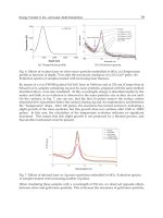
Fig. 7. Inhibition of the CLIO-D-Phe/anti-D-AA self assembly in the presence of increasing
concentrations of L- or D-Phe as detected by changes in the T2 relaxation time (Tsourkas A.
et al 2004)
Biosensors – Emerging Materials and Applications
112
Fig. 8. Time trace of cantilever defections resulting from the binding of enantiomers of
amino acids to micro cantilevers modified with covalently anti-L-amino acid antibody (1-4)
or human immunoglobulin G (5,6) 1-50 mg/L L-tryptophan, 2,5- 50mg/L L-phenylalanine,
3- 50 mg/L D-tryptophan, 4,6- 50 mg/L D-phenylalanine. (Dutta et al. 2003).
These antibodies have been also employed for enantioselective sequential-injection
chemiluminescence immunoassay of triiodothhyronine and tetraiodothyronine with
immunoreactor with immobilized haptens. It has been shown that the detection of <0.01% of
the L enantiomers in samples of D enantiomers is possible in less than 5 minutes including
regeneration of immunoreactor (Silvaieh et al. 2002). Anti-D-AA was used in
microfabricated cantilevers for enantioselective detection of amino acids based on inducing
surface stress by intermolecular forces arising from analyte adsorption on surface-
immobilized antibodies (Dutta et al. 2003). The temporal response of the cantilever allowed
the quantitative determination of enantiomeric purity up to an enantiomeric excess of 99.8%.
Based on the slope of response curves or anti-D-amino acid antibody, the selectivity
coefficients for D- enantiomer towards L-isomer were 6.5, 7.7, and 37.5 for D-phenylalanine,
D-tryptophan (Fig 8.) and D-methionine respectively. The largest enantioselectivity has been
observed for D-valine (104).
4. Enantioselective bioreceptors
4.1 Mass-based biosensors
There are many examples of sensors exhibiting the enantioselective properties based on
quartz crystal microbalance technique for example sensor for L-histidine (Zhang Z. et al.
2005), (+) methyl lactate (Ng et al. 2002), L-cysteine (Chen Z. et al. 2000), L-phenylalanine
(Huang et al. 2003) or (-) menthol (Tanese et al. 2004). However the combination of
biological macromolecules and QCM technique has been rarely reported for the studies of
chiral discrimination.
Two sensors were developed by immobilization of human serum albumin (HAS) and
bovine serum albumin (BSA) onto gold electrode combined with quartz plate by self-
Chiral Biosensors and Immunosensors
113
assembled monolayer technique. The decreased frequency demonstrated interactions
between albumines and enantiomers of R,S-1-(3-Metoxyphenyl)ethylamine (R,S-3-MPEA),
R,S-1-(4-Metoxyphenyl)ethylamine (R,S-4-MPEA), R,S-tetrahydronaphthylamine (R,S-TNA),
R,S-2-octanol (R,S-2-OT) and R,S-methyl lactate (R,S-MEL). The binding affinity of BSA and
HSA for all five pairs of enantiomers was stereodependent. The effectiveness of the QCM
sensor was described by the chiral discrimination factor α
QCM,
defined as a quotient of the
frequency decrease for enantiomer R and S respectively. For both sensors the highest
discrimination factor were obtained for R,S-TNA. The value were for BSA sensor α
QCM
=1.34
while for HSA sensor α
QCM
=1.57 (Su et al. 2009).
4.2 Optical biosensors
The Surface Plasmon Resonance method was used for monitoring real time interactions of
enantiomeric drug compounds to biomolecules immobilized on the surface of the sensor
chip. The example of such biosensor for the first time was used to check the binding of the
unnamed chiral drugs to human and rat albumins. However the enantiomers showed slight
differences in their affinities towards the immobilized albumins, authors admitted that they
were not able to detect whatever subtle differences could be due to differences in the
enantiomers or it could be due to experimental errors (Ahmad et al. 2003). The next SPR
biosensors were used to a detailed investigation of enantioselective interactions between
protein and chiral small drugs. The binding of β-blockers alprenolol and propranolol to
Cel7a cellulase was used as a model system. Cel7a was immobilized onto the sensor chip by
PDEA-mediated thiol coupling. The single enantiomers of β-blockers were injected in a
series with broad concentration range and a different pH of the solution was examined. The
results were compared with the previously validated HPLC perturbation method. (Arnell et
al., 2006). Similar interactions of drugs were examined for the SPR biosensors with two
types of proteins-transport and target, immobilized onto the sensor chip. Different type of
strong, intermediated and week interactions were exhibited by the models of binding of
propranolol enantiomers to α
1
-acid glycoprotein (AGP), R- and S-warfarin to human serum
albumin (HSA) and RS and SR-melagratan to thrombin, AGP and HSA. Strong binding
occurred in the case of RS-melagratan-trombin interaction. The other enantiomer did not
interact at all with the protein (Sandblad et al., 2009)
4.3 Ion channel biosensors
The enantioselectivity was also reported for coulometric ion channel sensor for glutamic
acid. The sensor was based on the use of glutamate receptor ion channel protein. The
glutamate receptor was immobilized within an artificial bilayer lipid membrane formed by
applying the folding method across a small circular aperture bored through a thin
polyimide-film. The detection of L-glutamic acid was performed at a concentration as low as
10
-8
M. The observed enantioselectivity for the channel activation was attributed to a
combined effect of both the relative strength of binding isomers to the receptor protein and
the relative potency of bound isomers to induce the ion channel current (Minami et al.,
1991).
5. Enantioselective aptamers
DNA aptamers are a new group of chiral selectors. They are a single-stranded
oligonucleotide sequences that can fold into a 3D shape with binding pocket and clefts that
Biosensors – Emerging Materials and Applications
114
allow them to bind many molecular targets as proteins, amino acids, peptides, cells and
viruses with specificity that allows them to distinguish even strictly structurally related
molecules. Aptamers are able to bind the target molecules with a very high affinity, equal or
sometimes even superior to those of antibodies. Comparing to antibodies they present also
some important advantages as well defined sequences produced by reproducible solid
phase synthesis which allows an accurate modulation of their selectivity and binding
parameters. Aptamers are much smaller than antibodies, permitting a higher density of
molecules to be attached to surfaces. Their production does not require animal’s
immunization. It’s also possible to obtain aptamers towards molecules that do not stimulate
immunoresponce or that are toxic. Selections are not limited by physiological constraints
allowing aptamers that bind their targets in extreme conditions to be isolated. Aptamers will
refold to regain functionality after exposure to denaturing conditions (Mosing & Bowser,
2007). They are attractive host molecules, because they can be tailored to a variety of guest
targets by the method of systematic evolution of ligands by exponential enrichment (SELEX)
(Giovannoli et al., 2008).
Fig. 9. SPR analyses of enantioselective binding interactions of selected aptamer with
complex of avidine and biotinylated L-glutamic acid- α,γ-di-t-butylester (closed circles), D-
glutamic acid- α,γ-di-t-butylester (open circles), glycine t-butyl ester (open triangles) and
aptamers complex with avidine and biotin (open diamonds) (Ohsawa et al., 2008)
Aptamers can be successfully used to the biosensor design. As a biocomponents in
biosensors they offers a multitude of advantages, such as the possibility of easily regenerate
the function of immobilized aptamers, their homogeneous preparation and the possibility of
using different detection methods due to easy labeling (Tombelli et al., 2005). A different
detection techniques can be use for the aptasensor design as for example electrochemical
(Liu et al., 2010), optical (Lee & Walt, 2000) or mass-based (Minunni et al., 2004). Although
many examples of aptamer biosensor are presented in the literature only few of them
considers the enantioselective properties.
The enzymatically prepared the biotinylated aptamers were immobilized on the sensor chip
attached with streptavidin. Two of three selected amptamers showed enantioselective
Chiral Biosensors and Immunosensors
115
recognition of the dicarboxylic acid moiety of glutamic acid. The binding affinity and
enantioselectivity were successfully evaluated by SPR measurements, and the binding
ability of these aptamers was eliminated by the absence of arginyl groups, indicating that
modified groups are indispensable due to their binding affinity and enantioselectivity. The
enantioselective response of selected aptamer is presented in Fig 9. (Ohsawa et al., 2008).
Another example presented in (Perrier et al., 2010) is based on the induced-fit binding
mechanism of end-labelled nucleic acid aptamers to the small molecule. The anti-adenosine
DNA aptamer, labelled by a single fluorescein dye was employed as a model functional
nucleic acid probe. Target binding is converted into a significant increase of the fluorescence
anisotropy signal presumably produced by the reduction of the local motional freedom of
the dye and detected by fluorescence polarization sensor. In case of target molecule the
difference in the anisotropy fluorescence signal generated by D and L enantiomers was not
enough to allow the enantioselective detection of adenosine. The presented DNA aptamer
was also able to bind the adenine nucleotides such as adenosine monophosphate AMP. In
latter case aptasensor exhibited important enantioselective properties. Titration curves
obtained by the addition of D-AMP show an FP response while for L-AMP does not cause
any significant response Fig 10.
Fig. 10. Titration curves of the 3’-F-21-Apt probe with increasing concentration of
enantiomers D-Ade (closed squares), L-Ade (open squares), D-AMP (closed triangles) and
L-AMP (open triangles). Δr is a difference between the measured anisotrophy in the
presence and in the absence of analyte (Perrier et al., 2010).
Aptamers are increasingly being used as chiral selectors in separation techniques as
capillary electrophoresis or HPLC. Recently new aptamers for different specific molecular
targets are selected. Some of them posses enantioselective properties for example for D-
peptides (Michaud et al., 2003), histidine (Ruta et al., 2007a), arginine ( Ruta et al., 2007b;
Brumbt et al., 2005), thalidomide (Shoji et al., 2007) or ibuprofen (Kim Y. S. et al., 2010).
These aptamers can potentially be used to construct chiral biosensors. Despite of successful
chiral separation by aptamer modified stationary phase (Ravelet et al., 2005) or aptamers
Biosensors – Emerging Materials and Applications
116
based capillary electrophoresis there still exists deficiencies in the understanding of the
molecular basis of their chiral recognition. In (Lin P. H. et al., 2009) authors study the
binding mechanism of DNA aptamers with L-argininamide by spectroscopic and
calorimetric methods.
5. Conclusion
The design and optimization of sensors based on the use of active biological materials,
biosensors and immunosensors for rapid, selective and sensitive determination of chiral
compounds seems to be an extremely promising direction of development. As it was
presented to the construction of such sensors a different detection methods may be
involved. Guideline in the selection of biologically active material can be results of research
conducted by separation methods using chiral antibodies or aptamers. Especially
development of aptasensor which are a relatively new technique seems to be promising. The
number of available biological active materials suitable to the construction of biosensors
could be increased by enzyme screening and protein design. It is quite possible that with
very well optimized enantioselectivity, stability and reproducibility biochemical sensors
may become in the future valuable instruments for quick control of chiral purity for
biotechnology and pharmaceutical industry.
6. References
Aboul-Enein, H. Y., Stefan, R. I. & Radu, G. L. (1999a) Biosensor for enantioselective analysis
of S-cilazapril, S-trandolapril, and S-pentopril Pharmacuetical Development and
Technology Vol. 4, No. 2, (May 1999), pp. 251-255, ISSN 1083-7450
Aboul-Enein, H. Y., Stefan, R. I. & Radu, G. L. (1999b) Biosensor for the enantioselective
analysis of S-perindopril Preparative Biochemistry & Biotechnology Vol. 29, No 1,
(February 1999), pp. 55-61, ISSN 1082-6068
Agranat, I., Caner, H. & Caldwell, J. (2002) Putting chirality to work: the strategy of chiral
switches. Nature Reviews Drug Discovery, Vol. 1, No. 10, (October 2002), pp. 753-768,
ISSN 1474-1776
Ahmad, A., Ramakrishnan, A., McLean, M. A. & Breau, A. P. (2003) Use of surface plasmon
resonance biosensor technology as a possible alternative to detect differences in
binding of enantiomeric drug compounds to immobilized albumins. Biosensors and
Bioelectronics, Vol. 18, No. 4, (April 2003), pp. 399-404, ISSN 0956-5663
Arnell, R., Ferraz, N. & Fornstedt, T. (2006) Analytical characterization of chiral drug-
protein interactions: Comparison between the optical biosensor (Surface Plasmon
Resonance) assay and HPLC perturbation method. Analytical Chemistry Vol. 78, No.
5, (March 2006), pp. 1682-1689, ISSN 00032700
Baumann, P., Zullino, D. F. & Eap, C. E. (2002) Enantiomers’ potential in
psychopharmacology. A critical analysis with special emphasis on the
antidepressant escitalopram., European Neuropsychopharmacology, Vol. 12, No. 5,
(October 2002), pp.433-444, ISSN 0924-977
Berkman, C. E., Quinn, D. A. & Thompson, C. M. (1993) Synthesis, absolute configuration,
and analysis of malathion, malaoxon, and isomalathion enantiomers. Chemical
Research in Toxicology. Vol. 6, No. 5, (September 1993), pp. 724-730. ISSN 0893-228X
Chiral Biosensors and Immunosensors
117
Brumbt, A., Ravelet, C., Grosset, C., Ravel, A., Villet, A. & Peyrin, E. (2005) Chiral stationary
phase based on a biostable L-RNA aptamers. Analytical Chemistry Vol. 77, No. 7,
(April 2005), pp. 1993-1998, ISSN 00032700
Bucheli, T., Müller, S. R., Voegelin, A. & Schwarzenbach, R. P. (1998) Bituminous Roof
Sealing Membranes as Major Source of Herbicide (R, S)-Mecoprop in roof Runoff
waters: Potential contamination of groundwater and surface waters., Environmental
Science and Technology Vol. 32, No. 22, (September 1998), pp. 3465-3471, ISSN 0013-
936X
Caner, H., Groner, E. & Levy, L. (2004) Trends in the development of chiral drugs. Drug
Discovery Today, Vol. 9, No. 3, (February 2004), pp. 105-110, ISSN 1359-6446
Chen, C. S., Fujimoto, Y., Girdaukas, G. & Sih C. J. (1982) Quantitative analyses of
biochemical kinetic resolution of enantiomers Journal of the American Chemical
Society Vol. 104, No. 25, (December 1982), pp. 7294-7299, ISSN 0002-7863
Chen, M., Fu, Y., Ciu, X., Wang, L. & Li, M. (2009) Comparative Analysis of Proline
Enantiomer in Chiral Recognition of Biological Macromolecule in Immunoassay
Electroanalysis Vol. 21, No. 21, (November 2009), pp. 2339-2344, ISSN 1040-0397
Chen, Z., Takei, Y., Deore, B. A. & Nagaoka, T. (2000) Enantioselective uptake of amino acid
with overoxidized polypyrrole colloid templated with L-lactate. Analyst, Vol. 125,
No. 12, (n.d.), pp. 2249-2254, ISSN 0003-2654
Cosnier, S., Le Pellec, A., Marks, R.S., Périé, K. & Lellouche, J-P. (2003) A permselective
biotinylated polydicarbazole film for the fabrication of amperometric enzyme
electrodes. Electrochemistry Communications, Vol. 5, No. 11, (November 2003), pp.
973-977, ISSN 1388-2481
Dominguez, R., Serra, B., Reviejo, A.J. & Pingarrón, J.M. (2001) Chiral analysis of amino
acids using electrochemical composite bienzyme biosensors. Analytical Biochemistry,
Vol. 298, No. 2, (November 2001), pp. 275-282, ISSN 0003-2697
Dutta, P., Tipple, C. A., Lavrik, N. V., Datskos, P. G., Hofstetter, H., Hofstetter, O. &
Sepaniak, M. J. (2003) Enantioselective Sensors Based on Antibody-Mediated
Nanomechanics. Analytical Chemistry, Vol. 75, No. 10, (May 2003), pp. 2342-2348,
ISSN 00032700
Eichelbaum, M. & Gross, S. A. (1996) Sterochemical aspects of drug action and disposition.,
Advances in Drug Research. Vol. 28, (n.d.). pp. 1-64, ISSN 0065-2490
Fono, L. J. & Sedlak, D. L. (2005) Use of the Chiral Pharmaceutical Propranolol to Identify
Sewage Discharges into Surface Waters., Environmental Science and Technology, Vol.
39, No. 23, (December 2005), pp. 9244-9252, ISSN 0013-936X
Gadepalli, R.S., Rimoldi, J.M., Fronczek, F.R., Nillos, M., Gan, J., Deng, X., Rodriguez-
Fuentes, G. & Schlenk, D. (2007) Synthesis of fenthion sulfoxide and fenoxon
sulfoxide enantiomers: Effect of sulfur chirality on acetylcholinesterase activity.
Chemical Research in Toxicology, Vol. 20, No. 2, (January 2007), pp. 257-262, ISSN
0893-228X
Garrison, A.W. (2006) Probing the enantioselectivity of chiral pesticides. Environmental
Science and Technology, Vol. 40, No. 1, (January 2006), pp. 16-23. ISSN 0013-936X
Giovannoli, C., Baggiani, C., Anfossi, L. & Giraudi, G. (2008) Aptamers and molecularly
imprinted polymers as artificial biomimetic receptors in affinity capillary
electrophoresis and electrochromatography. Electrophoresis, Vol. 29, No. 16, (August
2008), pp. 3349-3365, ISSN 0173-0835
Biosensors – Emerging Materials and Applications
118
Herráez-Hernández, R. & Campíns-Falco, P. (2001) Chiral Separation of Ephedrines by
Liquid Chromatography Using β-cyclodextrins., Analytica Chimica Acta, Vol. 434,
No. 2, (May 2001), pp. 315-324, ISSN 0003-2670
Hofstetter, O., Hertweck, J. K. & Hofstetter, H. (2005) Detection of enantiomeric impurities
in a simple membrane-based optical immunosensors Journal of Biochemical and
Biophysical Methods Vol. 63, No. 2, (May 2005), pp. 91-99, ISSN 0165-022X
Hofstetter, O., Hofstetter, H., Schurig, V., Wilchek, M. & Green, B. S. (1998) Antibodies Can
Recognize the Chiral Center of Free α-Amino Acids. Journal of the American Chemical
Society, Vol. 120, No. 13, (April 1998), pp. 3251-3252, ISSN 0002-7863
Hofstetter, O., Hofstetter, H., Wilchek, M., Schurig, V. & Green, B. S. (2000) An
immunochemical approach for the determination of trace amounts of enantiomeric
impurities. Chemical Communications, Vol. n. a., Iss 17, (n.d.) pp. 1581-1582, ISSN
1359-7345
Hofstetter, O., Hofstetter, H., Wilchek, M., Schurig, V. & Green, B. S. (1999) Chiral
discrimination using an immunosensor. Nature Biotechnology, Vol. 17, No. 4, (April
1999), pp. 371-374, ISSN 1087-0156
Hofstetter, O., Lindstrom, H. & Hofstetter, H. (2002) Direct Resolution of Enantiomers in
High-Performance Immunoaffinity Chromatography under Isocratic Conditions.
Analytical Chemistry; Vol 74, No. 9, (May 2002), pp. 2119-2125, ISSN 00032700
Huang, J., Egan, V., Guo, H., Yoon,J. Y., Briseno, A. L., Rauda, I. E., Garrell, R. L., Knobler,
C. M., Zhou, F. & Kaner, R. B. (2003) Enantioselective discrimination of D- and L-
phenylalanine by chiral polyaniniline thin films. Advanced Materials. Vol. 15, No. 14,
(July 2003) pp. 1158-1161, ISSN 0935-9648
Hutt, A.J. & Valentová, J. (2003) The Chiral Switch: The Development of single enantiomer
drugs from racemates. Acta Facultatis Pharmaceuticae Universitatis Comenianae, Vol.
50, (n.d.), pp. 7-23, ISSN 0301-2298
Inaba, Y., Hamada-Sato, N., Kobayashi, T., Imada, C. & Watanabe, E. (2003b) Determination
of D- and L-alanine concentrations using a pyruvic acid sensor., Biosensors and
Bioelectronics, Vol. 18, No. 8, (August 2003), pp. 963-971, ISSN 0956-5663
Inaba, Y., Mizukami, K., Hamada-Sato, N., Kobayashi, T., Imada, C. & Watanabe, E. (2003a)
Development of a D-alanine sensor for the monitoring of a fermentation using
improved selectivity by the combination of D-amino acid oxidase and pyruvate
oxidase Biosensors and Bioelectronics, Vol. 19, No. 5, (December 2003), pp. 423-431,
ISSN 0956-5663
Jiang, X.Y., Zhu, L.D., Yang, D.X., Mao, X.Y. & Wu, Y.H. (2009) Amperometric Ethanol
Biosensor Based on Integration of Alcohol Dehydrogenase with Meldola's
Blue/Ordered Mesoporous Carbon Electrode. Electroanalysis Vol. 21, Iss 14, (July
2009), pp. 1617-1623, ISSN 1040-0397
Jongejan, A., Machado, S. S. & Jongejan, J. A., (2000) The enantioselectivity of
quinohaemoprotein alcohol dehydrogenases: mechanistic and structural aspects,
Journal of Molecular Catalysis B: Enzymatic, Vol. 8, No. 1-3, (January 2000), pp. 121-
163, ISSN 1381-1177
Kacaniklic, V., Johansson, K., Marko-Varga, G., Gorton, I., Jonsson-Pettersson G. & Csoregi,
E. (1994) Amperometric biosensors for detection of L- and D-amino acids based on
coimmobilized peroxidase and L and D- amino acid oxidase in carbon paste
electrodes. Electroanalysis, Vol. 6, No. 5-6, (May-June 1994), pp. 381-390, ISSN 1040-
0397
Chiral Biosensors and Immunosensors
119
Kaniewska M. PhD Thesis, University of Warsaw, 2009
Killard, A.J. & Smyth, M.R. (2000) Separation-free electrochemical immunosensor strategies.
Analytical Letters, Vol. 33, No. 8, (n.d.), pp. 1451-1465, ISSN 0003-2719
Kim, H., Gritti, F. & Guiochon, G. (2004) Effect of the temperature on the isotherm
parameters of phenol in reversed-phase liquid chromatography. Journal of
Chromatography A, Vol. 1049, No. 1-2, (September 2004), pp. 25-36, ISSN 0021-9673
Kim, Y. S., Hyun, C. J., Kim, I. A. & Gu, M. B. (2010) Isolation and characterization of
enantioselective DNA aptamers for ibuprofen Bioorganic & Medicinal Chemistry. Vol.
18, No. 10, (May 2010), pp. 3467-3473, ISSN 0968-0896
Kullick, T., Ulber, R., Meyer, H. H., Scheper, T. & Schlügerl, K. (1994) Biosensors for
enantioselective analysis. Analytica Chimica Acta, Vol. 293, No. 3, (July 1994), pp.
271-276, ISSN 0003-2670
Landsteiner, K. & van der Scheer, J. (1928) Serological Differentiation of steric isomers.
Journal of Experimental Medicine, Vol. 48, No. 3, (September 1928), pp. 315-320, ISSN
0022-1007
Lane, R. M. & Baker, G. B., (1999) Chirality and Drugs Used in Psychiatry: Nice to Know or
Need to Know?, Cellular and Molecular Neurobiology, Vol. 19,No. 3, (June 1999), pp.
355-372 ISSN 0272-4340
Laska, M., Liesen, A. & Teubner, P. (1999) Enantioselectivity of odor perception in squirrel
monkey and humans. American Journal of Physiology Regulatory Integrative
Comperative Physiology, Vol. 277, No. 4, (October 1999), pp. R1098-R1103, ISSN 0363-
6119
Lee, M. & Walt, D.R. (2000) A fiber-optic microarray biosensor using aptamers as receptors.
Analytical Biochemistry Vol. 282, No. 1, (June 2000), pp. 142–146, ISSN 0003-2697
Lin, K. D., Zang, F., Zhou, S.S., Lui, W., Gan, J. & Pan, Z. (2007) Stereoisomeric separation
and toxicity of the namaticide fosthiazate Environmental Toxicology and Chemistry,
Vol. 26, No. 11, (November 2007), pp. 2339-2344, ISSN 0730-7268
Lin, P. H., Tong, S. J., Louis, S. R., Chang, Y., Chen, W. Y. (2009) Thermodynamic basis of
chiral recognition in a DNA aptamers. Physical Chemistry Chemical Physics Vol. 11,
No. 42, (November 2009), pp. 9744-9750, ISSN 1463-9076
Liu, Y., Tuleouva, N., Ramanculov, E. & Revzin, A. (2010) Aptamer-Based Electrochemical
Biosensor for Interferon Gamma Detection. Analytical Chemistry, Vol. 82, No. 19,
(October 2010), pp. 8131-8136 , ISSN 0003-2697
Lenz, W. (1988) A short history of thalidomide embryopathy. Teratology, Vol. 38, No. 3,
(n.d.), pp. 203-215, ISSN 0040-3709
Marchelli, R., Dossena, A. & Palla, G. (1996) The potential of enantioselective analysis as a
quality control tool., Trends in Food Science and Technology, Vol. 7, No. 4, (April
1996), pp. 113-119, ISSN 0924-2244
Michaud, M., Jourdan, E., Villet, A., Ravel, A., Grosset, C. & Peyrin, E.(2003) A DNA
aptamers as a new target-specific chiral selector for HPLC. Journal of the American
Chemical Society, Vol. 125, No. 28, (July 2003), pp. 8672-8679, ISSN 0002-7863
Minami, H., Sugawara, M., Odashima, K., Umezawa, Y., Uto, M., Michaelis, E. K. &
Kuwana, T. (1991) Ion channel sensors for glutamic acid. Analytical Chemistry, Vol.
63, No. 23, (December 1991), pp. 2787-2795, ISSN 00032700
Minunni, M., Tombelli, S., Gullotto, A., Luzi, E. & Mascini, M., 2004. Development of
biosensors with aptamers as bio-recognition element: the case of HIV-1 Tat protein.
Biosensors – Emerging Materials and Applications
120
Biosensors and Bioelectronics, Vol. 20, No. 6, (December 2004), pp. 1149–1156, ISSN
0956-5663
Miyazaki, A., Nakamura, T., Kawaradani, M. & Marumo, S. (1988) Resolution and Biological
Activity of Both Enantiomers of Methamidophos and Acephate. Journal of Agricultural
and Food Chemistry, Vol. 36, No. 4, (July 1988), pp. 835-837, ISSN 0021-8561
Moreno, J. A., Montes, F. J., Catakin, J. & Gahln M. A. (1996) Inhibition of D-amino acid
oxidase by alpha-keto acids analogs of amino acids. Enzyme and Microbial
Technology, Vol. 18, No. 5, (April 1996), pp. 379-382, ISSN 0141-0229
Mosing, R. K. & Bowser, M. T. (2007) Microfluidic selection and applications of aptamers.
Journal of Separation Science, Vol. 30, No. 10, (July 2007), pp. 1420-1426, ISSN 1615-9306
Motonaka, J., Katumoto, Y. & Ikeda, S. (1998) Preparation and properties of enzyme sensors
for L-lactic and D-lactic acids in optical isomers. Analytica Chimica Acta. Vol. 368,
No. 1-2, (July 1998), pp. 91-95, ISSN 0003-2670
Nakanishi, T., Yamakawa, N., Asahi, T., Shibata, N., Ohtani, B. & Osaka, T. (2004) Chiral
Discrimination Between Thalidomide Enantiomers Using a Solid Surface With
Two-Dimensional Chirality. Chirality Vol. 16, (n.d.), pp.S36-S39, ISSN 0899-0042
Ng, S-C., Sun, T. & Chan, H.S.O. (2002) Chiral discrimination of enantiomers with a self-
assembled monolayer of functionalized β-cyclodextrins on Au surfaces. Tetrahedron
Letters, Vol. 43, No. 15, (April 2002), pp. 2863-2866. ISSN 0040-4039
Nillos, M.G., Rodriguez-Fuentes, G., Gan, J. & Schlenk, D. (2007) Enantioselective
acetylcholinesterase inhibition of the organophosphorous insecticides profenofos,
fonofos, and crotoxyphos. Environmental Toxicology and Chemistry, Vol. 26, No. 9,
(September 2007), pp. 1949-1954, ISSN 0730-7268
Ohsawa, K., Kasamatsu, T., Nagashima, J., Hanawa, K., Kuwahara, M., Ozaki, H. & Sawai,
H. (2008) Arginine-modified DNA aptamers that show enantioselective recognition
of the dicarboxylic acid moiety of glutamic acid Analytical Sciences, Vol. 24, No. 1,
(January 2008), pp. 167-172, ISSN 0910-6340
Overbeeke, P.L.A., Orrenius, S.C., Jongejan, J.A. & Duine J.A. (1998) Enthalpic and entropic
contributions to lipase enantioselectivity Chemistry and Physics of Lipids, Vol. 93, No.
1, (June 1998), pp 81-93, ISSN 0009-3084
Perrier, S., Ravelet, C., Guieu, V., Fize, J., Roy, B., Perigaud, C. & Peyrin, E. (2010) Rationally
designed aptamer-based fluorescence polarization sensor dedicated to the small
target analysis. Biosensors and Bioelectronics, Vol. 25, No. 7, (March 2010), pp. 1652-
1657, ISSN 0956-5663
Pollegioni, L., Diederichs, K, Molla, G., Umhau, S., Welte, W., Ghisla, S. & Pilone, M. S.
(2002) Yeast D amino Acid Oxidase: Structural basis of its catalytic properties.,
Journal of Molecular Biology, Vol. 324, No. 3, (November 2002), pp. 535-546, ISSN
0022-2836
Ravelet, C., Boulkedid, R., Ravel, A., Grosset, C., Villet, A., Fize, J. & Peyrin, E. (2005) A L-
RNA aptamer chiral stationary phase for the resolution of target and related
compounds. Journal of Chromatography A, Vol. 1076, Iss 1-2, (May 2005), pp. 62-70,
ISSN 0021-9673
Robins, J., Jones, M. & Matisoo-Smith, E., 2001, Amino Acid Racemization Dating in New
Zealand: An Overview., Auckland University, Private Bag 92019, pp.1-45 Available
from
/>%20dating.pdf
Chiral Biosensors and Immunosensors
121
Rodriguez, O.P., Muth, G.W., Berkman, C.E., Kim, K. & Thompson, C.M. (1997) Inhibition of
various cholinesterases with the enantiomers of malaoxon. Bulletin of Environmental
Contamination and Toxicology, Vol. 58, No. 2, (n.d.), pp. 171-176, ISSN 0007-4861
Rogers, K.R. (2000) Principles of affinity-based biosensors. Molecular Biotechnology, Vol. 14,
No. 2, (February 2000), pp. 109-129, ISSN 1073-6085
Ruta, J., Grosset, .C, Ravelet, C., Fize, J., Villet, A., Ravel, A. & Peyrin, E. (2007a) Chiral
resolution of histidine using an anti-D-histidine L-RNA aptamers microbore
column. Journal of Chromatography B-Analytical Technologies in the Biomedical and Life
Sciences, Vol. 845 No. 2, (January 2007), pp. 186-190 ISSN 1570-0232
Ruta, J., Ravelet, C., Baussanne, I., Decout, J.L. & Peyrin, E. (2007b) Aptamer-based
enantioselective competitive binding assay for the trace enantiomer detection.
Analytical Chemistry, Vol. 79, No. 12, (June 2007), pp. 4716-4719, ISSN 00032700
Sandblad, P., Arnell, R., Samuelsson, J. & Fornstadt, T. (2009) Approach for Reliable
Evaluation of Drug Proteins Interactions Using Surface Plasmon Resonance
Technology. Analytical Chemistry, Vol. 81, No. 9, (May 2009), pp. 3551-3559, ISSN
00032700
Sarkar, P., Tothill, I. E., Setford, S. J. & Turner, A. P. F. (1999) Screen-printed amperometric
biosensors for the rapid measurement of l- and d-amino acids. The Analyst, Vol. 124,
No. 6, (June 1999), pp. 865-870, ISSN 0003-2654
Schlügerl, K., Ulber, R., & Scheper, T. (1996) Developments of biosensors for enantiomeric
analysis. Trends in analytical chemistry, Vol. 15, No. 2, (n.d.), pp. 56-62, ISSN 0165-9936
Shi, Q. & Dong, S. (1995) Amperometric biosensors based on the immobilization of oxidases
in Prussian blue film by electrochemical codeposition. Analytica Chimica Acta, Vol.
310, No. 3, (July 1995), pp. 429-436, ISSN 0003-2670
Shoji, A., Kuwahara, M., Ozaki, H. & Sawai, H. (2007) Modified DNA aptamers that binds
the (R)-Isomer of a thalidomide derivative with high enantioselectivity Journal of the
American Chemical Society Vol. 129, No. 5, (February 2007), pp. 1456-1464, ISSN
0002-7863
Silvaieh, H., Wintersteiger, R., Schmid, M.G., Hofstetter, O. & Schurig, V. (2002)
Enantioselective sequential-injection chemiluminescence immunoassays for 3,3′,5-
triiodothyronine (T
3
) and thyroxine (T
4
) Analytica Chimica Acta, Vol. 463, No. 1,
(July 2002), pp. 5-14, ISSN 0003-2670
Somers, W. A. C., Stigter, E. C. A, van Hartingsveldt, W. & van der Lugt, J. P. (1998)
Enantioselective oxidation of secondary alcohols at a quinohaemoprotein alcohol
dehydrogenase electrode. Applied Biochemistry and Biotechnology, Vol. 75, No. 2-3,
(November/December 1998), pp. 151-162, ISSN 0273-2289
Stefan, R.I., Aboul-Enein, H.Y. & Radu, G.L. (1998) Biosensors for the enantioselective
analysis of S-enalapril and S-ramipril Preparative Biochemistry & Biotechnology Vol.
28, No. 4, (n.d.), pp. 305-312, ISSN 1082-6068
Stefan, R.I. & Aboul-Enein, H.Y. (2002) The construction and characterization of an
amperometric immunosensor for the thyroid hormone (+)-3,3 ',5,5 '- tetraiodo-L-
thyronine (L-T-4) Journal of Immunoassay & Immunochemistry Vol. 23, No. 4,
(November 2002) pp. 429-437, ISSN 1532-1819
Stefan, R.I., Bala, C. & Aboul-Enein, H.Y. (2003a) Biosensor for the enantioselective analysis
of S- captopril. Sensors and Actuators B, Vol. 92, No. 1-2, (July 2003), pp. 228-231,
ISSN 0925-4005
Biosensors – Emerging Materials and Applications
122
Stefan, R.I., Bokretsion, R.G., van Staden, J.F. & Aboul-Enein, H.Y. (2003b) Determination of
L- and D- enantiomers of methotrexate using amperometric biosensors. Talanta,
Vol. 60, No. 5, (July 2003), pp. 983-990. ISSN 0039-9140
Stefan, R.I., Najem, R.M., van Staden, J.F. & Aboul-Enein, H.Y. (2003c) Biosensors for the
enantioselective analysis of pipecolic acid Sensors and Actuators B, Vol. 94, No. 3,
(October 2003), pp. 271-275, ISSN 0925-4005
Stefan, R.I., van Staden, J.F. & Aboul-Enain, H.Y. (1999) Analysis of chiral drugs with
enantioselective biosensors. An overview. Electroanalysis, Vol. 11, No. 16,
(November 1999), pp. 1233-1235, ISSN 1040-0397
Su, W. C., Zhang, W.G., Zhang, S., Fan, J., Yin, X.,. Luo, M.L & Ng, S.C. (2009) A novel
strategy for rapid real-time chiral discrimination of enantiomers using serum
albumin functionalized QCM biosensor. Biosensensors and Bioelectronics Vol. 25, No.
2, (October 2009), pp. 488-492, ISSN 0956-5663
Subrahmanyam, S., Piletsky, S. A & Turner, A. P. F. (2002) Application of Natural Receptors
in Sensors and Assays- Review Analytical Chemistry; Vol. 74, No. 16, (August 2002),
pp. 3942-3951, ISSN 00032700
Tanese, M.C., Torsi, L., Cioffi, N., Zotti, L.A., Colangiuli, D., Farinola, G.M., Babudri, F.,
Naso, F., Giangregorio, M.M., Sabbatini, L. & Zambonin, P.G. (2004)
Poly(phenyleneethynylene) polymers bearing glucose substituents as promising
active layers in enantioselective chemiresistors., Sensors and Actuators B, Vol. 100,
No. 1-2, (June 2004) pp. 17-21, ISSN 0925-4005
Tombelli, S., Minunni, M. & Mascini, M. (2005) Review. Analytical applications of aptamers.
Biosensors and Bioelectronics, Vol. 20, No. 12, (June 2005), pp. 2424–2434, ISSN 0956-
5663
Tsourkas, A., Hofstetter, O., Hofstetter, H. Weissleder, R. & Josephson, L. (2004) Magnetic
relaxation swich immunosensors detect anantiomeric impurities. Angewandte
Chemie-International Edition, Vol. 43, No. 18, (n.d.), pp. 2395-2399, ISSN 0044-8249
Varadi, M., Adanyi, N., Szabó, E.E. & Trummer, N. (1999) Determination of the ratio of D-
and L-amino acids in brewing by an immobilised amino acid oxidase enzyme
reactor coupled to amperometric detection. Biosensors and Bioelectronics, Vol. 14, No.
3, (March 1999), pp. 335-340, ISSN 0956-5663
Wcisło, M., Compagnone, D. & Trojanowicz, M. (2007) Enantioselective screen-printed
amperometric biosensor for determination of D-amino acids. Bioelectrochemistry
Vol. 71, No. 1, (September 2007), pp. 91-98, ISSN 1567-5394
Zawirska-Wojtasiak, R. (2006) Chirality and the nature of food authenticity of aroma., Acta
Scientiarum Polonorum, Technologia Alimentaria, Vol. 5, No. 1, (n.d.), pp. 21-36, ISSN
1644-0730
Zhang, S., Ding, J., Liu, Y., Kong, J. & Hofstetter, O. (2006) Development of a highly
enantioselective capacitive immunosensor for the detection of alpha-amino acids.
Analytical Chemistry Vol. 78, No. 21, (November 2006), pp. 7592-7596, ISSN 0003-2700
Zhang, Z., Liao, H., Li,H., Nie, L. & Yao, S. (2005) Stereoselective histidine sensor based on
molecularly imprinted sol-gel films., Analytical Biochemistry, Vol. 336, No. 1,
(January 2005), pp. 108-116, ISSN 0003-2697
Zhou, S., Lin, K., Yang, H., Li, L., Liu, W. & Li, J. (2007) Stereoisomeric Separation and
Toxicity of a New Organophosphorus Insecticide Chloramidophos Chemical
Research in Toxicology, Vol. 20, No. 3, (March 2003), pp. 400-405, ISSN 0893-228X
7
Recent Progress in the Construction
Methodology of Fluorescent
Biosensors Based on
Biomolecules
Eiji Nakata, FongFong Liew, Shun Nakano and Takashi Morii
Institute of Advanced Energy, Kyoto University
Kyoto, Japan
1. Introduction
The creation of novel molecular tools for detection and monitoring of the transitional
concentration and localization changes of biologically important molecules, such as
biomacromolecules, signaling small molecules and biologically important ions, is a great
challenge in the field of chemical biology. Therefore, much attention has been devoted by
chemists and biologists to develop sensing tools that allow real-time tracking of the
molecules of interests in vivo or in vitro. (Thevenot, D. R. et al., 2001; Jelinek, R. et al., 2004;
Borisov, S. M. et al., 2008) Among them, the fluorescent biosensor, which is defined as the
sensor that converts a molecular recognition event to a measurable fluorescent signal
change, has recently emerged as a powerful tool for the following reasons. (Hellinga, H.W.
et al., 1998; Johnsson, N. et al., 2007; Johnsson, K., 2009; Wang, H. et al., 2009; Liu, J. et al.,
2009) Biomacromolecular receptors, such as nucleic acids (DNA or RNA) or proteins, have
superior characteristics as the recognition platform because they play crucial roles in
numerous biological processes to mediate and regulate a range of strict recognition and
chemical reactions within cells. As for the tools for the transducer, the fluorescence
detection has the superior physical properties, such as high sensitivity, excellent spatial
resolution, good tissue penetration and low cost for the detection system, in contrast to the
other detection method including optical, electrical, electrochemical, thermal, magnetic
detections. Thus, transducing the molecular recognition events with the fluorescence
signals is very appealing and has been one of the most widely adapted methods. (Giepmans,
B.N. et al., 2006; Rao, J. et al., 2007) The rational design strategies of fluorescent biosensors
have not been matured as generally considered by the researchers in the biological field. A
simple strategy to construct a biosensor with tailored characteristics would be to conjugate a
recognition module with a signal transducer unit, although there is no simple methodology
to conjugate the recognition module and the transducer unit to afford a usable fluorescent
biosensor. Here we focus to overview the progress in the design strategy of fluorescent
biosensors, such as the auto-fluorescent protein-based biosensor, protein-based biosensor
covalently modified with synthetic fluorophores and signaling aptamers.
Biosensors – Emerging Materials and Applications
124
2. Auto-fluorescent proteins (AFPs) based biosensors
Auto-fluorescent proteins (AFPs) such as green fluorescent protein (GFP) from jellyfish
(Shimomura, O. et al., 1962) are widely used as noninvasive fluorescent markers for gene
expression, protein localization, and intracellular protein targeting (Chalfie, M. et al., 1994;
Lippincott-Schwartz, J. et al., 2001). The application of AFPs is not limited to the fluorescent
markers. Various kinds of AFP-based biosensors have recently been developed by fusion of
reporter proteins or mutation of AFPs for imaging and sensing important molecules and key
events in living cell. ( Zhang, J. et al. 2002; Zhang, J. et al. 2007; Mank, M. et al., 2008;
VanEngelenburg, S. B. et al. 2008; Lawrence, D. S. et al. 2007; Ozawa, T. 2006; Prinz, A. et al.
2008) The advantage of AFP-based biosensor is that it can be endogenously expressed in
cells or tissues simply by transfection of the plasmid DNA encoding it. This approach is a
noninvasive method and therefore avoids damage to the cell. Because AFPs based
biosensor can be produced automatically, the influence of dilution due to vital activity, such
as cell growth and division, is minimal. Moreover, it is possible to control the localization of
biosensors to the sites of interest within cell by introducing a certain organelle-specific
targeting signal. These biosensors have been powerful tool for in vivo applications.
2.1 Single AFP based biosensor
In the case of biosensors based on a single AFP, analyte binding events affect directly or
indirectly to fluorescent properties or formation, respectively, of the chromophore moiety of
AFP. The former is classified as analyte-sensitive sensors and the latter as conformation-
sensitive sensors.
The design of analyte-sensitive sensors utilizes AFP variants, whose fluorescent properties
are directly affected by the interaction between a target molecule and a chromophore moiety
in AFP. In general, the fluorescence of most of AFP variants is affected reversibly by
moderate acidification of the chromophore. To exploit such intrinsic properties of AFPs, pH
sensitive AFP variants have been developed. (Kneen, M. et al. 1998; Llopis, J. et al. 1998;
Miesenbock, G. et al. 1998; Matsuyama, S. et al. 2000) Mutants of YFPs showing pH
sensitivity bind to halide ion selectively and the binding of anion leads to fluorescence
quenching due to the induced pKa shift. (Wachter, R. M. et al. 1999; Jayaraman, S. et al. 2000;
Wachter, R. M. et al. 2000) The fluorescence of AFP becomes sensitive to other signals by the
introduction of specific mutation in close proximity to the chromophore or within the barrel
structure. In this manner, biosensors specific for Mercury (II) ion (Chapleau, R.R. et al. 2008)
and Zinc (II) ion (Barondeau, D. P. et al. 2002) have been created. The receptor function of
the sensor was directly integrated into the chromophore by alteration of the chemical nature
around the chromophore.
Another design strategy of a single AFP based biosensor relies on circularly permutated
AFP (cpAFP), which is classified as a conformation-sensitive sensor, that is, a
conformational change of the receptor associated with the ligand-binding event results a
formation of the AFP chromophore. The cpAFP is a regenerated AFP variant, in which the
original N- and C termini are connected with a flexible peptide linker to regenerate novel N
and C termini at specific positions. (Baird, G. S. et al. 1999) A number of cpAFPs with novel
termini retained their fluorescence even when a foreign receptor was inserted at the termini.
Indeed, cpAFP variants that detect Ca
2+
(Nakai, J. et al. 2001; Souslova, E. A. et al. 2007;
Baird, G. S. et al. 1999, Nagai, T. et al. 2001), cGMP (Nausch, L. W. et al. 2008), H
2
O
2
(Belousov, V. V. et al. 2006; Dooley, C. T. 2004), Zn
2+
(Mizuno, T. et al. 2007) and an inositol
Recent Progress in the Construction Methodology
of Fluorescent Biosensors Based on Biomolecules
125
phosphate derivative (Sakaguchi, R. et al. 2009), have been developed by inserting
appropriate receptor modules.
Morii and coworkers developed a cpAFP-based sensor for D-myo-inositol-1,3,4,5-
tetrakisphosphate, Ins(1,3,4,5)P
4
, by utilizing a newly designed split PH domain of Bruton’s
tyrosine kinase (Btk) and cpGFP (Sakaguchi, R. et al. 2009) (Figure 1). Interestingly, the
conjugate Btk-cpGFP realized a ratiometric fluorescence detection of Ins(1,3,4,5)P
4
by the
excitation of each distinct absorption band, and retained ligand affinity and selectivity of the
original PH domain.
Fig. 1. Schematic illustration shows a fluorescent biosensor for Ins(1,3,4,5)P
4
based on the
split Btk PH domain-cpGFP conjugate (Sakaguchi, R. et al. 2009). The original N and C
termini of GFP were linked with a short peptide linker (orange), and the novel terminal of
cpGFP (purple) was fused to the split Btk PH domain (blue). The conformational change of
the PH domain induced by the ligand-binding event was transduced to the structural
change of the chromophore of cpGFP, and then resulted in the ratiometric fluorescence
change of cpGFP.
2.2 Split AFP based biosensor
It is considered that the formation of a AFPs chromophore requires a properly folded and an
intact structure. However, many experimental data indicate that slight structural
modifications of AFPs, like circular permutation and insertion of recognition domains as
described in the previous section, still give fluorescent AFPs constructs. Therefore, AFP
sensors in the absence of targets often reveal unavoidable background fluorescence. An
excellent strategy to accomplish full suppression of the initial fluorescence utilizes an AFP
variant that was split into two non-fluorescent fragments.( Shyu, Y.J. et al. 2008; Kerppola T.
K. 2006 ) Regan and co-workers first demonstrated that a split GFP displayed a quite low
background fluorescence in the separated state and a fluorescence emission was
significantly recovered by the reassembly of the two fragments when they were placed in
close proximity by strongly interacting antiparallel leucine zippers.(Ghosh, I. et al. 2000)
Based on this strategy, a receptor composed of two subunits that are associated by binding
to the analyte can be converted into a fluorescent biosensor by connecting each of the two
subunits with each split AFP fragment (Figure 2). Actually, several types of biosensors have
Biosensors – Emerging Materials and Applications
126
been developed for fluorescent detection of specific DNA sequences (Stains, C. I. et al. 2005;
Demidov, V. V. et al. 2006), DNA methylation(Stains, C. I. et al. 2006), mRNA(Ozawa, T. et
al. 2007; Valencia-Burton, M. et al. 2007) and protein interactions (Nyfeler, B. et al. 2005; Hu,
C. -D. et al. 2003; Wilson, C. G. et al, 2004).
Unlike the above-mentioned split AFP reconstitution, in which split AFP halves reform into
a fluorescent structure via noncovalent association, another reconstitution strategy, intein-
mediated reconstitution, has been developed by Ozawa and co-workers (Ozawa, T. et al.
2000). In this strategy, split inteins were fused to split EGFPs. Each split intein-EGFP
fusion is attached to a protein of interest. The split inteins are brought into close proximity
to trigger protein splicing when an analyte induces the association between proteins of
interest. As a result, the two EGFP fragments are linked with a covalent bond and emit
fluorescence. More comprehensive information on this reconstitution strategy is available in
other excellent reviews (Ozawa, T. 2006; Awais, M. et al. 2011).
Fig. 2. Schematic illustration shows split AFP based fluorescent biosensor. A fluorescent
protein such as GFP is split into two halves [GFP(N) and GFP(C)], which connect each of the
two binding subunits, are associated by binding to the analyte.
2.3 FRET based biosensor
Non-radiative transfer of energy from an excited donor fluorophore to an acceptor
chromophore is known as fluorescence resonance energy transfer (FRET). In order to
induce FRET, the excitation spectrum of the acceptor must overlap with the emission
spectrum of the donor, and the two fluorophores must be close in proximity (< 10 nm) and
in a favorable orientation (Sapsford, K. E. et al. 2006). The efficiency of FRET is sensitive to
the distance and the orientation between the donor and acceptor groups. To obtain the
expected energy transfer efficiency for biological applications, the following two issues in
the sensor design should be considered. First, suitable FRET pairs in which the donor
emission spectrum overlaps the acceptor absorption spectrum should be chosen. In the
AFP-based FRET strategy, CFP and YFP mutants have been favorably utilized as a FRET
donor and an acceptor, respectively (Piston, D.W. et al. 2007). Second, the donor and the
acceptor fluorophores should be placed at a rational distance which can drastically change
Recent Progress in the Construction Methodology
of Fluorescent Biosensors Based on Biomolecules
127
the efficiency of FRET before and after the sensing event.(Ohashi, T. et al. 2007) Therefore, a
FRET based biosensor can sense the analyte in a ratiometric manner by comparing the donor
and acceptor emission intensities that are result from the analyte induced distance and/or
conformational changes. Based on the mechanism by which FRET efficiency changes, AFP-
based FRET biosensors can be divided into two classes, that is, an intramolecular and an
intermolecular FRET systems (Figure 3). In the case of intramolecular FRET biosensors, the
two fluorophores are attached at two ends of a peptide sequence in the receptor protein or
the concatenation of interacting domains. The feasibility of this strategy strongly depends
on the magnitude of the structural change of the receptor. In the case of a receptor that
displays a large structural change upon binding to the substrate, this strategy would be the
most straightforward way to integrate the signal transduction function into the receptor of
interest. Based on this strategy, various FRET biosensor for imaging intracellular events
such as enzyme activities [e.g. protease (Mahajan, N. P. et al. 1999; Luo, K. Q. et al. 2001;
Rehm, M. et al. 2002; Ai, H. W. et al. 2008), kinase (Sato, M. et al. 2002; Nagai, Y., et al. 2000),
phosphatase (Newman, R. H. et al. 2008)] and dynamics of intracellular second messengers
[e.g.Ca
2+
(Miyawaki, A. et al. 1997; Romoser, V. A. et al. 1997), cAMP (Nikolaev,V. et al.
2004), cGMP (Sato, M. et al. 2000), IP
3
(Sato, M. et al. 2005)] have been developed. It should
be noted that careful optimization, such as tuning the position of AFPs relative to the
sensing domain by changing the linker between each of protein units, is frequently
necessary to realize the satisfactory response of the FRET change. Most importantly, the
Fig. 3. AFP-fused FRET based biosensors. (a) Intramolecular FRET-based biosensor: The
protein domains with a large structural change upon the analyte binding event. (b)
Intermolecular FRET-based biosensor: The change of FRET efficiency is induced by the
dissociation or association of the subunit upon the analyte-binding event.
Biosensors – Emerging Materials and Applications
128
obligatory conformational change in the receptor protein severely limits the choice of
proteins available for the construction of FRET biosensors by this strategy. Recently,
Johnsson and co-workers have demonstrated a new type of FRET biosensor based on their
SNAP-tag technique, for which conformational changes upon analyte binding were not
required (Brun, M. A. et al. 2009). Intermolecular FRET biosensors have been developed by
employing two protein domains separated from each other, to which AFPs of FRET donor
and acceptor are attached, respectively. Zaccoro and co-workers constructed FRET
biosensor for cAMP by applying this strategy to the regulatory and catalytic subunit of
protein kinase A (PKA) (Zaccolo, M. et al. 2000; Zaccolo, M et al. 2002). This biosensor can
detect the rise of intracellular cAMP concentration by the decrease in the FRET efficiency
induced by dissociation of the catalytic subunit from the regulatory subunit. Although this
strategy shows a potential to effect a dynamic FRET change by the analyte-induced
association and/or dissociation of protein subunits, the stoichiometry of the FRET donor
and acceptor may vary between either cells or intracellular compartments. In these cases,
they cause difficulty in analysis of the FRET efficiency changes. More comprehensive
information on dual FRET-based biosensors is available in other excellent reviews
(Souslova, E. A. et al. 2007; VanDngelenburg, S. B. et al. 2008; Carlson, H. J. et al. 2009).
3. Protein-based biosensor covalently modified with fluorescent artificial
molecules
Another useful strategy to construct fluorescent biosensors is a structure-based design of a
protein covalently modified with a fluorescent dye. Advantages for the use of fluorescent
dyes are as follows. First, the relatively smaller size of the synthetic fluorophore is likely to
less perturb the property of the original receptor protein. Second, a superior characteristic of
dye, that is, the fluorescence changes in intensity and wavelength and the
microenvironmental sensitivity such as pH, polarity or molecular recognition, could be
introduced to the receptor protein. Not only simple dyes but also functional molecules, such
as artificial receptors, can be incorporated. Third, the attachment of dye to the protein
framework is more flexible than the use of AFPs. While the attaching positions of AFP are
generally limited to the N- and C termini of receptor proteins, the incorporation of small dye
to proteins is also possible in the middle of loop regions or at close proximity to the binding
pocket. On the other hands, unlike AFPs based biosensor, this type of protein-based
biosensor generally require the invasive technique for translocating across the plasma
membrane, such as electroporation (Marrero, M.B. et al. 1995; Fenton, M. et al. 1998;
Sakaguchi, R. et al. 2010), lipofection (Zelphati, O., et al. 2001; Zheng, X. et al. 2003),
microinjection (Abarzua, P. et al. 1995), and tagging cell-permeable peptide sequences
(Wadia, J.S. et al 2005; Sugimoto, K. et al 2004). In addition, the central issue for the
construction of these types of biosensors is the way to introduce a dye into the receptor
protein site-selectively. Here, a variety of fluorescent biosensors that use fluorescent
molecules is described according to a classification of the incorporation methodologies of
fluorescent dye.
3.1 Introduction of a thiol reactive fluorophore on a unique cysteine residue of
engineered receptor protein
The most important process to success this methodology is that all of the original cysteine
residue of the receptor protein must be initially substituted with other amino acids to avoid
Recent Progress in the Construction Methodology
of Fluorescent Biosensors Based on Biomolecules
129
the nonspecific labeling of cysteine reactive fluorophores. Following the process, a unique
cysteine residue was introduced at specific position. The position to introduce a fluorophore
is most conveniently determined by the three-dimensional structure of the receptor protein.
As a pioneering work, bPBPs (bacteria periplasmic binding protein), a representative
protein scaffold, were converted to fluorophore-modified biosensors by Hellinga et al.
(Dwyer, M. A. et al. 2004) or others (Gilardi, G. et al. 1994; Brune, M. et al. 1998; Hirshberg,
M. et al. 1998). Most of bPBPs consist of two domains connected by a hinge region, with a
ligand binding site located at the interface between the two domains, which can permit
dynamic conformational changes induced upon ligand binding. Therefore, two distinct
approaches are used to establish an efficient signal transduction mechanism that would
sense the ligand-binding event. In the first approach, an environmentally sensitive
fluorophore is positioned in the binding pocket so that the ligand-induced changes in the
fluorescence are produced by the direct fluorophore-ligand interactions. This approach
often has a disadvantage that unfavorable steric interactions between the introduced
fluorophore and the ligand lower the binding affinity. The second approach introduces
environmentally sensitive fluorophore at the region that is distant from the ligand-binding
site but exhibits dynamic domain movement in response to the ligand binding. This
allosteric sensing mechanism shows an advantage that the ligand binding is essentially
unaffected by introducing a fluorophore.
On the other hand, there are number of proteins that do not undergo such a dynamic
conformational change upon ligand binding, but they are capable of recognizing the various
substances of biological importance. The useful methodology to convert such non-allosteric
proteins to fluorescent biosensors is to introduce an environmentally sensitive fluorophore
within the proximity of the ligand-binding site, though this strategy might have some
drawbacks as mentioned above. But several successful examples demonstrated that such a
methodology is applicable for obtaining biosensors (Chan, P. H. et al. 2004; Nalbant, P. et al.
2004; Chan, P. H. 2008). Morii and coworkers constructed novel biosensors for inositol 1,4,5-
trisphosphate [Ins(1,4,5)P
3
] and 1,3,4,5-tetrakisphosphate [Ins(1,3,4,5)P
4
] by utilizing the
pleckstrin homology (PH) domain of phospholipase C (PLC)
1
(Morii, T. et al. 2002) and the
general receptor for phosphoinositides 1 (GRP1) (Sakaguchi, R. et al. 2010) (Figure 4),
respectively. In these biosensors a synthetic fluorophore was attached at the proximity of the
ligand-binding site based on the three dimensional structures of proteins so that the changes
in orientation of the fluorophore induced by the substrate binding lead to a sufficient
fluorescence response. This structure-based design of synthetic fluorophore-modified
biosensors is a powerful method to produce biosensors with high selectivity and
appropriate affinity to target inositol derivatives in living cells (Sakaguchi, R. et al. 2010;
Sugimoto, K. et al. 2004; Nishida, M. et al. 2003).
3.2 Site-specific unnatural amino acid mutagenesis with an expanded genetic code
As mentioned above, the post-labeling of unique cysteine residues required preliminary
preparation that all of the original cysteine residue of the receptor protein must be
substituted with other amino acids. The process might cause the instability of the receptor
protein mutant. A mutagenesis technique for direct incorporation of synthetic fluorophores
as unnatural amino acids into desired positions in proteins can avoid such a problem. A site-
specific mutagenesis with an expanded genetic code that employed an amber suppression
method (Wang, L. 2005; et al. Xie, J. et al. 2006) or a four-base codon method (Hohsaka, T. et
al., 2002) in cell-free translation systems has provided a variety of fluorescently modified
Biosensors – Emerging Materials and Applications
130
Fig. 4. A schematic illustration shows a fluorescent biosensor for Ins(1,3,4,5)P
4
based on the
GRP1 PH domain (Sakaguchi, R. et al. 2010). Firstly, the original cysteine residues (cyan) of
GRP PH domain were replaced with other amino acids. Second, a unique cysteine residue
(magenta) was introduced to the resultant mutant followed by labeling with thiol reactive
fluorescein (green) as an environment sensitive fluorophore to give Ins(1,3,4,5)P
4
sensor. The
local environmental change of the fluorophore induced by the ligand-binding event was
transduced to the fluorescence enhancement.
proteins (Anderson, R. D. et al. 2002; Taki, M. et al. 2002; Kajihara D. et al. 2006). As an
excellent example, Hohsaka and co-workers prepared a series of semisynthetic calmodulins,
two different position of which were replaced with unnatural amino acids bearing a FRET
pair of BODIPY derivatives by using two sets of four-base codons. Some of the doubly
modified calmodulin sensed calmodulin-binding peptide by substantial FRET signal
changes. This is a powerful tool for site-specific introduction of unnatural amino acids into
protein, though the examples of the construction of fluorescent biosensor based on these
methods are still limited.
3.3 Covalent introduction of fluorescent molecules by chemical modification
Modification of a protein by using genetic method often causes the lower activity or instability
of the mutated protein as mentioned in the previous section. In addition, the method is not
appropriate when the three dimensional structure of a receptor protein is not known. In that
case, an approach to site-specifically incorporate a signal transducer proximal to the binding
pocket of intact receptor protein by using selective chemical modifications is valid.
As the primary example, Schultz and co-workers constructed an antibody-based fluorescent
biosensor by using an affinity-labeling method (Pollack, S. J. et al. 1988). The chemically
engineered antibody, of which the proximal antigen-recognition site was modified by
fluorescent molecule, can detect antigen binding by fluorescence decrease. Hamachi and co-
workers constructed a lectin-based fluorescent biosensor using an improved photo affinity
labeling method, termed as P-PALM (post-photoaffinity labeling modification) (Hamachi, I.
et al. 2000; Nagase, T. et al. 2001, Nagase, T. et al. 2003). This methodology can introduce
artificial molecules (e.g. fluorophore, artificial receptor) proximal to the active site of a target
protein without genetically modifying the protein framework. In a proof-of-principle
experiment, P-PALM was demonstrated by using concanavalin A (Con A), an extensively
Recent Progress in the Construction Methodology
of Fluorescent Biosensors Based on Biomolecules
131
studied lectin (saccharide-binding protein). Introduction of a thiol group as a
chemoselective modification site in proximity of the ligand-binding pocket of Con A is
conducted by a designed photoaffinity labeling molecule, which is composed of a ligand
module, a photo reactive module and a cleavable disulfide module. Depending on the
nature of the subsequent modification by a thiol reactive artificial molecule, not only
environmental sensitive fluorophore (Koshi, Y. et al. 2005; Nakata, E. et al. 2005; Nakata, E.
et al. 2008) but also fluorescent artificial receptor (Nakata, E. et al. 2004) can be introduced to
Con A. Intact Con A can be converted to a various type of fluorescent biosensors that
successfully sense the saccharide derivatives in different manners. Because the initial P-
PALM strategy based on thiol chemistry shows limited bioorthogonality, this method is not
applicable to many proteins. To overcome this drawback, Hamachi group adopted the
ketone/aldehyde-based hydrazone/oxime exchange reaction (Takaoka, Y. et al. 2006) and
the organometallic Suzuki reaction (Wakabayashi, H. 2008) as bioorthogonal chemoselective
modifications. Recently, Hamachi and co-workers also developed ligand-directed tosyl
(LDT) chemistry-based approach as a more general and simple strategy of target selective
chemical modification (Tsukiji, S. 2009). A detailed description of their strategies is
described in other review articles (Nakata, E. et al. 2007; Wang. H. et al. 2009).
4. Signaling aptamers
Protein based biosensors are generally constructed by using native or slightly modified
proteins as the scaffold. Therefore, the function of the constructed biosensor, such as the
specificity and the affinity toward the substrate, depends on that of the native receptor.
Unlike receptor proteins, DNA or RNA based receptors (aptamers) which have appropriate
affinity and specificity for various targets ranging from small molecules to proteins can be
generated by using in vitro selection, also known as SELEX (systematic evolution of ligands
by exponential enrichment) (Ellington, A. D. 1994; Ellington, A. D. et al. 1990; Gold, L. et al.
1995; Osborne, S. E. et al. 1997; Wilson, D. S. et al. 1999). That is, aptamers that bind to the
substrate of interest with tailor made functions, such as the specificity and the affinity, can
potentially be generated through in vitro selection. Previous work indicated that most of the
structurally characterized aptamers underwent induced-fit type of conformational change
upon ligand binding [Westhof, E. et al. 1997]. Introduction of the signal transduction
module such as a fluorophore at an appropriate site of the aptamer enables a read out of the
ligand-binding event as a local environmental change of the fluorophore. Thus, the design
of aptamer-based fluorescent sensors represents an attractive and promising alternative to
the protein-based sensors. Some excellent reviews of aptamer sensors have already covered
the selection and evolution techniques and sophisticated applications of the aptamer sensors
[Liu, J. et al 2009; Mok, W. et al. 2008]. Here we focus on unique modular strategies to
construct aptamer sensors, which would avoid the cumbersome trial-and-error process to
construct a sensor with an optimized function.
4.1 Modular strategies for tailoring aptamer sensors
Sophisticated design strategies have successfully provided fluorescent biosensors based on
biomolecules such as DNA, RNA or proteins, but these strategies usually require the
redundant optimization of sensor functions. For example, introduction of the fluorophore
often impairs the original receptor function and does not always ensure the fluorophore-
labelled receptor to act as an expected sensor. It is quite difficult to empirically apply the
Biosensors – Emerging Materials and Applications
132
obtained findings from the previously constructed biosensor to the other one, because the
communication between the substrate binding and the signal-transduction is not so simple
and is unique to the individual biosensor. On the other hand, a modular strategy that
permits facile preparation of biosensors with tailored characteristics by a simple
combination of a receptor and a signal transducer has recently emerged as a new paradigm
for a versatile design of fluorescent biosensors. Stojanovic and co-workers have proposed a
modular design of signaling aptamers based on the allosteric regulation of binding events
(Stojanovic, M. N. et al. 2004). The target binding aptamers were fused with the reporter dye
binding aptamers, which can drastically increase the fluorescent intensity of reporter dye,
and the reporter dye binding was significantly enhanced upon target binding. Fluorescent
sensors for adenosine triphosphate (ATP), flavin mononucleotide (FMN) and theophylline
have been demonstrated based on this design, showing the generality of the approach.
Later, several groups reported various allosteric aptamer sensors based on the methodology
(Kolpashchikov, D. M. 2005; Xu, W. et al. 2010; Furutani, C. et al. 2010).
The application of the selection and evolution technique is not limited to obtain functional
macromolecules solely composed of RNA or DNA. Morii and co-workers have recently
developed a conceptually new strategy for preparation of fluorescent biosensors with
diverse functions based on a framework of ribonucleopeptide (RNP), such as the
structurally well characterized complex of the Rev Responsive Element (RRE)-HIV Rev
peptide (Rev peptide) and RRE RNA (Figure 5) (Tainaka, K. et al. 2010). In the first step to
construct the fluorescent RNP sensor, a randomized nucleotide sequence was introduced
into the RNA subunit of RNP to construct RNP library. In vitro selection method was
applied to the RNP library to afford a series of RNP receptors for a given target (Morii, T. et
al. 2002). In the second step, the Rev peptide was modified with a fluorophore as the
transducer of binding event without greatly disturbing the affinity and specificity of the
RNP receptor. The constructed fluorescent RNP sensor showed the fluorescent intensity
changing upon binding to the target molecule as the result of the conformational change of
RNA subunit by inducing target binding. In similar to RNA aptamers, the RNP receptors,
which obtained by in vitro selection, are considered as a RNP receptor library, because a
variety of RNA structures and reveal different affinity to the target molecule were included.
The combined peptide subunit is also easily converted to functionalized Rev peptide
libraries, such as various fluorophore modified Rev peptide libraries with a variety of
excitation and emission wavelengths. By taking the advantage of such the noncovalent
nature of the RNP complex, RNP sensors with desired affinity, selectivity and optical
sensing properties could be selected in a high-throughput manner by combining a series of
RNA subunits derived from each of the library. Actually, a variety of fluorescent biosensors
for targeting ATP (Hagihara, M. et al. 2006), GTP (Hagihara, M. et al. 2006), histamine
(Fukuda, M. et al. 2009), phosphotyrosine (Hasegawa, T. et al. 2005), and phosphotyrosine-
containing peptide fragment (Hasegawa, T. et al. 2008) have been produced by the group,
showing the generality of the approach. Recently, the group showed that ATP-binding RNP
sensor was rationally converted to GTP-binding RNP sensor to have realized the detail of
the recognition mechanism (Nakano, S. et al. 2011). Though the noncovalent configuration
conveniently provides fluorescent RNP sensors in the selection stage, it have a possibility to
becomes a disadvantage for the practical measurements after optimization of the sensor
function, for instance, the RNP complex would dissociate to each component under
reducing condition such as the nanomolar range. A covalently linking of RNA and peptide
subunits without sacrificing the sensing function would overcome such disadvantages.
Recent Progress in the Construction Methodology
of Fluorescent Biosensors Based on Biomolecules
133
Fig. 5. Screening methodology of a tailor-made RNP fluorescent sensor [Hagihara, M. et al.
2006]. Combination between the RNA subunit library and several dye-labeled Rev peptide
subunits generates combinatorial fluorescent RNP receptor libraries, from which RNP
sensors with desired function, such as optical property, affinity and selectivity, are selected.
5. Perspective
Here we overviewed construction methodologies of fluorescent biosensor based on
biomolecules, that is, protein-based biosensor and aptamer-based biosensor. The systematic
developments of these technologies have expanded the applicability of fluorescent
biosensors. In the case of the protein based biosensor, there is no doubt that these sensors
represent the most practical and reliable tools for the real-time measurements of various
biologically important molecules in living cells. Actually, the function of second
messengers, for example, in the cell has been progressively clarified owing to significant
contribution of these new biosensors. However, the wide varieties of the construction
strategies, which have both the advantages and drawbacks as mentioned above, strongly
indicated the lack of general approach to conjugate a recognition module with a signal
transducer unit. Further effort in the fields for establishing a general and simple strategy to
construct usable biosensors will realize tailor-made fluorescent biosensors.
Aptamer-based biosensors have potential to realize the tailor-made biosensor with finely
tuneable affinity and selectivity based on in vitro selection technique, and to visualize
intracellular molecules. However, this type of sensor is practically passive with challenges
in cell application owing to the inherent liability of RNA molecules in the intracellular
condition. Such the drawbacks will be overcome by the improved selection and evolution
technique to construct the aptamers that resist to the cellular degradation activity.
6. Reference
Abarzua, P., LoSardo, J. E., Gubler, M. L., & Neri, A. (1995). Microinjection of Monoclonal
Antibody PAb421 into Human SW480 Colorectal Carcinoma Cells Restores the
Transcription Activation Function to Mutant p53. Cancer Res., 55(16), 3490–3494.
Biosensors – Emerging Materials and Applications
134
Ai, H. W., Hazelwood, K. L., Davidson, M. W., & Campbell, R. E. (2008). Fluorescent protein
FRET pairs for ratiometric imaging of dual biosensors. Nat. Methods, 5(5), 401–403.
Anderson, R. D., 3rd, Zhou, J., & Hecht, S. M. (2002). Fluorescence Resonance Energy
Transfer between Unnatural Amino Acids in a Structurally Modified Dihydrofolate
Reductase. J. Am. Chem. Soc. 124(33), 9674–9675.
Awasis, M., & Ozawa, T. (2011). Illuminating Intracellular Signaling and Molecules for
Single Cell Analysis. Molecular Biosystems, in press
Baird, G.S., Zacharias, D.A., & Tsien, R.Y. (1999). Circular Permutation and Receptor
Insertion within Green Fluorescent Proteins. Proc. Natl. Acad. Sci. USA, 96(20),
11241–11246.
Barondeau, D. P., Kassmann, C. J., Tainer, J. A., & Getzoff, E. D. (2002). Structural Chemistry
of a Green Fluorescent Protein Zn Biosensor. J. Am. Chem. Soc., 124(14), 3522–3524.
Belousov, V. V., Fradkov, A. F., Lukyanov, K. A., Staroverov, D. B., Shakhbazov, K. S.,
Terskikh, A. V., & Lukyanov, S. (2006). Genetically Encoded Fluorescent Indicator
for Intracellular Hydrogen Peroxide. Nat. Methods, 3(4), 281–286.
Borisov, S. M., & Wolfbeis,O. S., S. M. & Wolfbeis,O. S. (2008) Optical Biosensors. Chem. Rev.,
108(2), 423–461.
Brun, M. A., Tan, K. T., Nakata, E., Hinner, M. J., Johnsson, K. (2009). Semisynthetic
Fluorescent Sensor Proteins Based on Self-Labeling Protein Tags. J. Am. Chem. Soc.,
131(16), 5873–5884.
Brune, M., Hunter, J. L., Howell, S. A., Martin, S. R., Hazlett, T. L., Corrie, J. E., & Webb, M.
R. (1998). Mechanism of Inorganic Phosphate Interaction with Phosphate Binding
Protein from Escherichia Coli. Biochemistry, 37(29), 10370–10380.
Carlson, H. J., &Campbell, R. E., (2009) Genetically Encoded FRET-Based Biosensors for
Multiparameter Fluorescence Imaging. Curr. Opin. Biotechnol. 20(1), 19–27.
Chalfie, M., Tu, Y., Euskirchen, G., Ward, W. W., & Prasher, D. C. (1994). Green Fluorescent
Protein as a Marker for Gene Expression. Science, 263(5148), 802–805.
Chan, P. H., Liu, H. B., Chen, Y. W., Chan, K. C., Tsang, C. W., Leung, Y. C.,& Wong, K. Y.
(2004). Rational Design of a Novel Fluorescent Biosensor for β-Lactam Antibiotics
from a Class a β-Lactamase. J. Am. Chem. Soc., 126(13), 4074–4075.
Chan, P. H., So, P. K., Ma, D. L., Zhao, Y., Lai, T. S., Chung, W. H., Chan, K. C., Yiu, K. F.,
Chan, H. W., Siu, F. M., Tsang, C. W., Leung, Y. C., Wong, K. Y. (2008).
Fluorophore-Labeled β-Lactamase as a Biosensor for β-Lactam Antibiotics: A Study
of the Biosensing Process. J. Am. Chem. Soc., 130(20), 6351–6361.
Chapleau, R. R., Blomberg, R., Ford, P. C., Sagermann, M., (2008). Design of a Highly
Specific and Noninvasive Biosensor Suitable for Real-Time in Vivo Imaging of
Mercury (II) Uptake. Protein Sci., 17(4), 614–622.
Demidov, V. V., Dokholyan, N. V., Witte-Hoffmann, C., Chalasani, P., Yiu, H. W., Ding, F.,
Yu, Y., Cantor, C. R., & Broude, N. E. (2006). Fast Complementation of Split
Fluorescent Protein Triggered by DNA Hybridization. Proc. Natl. Acad. Sci. USA
,103(7), 2052–2056.
Dooley, C. T., Dore, T. M., Hanson, G. T., Jackson, W. C., Remington, S.J., & Tsien, R. Y.
(2004). Imaging Dynamic Redox Changes in Mammalian Cells with Green
Fluorescent Protein Indicators. J. Biol. Chem. 279(21), 22284–22293.
Dwyer, M. A., & Hellinga, H. W. (2004). Periplasmic Binding Proteins: A Versatile
Superfamily for Protein Engineering. Curr. Opin. Struct. Biol., 14(4)
, 495–504.
Ellington, A. D. (1994). RNA Selection. Aptamers Achieve the Desired Recognition. Curr.
Biol. 4(5), 427–429.
Recent Progress in the Construction Methodology
of Fluorescent Biosensors Based on Biomolecules
135
Ellington, A. D., & Szostak, J. W. (1990). In Vitro Selection of RNA Molecules That Bind
Specific Ligands. Nature, 346(6287), 818–822.
Fenton, M., Bone, N., & Sinclair, A. J. (1998). The Efficient and Rapid Import of a Peptide
into Primary B and T Lymphocytes and a Lymphoblastoid Cell Line. J. Immunol.
Methods, 212(1), 41–48.
Fukuda, M., Hayashi, H., Hasegawa, T., & Morii, T. (2009). Development of a Fluorescent
Ribonucleopeptide Sensor for Histamine. Trans. Mat. Res. Soc. Jpn. 34, 525–527.
Furutani, C., Shinomiya, K., Aoyama, Y., Yamada, K., & Sando, S. (2010). Modular blue
fluorescent RNA sensors for label-free detection of target molecules. Mol Biosyst.,
6(9),1569-1571.
Ghosh, I., Hamilton, A.D., & Regan, L. (2000) Antiparallel Leucine Zipper-Directed Protein
Reassembly: Application to the Green Fluorescent Protein. J. Am. Chem. Soc.
122(23), 5658–5659.
Giepmans, B.N., Adams, S.R.,Ellisman, M.H., & Tsien, R.Y. (2006) The Fluorescent Toolbox
for Assessing Protein Location and Function. Science, 312(5771), 217–224.
Gilardi, G., Zhou, L. Q., Hibbert, L., & Cass, A. E. (1994). Engineering the Maltose Binding
Protein for Reagentless Fluorescence Sensing. Anal. Chem. 66(21), 3840–3847.
Gold, L., Polisky, B., Uhlenbeck, O., & Yarus, M. (1995). Diversity of Oligonucleotide
Functions. Annu. Rev. Biochem., 64, 763–797.
Hagihara, M., Fukuda, M., Hasegawa, T., & Morii, T. (2006). A Modular Strategy for
Tailoring Fluorescent Biosensors from Ribonucleopeptide Complexes. J. Am. Chem.
Soc. 128(39), 12932–12940.
Hamachi, I., Nagase, T., & Shinkai, S. (2000). A General Semisynthetic Method for
Fluorescent Saccharide-Biosensors Based on a Lectin. J. Am. Chem. Soc. 122(48),
12065–12066.
Hasegawa, T., Hagihara, M., Fukuda, M., Nakano, S., Fujieda, N., Morii, T. (2008). Context-
Dependent Fluorescence Detection of a Phosphorylated Tyrosine Residue by a
Ribonucleopeptide. J. Am. Chem. Soc. 130(27), 8804–8812.
Hasegawa, T., Ohkubo, K., Yoshikawa, S., & Morii, T. (2005). A Ribonucleopeptide Receptor
Targets Phosphotyrosine. J. Surf. Sci. Nanotech., 3, 33–37.
Hellinga, H.W. & Marvin, J.S. (1998) Protein Engineering and the Development of Generic
Biosensors. Trends Biotechnol. 16(4), 183–189.
Hirshberg, M., Henrick, K., Haire, L. L., Vasisht, N., Brune, M., Corrie, J. E., & Webb, M. R.
(1998). Crystal Structure of Phosphate Binding Protein Labeled with a Coumarin
Fluorophore, a Probe for Inorganic Phosphate. Biochemistry, 37(29), 10381–10385.
Hohsaka, T., &Sisido, M. (2002). Incorporation of Non-Natural Amino Acids into Proteins.
Curr. Opin. Chem. Biol. 6(6), 809–815.
Hu, C D. T. & Kerppola, K. (2003). Simultaneous visualization of multiple protein
interactions in living cells using multicolor fluorescence complementation analysis.
Nat. Biotechnol., 21(5), 539–545.
Jayaraman, S., Haggie, P., Wachter, R. M., Remington, S. J., &Verkman, A. S. (2000).
Mechanism and Cellular Applications of a Green Fluorescent Protein-based Halide
Sensor. J. Biol. Chem. 275(9), 6047–6050.
Jelinek, R. & Kolusheva, S. (2004). Carbohydrate biosensors. Chem. Rev. 104(12), 5987–6015.
Johnsson, K. (2009). Visualizing Biochemical Activities in Living Cells. Nat. Chem. Biol. 5(
2),
63–65.
Johnsson, N., & Johnsson, K. (2007) Chemical Tools for Biomolecular Imaging. ACS Chem.
Biol., 2(1), 31–38.