Biosensors for Health Environment and Biosecurity Part 2 docx
Bạn đang xem bản rút gọn của tài liệu. Xem và tải ngay bản đầy đủ của tài liệu tại đây (4.99 MB, 35 trang )
Biosensors for Health, Environment and Biosecurity
26
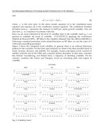
Fig. 6. SEM image of an integrated pH and glucose sensor. The insets show a schematic
cross-section of the pH sensor and also an SEM of the ZnO nanorods grown in the gate
region of the glucose sensor.
For the glucose detection, a highly dense array of 20-30 nm diameter and 2 µm tall ZnO
nanorods were grown on the 20 × 50 µm
2
gate area. The lower right inset in Figure 6 shows
closer view of the ZnO nanorod arrays grown on the gate area. The total area of the ZnO
was increased significantly with the ZnO nanorods. The ZnO nanorod matrix provides a
microenvironment for immobilizing negatively charged GO
x
while retaining its bioactivity,
and passes charges produced during the GO
x
and glucose interaction to the AlGaN/GaN
HEMT. The GOx solution was prepared with concentration of 10 mg/mL in 10 mM
phosphate buffer saline (pH value of 7.4, Sigma Aldrich). After fabricating the device, 5 μl
GO
x
(~100 U/mg, Sigma Aldrich) solution was precisely introduced to the surface of the
HEMT using a pico-liter plotter. The sensor chip was kept at 4
o
C in the solution for 48 hours
for GO
x
immobilization on the ZnO nanorod arrays followed by an extensively washing to
remove the un-immobilized GO
x
.
To take the advantage of quick response (less than 1 sec) of the HEMT sensor, a real-time
EBC collector is needed (Montuschi and Barnes 2002, Anh, Olthuis and Bergveld 2005). The
amount of the EBC required to cover the HEMT sensing area is very small. Each tidal
breath contains around 3 l of the EBC. The contact angle of EBC on Sc
2
O
3
has been
measured to be less than 45
o
, and it is reasonable to assume a perfect half sphere of EBC
droplet formed to cover the sensing area 4 × 50 µm
2
gate area. The volume of a half sphere
with a diameter of 50 µm is around 3 × 10
-11
liter. Therefore, 100,000 of 50 µm diameter
droplets of EBC can be formed from each tidal breath. To condense entire 3 l of water
vapor, only ~ 7 J of energy need to be removed for each tidal breath, which can be easily
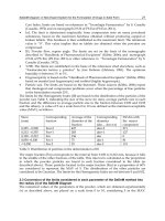
achieved with a thermal electric module, a Peltier device, as shown in Figure 7. The
AlGaN/GaN High Electron Mobility Transistor Based Sensors for Bio-Application
27
schematic of the system for collecting the EBC is illustrated in Figure 8. The AlGaN/GaN
HEMT sensor is directly mounted on the top of the Peltier unit (TB-8-0.45-1.3 HT 232,
Kryotherm), as also shown in Figure 7, which can be cooled to precise temperatures by
applying known voltages and currents to the unit. During our measurements, the hotter
plate of the Peltier unit was kept at 21
o
C, and the colder plate was kept at 7
o
C by applying
bias of 0.7 V at 0.2 A. The sensor takes less than 2 sec to reach thermal equilibrium with the
Peltier unit. This allows the exhaled breath to immediately condense on the gate region of
the HEMT sensor.
Fig. 7. Optical image of sensor mounted on Peltier cooler.
Prior to pH measurements of the EBC, a Hewlett Packard soap film flow meter and a mass
flow controller were used to calibrate the flow rate of exhaled breath. The HEMT sensors
were also calibrated and exhibited a linear change in current between pH 3-10 of 37µA/pH.
Due to the difficulty to collect the EBC with different glucose concentration, the samples for
glucose concentration detection were prepare from glucose diluted in PBS or DI water.
The HEMT sensors were not sensitive to switching of N
2
gas, but responded to applications
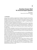
of exhaled breath pulse inputs from a human test subject, as shown at the top of Figure 9
(top), which shows the current of a Sc
2
O
3
capped HEMT sensor biased at 0.5V for exposure
to different flow rates of exhaled breath (0.5-3.0 l/min). The flow rates are directly
proportional to the intensity exhalation. Deep breath provides a higher flow rate. A similar
study was conducted with pure N
2
to eliminate the flow rate effect on sensor sensitivity. The
N
2
did not cause any change of drain current, but the increase of exhaled breath flow rate
decreased the drain current proportionally from 0.5 L/min to a saturation value of 1 L/min.
For every tidal breath, the beginning portion of the exhalation is from the physiologic dead
space, and the gases in this space do not participate in CO
2
and O
2
exchange in the lungs.
Therefore, the contents in the tidal breath are diluted by the gases from this dead space. For
higher flow rate exhalation, this dilution effect is less effective. Once the exhaled breath
flow rate is above 1L/min, the sensor current change reaches a limit. As a result, the test
subject experiences hyper ventilation and the dilution becomes insignificant. Figure 9
(bottom) shows the time response of the sensors to much longer exhaled breaths.
Biosensors for Health, Environment and Biosecurity
28
Fig. 8. Schematic of the system for collecting EBC.
The characteristic shape of the response curves is similar and is determined by the
evaporation of the condensed EBC from the gate region of the HEMT sensor. The sensor is
operated at 50 Hz and 10% duty cycle, which produces heat during operation. It only takes a
few seconds for the EBC to vaporize from the sensing area and causes the spike-like
response. The principal component of the EBC is water vapor, which represents nearly all of
the volume (>99%) of the fluid collected in the EBC. The measured current change of the
exhale breath condensate shows that the pH values are within the range between pH 7 and
8. This range is the typical pH range of human blood.
5. Glucose sensing
The glucose was sensed by ZnO nanorod functionalized HEMTs with glucose oxidase
enzyme localized on the nanorods, shown in Figure 10. This catalyzes the reaction of
glucose and oxygen to form gluconic acid and hydrogen peroxide. Figure 11 shows the real
time glucose detection in PBS buffer solution using the drain current change in the HEMT
sensor with constant bias of 250 mV. No current change can be seen with the addition of
buffer solution at around 200 sec, showing the specificity and stability of the device. By
sharp contrast, the current change showed a rapid response of less than 5 seconds when
target glucose was added to the surface. So far, the glucose detection using Au nano-
particle, ZnO nanorod and nanocomb, or carbon nanotube material with GOx
immobilization is based on electrochemical measurement (Wang et al. 2006b, Wei et al. 2006,
Yang et al. 2004, Hrapovic et al. 2004).
37
o
C heating
Air 2
L/min
DC power supply
+
-
pH sensor
GaN FET
Thermoelectric
cooler
4156C parameter analyzer
pH 7
pH 8
37
o
C heating
Air 2
L/min
DC power supply
+
-
+
-
pH sensor
GaN FET
Thermoelectric
cooler
4156C parameter analyzer
pH 7
pH 8
pH 7
pH 8
AlGaN/GaN High Electron Mobility Transistor Based Sensors for Bio-Application
29
Fig. 9. Changes of drain current for HEMT sensor at fixed drain-source bias of 0.5 V with
different flow rates or durations of exhaled breath from tidal breath to hyperventilation .The
duration of the breath is 5 secs in the bottom figure.
Biosensors for Health, Environment and Biosecurity
30
Since there is a reference electrode required in the solution, the volume of sample can not be
easily minimized. The current density is measured when a fixed potential applied between
nano-materials and the reference electrode. This is a first order detection and the range of
detection limit of these sensors is 0.5-70 µM. Even though the AlGaN/GaN HEMT based
sensor used the same GOx immobilization, the ZnO nanorods were used as the gate of the
HEMT. The glucose sensing was measured through the drain current of HEMT with a
change of the charges on the ZnO nano-rods and the detection signal was amplified through
the HEMT. Although the response of the HEMT based sensor is similar to that of an
electrochemical based sensor, a much lower detection limit of 0.5 nM was achieved for the
HEMT based sensor due to this amplification effect. Since there is no reference electrode
required for the HEMT based sensor, the amount of sample only depends on the area of gate
dimension and can be minimized. The sensors do not respond to glucose unless the enzyme
is present, as shown in Figure 12.
Although measuring the glucose in the EBC is a noninvasive and convenient method for the
diabetic application, the activity of the immobilized GO
x
is highly dependent on the pH
value of the solution. The GOx activity can be reduced to 80% for pH = 5 to 6. If the pH
value of the glucose solution is larger than 8, the activity drops off very quickly (Kouassi et
al. 2005). Figure 31 shows the time dependent source-drain current signals with constant
drain bias of 500 mV for glucose detection in DI water and PBS buffer solution. 50 l of PBS
solution was introduced on the glucose sensor and no current change can be seen with the
addition of buffer solution at 20 and 30 min. This stability is important to exclude possible
noise from the mechanical change of the buffer solution. By sharp contrast, the current
change showed a rapid response in less than 20 seconds when the sensor was dipped into
the 100 ml of 10 mM glucose solution using DI water as the solvent. This sudden drain
current increase indicated that GOx immediately reacted with glucose and oxygen was
produced as a by-production of this reaction. However, the drain current gradually
decreased. This was due to the oxygen produced in the GOx-glucose interaction reacting
with water and changing the pH value adjacent the gate area. Since there was not agitation
in the glucose solution, the solution around gate area became more basic and the activity of
GOx decreased due to the high pH value environment from 60 to 85 min.
Because the lower activity of GOx in the high pH value condition, the amount of oxygen
produced from GOx and glucose decreased as well during the period of 60-85 min. Once
the OH
-
ions produce from reaction between oxygen and water diffused away the gate area,
the pH value decreased. Thus around 85 min, the pH value of the glucose solution around
gate area decreased low enough to allow the activity of GOx to resume and the drain
current of the glucose sensor showed another sudden increase. Then, the same process
happened again and drain current of the glucose current gradually decreased for a second
time.
On the contrary, when the glucose sensor was used in a pH controlled environment, the
drain current stayed fairly constant, as shown in Figure 13. In this experiment, 50 l of PBS
solution was introduced on the glucose sensor to establish the base line of the sensor as in
the previous experiment. Then, glucose of 10 nM concentration prepared in PBS solution
was introduced to the gate area of the glucose sensor through a micro-injector. There was
no glucose in the 50 l PBS solution and the PBS solution was added at 20 and 30 min. It
took time for the glucose solution to diffuse to the gate area of the sensor through the blank
PBS and the drain current gradually increased corresponding to the glucose diffusion
process. Since the fresh glucose was continuously provided to the sensor surface and the
AlGaN/GaN High Electron Mobility Transistor Based Sensors for Bio-Application
31
pH value of the glucose was controlled, once the concentration of the glucose reached
equilibrium at the gate of the glucose sensor, the drain current of the glucose remained
constant except in the presence of glucose solution, which was taken out from time to time
using a micro-pipette. There were small oscillations of the drain current observed, which
could be eliminated by using a microfluidic device for this experiment.
Fig. 10. (left) Schematic of ZnO nanorod functionalized HEMT and (right) SEM of nanorods
on gate area.
Fig. 11. Plot of drain current versus time with successive exposure of glucose from 500 pM
to 125 M in 10 mM phosphate buffer saline with a pH value of 7.4, both with and without
the enzyme located on the nanorods.
Biosensors for Health, Environment and Biosecurity
32
10
0
10
1
10
2
10
3
10
4
10
5
0
5
10
15
20
no enzyme
with enzyme
Change of I
ds
(A)
Concentration (nM)
Fig. 12. Change in drain-source current in HEMT glucose sensors both with and without
localized enzyme.
Fig. 13. Plot of drain current versus time by dipping the glucose sensor in 10 mM of glucose
dissolved in DI water (black line) and exposing the sensor to continuously flow of 10 mM of
glucose dissolved in phosphate buffer saline with a pH value of 7.4(red line).
AlGaN/GaN High Electron Mobility Transistor Based Sensors for Bio-Application
33
The human pH value can vary significantly depending on the health condition. Since we
cannot control the pH value of the EBC samples, we needed to measure the pH value while
determine the glucose concentration in the EBC. With the fast response time and low
volume of the EBC required for HEMT based sensor, a handheld and real-time glucose
sensing technology can be realized.
6. Bio-toxin sensing
Reliable detection of biological agents in the field and in real time is challenging. Given the
adverse consequences of a lack of reliable biological agent sensing on national security, there
is a critical need to develop novel, more sensitive and reliable technologies for biological
detection in the field (Arnon et al. 2001, Greenfield et al. 2002, Michaelson, Halpern and
Kopans 1999, Harrison et al. 1998, McIntyre et al. 1999, Bigler et al. 2002, Paige and Streckfus
2007, Streckfus et al. 1999, Streckfus et al. 2000b, Streckfus et al. 2000a, Streckfus et al. 2001,
Streckfus and Bigler 2005, Streckfus, Bigler and Zwick 2006, Chase 2000, Navarro et al. 1997,
Bagramyan et al. 2008). The objective of this application is to develop and test a wireless
sensing technology for detecting logicalb toxins. To achieve this objective, we have
developed high electron mobility transistors (HEMTs) that have been demonstrated to
exhibit some of the highest sensitivities for biological agents. Specific antibodies targeting
Enterotoxin type B (Category B, NIAID), Botulinum toxin (Category A NIAID) and ricin
(Category B NIAID), or peptide substrates for testing the toxin’s enzymatic activity, have
been conjugated to the HEMT surface. While testing still needs to be performed in the
presence of cross-contaminants in biologically relevant samples, the initial results are very
promising. A significant issue is the absence of a definite diagnostic method and the
difficulty in differential diagnosis from other pathogens that would slow the response in
case of a terror attack. Our aim is to develop reliable, inexpensive, highly sensitive, hand-
held sensors with response times on the order of a few seconds, which can be used in the
field for detecting biological toxins. This is significant because it would greatly improve our
effectiveness in responding to terrorist attacks.
The current methods for toxin sensing in the field are generally not suited for field
deployment and there is a need for new technologies. The current methods involve the use
of HPLC, mass spectrometry and colorimetric ELISAs which are impractical because such
tests can only be carried out at centralized locations, and are too slow to be of practical value
in the field. These still tend to be the methods of choice in current detection of toxins, e.g. the
standard test for botulinum toxin detection is the ‘mouse assay’, which relies on the death of
mice as an indicator of toxin presence (Bagramyan et al. 2008). Clearly, such methods are
slow and impractical in the field.
Antibody-functionalized Au-gated AlGaN/GaN high electron mobility transistors (HEMTs)
show great sensitivity for detecting botulinum toxin. The botulinum toxin was specifically
recognized through botulinum antibody, anchored to the gate area, as shown in Figure 14.
We investigated a range of concentrations from 0.1 ng/ml to 100 ng/ml. The source and
drain current from the HEMT were measured before and after the sensor was exposed to
100 ng/ml of botulinum toxin at a constant drain bias voltage of 500 mV, as shown in Figure
16 (top). Any slight changes in the ambient of the HEMT affect the surface charges on the
AlGaN/GaN. These changes in the surface charge are transduced into a change in the
concentration of the 2DEG in the AlGaN/GaN HEMTs, leading to the decrease in the
conductance for the device after exposure to botulinum toxin.
Biosensors for Health, Environment and Biosecurity
34
Fig. 14. Schematic of functionalized HEMT for botulinum detection.
Figure 16 (bottom) shows a real time botulinum toxin detection in PBS buffer solution using
the source and drain current change with constant bias of 500 mV. No current change can
be seen with the addition of buffer solution around 100 seconds, showing the specificity and
stability of the device. In clear contrast, the current change showed a rapid response in less
than 5 seconds when target 1 ng/ml botulinum toxin was added to the surface. The abrupt
current change due to the exposure of botulinum toxin in a buffer solution was stabilized
after the botulinum toxin thoroughly diffused into the buffer solution. Different
concentrations (from 0.1 ng/ml to 100 ng/ml) of the exposed target botulinum toxin in a
buffer solution were detected. The sensor saturates above 10ng/ml of the toxin. The
experiment at each concentration was repeated four times to calculate the standard
deviation of source-drain current response. The limit of detection of this device was below 1
ng/ml of botulinum toxin in PBS buffer solution. The source-drain current change was
nonlinearly proportional to botulinum toxin concentration, as shown in Figure 15.
Figure 16 shows a real time test of botulinum toxin at different toxin concentrations with
intervening washes to break antibody-antigen bonds. This result demonstrates the real-time
capabilities and recyclability of the chip. Long term stability of the botulinum toxin sensor
was also investigated with a package sensor. Figure 17 shows a photograph of the packaged
sensor placed in a Petri dish for long term storage. PBS buffer solution was dropped on the
active region of the sensor and the Petri dish as well. The Petri dish was then covered and
sealed in order to keep the antibodies on the sensor in a PBS environment.
AlGaN/GaN High Electron Mobility Transistor Based Sensors for Bio-Application
35
Fig. 15. Drain current of an AlGaN/GaN HEMT versus time for botulinum toxin from 0.1
ng/ml to 100 ng/ml(top) and change of drain current versus different concentrations from
0.1 ng/ml to 100 ng/ml of botulinum toxin (bottom).
Sensors were re-tested for the botulinum detection every three months. For those tests, the
procedures of toxin detection and sensor surface reactivation were repeated for five times.
This experiment demonstrated that after 9 month storage, the sensor still could detect the
toxin and could be reactivated right after the test with PBS buffer solution rinse. This
indicated that the toxin could be completely washed away from the antibodies. However, it
was obvious that the detection sensitivity decreased after 9 months of storage. The decrease
of the detection sensitivity drop after 9 month storage was not caused by the existence of the
un-breakable toxin-antibody binding, but was rather due to the decrease of antibody
activity. Another important finding was that the response time of the 9 month stored sensor
increased from 5 seconds of the fresh sensor to around 10 seconds, when target toxins were
exposed to the sensor. The longer response time may be also due to the decreased number of
Biosensors for Health, Environment and Biosecurity
36
highly active sites on the antibodies after long term storage. This corresponds to the lower
sensitivity of the sensor. The detailed mechanism needs further investigation.
Fig. 16. Real-time test from a used botulinum sensor which was washed with PBS in pH 5 to
refresh the sensor.
Fig. 17. Photograph of a packaged sensor placed in a Petri dish for long term storage.
Figure 18 shows the current changes of the sensors tested after different storage times at a
fixed toxin concentration of 10 ng/ml against the first drain current measurement of the
sensor. After 3, 6 and 9 months of storage, the current change drops 2%, 12% and 28%,
respectively. Within 3 months of storage, this sensor showed almost the same sensitivity as
when it was fresh. Although, after 6 and 9 months of storage, the sensor would need to be
re-calibrated for toxin concentration determination usage, there is no need for recalibration
for the use as the first responder of the detection for the presence or absence of the toxin.
AlGaN/GaN High Electron Mobility Transistor Based Sensors for Bio-Application
37
Fig. 18. The drain-source current change percentages of the initial, 3, 6 and 9 month stored
sensors. The current change in the initial test is defined as 100%. The testing at the
subsequent periods was defined relative to the initial test.
In summary, we have shown that through a chemical modification method, the Au-gated
region of an AlGaN/GaN HEMT structure can be functionalized for the detection of
botulinum toxin with a limit of detection less than 1 ng/ml. This electronic detection of
biomolecules is a significant step towards a field-deployed sensor chip, which can be
integrated with a commercial available wireless transmitter to realize a real-time, fast
response and high sensitivity botulinum toxin detector.
7. Biomedical applications
7.1 Prostate cancer detection
Prostate cancer is the second most common cause of cancer death among men in the United
States (Kelloff et al. 2004). The most commonly used serum marker for diagnosis of prostate
cancer is prostate specific antigen (PSA) (Thompson and Ankerst 2007, Healy et al. 2007).
The market size for prostate cancer testing is enormous. According to the American Cancer
Society, prostate cancer is the most common form of cancer among men, other than skin
cancer. It is estimated that during 2007, in the United States alone, 218,890 new cases of
prostate cancer will be diagnosed and 1 in 6 men will be diagnosed with prostate cancer
during his lifetime .
The American Cancer Society recommends health care professionals to offer the prostate-
specific antigen (PSA) blood test and the digital rectal exam (DRE) yearly for men above the
age of 50. Those men who have a higher risk, such as African Americans and men who have
a first-degree relative diagnosed with prostate cancer should start testing at 45. Men who
have several first-degree relatives diagnosed with prostate cancer should begin testing at 40.
Since 1990, a recent awareness of cancers and the benefits of early detection have increased
Biosensors for Health, Environment and Biosecurity
38
early detection tests for prostate cancer and they have grown to become fairly common.
Prostate cancer can often be found early by testing the amount of prostate-specific antigen
(PSA) in the patient’s blood. It can also be detected on a digital rectal exam (DRE). If you
have routine yearly exams and either one of these test results becomes abnormal, then any
cancer you might have has likely been found at an early, more treatable stage.
The prostate cancer testing market is expected to grow over the upcoming years. As
awareness of cancer and early detection increases, so too will the need for testing. Given the
high demand for prostate cancer testing, one would think that there are many options for
early detection. However, there are only two main ways for preliminary testing for prostate
cancer: the prostate cancer antigen blood test and the digital rectal exam. Prostate-specific
antigen (PSA) is made by cells in the prostate gland and although PSA is mostly found in
semen, a certain amount is found in the blood as well. Most men have PSA levels under 4
nanograms per milliliter of blood. When prostate cancer develops, the PSA level usually
goes up above 4 nanograms per milliliter; however, about 15% of men with a PSA below 4
will have prostate cancer on biopsy. If the patient’s PSA level is between 4 and 10, their
chance of having prostate cancer is about 25%. If the patient’s PSA level is above 10, there is
more than a 50% chance they have prostate cancer, which increases as the PSA level goes
up. If the patient’s PSA level is high, the doctor may advise a prostate biopsy to find out if
they have cancer.
Generally PSA testing approaches are costly, time-consuming and need sample
transportation. A number of different electrical measurements have been used for rapid
detection of PSA(Wang 2006, Fernández-Sánchez et al. 2004, Hwang et al. 2004, Wee et al.
2005, Wang et al. 2009, Anderson et al. 2009). For example, electrochemical measurements
based on impedance and capacitance are simple and inexpensive but need improved
sensitivities for use with clinical samples (Wang 2006, Fernández-Sánchez et al. 2004).
Resonant frequency changes of an anti-PSA antibody coated microcantilever enable a
detection sensitivity of ~ 10 pg/ml but this micro-balance approach has issues with the
effect of the solution on resonant frequency and cantilever damping (Fernández-Sánchez et
al. 2004, Hwang et al. 2004). Antibody-functionalized nanowire FETs coated with antibody
provide for low detection levels of PSA (Wang et al. 2009, Anderson et al. 2009), but the
scale-up potential is limited by the expensive e-beam lithography requirements. Antibody
functionalized Au-gated AlGaN/GaN HEMTs shown schematically in Figure 19 were found
to be effective for detecting PSA at low concentration levels.
The PSA antibody was anchored to the gate area through the formation of carboxylate
succinimdyl ester bonds with immobilized thioglycolic acid. The HEMT drain-source
current showed a response time of less than 5 seconds when target PSA in a buffer at clinical
concentrations was added to the antibody-immobilized surface. The devices could detect a
range of concentrations from 1 μg/ml to 10 pg/ml. The lowest detectable concentration was
two orders of magnitude lower than the cut-off value of PSA measurements for clinical
detection of prostate cancer. Figure 20 shows the real time PSA detection in PBS buffer
solution using the source and drain current change with constant bias of 0.5V(Kang et al.
2007c). No current change can be seen with the addition of buffer solution or nonspecific
bovine serum albumin (BSA), but there was a rapid change when10 ng/ml PSA was added
to the surface. The abrupt current change due to the exposure of PSA in a buffer solution
could be stabilized after the PSA diffused into the buffer solution. The ultimate detection
limit appears to be a few pg/ml (Kang et al. 2007c).
AlGaN/GaN High Electron Mobility Transistor Based Sensors for Bio-Application
39
Fig. 19. Schematic of HEMT sensor functionalized for PSA detection.
Fig. 20. Drain current versus time for PSA detection when sequentially exposed to PBS, BSA,
and PSA
Biosensors for Health, Environment and Biosecurity
40
7.2 Kidney injury molecule detection
Problems such as Acute Kidney Injury (AKI) or Acute Renal Failure (ARF) are unfortunately
still associated with a high mortality rate (Thadhani, Pascual and Bonventre 1996, Chertow
et al. 1998, Bonventre and Weinberg 2003). An important biomarker for early detection of
AKI is the urinary antigen known as kidney injury molecule-1 or KIM-1(Ichimura et al.
1998) and this is generally carried out with the ELISA technique discussed earlier (Vaidya
and Bonventre 2006, Vaidya et al. 2006, Lequin 2005). The biomarker can also be detected
with particle-based flow cytometric assay, but the cycle time is several hours (Vignali 2000).
Electrical measurement approaches based on carbon nanotubes (Chen et al. 2003),
nanowires of In
2
O
3
(Li et al. 2005) or Si (Zheng et al. 2005b, Patolsky, Zheng and Lieber
2006a, Patolsky, Zheng and Lieber 2006b, Patolsky et al. 2007, Han et al. 2005), or Si or GaN
FETs look promising for fast and sensitive detection of anibodies and potentially for
molecules such as KIM-1(Thadhani et al. 1996, Chertow et al. 1998, Bonventre and Weinberg
2003, Ichimura et al. 1998, Vaidya and Bonventre 2006, Vaidya et al. 2006, Lequin 2005,
Vignali 2000, Chen et al. 2003, Li et al. 2005, Zheng et al. 2005b, Patolsky et al. 2006a,
Patolsky et al. 2006b, Patolsky et al. 2007, Han et al. 2005).
The functionalization scheme in the gate region began with thioglycolic acid followed by
KIM-1 antibody coating (Wang et al. 2007d). The gate region was deposited with a 5 nm
thick Au film. Then the Au was conjugated to specific KIM-1 antibodies with a self-
assembled monolayer of thioglycolic acid. The HEMT source-drain current showed a clear
dependence on the KIM-1 concentration in phosphate-buffered saline (PBS) buffer solution,
as shown in Figure 21 where the time dependent source-drain current at a bias of 0.5 V is
plotted for KIM-1 detection in PBS buffer solution. The limit of detection (LOD) was 1ng/ml
using a 20 µm ×50 μm gate sensing area (Wang et al. 2007d).
7.3 Breast cancer detection
The market size for breast cancer testing is vast – nearly 200,000 women and 1,700 men were
diagnosed in 2006 alone. Although lucrative, competition in this industry is strong. Growth
potential is possible, however, as the most effective and widely used diagnostic exam for
breast cancer, the mammogram, is potentially harmful due to radiation exposure. Other, less
popular, exams that do not involve radiation tend to be both invasive and expensive.
Currently, the overwhelming majority of patients are screened for breast cancer by
mammography . This procedure involves a high cost to the patient and is invasive
(radiation) which limits the frequency of screening. Breast cancer is currently the most
common female malignancy in the world, representing 7% of the more than 7.6 million
cancer-related deaths worldwide. Breast cancer accounts for over 30% of all new diagnoses
in women aged 20-49 and 50-69, and 20% among older women. As a result, more than one
million mammograms are performed each year. According to the National Breast Cancer
Foundation, it is estimated that nearly 200,000 women and 1,700 men will be diagnosed with
breast cancer this year.
When breast cancer is discovered early on, there is a much better chance of successful
treatment. Therefore it is highly recommended that women check their breasts monthly
from the age of 20. Clinical breast examinations should be conducted every three years from
ages 20-39 and an annual mammogram for women 50 and older. Work by Michaelson et al.
(Michaelson et al. 1999) indicates a 96% survival rate if patients could be screened every
three months. Thus, mortality in breast cancer patients could be reduced by increasing the
frequency of screening. However this is not feasible presently due to the lack of cheap and
reliable technologies that can screen breast cancer non-invasively.
AlGaN/GaN High Electron Mobility Transistor Based Sensors for Bio-Application
41
Fig. 21. Time dependent current signal when exposing the HEMT to 1ng/ml and 10ng/ml
KIM-1 in PBS buffer.
There is recent evidence to suggest that salivary testing for makers of breast cancer may be
used in conjunction with mammography (Bigler et al. 2002, Harrison et al. 1998, McIntyre et
al. 1999, Streckfus et al. 1999, Streckfus et al. 2000b, Streckfus et al. 2000a, Streckfus et al.
2001, Streckfus and Bigler 2005, Streckfus et al. 2006, Chase 2000). Saliva-based diagnostics
for the protein c-erbB-2, have tremendous prognostic potential (Streckfus and Bigler 2005,
Biosensors for Health, Environment and Biosecurity
42
Paige and Streckfus 2007). Soluble fragments of the c-erbB-2 oncoprotein and the cancer
antigen 15-3 were found to be significantly higher in the saliva of women who had breast
cancer than in those patients with benign tumors
(Streckfus et al. 2006). Other studies have
shown that epidermal growth factor (EGF) is a promising marker in saliva for breast cancer
detection (Paige and Streckfus 2007, Navarro et al. 1997). These initial studies indicate that
the saliva test is both sensitive and reliable and can be potentially useful in initial detection
and follow-up screening for breast cancer. However, to fully realize the potential of salivary
biomarkers, technologies are needed that will enable facile, sensitive, specific detection of
breast cancer.
Antibody-functionalized Au-gated AlGaN/GaN high electron mobility transistors (HEMTs)
show promise for detecting c-erbB-2 antigen. The c-erbB-2 antigen was specifically
recognized through c-erbB antibody, anchored to the gate area. We investigated a range of
clinically relevant concentrations from 16.7 μg/ml to 0.25 μg/ml.
The Au surface was functionalized with a specific bi-functional molecule, thioglycolic acid.
We anchored a self-assembled monolayer of thioglycolic acid, HSCH
2
COOH, an organic
compound and containing both a thiol (mercaptan) and a carboxylic acid functional group,
on the Au surface in the gate area through strong interaction between gold and the thiol-
group of the thioglycolic acid. The devices were first placed in the ozone/UV chamber and
then submerged in 1 mM aqueous solution of thioglycolic acid at room temperature. This
resulted in binding of the thioglycolic acid to the Au surface in the gate area with the COOH
groups available for further chemical linking of other functional groups. The device was
incubated in a phosphate buffered saline (PBS) solution of 500 μg/ml c-erbB-2 monoclonal
antibody for 18 hours before real time measurement of c-erbB-2 antigen.
After incubation with a PBS buffered solution containing c-erbB-2 antibody at a
concentration of 1 μg/ml, the device surface was thoroughly rinsed off with deionized
water and dried by a nitrogen blower. The source and drain current from the HEMT were
measured before and after the sensor was exposed to 0.25 µg/ml of c-erbB-2 antigen at a
constant drain bias voltage of 500 mV. Any slight changes in the ambient of the HEMT
affect the surface charges on the AlGaN/GaN. These changes in the surface charge are
transduced into a change in the concentration of the 2DEG in the AlGaN/GaN HEMTs,
leading to the slight decrease in the conductance for the device after exposure to c-erbB-2
antigen.
Figure 22 shows real time c-erbB-2 antigen detection in PBS buffer solution using the source
and drain current change with constant bias of 500 mV. No current change can be seen with
the addition of buffer solution around 50 seconds, showing the specificity and stability of
the device. In clear contrast, the current change showed a rapid response in less than 5
seconds when target 0.25 µg/ml c-erbB-2 antigen was added to the surface. The abrupt
current change due to the exposure of c-erbB-2 antigen in a buffer solution was stabilized
after the c-erbB-2 antigen thoroughly diffused into the buffer solution. Three different
concentrations (from 0.25 µg/ml to 16.7 µg/ml) of the exposed target c-erbB-2 antigen in a
buffer solution were detected. The experiment at each concentration was repeated five
times to calculate the standard deviation of source-drain current response.
The limit of detection of this device was 0.25 µg/ml c-erbB-2 antigen in PBS buffer solution.
The source-drain current change was nonlinearly proportional to c-erbB-2 antigen
concentration, as shown in Figure 23. Between each test, the device was rinsed with a wash
buffer of 10 mM, pH 6.0 phosphate buffer solution containing 10 mM KCl to strip the
antibody from the antigen.
AlGaN/GaN High Electron Mobility Transistor Based Sensors for Bio-Application
43
Clinically relevant concentrations of the c-erbB-2 antigen in the saliva and serum of normal
patients are 4-6 μg/ml and 60-90 μg/ml respectively. For breast cancer patients, the c-erbB-2
antigen concentrations in the saliva and serum are 9-13 μg/ml and 140-210 μg/ml,
respectively. Our detection limit suggests that HEMTs can be easily used for detection of
clinically relevant concentrations of biomarkers. Similar methods can be used for detecting
other important disease biomarkers and a compact disease diagnosis array can be realized
for multiplex disease analysis.
Fig. 22. Drain current of an AlGaN/GaN HEMT over time for c-erbB-2 antigen from 0.25
μg/ml to 17 μg/ml.
Fig. 23. Drain current as a function of c-erbB-2 antigen concentrations from 0.25 μg/ml to 17
μg/ml.
Biosensors for Health, Environment and Biosecurity
44
7.4 Lactic acid detection
Lactic acid can also be detected with ZnO nanorod-gated AlGaN/GaN HEMTs. Interest in
developing improved methods for detecting lactate acid has been increasing due to its
importance in areas such as clinical diagnostics, sports medicine, and food analysis. An
accurate measurement of the concentration of lactate acid in blood is critical to patients that
are in intensive care or undergoing surgical operations as abnormal concentrations may lead
to shock, metabolic disorder, respiratory insufficiency, and heart failure. Lactate acid
concentration can also be used to monitor the physical condition of athletes or of patients
with chronic diseases such as diabetes and chronic renal failure.
In the food industry, lactate
level can serve as an indicator of the freshness, stability and storage quality. For the reasons
above, it is desirable to develop a sensor capable of simple and direct measurements, rapid
response, high specificity, and low cost. Recent researches on lactate acid detection mainly
focus on amperometric sensors with lactate acid specific enzymes attached to an electrode
with a mediator (Parra et al. 2006, Phypers and Pierce 2006, Lin, Shih and Chau 2007, Spohn
et al. 1996, Pohanka and Zboril 2008, Suman et al. 2005, Haccoun et al. 2006, Di et al. 2007).
Examples of materials used as mediators include carbon paste, conducting copolymer,
nanostructured Si
3
N
4
and silica materials. Other methods of detecting lactate acid include
utilizing semiconductors (Lupu et al. 2007) and electro-chemiluminescent materials
(Marquette, Degiuli and Blum 2000).
A ZnO nanorod array, which was used to immobilize lactate oxidase oxidase (LOx), was
selectively grown on the gate area using low temperature hydrothermal decomposition as
illustrated in Figure 24. The array of one-dimensional ZnO nanorods provided a large
effective surface area with high surface-to-volume ratio and a favorable environment for the
immobilization of LOx.
Fig. 24. Schematic cross sectional view of the ZnO nanorod gated HEMT for lactic acid
detection.
AlGaN/GaN High Electron Mobility Transistor Based Sensors for Bio-Application
45
The AlGaN/GaN HEMT drain-source current showed a rapid response when various
concentrations of lactate acid solutions were introduced to the gate area of the HEMT
sensor. The HEMT could detect lactate acid concentrations from 167 nM to 139 μM. Figure
25 shows a real time detection of lactate acid by measuring the HEMT drain current at a
constant drain-source bias voltage of 500 mV during exposure of HEMT sensor to solutions
with different concentrations of lactate acid. The sensor was first exposed to 20 l of 10 mM
PBS and no current change could be detected with the addition of 10 l of PBS at
approximately 40 seconds, showing the specificity and stability of the device. By contrast, a
rapid increase in the drain current was observed when target lactate acid was introduced to
the device surface. The sensor was continuously exposed to lactate acid concentrations from
167 nM to 139 μM.
Fig. 25. Drain current as a function of the time with successive exposure to lactate acid from
167 nM to 139 M.
As compared with the amperometric measurement based lactate acid sensors, our HEMT
sensors do not require a fixed reference electrode in the solution to measure the potential
applied between the nano-materials and the reference electrode. The lactate acid sensing
with the HEMT sensor was measured through the drain current of HEMT with a change of
the charges on the ZnO nanorods and the detection signal was amplified through the
HEMT. Although the time response of the HEMT sensors is similar to that of
electrochemical based sensors, a significant change of drain current was observed for
exposing the HEMT to the lactate acid at a low concentration of 167 nM due to this
amplification effect. In addition, the amount of sample, which is dependent on the area of
gate dimension, can be minimized for the HEMT sensor due to fact no reference electrode is
required. Thus, measuring lactate acid in the exhaled breath condensate (EBC) can be
achieved as a noninvasive method.
Biosensors for Health, Environment and Biosecurity
46
7.5 Chloride ion detection
Chlorine is widely used in the manufacture of many products and items directly or
indirectly, i.e. in paper product production, antiseptic, dye-stuffs, food, insecticides, paints,
petroleum products, plastics, medicines, textiles, solvents, and many other consumer
products. It is used to kill bacteria and other microbes in drinking water supplies and waste
water treatment. Excess chlorine also reacts with organics and forms disinfection by-
products such as carcinogenic chloroform, which is harmful to human health. Thus, to
ensure the safety of public health, it is very important to accurately and effectively monitor
chlorine residues, typically in the form of chloride ion concentration, during the treatment
and transport of drinking water (Taylor and Hong 2000, Walker, Hall and Hurst 1990, Cook
and Miles 1980, Elsheimer 1987, Verma and Parthasarathy 1996, Graule et al. 1989, Kumar,
Venkatesh and Maiti 2004, Blackwell et al. 1997). In addition, the chloride ion is an essential
mineral for humans, and is maintained to a total body chloride balance in body fluids such
as serum, blood, urine, exhaled breath condensate etc., by the kidneys. Variations in the
chloride ion concentration in serum may serve as an index of renal diseases, adrenalism,
pneumonia and, thus, the measurement of this parameter is clinically important (Walker et
al. 1990, Davidsson et al. 2005, Davidsson et al. 2007, Niimi et al. 2004, Effros et al. 2002).
7.5.1 HEMT Functionalized with Ag/AgCl
(HEMTs) with a Ag/AgCl gate are found to exhibit significant changes in channel
conductance upon exposing the gate region to various concentrations of chorine ion
solutions, as shown in Figure 26. The Ag/AgCl gate electrode, prepared by potentiostatic
anodization, changes electrical potential when it encounters chorine ions. This gate
potential changes lead to a change of surface charge in the gate region of the HEMT,
inducing a higher positive charge on the AlGaN surface, and increasing the pizeo-induced
charge density in the HEMT channel. These anions create an image positive charge on the
Ag gate metal for the required neutrality, thus increasing the drain current of the HEMT.
The HEMT source-drain current showed a clear dependence on the chorine concentration
(Walker et al. 1990).
Figure 27 shows the time dependence of Ag/AgCl HEMT drain current at a constant drain
bias voltage of 500mV during exposure to solutions with different chlorine ion
concentrations. The HEMT sensor was first exposed to DI water and no change of the drain
current was detected with the addition of DI water at 100 seconds. This stability was
important to exclude possible noise from the mechanical change of the NaCl solution. By
sharp contrast, there was a rapid response of HEMT drain current observed in less than 30
seconds when target of 1×10
-8
M NaCl solution was switched to the surface at 175 sec. The
abrupt current change due to the exposure of chlorine in NaCl solution stabilized after the
chlorine thoroughly diffused into the water to reach a steady state. When Ag/AgCl gate
metal encountered chorine ions, the electrical potential of the gate was changed, inducing a
higher positive charge on the AlGaN surface, and increased the pizeo-induced charge
density in the HEMT channel. 1×10
-7
M of NaCl solution was then applied at 382 second
and it was accompanied with a larger signal corresponding to the higher chlorine
concentration. Further real time tests were carried out to explore the detection of higher Cl
-
ion concentrations. The sensors was exposed to 10
-8
M, 10
-7
M, 10
-6
M, 10
-5
M and 10
-4
M
solutions continuously and repeated five times to obtain the standard deviation of source-
drain current response for each concentration. The limit of detection of this device was 1×10
-
AlGaN/GaN High Electron Mobility Transistor Based Sensors for Bio-Application
47
8
M chlorine in DI-water. Between each test, the device was rinsed with DI water. These
results suggest that our HEMT sensors are recyclable with simple DI water rinse. The
presence of the Ag/AgCl gate leads to a logarithmic dependence of current on the
concentration of NaCl.
Fig. 26. Schematic cross sectional view of a Ag/AgCl gated HEMT.
0 200 400 600 800 1000 1200 1400
3.04
3.06
3.08
3.10
3.12
3.14
3.16
1×10
-4
M
1×10
-5
M
1×10
-6
M
1×10
-7
M
1×10
-8
M
I
ds
(mA)
Time (sec.)
water
Fig. 27. Time dependent drain current of a Ag/AgCl gated AlGaN/GaN HEMT exposed to
different concentrations of NaCl solutions (bottom).
Biosensors for Health, Environment and Biosecurity
48
7.5.2 HEMT Functionalized with InN
Real time detection of chloride ion detection with AlGaN/GaN high electron mobility
transistors (HEMTs) with an InN thin film in the gate region has also been demonstrated.
The sensor, shown schematically in Figure 28, exhibited significant changes in channel
conductance upon exposure to various concentrations of NaCl solutions. The InN thin film,
deposited by Molecular Beam Epitaxy, provided fixed surface sites for reversible anion
coordination. The potential change in the gate area induced a change of the piezo-induced
charge density in the electron channel in the HEMT. The sensor was tested over the range of
100nM to 100µM NaCl solutions.
Fig. 28. Cross section schematic of the InN-gated HEMT.
Figure 29 also shows the results of real time detection of Cl
-
ions by measuring the HEMT
drain current at a constant drain bias voltage of 500mV during exposure to solutions of
different chloride ion concentrations. The HEMT sensor was first exposed to DI water and
no change of the drain current was detected with the addition of DI water at 100 seconds.
The small spike in the current is due to mechanical disturbance of the HEMT surface when
the water was added. By sharp contrast, a rapid response of HEMT drain current was
observed in less than 20 seconds when target of 100 nM NaCl solution was exposed to the
surface at 200 seconds. The abrupt current change stabilized after the sodium chloride
solution thoroughly diffused into water and reached a steady state. When the InN gate
metal encountered chloride ion, the electrical potential of the gate was changed and resulted
in the increase the pizeo-induced charge density in the HEMT channel. A larger signal
change was observed when 1µM of NaCl solution was applied at 300 seconds. The sensor
was exposed to higher Cl
-
ion concentrations of 10µM and 100µM sequentially for a further
real time test. The test was repeated with the same sensor for five times to obtain the
standard deviation of source-drain current response for each concentration. The sensor can
be reusable by washing it with DI water and drying with nitrogen gas. The limit of detection
of this device was 100nM chloride ions in DI water. The presence of the InN gate leads to a
logarithmic dependence of current on the concentration for NaCl.
AlGaN/GaN High Electron Mobility Transistor Based Sensors for Bio-Application
49
Fig. 29. Real time source-drain current at a constant bias of 500mV as different
concentrations of Cl- ions were added.
7.6 Traumatic Brain Injury
TBI is one of the most frequent causes of morbidity and mortality on the modern battlefield.
U.S. casualties in Iraq are suffering a greater percentage of brain injuries than in previous
wars. One of the contributing factors is the proliferation of the use of Improvised Explosive
Device (IED) against US warfighters (Warden , Langlois, Rutland-Brown and Thomas 2004).
Recent assessments have indicated that about 65 % of casualties are correlated with brain
injuries. Traumatic brain injury, including concussion are also growing medical problem
among civilians, with almost 2 million cases in the US each year (Langlois et al. 2004). The
development of a fast response and portable TBI sensor can have tremendous impact in
early diagnosis, and proper management of TBI. Accurate and early diagnosis of a soldier’s
health in acute care environments can significantly simplify decisions about situation
management. For example, decisions need to be made about whether to admit or discharge
injured soldiers or to transfer other facility with advanced diagonal system, such as
computer tomography (CT) and magnetic resonance imaging (MRI) scans. The capability to
detect, in real time, markers in body fluids of soldiers can result in better patient outcomes
especially in the battlefield or remote areas, where complicated and expensive CT and MRI
scans are not available. For example, traumatic brain injury (TBI) is one of the most
frequent causes of morbidity and mortality on the modern battlefield (Warden , Langlois et
al. 2004). U.S. casualties in Iraq are suffering a greater percentage of brain injuries than in
previous wars. Recent assessments have indicated that about 65 % of casualties are
correlated with brain injuries, and concussion is a growing medical problem. The
development of a fast response and portable TBI sensor can have tremendous impact in
early diagnosis, and proper management of TBI
Biosensors for Health, Environment and Biosecurity
50
Preliminary results show the TBI antibody can be functionalized on the HEMT surface and
fast response of TBI antigen was achieved. The detection of limit of detection (LOD) was in
the 10
th
of g/ml range, however this is not low enough for practical use. The typical TBI
antigen concentration in the TBI patient’s serum is in the range of ng/ml. We have used
HEMT sensors to detect the kidney injury molecules and prostate specific antigen and
achieved the LOD in the range of 1-10 pg/ml range. The reason for higher LOD for the TBI
antigen detection was due to the much smaller size of the TBI antigen. Smaller antigens
carry less charges, thus provide less effect on the drain current of the HEMT sensor. Based
on the promising biomarker and device data, we have recently used HEMTs for detecting a
biomarker UCH-L1 (BA0127) antigen involved in Traumatic Injury Molecule. The gate
region was functionalized with a specific antibody to traumatic brain injury antigen. The
HEMT current showed a decrease as a function of TBI antigen concentration in PBS buffer
(Figure 30). This shows the time dependent current change in BA0127 (UCH-L1) antibody
modified HEMTs upon exposure to 2 µg/ml, and then to 16.9, 80, and 188 µg/ml of BA0127
(UCH-L1) in PBS buffer. The response time is around 6 seconds. The preliminary limit of
detection (LOD) was found to be 20 µg/ml, demonstrating the potential for TBI detection
with accurate, rapid, noninvasive, and high throughput capabilities.
Fig. 30. Time dependent current signal when exposing the HEMT to 2 µg/ml to 188 µg/ml
BA0127 TBI antigen in PBS buffer.
8. Endocrine disrupter exposure level meassurement
There have been many reports evaluating the adverse effects of endocrine disrupters (ED)
on reproduction in wild animals, especially in aquatic environments (Mosconi et al. 2002,
Sumpter and Jobling 1995, Matozzo et al. 2008a, Watson et al. 2007, Garcia-Reyero et al.
2006, Porte et al. 2006, Hinck et al. 2008). A wide range of chemicals are considered EDs,
including naturally occurring or improperly disposed estrogens and anthropogenic
chemicals that were heavily used in the past. These chemicals promote feminization in wild
life and also pose a threat to public health. Some reports suggest that ED can influence fetal
development (Bern 1998) or act as a carcinogen (Davis et al. 1993, Yager and Liehr 1996,