New Perspectives in Biosensors Technology and Applications Part 4 pot
Bạn đang xem bản rút gọn của tài liệu. Xem và tải ngay bản đầy đủ của tài liệu tại đây (19.57 MB, 30 trang )
New Perspectives in Biosensors Technology and Applications
82
Walkowiak, B., Kochmanska, V., Jakubowski, W., Okroj, W. & Kroliczak, V. (2002).
Interaction of Body Fluids with Carbon Surfaces, J. Wide Bandgap Materials, Vol. 9,
pp. 231-242, ISSN 1524-511X
Walkowiak, B., Michalak, E., Borkowska, E., Koziolkiewicz, W. & Cierniewski, CS. (1994).
Concentration of RGDS-Containing Degradation Products in Uremic Plasma is
Correlated with Progression in Renal Failure, Thromb Res, Vol. 76, pp. 133-145, ISSN
0049-3848
Wendler, B., Kaczmarek, Ł., Klimek, L., Rylski, A., & Jachowicz, M. (2004). Nanocrystalline
γ-TiAl Based Microalloyed Coatings as Gas Corrosion Barriers, Rev. Adv. Mater.
Sci., Vol. 8, pp. 116-121, ISSN 1605-8127
Woollam, JA., Johs, BD., Tiwald, TE., Liphardt, MM., Welch, JD. (2007). Use of Elipsometry
and Surface Plasmon Resonance in Monitorin Thin Film Deposition or Removal
from a Substrate Surface. United State Patent US 7,283,234 B1
Wood, RW. (1902). On a Remarkable Case of Uneven Distribution of Light in a Diffraction
Spectrum, Proc. Phys. Soc. (London), Vol. 18, pp. 269-275
4
Highly Sensitive SPR Biosensor
Based on Nanoimprinting Technology
Satoshi Fujita
1,3
and Takeo Nishikawa
2,3
1
OPTOQUEST Corporation
2
OMRON Corporation
3
CREST, JST
Japan
1. Introduction
Detection of biomolecular interactions is becoming more important as a technique to achieve
rapid diagnoses of incipient diseases and preventive medical care. Among various detection
techniques currently available (e.g., fluorometry, quartz crystal microbalance, etc.), surface
plasmon resonance (SPR)-based biosensing has received much attention since it does not
require any labelling of the analytes and enables high-throughput real-time sensing. The
SPR technique allows very fast measurements of the order of several minutes, whereas
conventional enzyme-linked immunosorbent assay (ELISA) methods are often lengthy
processes. Recently, SPR-based biosensors have been extensively applied to analyses in
biomedical (Vaisocherová et al., 2006), environmental (Dostálek et al., 2006), and food
sciences (Ladd et al., 2006).
In conventional SPR, the evanescent field penetrates into the metal surface by as much as
~300 nm (Stenberg et al., 1991, Homola, 2003). When the target analyte binds to the metal
surface, changes in the local refractive index occur, which in turn causes the SPR angle to
shift. However, the sensing of target molecules suffers due to unwanted noise factors such
as the instability of the temperature and the change in the refractive index of the mobile
phase. Thus, the “sensing depth” of conventional SPR is significantly larger than the range
required for practical use such as in clinical diagnoses. In this paper, we demonstrate that
the sensing depth of SPR can be controlled by producing a pattern of periodic metal
nanogrooves on the sensor surface.
2. Advantages of SPR biosensor based on nanoimprinting technology
2.1 Surface plasmon resonance (SPR)
Surface plasmon resonance (SPR) is an interactive coupling phenomenon between light
(electric field) and free electrons in metal. When the wave number and the frequency of
propagating light match those of the eigenmode of the free electrons, the energy of the
propagating light is transferred to the oscillation of free electrons. The coupling that occurs
near the surface of the metal is called surface plasmon resonance (SPR) (Homola, 2003). It is
known that SPR can be roughly classified in two types; the first type is propagating SPR and
the second type is localized SPR.
New Perspectives in Biosensors Technology and Applications
84
To generate propagating SPR, light must be translated in the evanescent field for the matching
of the wavenumber and frequency of the propagating light. In general, propagating SPR is
generated using a prism with a Kretschmann configuration (Kretschmann & Raether, 1968).
A thin gold layer (thickness, about 50 nm) is prepared on a glass substrate, which is then
attached on the prism surface with matching oil. When light enters into the prism, total
internal reflection of light occurs on the glass surface as a result of the thin gold layer. By
changing the incident angle or the wavelength of the incident light, propagating SPR can be
generated when the coupling condition is satisfied. The generated SPR propagates along the
gold surface as the collective oscillation of the free electrons near the gold surface. At that
time, the reflection of the incident light is almost absorbed for SPR generation.
Localized SPR occurs on nano-metal structures such as metal nanocolloids (diameter of
several tens of nanometres) (Nath & Chilkoti, 2004) and metal nanorods (Huang et al., 2011)
and so on. Localized SPR does not require an evanescent field. And the propagating light
can couple with the eigenmode of the free electrons in the metal nanostructure. One main
difference between localized SPR and propagating SPR is that the localized SPR does not
propagate along the metal surface, and that the electric field generated by the localized SPR
is much smaller than that of the propagating SPR. The depth of the electric field generated
by the localized SPR is several tens of nanometres in size, which means it is smaller than the
diffraction limit of light.
These two types of SPR have been extensively studied in physics, and are now demonstrated
for the application of practical biosensors.
2.2 SPR biosensor
One important characteristic of SPR is that its coupling condition sensitively depends on the
refractive index of a dielectric material located in close proximity to the metal substrate.
Therefore, we can find the binding of biomolecules whose refractive index is larger than that
of water on the metal surface by detecting the change of coupling angle or wavelength of
incident light. To realize a practical biosensor, we immobilize probe molecules, such as
antibodies that can capture specific target molecules, on the metal surface. And after that,
the sample reagent is applied on its surface. When the target molecules are included, the
signal change, which can be expressed as a shift of the resonant angle or wavelength, can be
observed according to the concentration of the target molecules. The strong points of the
SPR biosensor are that detection can be achieved without any labelling of fluorescent
molecules on the target biomolecules and that it realizes quantitative and real-time sensing.
As a result, it can also provide the dissociation/association coefficients that cannot be
obtained by conventional detection methods.
The biosensor based on the propagating SPR principle was first commercialized by
Pharmacia Biosensor AB in 1990 (Homola et al., 1999). And the SPR biosensors are widely
used in the pharmaceutical field and research field now. However, the commercialized SPR
biosensors are generally very expensive. Thus, low-cost and high sensitivity SPR biosensors
have been demanded for a long time.
Some researchers have already started to use localized SPR in biosensors. As mentioned
above, localized SPR has very unique physical characteristics that rely on the coupling
between light and free electrons that occurs without the need for an optical prism. Also, the
resonant electric field, “sensing depth” is much smaller than the diffraction limit of light,
which means that areas further than several tens of nanometres from the metal surface are
not detected by this sensor. This provides a unique advantage for biosensors as the size of
Highly Sensitive SPR Biosensor Based on Nanoimprinting Technology
85
the biomolecules is generally only about ten nanometres and the background noise can be
all but eliminated by localized SPR rather than propagating SPR (Fig. 1). As a result, the
detection system can be much simpler and the signal-to-noise ratio can be high as a result of
using localized SPR.
In recent studies, biosensors using localized SPR have been keenly studied and some groups
have reported that they could detect disease related biomolecules by using localized SPR.
Fig. 1. Reduction of the background noise by localizing the “sensing depth” close to the
surface.
2.3 SPR biosensor based on nanoimprinting technique
Localized SPR has a great potential to realize small-sized, easy operation, low-cost and high
sensitivity biosensors. However, it is still challenging to fabricate uniform nanopatterns on a
wide area of the metal surface. For instance, metal colloids immobilized on a substrate are
commonly used as a sensor substrate. To realize a uniform quality in colloid diameter and
shape, high process control in deoxidization of metal ions is necessary. In addition, the
uniform immobilization of colloids on the sensor surface while avoiding aggregation and
density fluctuations are still challenging in mass production. As a stable nanofabrication
method, electron beam lithography is a viable candidate. However, the patterns are
produced by scanning a single electron beam across a wafer, which is a time consuming and
costly process. Other methods such as nano-sphere lithography etc. also have low pattern
reproducibility and process throughput. To overcome these conventional problems, our
group has proposed a unique way to prepare the metal nanostructures for localized SPR by
using nanoimprinting technology (Table 1).
New Perspectives in Biosensors Technology and Applications
86
Table 1. Advantages of Nanoimprint method compared with conventional methods.
3. Fabrication procedure of nanoimprinting SPR biosensor device
3.1 Nanoimprint method
Nanoimprinting technology was first proposed by S. Y. Chou et al. in 1995 (Chou et al.,
1995). Prior to this, nanoscale patterns were fabricated using time consuming nanopatterning
techniques such as X-ray lithography, electron beam lithography etc. Nanoimprinting
technology basically uses the pattern transfer principle and it can foreshorten the process
time. Its general process is below.
1. Prepare the master substrate with nanoscale patterns on its surface by using electron
beam lithography etc.
2. Make a metal mould from the master substrate by an electroforming process.
3. Press the metal mould onto a polymer surface with heating produced during ultraviolet
(UV) irradiation.
4. Peel off the metal mould from the solidified polymer.
5. Repeat steps 3) and 4) for each new polymer surface.
The preparation of the master substrate involves a conventional nano-fabrication technique,
which is time consuming. However, the fabricated master substrate can be used to produce
the metal mould, which can be used repeatedly. The replication process time with the metal
mould is much shorter than the master fabrication process, and generally takes only several
tens of seconds. It is demonstrated that nanoscale patterns as small as 5 nm can be
successfully transferred by this method. By using this fabrication technology, devices with
nanoscale patterns can be fabricated with very high process throughput and in low-cost.
This process is keenly focused and has been demonstrated to have wide application in
various electrical (CMOS, FET, patterned media etc.), optical (anti-reflection structure etc.)
and energy devices (organic solar cells, fuel cells etc.) and so on.
3.2 Nanoimprinting process for SPR biosensor device
To generate localized SPR, nanosized metal colloids and metal nano rods have been used
and studied. High process reproducibility and stability are, however, demanded for the
Highly Sensitive SPR Biosensor Based on Nanoimprinting Technology
87
biosensor products. Furthermore, low-cost sensing devices are necessary to realise
disposable usage to avoid contamination resulting from repeated use of a sensing device.
These demands are not satisfied by the conventional methods as the chemical fabrication
process is still unstable and of high-cost.
To overcome these conventional problems, we have proposed to make a localized SPR
biosensor by using the nanoimprinting technique. The main process flow is shown in Fig. 2.
As a first step, nanopatterns were created in a photoresist, ZEP520A (Nippon Zeon, Japan)
coated on an 8 inch silicon wafer. The nanopatterning step typically takes around 9 hours to
pattern a 45 mm
2
area. After that, the nanopatterned area was sputtered with Ni (CS-200S,
ULVAC, Japan) and then electroformed with Ni (SA1m, Digital Matrix, USA) to produce a
metal mould having a thickness of 250-300 μm. This metal mould is used to replicate the
nanoscale patterns onto a polymer surface. Polymer resin was first deposited onto a glass
substrate and then the metal mould was pressed onto the polymer surface with heating or
UV irradiation. After solidification of the polymer resin, the metal mould was peeled off
from the replicated polymer surface. In general, this process takes only several tens of
nanometres. As a last step, a thin gold layer was sputtered onto the surface of the polymer
replica. The gold nanoscale patterns generate localized SPR when exposed to incident light.
By this process, the single metal mould can be used repeatedly. As a result, nanopatterns
having substantially the same dimensions can be fabricated on the surface of the replica,
which is difficult to achieve by using the conventional colloid base method. And the process
cost can be also very low.
Fig. 2. Fabrication process diagram for the nanoimprint method.
Fig. 3 shows the sensor chip fabricated by the nanoimprinting technique. The nanopatterned
areas are slightly red in colour, which means that green light is absorbed by the localized
SPR (Fig. 3a). The nanostructures on the sensor chip surface are produced by the
nanoimprint injection moulding method. The replication process takes 15 seconds. The
period (300 nm) of the nano patterns and the gap size (100-140 nm) of the nanogrooves was
confirmed by atomic force microscopy (AFM) image (Fig. 3b).
New Perspectives in Biosensors Technology and Applications
88
Fig. 3. The sensor chip fabricated by the nanoimprinting technique (a) and an AFM image of
the nanopatterned area (b).
4. Design of nanogroove structure for SPR biosensor
4.1 Simulation methods
An analysis of the physical interaction between the metal nanostructures and the incident
light (electric field) is necessary to design the shape, size, and period of the metal
nanostructures. Here, we have used two simulation methods, finite-difference time-domain
(FDTD) and rigorous coupled-wave analysis (RCWA) in this study.
FDTD is a major photonic analysis tool in which the space is divided into a small mesh, the
so-called “Yee mesh”. The electric and magnetic fields in each mesh are solved according to
Maxwell’s equation step by step. The dynamic behaviour of the electric field can be
calculated for an arbitrary material environment by this method. However, the simulation
time and memory space required for solving such complex structures are considerable. We,
therefore, used the RCWA method for a static analysis. In the RCWA method, the space is
transformed using the Fourier transfer method and solved. Though only the periodic
structure can be analysed, the simulation time and memory required for the RCWA method
are much smaller than that required for the FDTD method. We used these two methods for
each purpose complementarily and optimized the metal nano structures for application in a
high sensitivity biosensor.
4.2 Basic design of periodic nanogroove structure
While conducting the FDTD simulations, we found that the resonance occurs inside the
metal nano-gap when the periodic nanogrooves are prepared on the metal surface. This
resonance is a kind of SPR and its resonant electric field depends on the size of the
nanogroove. As shown in Fig. 4, when the gap size of the nanogroove is several tens of
nanometres in size, the depth of the resonant electric field is smaller than 100 nm, which
overcomes the diffraction limit of light. In this simulation, the light (wavelength, 670 nm) is
focused on the sensor substrate from the front side. This result means that localized SPR can
occur when periodic nanogroove structures are prepared on the metal surface.
It is also proved that the resonant wavelength can be tuned by changing the structural
parameters of the nanogroove. The relationship between the structural parameter and the
resonant wavelength is shown in Table 2. The depth of the resonant electric field, the so-
called “sensing depth”, is very important for a biosensor because when the sensing depth is
Highly Sensitive SPR Biosensor Based on Nanoimprinting Technology
89
Fig. 4. FDTD simulation of the electric field generated by nanogap structures.
Table 2. The relationship between the structural parameter and the resonant wavelength.
New Perspectives in Biosensors Technology and Applications
90
too small, the target molecules are not detected. Moreover, when the sensing depth is too
large, the background noise is included in the signal. The unique point of this nanogroove
SPR is that the sensing depth can be easily selected by changing the structural parameters.
The optimal sensing depth can be tuned according to the sizes of the probe molecule and
target molecule. Furthermore, the resonant wavelength can be tuned by adjusting the gap
size and the depth of the nanogroove. The wide-range tuning of the sensing depth and the
resonant wavelength are not easy to accomplish in conventional localized SPR.
4.3 Experimental evaluation of periodic nano-groove structure
As a next step, we evaluated the optical characteristics of the metal nanogroove structures
experimentally. Metal nanogroove structures were fabricated by the nanoimprinting process
to yield structures with different period and gap sizes. The depth of the nanogrooves was
found to be 50 nm, as determined by the thickness of the polymer photo-resist on the master
substrate. And the gap size of the groove is varied by changing the dose energy of the
electron beam on the master substrate. After making the nickel mould, the replica substrate
is produced by the replication process using UV irradiation. A thin gold layer (thickness 80-
100 nm) was then deposited on the replica’s surface. Fig. 5 shows the optical image of the
fabricated device. We can observe a reflection colour change by changing the period and
gap size of the nanogrooves. When the period gets shorter, the colour changes from green to
red. This result means that the absorption wavelength decreases (red to green). Also, when
the gap size gets smaller (dose energy gets smaller), the pattern colour changes from red to
green. This means that the resonant wavelength gets longer when the gap size gets smaller.
These results are well identical to the simulation results.
Fig. 5. The metal nanogroove structures changed their period and gap sizes by the
nanoimprinting process.
Highly Sensitive SPR Biosensor Based on Nanoimprinting Technology
91
5. Design of immobilization layer for an SPR biosensor
5.1 Three key factors for surface preparation
For SPR biosensing in solution, it is necessary that one interaction partner (probe molecules)
is immobilized on the sensor surface to capture the target molecules. Conventional
immunochemical methods such as ELISA are based on the simple physical adsorption of
probe molecules onto a plastic plate. However, it is thought that a more sophisticated
approach is required for surface preparation of a sensor surface for SPR biosensing. This is
because the sensitivity of SPR biosensors is highly dependent on the binding capacity of the
immobilized probe molecules on the sensor surface and on the resistance of the surface to
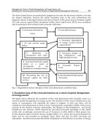
nonspecific protein adsorption. The performance of the sensor surface is supported by three
crucial factors (the capture agent, the surface chemistry, and the surface matrix). Fig. 6
shows a schematic diagram of an immobilization layer for SPR biosensing and the desired
characteristics of the three key factors.
Fig. 6. Schematic diagram of the immobilization layer (a) and desired characteristics for the
three key factors (b).
5.2 Basic design of the immobilization layer
5.2.1 Capture agent
In principle, it is difficult for SPR sensors to clearly distinguish the signal component of target
molecules from the background noise factors associated with non-specific absorption. First,
the capture agent must have the capability for specific recognition of the target molecules.
Proteins such as immunoglobulin (IgG), which are known as immune antibodies, are
frequently used as capture agents on account of their high specificity towards their target
antigens (Table 3, Besselink et al., 2004, Yang et al., 2005). Second, the selection of capture
agents with high affinity (equilibrium dissociation constant, in units of molar concentration;
K
D
< 10
-9
) is necessary to achieve high sensitivity. Recently, nucleic acid aptamers and
synthetic peptides have been developed as artificial antibodies with high specificity, high
New Perspectives in Biosensors Technology and Applications
92
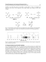
(1) Besselink et al., 2004; (2) Yang et al., 2005; (3) Kim et al., 2007; (4) Polonschii et al., 2010; (5) Katz et al.,
1995; (6) Torrance et al., 2006; (7) Huang et al., 2005; (8) Bonroy et al., 2006; (9) Johnsson et al., 1991; (10)
Lahiri et al, 1999; (11) Kwon et al., 2004; (12) Huang et al., 2002; (13) Lee et al., 2007; (14) Ha et al., 2007;
(15) Sigal et al., 1996; (16) Wazawa et al, 2006; (17) Masson et al., 2007; (18) Prime & Whitesides, 1993;
(19) Sigal et al., 1998; (20) Athey et al., 2005; (21) Shah et al., 2007; (22) Ishizuka-Katsura et al., 2008
Table 3. Examples of three factors (capture agent, surface chemistry, and surface matrix) that
are important in the formation of immobilization layers.
Highly Sensitive SPR Biosensor Based on Nanoimprinting Technology
93
affinity, and ease of size control, using the molecular evolutionary systematic evolution of
ligands by exponential enrichment (SELEX) process and phage display method. The obtained
artificial antibodies have been often used as capture agents in SPR (Katz et al., 1995,
Polonschii et al., 2010).
5.2.2 Surface chemistry
A coupling method involving activated N-hydroxysuccinimide esters is one of the most
commonly used surface chemistry techniques for anchoring capture molecules to a sensor
surface (Table 3, Johnsson et al., 1991, Lahiri et al, 1999). Since the target of this activated
ester is any amino group that is present on the protein molecule with high probability, this
coupling method is applicable to various capture agents used for SPR biosensing. To obtain
highly sensitive SPR signals, the orientation of capture agents should be considered. The
percentage of biochemically active capture agents that can interact with the target molecules
would be higher if the orientation of the capture agents on the sensor surface can be
improved. As a result of this improvement, the SPR response would be increased several
times. As a typical example, the surface chemistry to immobilize proteins via hexahistidine
tags (His-tag) has been used (Sigal et al., 1996). In recent years, mutated proteins, such as
functional fusion proteins, have been used for achieving oriented immobilization of capture
agents and simplification of the immobilization process (Terrettaz et al., 2002, Ha et al., 2007,
Park et al., 2009, Le Brun et al., 2011). Some mutated proteins are already on the market as
commercial layers (Athey et al., 2005).
5.2.3 Surface matrix
Polymers, polysaccharides, self-assembled monolayers, phospholipid and protein layers,
among other, have all been reported as surface matrices (Table 3). One of the most
important functions of the surface matrix in SPR biosensors is the suppression of non-
specific adsorption of contaminants to the sensor surface. For this purpose, the introduction
of oligo(ethylene glycol) molecules is highly effective (Prime & Whitesides, 1993, Sigal et al.,
1998). Moreover, it is also important to increase the binding capacity of the capture agents.
This factor, which determines the maximum signal variation of the SPR sensor, can control
the dynamic range of the biosensing. One example of a surface matrix that has been
successful in increasing the binding capacity of capture agents is the carboxymethylated
dextran matrix provided by GE Healthcare (Sweden). The carboxymethylated dextran
matrix provides a three-dimensional space with a thickness of 100 nm for target molecule
binding (Yang et al., 2005, Johnsson et al., 1991).
5.3 Preparation of an immobilization layer on the sensor surface
To accomplish highly sensitive SPR biosensing with a nanoimprinted sensor device, the
thickness of the probe layer using such as antibodies should be ~20 nm, as the sensing depth
is about 40–80 nm. For this purpose, we attempted to introduce self-assembling layers of
ORLA18 proteins (Orla Protein Technologies, UK) onto the sensor surface. The scaffold
structure of ORLA proteins is based on the stable structure of the beta-sheet and beta-barrel
mutated porin outer membrane protein (Omp) of Escherichia coli. It was firstly reported by
the research group of J.H. Lakey in 2002 that OmpF proteins can be immobilized directly on
a gold surface via thiol-gold bonds formed between the gold and the cysteine residues of the
Omp protein (Terrettaz et al., 2002). Moreover, the surface loops of the monomeric porin
OmpA can be replaced by anything from short peptides to larger protein domains. The
New Perspectives in Biosensors Technology and Applications
94
advanced ORLA protein (ORLA18) is designed to present precisely oriented antibody (IgG)-
binding domain structures (two Z-domains of protein A) as single layers with a thickness of
~10 nm on surfaces (Athey et al., 2005).
The surface preparation process of the ORLA18 protein layer is described below and shown
in Fig. 7.
1. Treatment of the gold surface on the nanoimprinted sensor device by injecting an
aqueous solution containing 1% (v/v), beta-mercaptoethanol.
2. Self assembly of the scaffold protein on the gold surface by injecting a 5 μM ORLA18
dissolved in ROG-8 buffer (Orla Protein Technologies).
3. Stabilization of the scaffold proteins and masking of the spaces between the proteins in
the monolayer using 1x filler solution (Orla Protein Technologies).
4. Antibody binding on the ORLA18 protein layers by injecting a 100 μg/mL antibody
dissolved in Tris-buffered saline (TBS; 10 mM Tris, pH 7.5, 150 mM NaCl) solution.
Fig. 7. Preparation process of an antibody-immobilization layer on the nanogroove sensor
surface.
6. SPR biosensing using the nanoimprinted sensor device
6.1 Development of the SPR biosensor system
As a prototype of the nanoimprinted SPR biosensor system, we developed a desktop model
for use in a laboratory (see Fig. 8). The dimensions of this model are W250mm × D250mm ×
H150mm, which is 10 times smaller than the commercialized SPR systems (Biacore-X, GE
Healthcare, US).
Fig. 9 shows the optical system employed inside the prototype. The white light from the
halogen lamp (LS1-LL, Ocean Optics Inc, US) is collimated and irradiated onto the sensor
Highly Sensitive SPR Biosensor Based on Nanoimprinting Technology
95
Fig. 8. Prototype of the nanoimprinted SPR biosensor system (a) and a schematic
representation of the sensor chip including a microchannel (b).
Fig. 9. Optical system inside the nanoimprinted SPR biosensor system.
surface through the objective lens (Plan N ×10, Olympus Co., Japan). The reflected light is
gathered by the object lens and split by the half prism (BS CUBE NON-POL VIS 47121,
Edmund Optics Inc., US) before reaching the spectrometer (USB4000, Ocean Optics Inc.,
USA). The resonant wavelength is analyzed by measuring the reflection spectrum data in
real time. The motorized linear XY stage (SGSP15-10, Sigma Koki Co., Ltd, Japan) is located
under the sensor device holder and is configured to enable multiple points on the sensor
chip to be monitored by programming the detection points in advance. A correct and stable
liquid flow control system is also very important. Several pumps, such as Veritas and
plunger pumps, were considered for use as a flow control system in the prototype. In the
New Perspectives in Biosensors Technology and Applications
96
end, an electro osmotic flow (EOF) pump was selected for use. The EOF pump (RP7SP,
Nano fusion Technology, Japan) is a very small-sized low-cost pump that provides a non-
pulsating flow.
We attached a flow sensor (ASL1430-24, Sensirion, US) downstream of the EOF pump. The
applied voltage to the EOF pump was determined under the feedback control of the flow
sensor to realize a small and stable flow.
6.2 Two-dimensional monitoring of antibody-binding on the ORLA18 surface
The ORLA18 protein was immobilized on the nanogroove sensor surface according to the
immobilization process shown in Fig. 7. To suppress non-specific adsorption, the remaining
area was blocked using an oligo(ethylene glycol) (OEG) self-assembled monolayer composed
of hydroxyl-terminated thiol (HS(CH
2
)
11
(OCH
2
CH
2
)
3
OH) (Dojindo, Japan) molecules. After
the docking of the ORLA18-immobilization gold substrate onto the sensor chip cassette (Fig.
8b), the SPR measurements were started. The two-dimensional variation of the SPR peak
wavelength shift on the nanogroove sensor surface was monitored by the nanoimprinted
SPR biosensor system of Fig. 8a. After the injection of 100 μg/ml rabbit polyclonal antibodies
and IgG (Monosan, Netherlands) dissolved in TBS (pH 7.5) buffer, the specific signal
corresponding to antibody-binding was confirmed in the ORLA18-immobilized area (Fig.
10, top left-hand corner of the immobilized area).
Fig. 10. 2D monitoring of the antibody-binding process on the ORLA18 surface using the
nanoimprinted SPR biosensor system.
6.3 Evaluation of the sensing depth on the nanoimprinted sensor device
To demonstrate that the sensing depth is confined to a much smaller region in this sensor,
we have attempted to compare the “bulk effect” between this sensor and the propagating
SPR. Undiluted fetal bovine serum (FBS) purchased from Japan Bioserum (Japan) was used
as a model of blood serum, which contains various proteins as background noise factors
(Fig. 11).
In the propagating SPR (Biacore-X, GE Healthcare Co.), whose sensing depth is several
hundred nanometers, the signal change caused by the undiluted FBS injection was about
8,000 RU, while the signal change from the binding of the antibody (IgG) dissolved in TBS
Highly Sensitive SPR Biosensor Based on Nanoimprinting Technology
97
(pH 7.5) buffer was 2,100 RU. In the nanoimprinted SPR sensor, the peak shift caused by the
undiluted FBS injection was 1.2 nm, while the peak shift caused by the binding of IgG
dissolved in TBS (pH 7.5) buffer was 3.5 nm. The signal-to-noise ratio (IgG binding/FBS
signal before washing) was 0.26 and 2.92 in the propagating SPR and the nanoimprinted
SPR, respectively (Fig. 11). This shows that the nanoimprinted SPR sensor is more than 10
times less subject to bulk effects, due, for example, to the other constituents of blood and
temperature fluctuations. This indicates that the sensing depth of the SPR is much smaller
than that of a conventional propagating SPR sensor. This will help fabricate small-sized
equipment in which temperature control is not necessary. Also, the washing protocol to
separate the signal caused by the binding of the target molecules from the signal caused by
the mixture of other floating biomolecules can be omitted. These advantages promise to lead
to the development of protein and point-of-care chips, which are expected to become
prevalent in diagnostic and healthcare applications.
Fig. 11. Comparison of the signal-to-noise ratios of the nanoimprinted SPR and propagating
SPR.
6.4 Detection of alpha-fetoprotein using the nanoimprinted SPR biosensor
Using the nanoimprinted SPR biosensor, we performed the quantitative detection of alpha-
fetoprotein (AFP), a tumor marker. The AFP concentration in healthy human serum is
approximately ~20 ng/ml, but its level increases markedly to over several hundred ng/ml
in patients with liver cancer (Teramura & Iwata, 2007). Currently, the cut-off-value of AFP
for clinical diagnosis is 200 ng/ml. Hence, the sensitive detection of the AFP using the
nanoimprinted SPR system must be useful in cancer diagnosis. For the highly sensitive
New Perspectives in Biosensors Technology and Applications
98
detection of AFP, an affinity purified rabbit polyclonal antibody (95% IgG) against human
AFP was purchased from Monosan (Netherlands). Pure human AFP (a single band on SDS-
PAGE) was obtained from Morinaga Institute of Biological Science (Japan). These AFP and
Anti-AFP were diluted in Hepes-buffered saline (10 mM HEPES, pH 7.4, 150 mM NaCl)
solution containing 0.005% (v/v) Surfactant P20 and 3 mM EDTA, which was used as a
running buffer in the flow cell of the SPR system.
At first, we attempted to detect the AFP molecules on the anti-AFP-immobilized ORLA18
surface under condition 1 in Fig. 12. However, degradation of the signal was observed in
low concentration. Then the detection limit of the nanoimprinted SPR biosensor was
estimated to be more than several hundred ng/mL (Fig. 13). To obtain a higher variation of
the SPR signal, we attempted to increase the efficiency of the antigen-antibody reaction by
five times higher flow rate (100 μl/min) and two times larger than the contact time (6 min)
of this reaction under condition 2. Fig. 14 shows the results of AFP detection at condition 2.
The error bar indicates three standard deviations of the base line signal after the injection of
a zero-concentration AFP solution. After several experiments, finally we realized that
reducing the diffusion time of AFP is essential. Under condition 1 (Fig. 12), the mass-
transport effect (Karlsson et al., 1991) can be larger because of the size of its flow cell.
Therefore, under condition 3, we decreased the flow cell size. In this condition, the signal
degradation at low concentration was observed under condition 1 to be significantly
improved (Fig. 13). Then we estimated that the detection limit of AFP by the nanoimprinted
SPR biosensor is ~20 ng/mL. This value already overcomes the cut-off value of 200 ng/mL
in a clinical diagnosis.
Fig. 12. Experimental conditions for AFP detection using the nanoimprinted SPR biosensor.
Highly Sensitive SPR Biosensor Based on Nanoimprinting Technology
99
Fig. 13. Standard curves for AFP detection at different experimental conditions.
Fig. 14. Results of AFP detection using the nanoimprinted SPR biosensor. The right graphs
show the expansion of the left graphs.
7. Present research - Development of palm-sized model -
Localized SPR has good potential to be used more widely as it can provide real-time,
quantitative and easy operation sensing without any labeling on the target molecules. For
various usages such as point-of-care testing, food analysis, and environmental tests, we have
developed a palm-sized prototype based on the nanoimprinted SPR biosensor.
Fig. 15 shows a picture of the prototype. The dimensions are W77mm × D52mm × H56mm
and its weight is only 240 g. The electric power used in this model is all supplied by the USB
cable connected to the PC. The sensor chip is inserted from the front side into the
equipment. The injection of the liquid sample through the micro channel is conducted by the
637.95
637.98
638.01
638.04
638.07
638.10
1700 1900 2100 2300 2500
Time [sec]
Peak wavelength [nm
]
637.95
638.05
638.15
638.25
638.35
638.45
1700 1900 2100 2300 2500
Time [sec]
Peak wavelength [nm
]
2.37 μg/mL
0.47 μg/mL
0.12 μg/mL
0.02 μg/mL
0 μg/mL
AFP
AFP
[nm]
[nm]
0.02μg/mL
0.12μg/mL
0.47μg/mL2.37μg/mL
0μg/mL
(±3SD)
637.95
637.98
638.01
638.04
638.07
638.10
1700 1900 2100 2300 2500
Time [sec]
Peak wavelength [nm
]
637.95
638.05
638.15
638.25
638.35
638.45
1700 1900 2100 2300 2500
Time [sec]
Peak wavelength [nm
]
2.37 μg/mL
0.47 μg/mL
0.12 μg/mL
0.02 μg/mL
0 μg/mL
AFP
AFP
[nm]
[nm]
[nm]
[nm]
[nm]
[nm]
0.02μg/mL
0.12μg/mL
0.47μg/mL2.37μg/mL
0μg/mL
(±3SD)
0μg/mL
(±3SD)
New Perspectives in Biosensors Technology and Applications
100
Fig. 15. Palm-sized model of the nanoimprinted SPR biosensor system (a) and the sensor
chip including a micro channel (b).
syringe pressure. Fig. 16 shows the optical system of this model. A red laser (LM10-650,
Kyoritsu-Electric Corporation, Japan) is used as a light source and the change of the
reflection power is detected in this model. The light from the light source is split into four
beams and four spot areas, which are detected at once. And the reflected light is detected by
the photo-diode (S8745-01, Hamamatsu Photonics, Japan).
Fig. 16. Optical system inside the palm-sized nanoimprinted SPR biosensor.
To demonstrate the detection of biomolecules by using this model, we have attempted to
detect avidin protein. The sensor surface is previously modified by the biotinylated alkyl
thiol (BAT) layer according to the protocol below.
1. Biotin-terminated tri(ethylene glycol)hexadecanethiol (OEG-BAT) (Assemblon, USA)
and 11-hydroxy-1-undecanethiol (HUT) (Dojindo, Japan) are solved in 99.5% ethanol.
2. The sensor chip was immediately immersed in the ethanol solution containing 0.95 mM
OEG-BAT and 0.05 mM HUT.
3. The sensor chip is rinsed with an ethanol solution after 10 minutes of immersion.
4. The sensor chip is dried under a stream of nitrogen.
Highly Sensitive SPR Biosensor Based on Nanoimprinting Technology
101
5. Inject 0.1 mg/mL NeutrAvidin (Thermo Scientific, USA) dissolved in Hepes-buffered
saline (10 mM HEPES, pH 7.4, 150 mM NaCl) solution containing 0.005% (v/v)
Surfactant P20 and 3 mM EDTA.
The result is shown in Fig. 17. NeutrAvidin was injected at about t=50 sec. As a result, the
reflection change of 6% was observed in this system. This result indicates that the detection
of biomolecules can be realized by this small and simple model.
Fig. 17. Avidin protein detection using the palm-sized nanoimprinted SPR biosensor.
8. Conclusions
In this study, we accomplished a 10-times-higher signal-to-noise ratio measurement than
conventional SPR using an SPR biosensor based on a gold nanogroove surface. Notably,
using nanoimprinting technology, we have developed metal nanopatterns on a sensor
surface with a process that has high reproducibility and throughput. Furthermore, the size
of the prototype based on this detection principle was 10 times smaller than in
commercialized SPR systems. Using the SPR prototype, we accomplished quantitative AFP
detection as low as ~20 ng/mL. For this, 1) the smaller thickness of the probe layer and 2)
the immobilization of antibodies on the sensor surface in a well-oriented manner were
essential. A self-assembled fusion protein, the ORLA18 layer, was useful for accomplishing
this purpose. Decreasing the size of the flow cell to reduce the diffusion time of the AFP was
also very important. To the best of our knowledge, the detection limit of 20 ng/mL is the
highest sensitivity achieved for direct detection of AFP using an SPR biosensor.
Recently, we were also successful in detecting AFP of about 100 pg/ml with an enhanced
assay method by stepwise application of a biotinylated secondary antibody and
streptavidin-bound colloidal gold. This suffices as a practical way to measure other
important biomarkers such as prostate-specific antigen and carcinoembryonic antigen. Thus,
the effectiveness of our SPR sensor has been demonstrated by the achievement of a high
signal-to-noise ratio and highly sensitive detection of tumor marker protein. Our
nanoimprinting technology-based SPR biosensor technology will have various useful
applications, such as for medical diagnoses, environmental monitoring, and in the food
industry.
New Perspectives in Biosensors Technology and Applications
102
9. Acknowledgments
We are grateful to Dr. Tetsuichi Wazawa (Graduate School of Engineering, Tohoku
University) for critical reading of the abstract. This work was supported by the Core
Research for Evolutional Science and Technology (CREST) project of the Japan Science and
Technology Agency (JST).
10. References
Athey, D.; Shah, D.S.H.; Phillips, S.R.; Lakey, J.H. (2005). A manufacturable surface-biology
platform for nano applications; cell culture, analyte detection, diagnostics sensors.
Ind. biotechnol., 1, 185-189
Besselink, G.A.J.; Kooyman, R.P.H.; van Os, P.J.H.J.; Engbers, G.H.M.; Schasfoort, R.B.M.
(2004). Signal amplification on planar and gel-type sensor surfaces in surface
plasmon resonance-based detection of prostate-specific antigen. Anal. Biochem., 333,
165–173
Bonroy, K.; Frederix, F.; Reekmans, G.; Dewolf, E.; Palma, R.D.; Borghs, G.; Declerck, P.;
Goddeeris, B. (2006). Comparison of random and oriented immobilisation of
antibody fragments on mixed self-assembled monolayers. J. Immunol. Methods, 312,
167–181
Chou, S.Y.; Krauss, P.R.; Renstrom, P. J. (1995). Imprint of sub-25 nm vias and trenches in
polymers. Appl. Phys. Lett., 67, 3114-3116
Dostálek, J. & Homola, J. (2006). SPR biosensors for environmental monitoring. Springer Ser.
Chem. Sens. Biosens., Vol. 4, 191–206, ISBN 978-3-540-33918-2, Springer Berlin
Heidelberg New York
Félidj, N.; Aubard, J.; Lévi, G. Krenn, J.R.; Salemo, M.; Schider, G.; Lamprecht, B.; Leitner, A.;
Aussenegg, F.R.; (2002). Controlling the optical response of regular arrays of gold
particles for surface-enhanced Raman scattering. Phys. Rev. B, 65, 075419 (1-9)
Ha, T.H.; Jung, S.O.; Lee, J.M.; Lee, K.Y.; Lee, Y.; Park, J.S.; Chung, B.H. (2007). Oriented
immobilization of antibodies with GST-fused multiple Fc-specific B-domains on a
gold surface. Anal. Chem., 79, 546–556
Homola, J.; Yee, S. S.; Gauglitz, G. (1999). Surface plasmon resonance sensors: review. Sens.
Actuators B, 54, 3–15
Homola, J. (2003). Present and future of surface plasmon resonance biosensors. Anal. Bioanal.
Chem., 377, 528–539
Huang, H.; Huang, S.; Yuan, S.; Qu, C.; Chen, Y.; Xu, Z., Liao, B.; Zeng, Y.; Paul K. Chu, P. K.
(2011). High-sensitivity biosensors fabricated by tailoring the localized surface
plasmon resonance property of core–shell gold nanorods. Anal. Chimi. Acta, 683,
242–247
Huang, L.; Reekmans, G.; Saerens, D.; Friedt, J M.; Frederix, F.; Francis, L.; Muyldermans,
S.; Campitelli, A.; Hoof, C.V. (2005). Prostate-specific antigen immunosensing
based on mixed self-assembled monolayers, camel antibodies, and colloidal gold
enhanced sandwich assays. Biosens. Bioelectron., 21, 483–490
Huang, N P.; Vörös, J.; De Paul, S.M.; Textor, M.; Spencer, N.D. (2002). Biotin-derivatized
poly(L-lysine)-g-poly(ethylene glycol): a novel polymeric interface for bioaffinity
sensing. Langmuir, 18, 220–230
Highly Sensitive SPR Biosensor Based on Nanoimprinting Technology
103
Ishizuka-Katsura, Y.; Wazawa, T.; Ban, T.; K. Morigaki, K.; Aoyama, S. (2008). Biotin-
containing phospholipid vesicle layer formed on self-assembled monolayer of a
saccharide-terminated alkyl disulfide for surface plasmon resonance biosensing. J.
Biosci. Bioeng., 105, 527–535
Johnsson, B.; Löfås, S.; Lindquist, G. (1991). Immobilization of proteins to a
carboxymethyldextran-modified gold surface for biospecific interaction analysis in
surface plasmon resonance sensors. Anal. Biochem., 198, 268–277
Karlsson, R.; Michaelsson, A.; Mattsson, L. (1991). Kinetic analysis of monoclonal antibody-
antigen interactions with a new biosensor based analytical system. J. Immunol.
Methods, 145, 229–240
Katz, B.A.; Cass, R.T.; Liu, B.; Arze, R.; Collins, N. (1995). Topochemical catalysis achieved
by structure-based ligand design. J. Biol. Chem., 270, 31210–31218
Kim, S.J.; Gobi, K.V.; Iwasaka, H.; Tanaka, H.; Miura, N. (2007). Novel miniature SPR
immunosensor equipped with all-in-one multi-microchannel sensor chip for
detecting low-molecular-weight analytes. Biosens. Bioelectron. 23, 701–707
Kretschmann, E. & Raether, H. (1968). Radiative decay of non-radiative surface plasmons
excited by light. Z. Naturforsch., 23A, 2135-2136
Kwon, Y.; Han, Z.; Karatan, E.; Mrksich, M.; Kay, B.K. (2004). Antibody arrays prepared by
cutinase-mediated immobilization on self-assembled monolayers. Anal. Chem., 76,
5713–5720
Ladd, J.; Taylor, A.; Jiang, S. (2006). SPR biosensors for food safety. Springer Ser. Chem. Sens.
Biosens., Vol. 4, 207–227, ISBN 978-3-540-33918-2, Springer Berlin Heidelberg New
York
Lahiri, J.; Isaacs, L.; Tien, J.; Whitesides, G.M. (1999). A strategy for the generation of
surfaces presenting ligands for studies of binding based on active aster as a
common reactive intermediate: a surface plasmon resonance study. Anal. Chem. 71,
777–790
Le Brun, A.P.; Holt, S.A.; Shah, D.S.H.; Majkrzak, C.F.; Lakey, J.H. (2011). The structural
orientation of antibody layers bound to engineered biosensor surfaces. Biomaterials
32, 3303–3311
Lee, J.M.; Park, H.K.; Jung, Y.; Kim, J.K.; Jung, S.O.; Chung, B.H. (2007). Direct
immobilization of protein G variants with various numbers of cysteine residues on
a gold surface. Anal. Chem., 79, 2680–2687
Masson, J F.; Battaglia, T.M.; Khairallah, P.; Beaudoin, S.; Booksh, K.S. (2007). Quantitative
measurement of cardiac markers in undiluted serum. Anal. Chem., 79, 612-619
Nath, N. & Chilkoti, A. (2004). Label-free biosensing by surface plasmon resonance of
nanoparticles on Glass: optimization of nanoparticle size. Anal. Chem., 76, 5370-5378
Park, T.J.; Hyun, M.S.; Lee, H.J.; Lee, S.Y.; Ko, S. (2009). A self-assembled fusion protein-
based surface plasmon resonance biosensor for rapid diagnosis of severe acute
respiratory syndrome. Talanta, 79, 295–301
Polonschii, C.; David, S.; Tombelli, S.; Mascini, M.; Gheorghiu, M. (2010). A novel low-cost
and easy to develop functionalization platform. Case study: Aptamer-based
detection of thrombin by surface plasmon resonance. Talanta, 80, 2157–2164
Prime, K.L.; Whitesides, G.M. (1993). Adsorption of proteins onto surfaces containing end-
attached oligo(ethylene oxide): a model system using self-assembled monolayers. J.
Am. Chem. Soc., 115, 10714–10721
New Perspectives in Biosensors Technology and Applications
104
Sigal, G.B.; Bamdad, C.; Barberis, A.; Strominger, J.; Whitesides, G.M. (1996). A self-
assembled monolayer for the binding and study of histidine-tagged proteins by
surface plasmon resonance. Anal. Chem., 68, 490–497
Sigal, G.B.; Mrksich, M.; Whitesides, G.M. (1998). Effect of surface wettability on the
adsorption of proteins and detergents. J. Am. Chem. Soc., 120, 3464–3473
Stenberg, E.; Persson, B.; Roos, H.; Urbaniczky, C. (1991). Quantitative determination of
surface concentration of protein with surface plasmon resonance using
radiolabelled proteins. J. Colloid Interface Sci., 143, 2, 513-526
Shah, D.S.; Thomas, M.B.; Phillips, S.; Cisneros, D.A.; Le Brun, A.P.; Holt, S.A.; Lakey, J.H.
(2007). Self-assembling layers created by membrane proteins on gold. Biochem. Soc.
Trans., 35, 522-526
Teramura, Y. & Iwata, H. (2007). Label-free immunosensing for α-fetoprotein in human
plasma using surface plasmon resonance. Anal. Biochem., 365, 201–207
Terrettaz, S.; Ulrich, W P.; Vogel, H.; Hong, Q.; Dover, L.G.; Lakey, J.H. (2002). Stable self-
assembly of a protein engineering scaffold on gold surfaces. Protein Sci., 11, 1917–
1925
Torrance, L.; Ziegler, A.; Pittman, H.; Paterson, M.; Toth, R.; Eggleston, I. (2006). Oriented
immobilisation of engineered single-chain antibodies to develop biosensors for
virus detection. J. Vir. Methods, 134, 164–170
Vaisocherová, H. & Homola, J. (2006). SPR biosensors for medical diagnostics. Springer Ser.
Chem. Sens. Biosens., Vol. 4, 229–247, ISBN 978-3-540-33918-2, Springer Berlin
Heidelberg New York
Wazawa, T.; Ishizuka-Katsura, Y.; Nishikawa, S.; Iwane, A.H.; Aoyama, S. (2006). Grafting
of poly(ethylene glycol) onto poly(acrylic acid)-coated glass for a protein-resistant
surface. Anal. Chem., 78, 2549–2556
Yang, C Y.; Brooks, E.; Li, Y.; Denny, P.; Ho, C M.; Qi, F.; Shi, W.; Wolinsky, L.; Wu, B.;
Wong, D.T.; Montemagno, C.D. (2005). Detection of picomolar levels of interleukin-
8 in human saliva by SPR. Lab Chip, 5, 1017–1023
0
Numerical Optimization of Plasmonic Biosensors
Dominique Barchiesi
GAMMA3 project (INRIA-UTT)- Charles Delaunay Institute - University of technology of
Troyes 12 rue Marie Curie - BP 2060 - 10010 TROYES cedex
France
1. Introduction
Since the engineering control of the deposition of nanometric gold plates on substrates
the Surface Plasmon Resonance (SPR) based sensor has become one of the most successful
label-free and commercially developed optical sensor, with applications to biology (Hoaa
et al., 2007; Kolomenskii et al., 1997; Kretschman & Raether, 1968; Lecaruyer et al., 2006).
This technique is currently employed in biomolecular engineering, drug design, monoclonal
antibody characterization, virus-protein i n teraction, environmental pollutants detection,
among other interesting problems. The basic principle of such transducer is the measurement
of the sudden absorbtion of light by the thin metallic layer, under particular illumination
conditions (p-polarization) and a specific angle of incidence of the illumination (Barchiesi,
Kremer, Mai & Grosges, 2008; Barchiesi, Macías, Belmar-Letellier, Van Labeke, Lamy de la
Chapelle, Toury, Kremer, Moreau & G rosges, 2008; Kre tschman & Raether, 1968), leading to
a highly sensitive device (Kolomenskii et al., 1997; Lecaruyer et al., 2006). The conditions of
such absorption are linked to the plasmon resonance in metallic structure, and therefore a tiny
change of the optical properties of medium above the gold plate, produces an angular shift of
this abs orption, due to the detuning of the resonance. The s ensing principle relies therefore
on the shift of the plasmonic resonance caused by the surrounding dielectric environmental
change in a binding event.
The Plasmonic biosensors use the property of resonance between an illumination and the
metallic part of the sensor. This resonance is used to increase the sensitivity of the biosensor
and the threshold of detection. Actually, a given set of parameters of the biosensor can lead
to a maximum of the absorption of the incoming light. A slight change of its immediate
environment (presence/Absence of biomoecules to be detected) produces a strong change of
the detected light due to the detuning of the resonance. This property is also used in cancer
therapy or imaging, through metallic nanoparticles or nanoshells (Grosges, Barchiesi, Toury &
Gréhan, 2008). The design of specific shapes for nanoparticles can help to tune the resonance
for specific applications (Billot et al., 2006).
In the case of planar SPR (Surface Plasmon Resonance) biosensors, the reflected intensity
vanishes for a specific angle of incidence. The illumination is almost totally coupled in the
metal layer. A tiny change of the optical index of the upper medium, due to the presence
of biomolecules, produces a measurable shift of this minimum. Therefore, to improve the
efficiency of the sensor, the material and geometrical characteristics of the materials involved
in the biosensor must be adjusted correctly. Surprisingly, the optimization of such structures
has rarely been addressed (Ekgasit et al., 2005; Ko lomenskii et al., 1997; Lecaruyer et al.,
5