Photodiodes World Activities in 2011 Part 10 ppt
Bạn đang xem bản rút gọn của tài liệu. Xem và tải ngay bản đầy đủ của tài liệu tại đây (4.73 MB, 30 trang )
Geiger Avalanche Photodiodes (G-APDs) and Their Characterization
261
Different configurations have been investigated and some other measurements were carried
out with the same results.
9. PDE measurements
Only a fraction of the photons impinging on the sensor will actually trigger an avalanche to
produce a detectable signal (Piemonte 2006). Essentially three effects influence a G-APD
response efficiency:
1. physical (reflection/absorption by passive layers, material), that is the so called net
quantum efficiency (QE);
2. electrical (photon arrival in regions where the electric field is not suitable for triggering
the avalanche), that represents in practice the probability that an event occurs and
generally is named Trigger probability (TP).
3. geometrical (dead areas between cells), and is generally known as fill factor (FF);
The overall efficiency of the sensor, as for the single element, is generally named Photo
Detection Efficiency (PDE), and it relates the real number of impinging photons to the
measured effect (photo-electrons) and is the product of the three above mentioned effects:
PDE QE TP FF
(1)
In the following sections the reader will be introduced into an important aspect to be
considered when the detector PDE has to be evaluated with high accuracy. The requirement
to have a well defined methodology, taking care, not only on the precision of all involved
instruments, but also on the implemented procedure, is crucial to obtain precise
measurements. Here we will demonstrate how the extra noise sources, optical cross-talk and
afterpulse, may influence the PDE measurements. In fact, to measure the detector PDE
essentially two approaches can be used:
1.
one consisting in measuring the generated charges considered as current, that we name:
“Photocurrent” method,
2.
and another consisting in counting each produced event, that we name “Photon
counting” method.
The PDE measurements for both methods have been carried out by using the optical setup
sketched in Fig. 14 and the electronic setup sketched in Fig. 15.
The first consideration, to obtain accurate measurements, is addressed to the different
dimensions of both detectors, the G-APD and the reference photodiode. In fact, while the
tested devices have dimensions of squared millimeter, the reference detector have a
sensitive area of 1 cm
2
(leakage current less than 1pA), thus in the “Photon counting” case,
we have to adjust the photon flux level (from about 10
5
to about 10
7
phs·mm
-2
·s
-1
) in such a
way that the reference detector was still sensitive and the detectors were safely in the single
photon regime with negligible pile-up.
9.1 Photocurrent method
The “Photocurrent” method consists in comparing the photocurrent of the characterized
detectors with respect to that of the NIST calibrated reference photodiode. In this case the
setup apparatus of Fig. 15 is simplified by substituting the amplifier, the discriminator and
the counter with an ammeter. In practice we have two identical systems, one for the tested
and one for the reference detector, and simply we have to do measurements of the photo-
generated current in both sensors. The following formula explains how the method works:
Photodiodes - World Activities in 2011
262
1
Det DarkDet) PhD DarkPhD PhD PhD Det
PDE [ I I / I I G PDE A /A
(2)
Where I
Det
-I
DarkDet
is the current measured in the tested detector, I
PhD
-I
DarkPhD
is the current
measured in the calibrated photodiode, G is the gain (N
el
/pe-), PDE
PhD
is the PDE of the
calibrated photodiode and A
PhD
/A
Det
is the detectors area ratio.
We operated the detectors at room temperature and measured the PDE of the STM SiPM
biased
at 32.5V (10% OV) and that of the 100 and 400 cells MPPC biased respectively at
69.8V (~2% OV) and at 69.4V (~2% OV). Using the G values obtained with our
measurements, we found unreasonable PDE values (higher than expected). Thus, the sole
alternative we had was using the G values given by the manufacturers. Despite a sort of
uncertainty of the method, due to the fact that we have to rely on manufacturer’s
measurements accuracy, we decide to compute the PDE. We made the PDE computation
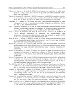
only on the two Hamamatsu MPPCs. The obtained values are plotted in Fig. 19.
As expected the PDE of the 100 cells MPPC at 450 nm has a peak of about 50%, while the 400
cells MPPC has a peak of 30% because of the different fill factor. Now we have to
investigate if these results are realistic or the noise contribution has to be taken into account
and avoided as much as possible. It is clear that a technique, based on photocurrent
measurements, is unable to discriminate from extra-generated pulses, i.e. afterpulses and
optical cross-talk pulses, and thus two questions rise:
Can we include in each PDE value an amount of pulses that is considered “noise”?
Can we say that the obtained PDE values are accurate?
Fig. 19. PDE plots of the two Hamamatsu G-APDs: the 100-cells MPPC and 400-cells MPPC
by using the “Photocurrent” method.
If it is impossible to discriminate the extra pulses with respect to the real signal, probably
the photocurrent method may lead to overestimated PDE values, and will be better to use
another method that can discriminate the real photo-events from extra pulses.
9.2 Photon counting method
The “Photon counting” method is based on measuring the G-APDs count rate due to the
real photo-events and comparing it to the photocurrent measured by the ammeter converted
into number of electrons per second. The formula of this method is:
Geiger Avalanche Photodiodes (G-APDs) and Their Characterization
263
Det DarkDet PhD DarkPhD PhD PhD Det
PDE CR CR / I I PDE e A /A
(3)
Where CR
Det
-CR
DarkDet
is the measured count rate, e
-
is the electron charge and I
PhD
-I
DarkPhD
,
PDE
PhD,
A
PhD
/A
Det
are the same as on formula (2).
By using this method the afterpulse and the cross-talk can be characterized and taken into
account in the right way, in fact we can set the threshold at a convenient value and can
acquire the signal at a selected time (by varying the time length of the digital output pulse
from the discriminator) away from the eventual afterpulse contribution. The first step to
carry out the PDE measurement is to analyse the count rates as a function of the threshold.
As seen in Fig.16 of section 7, a threshold equivalent to 0.5 photons can be selected as this
value is in a safe plateau region. In the tested devices we found that the afterpulse
probability is not appreciable after ≈100ns and thus we settled the output logic signal
duration from the discriminator longer than this value. We counted the number of pulses
per unit time both in dark conditions (~ 600 KCnts/s for the 100-cells MPPC, ~ 500 KCnts/s
for the 400-cells MPPC, ~ 500 KCnts/s for the 100-cells STMicroelectronics device) and with
monochromatic light conditions (photon signal ranging from ~ 100 KCnts/s to ~ 500
KCnts/s), recording at the same time the light level seen by the reference detector, for
several wavelengths. We also carefully tuned the light intensity to keep at negligible levels
the pile-up probability. As an example here the analysis made on both the
STMicroelectronics and Hamamatsu 100-cells G-APDs is presented. For both devices we
evaluated the PDE by measuring all the contributing signals, noise and photons with two
gate logic signal durations and accounted for the dead time. For the STMicroelectronics we
selected the duration of 50 ns and 500 ns and the resulting PDE plots are shown in Fig. 20,
while for the Hamamatsu device we selected the duration of 100 ns and 1000 ns and the
resulting plots are shown in Fig. 21. The unappreciable difference between the two sets of
Fig. 20. PDE of the 100-cells SiPM STMicroelectronics device biased at 32.5 V, measured and
reconstructed with our method using logic signal durations of 50ns and 500ns respectively.
As can be noted the difference between the two sets of measurements is unappreciable,
meaning that the afterpulse effect not influence each measure.
Photodiodes - World Activities in 2011
264
measurements, for both G-APDs, demonstrates that the afterpulses are not influent on each
measure and strongly supports the correctness of this method.
Fig. 21. PDE measured for the Hamamatsu 100 cells biased at 69.4 V using gate signals of
100ns and 1000ns. As can be noted the difference between the two sets of measurements,
also in this case, is within the error-bar, meaning that in these measurements the afterpulses
are not a problem.
As can be noted from Figs. 20 and 21 the PDE plots of the two G-APDs are quite different
specially in the 350 ÷ 450 nm spectral region. This is essentially due to the different
technology adopted by the two manufacturers. In the case of Hamamatsu device (that uses
the so called p-on-n junction technology) the photons impinging in the first layers of
material are absorbed more efficiently than those arriving in the same region of the
STMicroelectronics device (that uses the so called n-on-p junction technology).
10. Comparison between “photocurrent” and “photon counting” methods
In order to compare the photocurrent method with the photon counting one, we have plotted
in Fig. 22 the PDEs obtained with the two methods for the Hamamatsu MPPC 100-cells.
As can be seen from Fig. 22, the PDE obtained with the photocurrent method is
systematically higher than that measured with the photon-counting mode in all the spectral
range. Moreover the error-bars associated to the PDE values are very low (not exciding the
point itself) demonstrating the high accuracy of measurements and the real difference
between the two PDE curves. Unequivocally, Fig. 22 shows that each PDE value obtained
using the photocurrent method doubles that of the photon counting operating mode. We,
thus have to conclude that the extra noise pulses heavily influence the detector PDE
evaluation. A different way that allows us to better clarify the real difference between the
two methods, is to represent the two PDE plots as in Fig. 23 where the left axis is used to
represent the PDE values obtained with the photocurrent method and the right axis refers to
the PDE values obtained in photon counting mode. In order to better understand this figure,
it is extremely important to note that the right axis scale (that refers to the photon counting
mode) is exactly half of that of the principal axis (that refers to the photocurrent mode).
Geiger Avalanche Photodiodes (G-APDs) and Their Characterization
265
From the Fig. 23 we can observe that even if the two PDE plots came from different
methods, there’s an amazing over-position between the two plots. This demonstrates that at
each wavelength the PDE values obtained with the two different methods can be related
between themselves, and by noting the scale of the left axis respect to right axis, the relation
is that each value almost doubles the corresponding. And then, definitively, we can
conclude that the PDE of this device in photon counting mode is half of that in which we
can’t avoid the extra pulses contribute.
Fig. 22. PDE measurements for a 100-cells Hamamatsu MPPC. The solid line refers to the
PDE obtained with the photocurrent
method, while the dashed line refers to the PDE
obtained with the photon counting technique.
Unequivocally the PDE values obtained using
the photocurrent method doubles that of the photon counting.
Fig. 23. “Photocurrent” method versus “Photon counting” method. The solid line refers to
the PDE (values on the left axis) obtained with the photocurrent method, while the dashed
line refers to the PDE (values on the right axis) obtained in photon counting regime. The
right axis scale is half of that that refers to the PDE obtained with the photocurrent method.
Photodiodes - World Activities in 2011
266
11. Conclusion
In this chapter, a detailed description of a particular kind of photodiodes able to work in
Geiger avalanche mode recently named G-APDs has been described. Starting from a
description of the relevant characteristics of the single G-APD we extended to describing the
multi-element G-APD as a photodetector constituted by hundreds/thousands of single
elements. By discussing in detail the manufacturing technology and the relevant electro-
optical characteristics of these devices, we tried to give an idea of the real achievable
performance in application such as Nuclear Physics or Astrophysics. The characterisation in
terms of noise, and Photon-Detection Efficiency (PDE) has been treated in great detail for
both kind of devices together with the adopted experimental setups. Some measurements
and results on various single element G-APDs and multi-element G-APDs, manufactured by
various companies have been also presented. Finally, emphasis has been given to the
developed technique to obtain very accurate PDE measurements based on single photon
counting with subtraction of dark noise, and avoiding as much as possible cross-talk and
afterpulses. We discussed and compared the two commonly used techniques to measure the
PDE, the photocurrent consisting in measuring the photo-generated current in the detector,
and the photon counting consisting in measuring the signal considered as number of
photons. The comparison between the two methods has pointed out the vulnerability of the
photocurrent method that gives PDE values overestimated with respect to those from
photon counting. We demonstrated unequivocally that this is essentially due to the fact that
the photocurrent technique cannot discriminate the afterpulse and the cross-talk effects. On
the contrary, the photon counting method allows to characterize and accurately discriminate
the two noise effects providing PDE values quite close to the real ones, but needs to operate
in appropriate signal conditions, in fact very fast events can be lost and the total counted
events can be lower than those expected. Then we can conclude that the photon counting is
a method well suited for PDE measurements because it definitely deals with true photons,
reducing as much as possible the contribution of extra pulses.
12. References
S. Billotta et al., JMO, Vol. 56, 273–283 (2009).
G. Bonanno et al., SPIE Proceedings, 2808, p.242 (1996).
R.G. Brown et al., Appl. Opt. 26, 2383 (1987).
P. Buzhan et al
., Nucl. Instrum. Methods Phys. Res. A, Accel. Spectrom. Detect. Assoc.
Equip., vol. 504, no. 1–3, 48–52, (2003).
S. Cova et al., Appl. Opt. 35, 1956 (1996).
S. Cova et al., Rev. Sci. Instrum. 52, 408 (1981).
B. Dolgoshein et al., Nuclear Instruments and Methods in Physics Research A 563, 368–376 (2006)
P. Finocchiaro et al., IEEE Trans. on Electron Devices, Vol. 55, no. 10, 2757-2764 (2008).
P. Finocchiaro et al., IEEE Trans. on Nucl. Sci., Vol. 56 no. 3, 1033-1041 (2009).
M. Ghioni et al
., Rev. Sci. Instrum., vol. 67, no. 10, 3440–3448, (1996).
M. Ghioni and G. Ripamonti, Rev. Sci. Instrum. 62 163 (1991).
V. D. Kovaltchouk et al., Nucl. Instrum. Methods Phys. Res. A,
Accel. Spectrom. Detect.
Assoc. Equip., vol. 538, no. 1–3, 408–415 (2005).
M. Mazzillo et al., Nucl. Instrum. Methods A Vol. 591, 367–373 (2008).
M. Mazzillo et al., Sens. Actuators A, Vol.138, 306–312 (2007).
C. L. Melcher and J. S. Schweitzer, IEEE Trans. Nucl. Sci., vol. 39, no. 4, 502–505 (1992).
C. Piemonte, Nucl. Instrum. Methods Phys. Res. A, Accel. Spectrom. Detect. Assoc. Equip.,
vol. 568, no. 1, 224–232 (2006).
F Zappa et al., JMO Vol. 54, 163-189 (2007).
12
Design of High Quantum Efficiency and High
Resolution, Si/SiGe Avalanche Photodiode
Focal Plane Arrays Using Novel, Back-
Illuminated, Silicon-on-Sapphire
Substrates
Alvin G. Stern
AG STERN, LLC, Newton, MA 02467
USA
1. Introduction
The design and development of large scale, high quantum efficiency and high resolution
silicon and silicon-germanium (Si/SiGe) avalanche photodiode (APD) focal plane arrays
(FPAs) is an active topic of research due to the wide range of scientific, medical and
industrial applications for such high sensitivity imagers. Avalanche photodiodes can attain
single photon sensitive operation due the large internal device gain that compensates and
can fully eliminate the electronic readout noise normally limiting the sensitivity of solid-
state detector devices, hence their importance in electronic imaging. Large, wafer scale
arrays of ultra sensitive, high resolution silicon and silicon-germanium avalanche
photodiodes have not been developed yet, primarily due to the increased fabrication
complexity of such detector devices and arrays compared to the more common, non-
avalanching detectors such as CCDs and CMOS-APS devices. One major fabrication
challenge for avalanche type detectors is the requirement of providing effective optical
isolation between adjacent detectors in an array since the avalanche gain process produces
photons that could create false detection events in neighboring pixels and thereby increase
the noise. Providing effective optical crosstalk isolation becomes more difficult for higher
resolution arrays. While it is common for CCD arrays to have a pixel pitch between 12-30
µm and for CMOS-APS devices to have pixel pitch below 10 µm, it becomes more
challenging to architect arrays of avalanche photodiodes for example, having such a small
pitch due to optical crosstalk. The second major fabrication challenge for linear mode
avalanche type detectors, especially critical in arrays is the detector gain uniformity.
Detector gain uniformity is a critical performance parameter since an increase in gain excess
noise will make the detector arrays unsuitable for precision metrology applications. As
solid-state avalanche detectors are made smaller, it becomes more difficult to control the
gain excess noise due to smaller area multiplication regions where the effects from slight
variations in doping profiles and electric fields produce greater gain variability compared to
larger area detectors.
Photodiodes - World Activities in 2011
268
In this chapter, design aspects of a novel, back-illuminated silicon-on-sapphire material
system are presented and compared to present substrate technologies to illustrate the
capability of the novel substrates in solving optical crosstalk and detector gain uniformity
fabrication challenges for producing high quantum efficiency and high resolution wafer
scale arrays of Si/SiGe APD detector arrays. The novel substrate design incorporates a
single crystal, epitaxially grown aluminum nitride antireflective layer between sapphire and
silicon to improve optical transmittance into the silicon from sapphire. A
λ
/4-MgF
2
antireflective layer deposited on the backside of the sapphire improves optical transmittance
from the ambient into the sapphire. The high transmittance, back-illuminated silicon-(AlN)-
sapphire substrates represent an enabling technology for producing radiation tolerant, high
resolution, wafer scale arrays of solid-state light detectors. (Stern & Cole, 2008) The Si and
SiGe solid-state avalanche photodiodes for example, could be produced in highly uniform
wafer scale arrays by liquid crystallographic etching of mesa pixels due to sapphire acting as
a natural etch stopping layer. Mesa detectors and arrays would retain high quantum
efficiency and sensitive-area-fill-factor respectively, due to light focusing monolithic
sapphire microlenses beneath each pixel. The space between mesa detectors could be filled
with metal to form a low-resistance contact across the array and also block direct pixel-to-
pixel optical crosstalk. The closely integrated monolithic sapphire microlenses also help to
address detector gain uniformity by focusing optical k-vectors directly into the active
multiplication region of the avalanche photodiodes, thereby helping to improve the gain
uniformity of the detectors and arrays. Coupled with recent advances in dual linear and
Geiger-mode avalanche detector design, the novel substrates will enable wide dynamic
range focal plane arrays operating near room temperature, capable of imaging over the full
range of natural illumination conditions from AM 0 in space to a cloudy moonless night.
(Stern & Cole, 2010)
The novel, back-illuminated silicon-on-(AlN)-sapphire substrates offer the possibility of
solving the fabrication challenges currently limiting the low cost availability of highly
sensitive, wide dynamic range Si and SiGe avalanche photodiode arrays, including direct
pixel-to-pixel optical crosstalk and detector gain uniformity. There still exists however, the
phenomenon of indirect optical crosstalk by multiple reflections in the finite thickness, 50
µm thick sapphire substrate. It will be shown through detailed calculations and analysis
means that indirect optical crosstalk through the 50 µm thick sapphire substrate although
present, will not prevent high resolution, 27 µm pixel pitch Si/SiGe APD detector arrays
operating in the highest (Geiger-mode) gain regimes with low noise across the full
1024x1024 pixel FPA for a f/# = 5.6 optical system. This significant result confirms that the
novel substrates will enable a new class of highly sensitive, solid-state, wide dynamic range,
Si/SiGe detector arrays.
2. Technology of silicon avalanche photodiode focal plane arrays
The present approaches to fabricating solid-state Si/SiGe avalanche photodiode (APD)
arrays have been constrained by the less than optimal substrates available for fabricating
such specialized light detector arrays. Two prevailing approaches have been used in
fabricating such APD detector device arrays and both approaches borrow heavily from the
fabrication and substrate technology used in more common CCD and CMOS-APS sensor
arrays. The first approach shown in Fig. 1 is the simplest and uses conventional CMOS
foundry processing for electronic circuits that is also ordinarily used to fabricate low cost,
Design of High Quantum Efficiency and High Resolution, Si/SiGe Avalanche
Photodiode Focal Plane Arrays Using Novel, Back-Illuminated, Silicon-on-Sapphire Substrates
269
front-illuminated CMOS-APS sensor arrays, to fabricate front-illuminated avalanche
photodiode arrays. The silicon APD focal plane array design approach in Fig. 1 is known as
planar CMOS technology because the detector array is fabricated in the same silicon
substrate as the integrated pixel control readout electronics. The planar CMOS approach is
cost effective because new substrate technology is not needed and existing silicon IC
fabrication technology can be leveraged. Planar CMOS technology has been adapted in
novel ways for silicon APD arrays by researchers in Italy and Switzerland. (Charbon, 2008;
Guerrieri et al., 2009; Niclass et al., 2005) The usual limitations for solid-state detector arrays
apply in using the planar silicon CMOS approach including reduced quantum efficiency
inherent for front-illuminated devices and less than optimal array sensitive-area-fill-factor
due to the space taken up by the pixel electronics.
Fig. 1. Planar CMOS technology approach for fabricating cost effective silicon APD focal
plane arrays.
Fig. 2. Hybrid approach for fabricating high performance Si/SiGe APD focal plane arrays.
The second approach shown in Fig. 2, uses a hybridized focal plane array that consists of a
back-illuminated detector array chip which is flip-chip bump-bonded or otherwise electrically
mated to CMOS readout electronics. (Stern et al., 2003) The hybrid approach offers greater
flexibility than the planar CMOS approach because the detector array can be designed in a
different substrate material system from the CMOS control electronics. For example, the APD
detector array could be fabricated from silicon, silicon-germanium, indium phosphide, indium
gallium arsenide or mercury cadmium telluride. Moreover, back-illumination inherently
supports higher detector quantum efficiency and array sensitive-area-fill-factor compared to
Photodiodes - World Activities in 2011
270
front-illuminated planar arrays. The planar CMOS APD-FPA approach in Fig. 1 and the
hybrid approach in Fig. 2 can both support integration of light focusing microlens arrays to
increase the effective sensitive-area-fill-factor of the APD-FPAs, however, the planar CMOS
approach is less amenable to microlens integration for the APDs since they would need to be
epoxied to the CMOS chip and it is difficult to control epoxy thickness uniformity and
refractive index matching. The hybrid APD-FPA approach however, supports microlenses to
be monolithically integrated to the detectors without epoxy. The hybrid fabrication approach
for silicon APD arrays has been implemented in the United States and is the preferred
fabrication method resulting in higher performance arrays, albeit at increased cost. The hybrid
approach shown in Fig. 2, has been used to fabricate focal plane arrays of silicon APD
detectors using conventional silicon substrates that are back-thinned and either epoxied or
oxide bonded to optically transparent quartz substrates followed by flip-chip bump-bonding
to silicon CMOS readout ICs as shown in Figs. 3-4 respectively.
Fig. 3. Back-illuminated APD detector array silicon is thinned and epoxied to a quartz
support wafer.
Fig. 4. Back-illuminated APD detector array silicon is thinned and oxide bonded to a quartz
support wafer.
Design of High Quantum Efficiency and High Resolution, Si/SiGe Avalanche
Photodiode Focal Plane Arrays Using Novel, Back-Illuminated, Silicon-on-Sapphire Substrates
271
The approaches for manufacturing hybrid Si/SiGe APD-FPAs shown in Figs. 3-4 are still
non-optimal because the quartz substrate does not provide optimal light transmittance into
the device silicon and also because quartz is not resistant to the common hydrofluoric acid
(HF) etchant, used in silicon device processing. This may constrain silicon detector devices
to be processed in the bulk silicon wafer prior to silicon thinning and subsequent oxide
bonding or epoxying to the quartz substrate. As a result, the ultra sensitive detector devices
might become damaged during the epoxying or oxide bonding process.
2.1 Back-illuminated, silicon-on-sapphire substrates with improved antireflective
layers for Si/SiGe APD-FPAs
The silicon-on-sapphire material system is particularly well adapted for fabricating back-
illuminated, hybrid Si/SiGe APD-FPAs. Silicon-on-sapphire was discovered in 1963 by
researchers working at the Boeing Corporation. Workers experimented with thermal
decomposition of silane gas on a sapphire crystal polished into the shape of a sphere,
thereby exposing all possible crystal planes, and discovered that (100) Si resulted from
epitaxial growth on the R-plane surface of sapphire. (Manasevit & Simpson, 1964) The
advantages of (100) silicon-on-(R-plane)-sapphire (SOS) substrates soon became apparent in
fabricating high speed, radiation resistant SOS-CMOS circuits for space electronics including
the microprocessor of the Voyager I spacecraft launched in 1977. The problem of high
defect densities due to lattice mismatch in the silicon close to the sapphire interface where
FETs are fabricated, caused device reliability problems and kept integrated circuit
production yields low. The resulting increased cost of production prevented the technology
from gaining a wide market share for consumer electronics. In 1979, Lau discovered a
method to improve the epitaxial growth of (100) silicon on R-plane sapphire, resulting in
lower defect densities in the silicon near the sapphire interface. (Lau et al., 1979) In 1991,
Imthurn developed a method of directly bonding a silicon wafer to the sapphire R-plane
followed by thinning the silicon using chemical mechanical polishing to proper device
thickness. He subsequently fabricated silicon test diodes that exhibited reverse dark
currents one order of magnitude lower than similar devices fabricated in heteroepitaxially
grown SOS. (Imthurn et al., 1992)
Although silicon-on-sapphire was originally developed for integrated circuit applications, it
also has many ideal attributes for use as a substrate material, supporting back-illuminated,
solid-state, Si/SiGe detector arrays. Sapphire is an anisotropic, dielectric crystal of the
negative uniaxial type that is weakly birefringent (n
o
-n
e
= 0.008) and possesses broadband
optical transmittance ranging from the deep ultraviolet (
λ
0
= 200 nm) to the midwave IR (
λ
0
= 5500 nm). Sapphire is extremely resilient, supporting thinning below 100 μm which is an
important requirement for high resolution, back-illuminated detector arrays. Sapphire can
be optically polished to better than an 80-50 scratch and dig surface finish and can be etched
using inductively coupled plasma (ICP) to fabricate light focusing microlenses beneath the
silicon detectors. (Park et al., 2000) Sapphire is chemically resistant to most liquid etchants
at room temperature and therefore functions as an ideal etchstop material during liquid
crystallographic etching with tetramethyl ammonium hydroxide (TMAH) solution to define
the silicon pixel mesa arrays. To enable high quantum efficiency, back-illuminated silicon
detector arrays, the refractive index mismatch between air, sapphire and silicon has to be
corrected however. The wide bandgap semiconductor material aluminum nitride (AlN), is
closely lattice matched and refractive index matched to both sapphire and silicon and offers
Photodiodes - World Activities in 2011
272
the prospect of enabling fabrication of high transmittance (100) silicon-on-(AlN)-sapphire
substrates for back-illuminated silicon imagers. In 2008, Stern proposed introducing an
epitaxially grown lattice matched and refractive index matched, single crystal aluminum
nitride antireflective layer between the silicon and sapphire. (Stern & Cole, 2008) The
λ
/4-
AlN antireflective layer helps to improve the back-illuminated optical transmittance from
sapphire into the device silicon. A
λ
/4-MgF
2
antireflective layer can be deposited on the
back surface of the thinned sapphire substrate to improve optical transmittance from the
ambient into the sapphire. Figure 5 illustrates the back-illuminated silicon-on-sapphire
substrate with
λ
/4-AlN and
λ
/4-MgF
2
antireflective layers. Research shows that further
improvement on the Si-(AlN)-sapphire substrate design from Fig. 5 can be achieved by
incorporating an antireflective bilayer between sapphire and silicon, consisting of single
crystal AlN and amorphous silicon nitride (a-SiN
X
) as shown in Fig. 6. The design of the
novel AlN/a-SiN
X
antireflective bilayer is analyzed in detail in Sec. 2.2.
Fig. 5. Back-illuminated, hybrid, silicon-on-sapphire APD-FPA with
λ
/4-AlN and
λ
/4-MgF
2
antireflective layers.
Fig. 6. Back-illuminated, hybrid, silicon-on-sapphire APD-FPA with AlN, SiN
X
and
λ
/4-
MgF
2
antireflective layers.
Design of High Quantum Efficiency and High Resolution, Si/SiGe Avalanche
Photodiode Focal Plane Arrays Using Novel, Back-Illuminated, Silicon-on-Sapphire Substrates
273
In contrast to silicon on quartz shown in Fig. 4, the Si-(AlN)-sapphire and Si-(AlN/a-SiN
X
)-
sapphire substrates shown in Figs. 5-6 can be prepared prior to detector device fabrication
because the sapphire, AlN and a-SiN
X
material layers are not affected by hydrofluoric acid
(HF) or other etchants used in silicon device processing. Moreover, Si-(AlN/a-SiN
X
)-
sapphire substrates with
λ
/4-MgF
2
provide nearly optimal back-illuminated light
transmittance into silicon as will be shown in Sec. 2.2, and in addition, microlenses can be
directly fabricated in sapphire. (Park et al., 2000) Figure 7 shows a back-illuminated,
crystallographically etched silicon mesa APD pixel with monolithically integrated sapphire
microlens. Fig. 8 shows a crystallographically etched silicon mesa APD-FPA with
monolithic, light focusing sapphire microlenses.
Fig. 7. Back-illuminated, silicon-on-sapphire mesa APD detector pixel with monolithic
sapphire microlens.
Fig. 8. Back-illuminated, hybrid, silicon-on-sapphire APD-FPA with monolithic sapphire
microlenses.
Photodiodes - World Activities in 2011
274
The sapphire substrate shown in Fig. 7 incorporates an antireflective bilayer between
sapphire and silicon consisting of single crystal AlN and amorphous or a-SiN
X
to improve
optical transmittance into the device silicon. The space between mesa APD detector pixels is
filled by a low resistance Al or Cu metal anode grid that provides low resistance anode
contact at the base of each device mesa and also functions to block direct pixel-to-pixel
optical crosstalk by line of sight light propagation. The monolithic sapphire microlens
aligned beneath the mesa APD focuses light under the full height of the silicon mesa and
away from the reduced height sidewalls. (Stern & Cole, 2008)
2.2 Advanced, very high transmittance silicon-on-sapphire substrate design for
Si/SiGe APD-FPAs
A variation on the back-illuminated Si-(AlN)-sapphire substrate described in Sec. 2.1,
provides improved optical transmittance into the device silicon by using an advanced
antireflective bilayer design between sapphire and silicon consisting of single crystal AlN
and non-stoichiometric, silicon rich, amorphous (a-SiN
X
) with x < 1.33 as shown in Fig. 6.
Stoichiometric, fully dense, silicon nitride (Si
3
N
4
or SiN
1.33
) is an amorphous dielectric
having a high optical bandgap, E
g
= 5.3 eV and low optical absorption coefficient from UV
to infrared. (Sze, 1981) Amorphous silicon nitride or a-SiN
X
thin films have many
applications in silicon processing and device fabrication including surface and bulk
passivation of silicon, antireflective layers for silicon solar cells, barrier layers against Na
and K ion diffusion and CMOS transistor device isolation using the LOCOS method.
(Plummer et al., 2000) In addition, silicon rich a-SiN
X<1.33
that has a higher refractive index
and lower tensile strain than stoichiometric a-SiN
1.33
has important applications for high
speed optical interconnects in silicon nanophotonics and for silicon micromachining in
MEMS and MOEMS applications. (Gardeniers at al., 1996)
Due to the ubiquity and importance of a-SiN
X
thin films, much effort has been expended in
developing optimized, application specific deposition methods for such films. Deposition of
a-SiN
X
is most readily achieved using low pressure (< 1 Atm.) gaseous precursors reacting
either at low or high temperatures. High temperature, stoichiometric a-SiN
1.33
films are
most commonly deposited on substrates in a low pressure chemical vapor deposition
(LPCVD) reactor according to the chemical reaction in Eq. (1). (Rosler, 1977)
3SiH
2
Cl
2
+ 4NH
3
→ Si
3
N
4
+ 6HCl + 6H
2
+ 2N
2
(700 – 900 °C) (1)
3SiH
4
+ 4NH
3
→ Si
3
N
4
+ 12H
2
(700 – 900 °C) (2)
In Eq. (1), dichlorosilane (DCS) is shown as the silicon containing reactant species however,
silane (SiH
4
) can also be used as shown in Eq. (2). The advantage of DCS over silane is that
the HCl byproduct can help remove metallic impurities from substrate surfaces by reacting
to form volatile metal halides. Recently, much effort has been placed in developing low
substrate temperature a-SiN
X
deposition methods using plasma enhanced chemical vapor
deposition (PECVD) and hot filament chemical vapor deposition (HFCVD), as such methods
can be used to conserve valuable thermal budget during silicon device processing. In
PECVD, a plasma reactor is used to enhance the chemical deposition while allowing
substrate temperatures to remain in the low 200 – 450 °C temperature range. (Lowe et al.,
1986) In HFCVD, an energized tungsten or tantalum filament heats the reactant gases while
allowing low substrate temperatures to be used. (Verlaan et al., 2007)
Design of High Quantum Efficiency and High Resolution, Si/SiGe Avalanche
Photodiode Focal Plane Arrays Using Novel, Back-Illuminated, Silicon-on-Sapphire Substrates
275
For the present application however, conservation of thermal budget is not a concern
because the Si-(AlN/a-SiN
X
)-sapphire substrate can be fabricated before the mesa APD
detector device. The a-SiN
X
antireflective layer shown in Fig. 6, can be fabricated by direct
deposition using LPCVD at elevated temperature, on a full thickness (100) silicon wafer
according to the chemical reaction in Eq. (1). The sought after a-SiN
X
antireflective layer
characteristics listed in order from greatest to least in importance include, (1) refractive
index, (2) optical bandgap E
g
, (3) tensile strain in the layer and (4) surface and bulk
passivation properties for silicon. Each of the four characteristics of the a-SiN
X
antireflective
layer will be analyzed and/or discussed in order of importance for the present application.
The primary role of the a-SiN
X
is to function as an antireflective layer in conjunction with AlN as
shown in Fig. 6, therefore, it is critical to design the layer to have a refractive index meeting the
condition, n
a-SiN
= (n
AlN
⋅n
Si
)
0.5
over most of the wavelength range of interest, to yield maximum
optical transmittance from sapphire into the device silicon. The Sellmeier dispersion relation for
stoichiometric a-SiN
1.33
is given in Eq. (3), with constants for the equation listed in Table 1.
()
2
2
1
22
1
1
A
n
λ
λ
λλ
=+
−
→ (
λ
in nm) (3)
Parameter Value
A
1
2.8939
λ
1
139.67 x 10
-3
Table 1. Sellmeier dispersion relation constants for stoichiometric a-SiN
1.33
from Eq. (3).
The real refractive index of a-SiN
1.33
is plotted in Fig. 9 according to Eq. (3), using the
parameters in Table 1. The real refractive indices for MgF
2
, sapphire, AlN and Si are also
plotted for reference.
Fig. 9. Real refractive indices for MgF
2
, sapphire, AlN, stoichiometric a-SiN
1.33
and Si are
shown as a function of optical wavelength.
Photodiodes - World Activities in 2011
276
The Sellmeier relation given by Eq. (3) and plotted in Fig. 9, shows that stoichiometric a-
SiN
1.33
has a refractive index that is too low to provide refractive index matching
between sapphire and silicon in conjunction with AlN as shown in Fig. 6, and the arrow
in Fig. 9, represents the direction of vertical shift of the refractive index as a function of
wavelength curve that would be required to provide refractive index matching. The Si
content in a-SiN
1.33
therefore should be raised (x<1.33), to increase the refractive index
of the layer, by increasing the DCS:NH
3
gas flow ratio in Eq. (1). (Gardeniers et al.,
1996)
The statistical experiments performed by Gardeniers, studied the properties of silicon rich
a-SiN
X<1.33
films deposited according to Eq. (1), by varying the primary process
parameters including (1) temperature, (2) total pressure, (3) total gas flow, (4) DCS:NH
3
gas
flow ratio. Although their goal was to optimize the a-SiN
X
thin films for
micromechanical or MEMS applications requiring low tensile strain, their results also
confirmed an important theoretical model described by Makino, predicting the a-SiN
X
thin film refractive index as a function of the nitrogen to silicon ratio (x = N:Si) in the
film. The model assumes that the refractive index of a-SiN
X≤1.33
films is a “bond-
density-weighted linear combination” of a-Si and a-SiN
1.33
reference refractive indices
and is given by Eq. (4). (Makino, 1983)
()
()
01.33
4/ 3 6
4/ 3
xnn
n
x
− +
=
+
(4)
In Eq. (4), the quantity n
0
represents the refractive index of a-Si and n
1.33
represents the
refractive index of stoichiometric a-SiN
1.33
. Although the refractive index model in Eq. (4)
does not consider the presence of residual hydrogen in the a-SiN
X
thin film in the form of Si-
H and N-H bonds, this only becomes a problem when applying the model to low
temperature deposited films that usually contain larger amounts of residual hydrogen than
films deposited at high temperature. The a-SiN
X≤1.33
thin films deposited according to Eq.
(1) contain negligible amounts of hydrogen due to high temperature deposition and
therefore, the model given by Eq. (4) is valid for the proposed fabrication approach. The
experiments by Gardeniers, verified Eq. (4) using a value for the refractive index of a-Si
equal to that of crystalline silicon or n
0
= 3.9 (at
λ
0
= 633 nm), by measuring the values of
N:Si = x, in the films they grew, measuring the refractive indices of those films and
correlating the measured refractive indices to the calculated ones from Eq. (4) using the
measured values of x as input to Eq. (4). Using the results from Gardeniers together with
Eqs. (3-4), it becomes possible to calculate the N:Si ratio value x, in the silicon rich a-
SiN
X<1.33
, that yields the nearly optimal refractive index as a function of wavelength curve
shown in Fig. 10, for achieving refractive index matching with AlN between the sapphire
substrate and device silicon.
The N:Si ratio value in the nearly optimal thin film that yields the curve in Fig. 10 is given as
N:Si = 0.62, corresponding to a-SiN
0.62
. The nearly optimal refractive index as a function of
wavelength needed for refractive index matching between sapphire and silicon will be
provided by a-SiN
0.62
, however, it is also necessary to consider the extinction coefficient of
the antireflective layer now having a reduced optical bandgap compared to stoichiometric a-
Design of High Quantum Efficiency and High Resolution, Si/SiGe Avalanche
Photodiode Focal Plane Arrays Using Novel, Back-Illuminated, Silicon-on-Sapphire Substrates
277
Fig. 10. Real refractive indices for MgF
2
, sapphire, AlN, a-SiN
0.62
and silicon are shown as a
function of the optical wavelength.
SiN
1.33
, before calculating the back-illuminated optical transmittance of the novel Si-(AlN/a-
SiN
0.62
)-sapphire substrate.
Data for the extinction coefficient as a function of wavelength was not collected in the a-
SiN
X
samples deposited by high temperature LPCVD according to Eq. (1) by Gardeniers,
however, it is still possible to infer the absorbance of the a-SiN
0.62
antireflective layer from
Fig. 10, using data collected by Verlaan, who used HFCVD to deposit a-SiN
0.62
with identical
stoichiometry to the nearly optimal antireflective layer in Fig. 10, and measured the
extinction coefficient of the sample over the visible wavelength range from 400–650 nm.
(Verlaan et al., 2007) Although HFCVD used by Verlaan maintains the substrate at a lower
temperature of 230 °C during deposition compared to high temperature LPCVD used by
Gardeniers, the resulting a-SiN
X
from HFCVD has a density approaching the density of
material deposited by high temperature LPCVD while retaining more hydrogen. Despite
these differences between LPCVD and HFCVD deposited thin films, the a-SiN
0.62
sample
data from Verlaan may be used to infer the expected absorbance as a function of wavelength
of the LPCVD deposited a-SiN
0.62
antireflective layer from Fig. 10. To calculate the expected
absorbance as a function of wavelength for the a-SiN
0.62
antireflective layer in the Tauc
absorption region from 250 nm to
λ
Eg
, from Verlaan’s data, the optical bandgap E
g-opt
, of the
HFCVD deposited a-SiN
0.62
must first be calculated using the Tauc equation given in Eq. (5).
(Tauc, 1974)
()
()
g
o
p
t
BE
−
ωα ω = ω −
(5)
Photodiodes - World Activities in 2011
278
In Eq. (5), ħ is the reduced Planck constant,
ω
= 2
πν
is the angular frequency, E
g-opt
is the
optical bandgap in eV and B is a slope parameter with units of [cm
-1
eV
-1
]. Table 2 lists the
measured extinction coefficient as a function of the optical wavelength from 400-650 nm for
HFCVD deposited a-SiN
0.62
and substrate temperature of 230 °C.
Wavelength (nm) Extinction coefficient Wavelength (nm) Extinction coefficient
400 0.12 550 0.012
425 0.09 575 0.007
450 0.064 600 0.005
475 0.043 625 0.003
500 0.030 650 0.002
525 0.018
Table 2.
Extinction coefficient of HFCVD deposited a-SiN
0.62
(Verlaan et al., 2007)
Using the measured data for a-SiN
0.62
from Table 2 and knowing that the extinction
coefficient of a material is related to its absorption coefficient as 0.5(
α
/k
0
), it is possible to
plot the left side of Eq. (5) as a function of energy as shown in Fig. 11. Fitting a straight line
to the linear part of the scatter plot of points in Fig. 11 and extrapolating to the x-axis yields
the optical bandgap E
g-opt
= 2.1 eV for the HFCVD deposited a-SiN
0.62
from Verlaan. Using
the optical bandgap value E
g-opt
= 2.1 eV for a-SiN
0.62
and corresponding wavelength
λ
Eg
=
590 nm, the absorption coefficient as a function of wavelength for the Tauc absorption
region is calculated using Eq. (5) and plotted from 250 nm to
λ
Eg
= 590 nm in Fig. 12 by
fitting to the measured data from Table 2.
Fig. 11. Determination of the optical bandgap E
g-opt
using Tauc plot for a-SiN
0.62
deposited
by HFCVD.
Design of High Quantum Efficiency and High Resolution, Si/SiGe Avalanche
Photodiode Focal Plane Arrays Using Novel, Back-Illuminated, Silicon-on-Sapphire Substrates
279
Fig. 12. Tauc absorbance of a-SiN
0.62
calculated using the optical bandgap E
g-opt
= 2.1 eV from
Fig. 11.
It will be assumed that the high temperature LPCVD deposited a-SiN
0.62
antireflective layer
from Fig. 10 is characterized by a similar absorption coefficient over the Tauc absorption
region from 250 nm to
λ
Eg
= 590 nm as calculated in Fig. 12 for low temperature HFCVD
deposited material, where α ≈ 1×10
5
cm
-1
at 250 nm. In practice, the absorption coefficient in
the Tauc absorption region for high temperature LPCVD deposited a-SiN
0.62
should be
lower than the calculation in Fig. 12, which represents a worst case scenario. Assuming the
worst case of high absorption as calculated in Fig. 12, entails that for an a-SiN
0.62
antireflective layer thickness below 50 nm, the absorbance may still be considered negligible
for wavelengths between 250 nm to
λ
Eg
= 590 nm and the layer can therefore be modeled as a
lossless dielectric. Equation (6) expresses the impedance of a material as a function of the
real refractive index n(
λ
), and the absorption coefficient
α
(
λ
).
()
()
()
2
0
0
1
2
nj
k
ηλ
αλ
ελ
=
−
(6)
To calculate the optical power transmittance of TE and TM waves into silicon for the back-
illuminated (MgF
2
)-sapphire-(AlN/a-SiN
0.62
)-Si substrate, the full wave transfer matrix
M
STACK
for the material layers in the substrate has to be obtained. This result needs to be put
into a scattering matrix form that yields the reflection coefficients for the incident waves
which in turn allow the reflected and transmitted optical power to be calculated. The wave
transfer-scattering matrix theory is described in the text by Saleh & Teich. (Saleh & Teich,
2007) The matrix M
STACK
for air-(MgF
2
)-sapphire-(AlN/a-SiN
0.62
)-Si results from
multiplying together nine wave transfer matrices including four for propagation through
MgF
2
, sapphire, AlN, SiN
X
and five matrices for the material interfaces as shown in Eq. (7).
Photodiodes - World Activities in 2011
280
987654321STACK
AB
M
MMMMMMMMM
CD
η−
==
(7)
The matrices
M
1
, M
3
, M
5
, M
7
and M
9
represent wave transfer matrices at the air-MgF
2
, MgF
2
-
sapphire, sapphire-AlN, AlN-a-SiN
0.62
and a-SiN
0.62
-silicon interfaces while matrices M
2
, M
4
,
M
6
and M
8
are propagation matrices through MgF
2
, sapphire, AlN and a-SiN
0.62
. All nine
matrices are expressed in terms of the complex impedances of the materials given by Eq. (6).
Using a Monte Carlo integration approach, it is possible to calculate the back-illuminated
optical transmittance into the APD device silicon as a function of wavelength, for TE waves
normally incident to the sapphire substrate plane of the mesa APD pixel from Fig. 7, as
shown in Fig. 13.
Fig. 13. Optical power transmittance into silicon of a TE wave normally incident from air to
the back-illuminated APD substrate.
From the calculation in Fig. 13, it is evident that using silicon rich a-SiN
0.62
prepared by
high temperature LPCVD as an antireflective layer together with AlN, provides the
required refractive index matching to enable very high transmittance, back-illuminated
silicon-on-sapphire wafer substrates, while retaining a sufficiently high optical bandgap
to be treated as a lossless dielectric in this application, as calculated in Figs. 11-12. The
silicon-on-sapphire substrate represented by the thick solid curve in Fig. 13, having an
AlN/a-SiN
0.62
antireflective bilayer of 52/30 nm thickness respectively between sapphire
and silicon and 120 nm thick MgF
2
antireflective layer between air and sapphire, provides
significantly improved back-illuminated transmittance into silicon as compared to the
silicon-on-sapphire substrate represented by the dashed curved in Fig. 13, having only an
82 nm thick
λ
/4-AlN antireflective layer between sapphire and silicon and an 82 nm thick
λ
/4-MgF
2
antireflective layer between air and sapphire. Other advantages of using silicon
rich a-SiN
X<1.33
films include lower tensile strain than stoichiometric films which is
important for reducing the injection of vacancy and interstitial defects in silicon. High
temperature LPCVD a-SiN
X
films unfortunately contain only a trace amount of hydrogen
compared to PECVD and HFCVD films, and hydrogen is very useful for bulk and surface
Design of High Quantum Efficiency and High Resolution, Si/SiGe Avalanche
Photodiode Focal Plane Arrays Using Novel, Back-Illuminated, Silicon-on-Sapphire Substrates
281
passivation of silicon defects and the lengthening of carrier lifetimes in silicon. It is
particularly challenging to optimize a-SiN
X
films for high transmittance antireflective
layers, that are better prepared using high temperature LPCVD and also for bulk and
surface silicon passivation with hydrogen, which require low temperature deposition
using PECVD or HFCVD. Such novel substrates however, represent an enabling
technology for the next generation of high performance Si/SiGe APD focal plane array
imagers.
3. Optical crosstalk in silicon-on-sapphire APD-FPAs
The phenomenon of optical crosstalk in high resolution avalanche photodiode arrays is well
known and has been the subject of extensive theoretical and experimental study. (Akil et al.,
1998, 1999; Lahbabi et al., 2000; Rech et al., 2008) Optical crosstalk in APD arrays results
primarily from photon emission that occurs during impact ionization in the avalanche
carrier multiplication process, erroneously triggering neighboring APD pixels and thereby
producing noise. The increased APD-FPA noise from optical crosstalk might prevent high
sensitivity imaging. Past scientific literature has mainly addressed the phenomenon of
direct pixel-to-pixel optical crosstalk which occurs between immediately adjacent detector
pixels by line of sight light propagation, however, indirect optical crosstalk might also be
present. Indirect optical crosstalk is more difficult to block and occurs from light
undergoing multiple reflections in the planar sapphire substrate waveguide. Two forms of
indirect optical crosstalk exist in the silicon-on-sapphire APD-FPA due to multiple
reflections and they include reflected light generated in the multiplication region of the APD
during a detection event as well as reflected incident ambient illumination as shown in Figs.
14-15 respectively.
The back-illuminated silicon-on-sapphire material system readily enables the design of
Si/SiGe APD focal plane arrays with zero direct pixel-to-pixel optical crosstalk by line of
sight propagation as understood from Fig. 14, where the Al or Cu metal anode grid across
the mesa APD detector array performs the important secondary function of blocking direct
Fig. 14. Indirect optical crosstalk from APD emitted light coupled into the sapphire substrate
waveguide.
Photodiodes - World Activities in 2011
282
Fig. 15. Indirect optical crosstalk from ambient incident illumination coupled into the
sapphire substrate waveguide.
line of sight light propagation between adjacent detectors. In Fig. 14, the mesa APD is shown
by approximation to emit light isotropically from a central point in the high electric field
avalanche multiplication region. A fraction of the emitted photons are coupled into the planar
sapphire substrate waveguide where they might undergo multiple reflections and subsequent
transmission into the neighboring silicon mesa APD pixels. In Sec. 3.1-3.2, the fraction of APD
emitted light during linear or Geiger-mode detection events transmitted into neighboring 27
µm mesa APD pixels is calculated for planar Si-(AlN)-sapphire and Si-(AlN/a-SiN
0.62
)-
sapphire substrates with back-side
λ
/4-MgF
2
antireflective layer. In Sec. 4, the fraction of
incident light on a 27 µm mesa APD from an isotropic point source at infinity, transmitted into
neighboring detectors as shown in Fig. 15, is calculated for planar Si-(AlN)-sapphire and Si-
(AlN/SiN
0.62
)-sapphire substrates with back-side
λ
/4-MgF
2
antireflective layer.
Indirect optical crosstalk through the planar sapphire substrate can be reduced by thinning
the sapphire substrate and fabricating microlenses beneath each APD detector pixel as
shown in Fig. 7. Although microlenses increase APD-FPA performance as described in Sec.
2.1, they also increase the cost and fabrication complexity of the detector arrays. (Stern &
Cole, 2008) It will be shown in detailed analysis and calculations in Sec. 5, that indirect
optical crosstalk through the planar sapphire substrate waveguide from light generated by
impact ionization or from ambient illumination will not increase detector noise to levels that
prevent high sensitivity imaging and therefore, fabricating sapphire microlenses is not an
imperative for achieving low noise, high resolution Si/SiGe APD-FPAs using back-
illuminated silicon-on-sapphire.
3.1 Indirect optical crosstalk from APD emitted light, coupled into the sapphire
waveguide; Si-(AlN)-sapphire
The fraction of light emitted by an APD during detection events, coupled into the sapphire
substrate waveguide and transmitted to neighboring detectors thereby contributing to indirect
optical crosstalk, can be calculated by modeling and simulation of the mesa APD detector
pixel. Using the Monte Carlo method, it is possible to calculate how photons are emitted from
Design of High Quantum Efficiency and High Resolution, Si/SiGe Avalanche
Photodiode Focal Plane Arrays Using Novel, Back-Illuminated, Silicon-on-Sapphire Substrates
283
the APD multiplication region and transmitted into adjacent mesa APD detector pixels as
shown in Fig. 16, for Si-(AlN)-sapphire substrates with back-side
λ
/4-MgF
2
antireflective layer.
The thickness for both the AlN and MgF
2
antireflective layers is taken to be 82 nm as indicated
in Fig. 16. The high electric field mesa APD multiplication region shown in Fig. 16 is 300-500
nm thick and consists of more highly doped p-type silicon formed by boron impurity
diffusion. (Stern & Cole, 2008) Although impact ionization resulting in avalanche
multiplication of charge carriers as well as photon emission, occurs throughout the volume of
the high electric field region, for modeling purposes, it will be assumed that photons are only
emitted isotropically from a single point in the center of the multiplication region located at a
height
h
M
= 9 μm, above the silicon pixel base plane as shown in Fig. 16.
Using the Monte Carlo simulation approach, points are randomly generated in the silicon
base plane area of the 27 µm mesa APD. Trajectories of light propagation or optical k-
vectors are created by connecting straight lines from the isotropic point source in the
multiplication region to the randomly generated points in the silicon base plane area of the
27 µm mesa APD as shown in Fig. 17.
Fig. 16. Indirect optical crosstalk from APD emitted light coupled into the (AlN)-sapphire
substrate waveguide.
Fig. 17. 3-D ray tracing shows the optical k-vector transmission cone for 280<
λ
0
<1100 nm
light into the sapphire substrate.
Photodiodes - World Activities in 2011
284
In Fig. 17, 3-D ray tracing is used to calculate paths of light propagation for the randomly
generated optical k-vectors over a 250 <
λ
0
< 1100 nm wavelength range, emitted from the APD
multiplication region, transmitted into the sapphire substrate and undergoing multiple
reflections. The calculation in Fig. 17 shows that light between the wavelengths of 280-1100 nm
(left pixel in Fig. 17) emitted from the isotropic point source in the multiplication region, can
only exit the mesa APD through the sapphire substrate waveguide if the optical k-vector from
the point source is emitted into a cone characterized by a wavelength dependent solid angle
Ω(
λ
), subtended by a corresponding circular base area and having height h
M
= 9 μm of the
isotropic point source. The calculation also shows that for 250 <
λ
0
< 280 nm wavelengths (right
pixel in Fig. 17) light can be transmitted into the sapphire substrate through the major part of
the 27
μm mesa APD base area because the refractive index of Si is smaller than AlN as shown
in Fig. 10, and the incidence angle at the AlN-sapphire interface does not exceed the critical
angle
θ
C
except when optical k-vectors with 270 <
λ
0
< 280 nm, are incident near the corners of
the mesa APD base area. For 280 <
λ
0
< 1100 nm wavelengths, the refractive index of silicon is
larger than AlN as well as sapphire and total internal reflection (TIR) occurs if the incidence
angle of the optical k-vector at the silicon-AlN or AlN-sapphire interface exceeds the critical
angle
θ
C
. The critical angles
θ
C
for light emission out of the silicon mesa into the AlN and from
AlN into the sapphire substrate, depend on the refractive indices of the silicon, AlN and
sapphire material layers, that in turn depend on the optical wavelength as shown in Eq. (8).
()
()
2
1
sin
C
n
a
n
λ
θ
λ
=
; n
1
> n
2
(8)
In Eq. (8), the light propagates from a material with refractive index n
1
to a material with
refractive index n
2
where n
1
> n
2
. Using Eq. (8), the critical angle
θ
C
(
λ
) is calculated in Fig. 18 as
a function of wavelength at the Si-AlN (dashed line) and AlN-sapphire (thin solid line)
material interfaces. The effective critical angle
θ
C-eff
(
λ
), (thick solid line) shown in Fig. 18, for
light transmission from Si into sapphire through AlN, is equal to the critical angle for light
transmission directly from Si into sapphire. The corresponding effective, fractional solid angle
Ω
F
for light transmission from Si into sapphire through AlN, as a function of wavelength is
calculated as the effective light transmission cone solid angle Ω
eff
= π(
θ
C-eff
(
λ
))
2
divided by 4π sr
solid angle of the sphere into which the isotropic point source emits, as shown in Fig. 19.
Fig. 18. Critical angle for Si-AlN, AlN-Sapphire, Si-Sapphire.
Design of High Quantum Efficiency and High Resolution, Si/SiGe Avalanche
Photodiode Focal Plane Arrays Using Novel, Back-Illuminated, Silicon-on-Sapphire Substrates
285
Fig. 19. Fractional solid angle of the light transmission cone.
Reducing the fractional solid angle of the light transmission cone calculated in Fig. 19, would
help to prevent optical k-vectors with large incidence angle at the Si-AlN and AlN-sapphire
interfaces from propagating into the sapphire substrate where they can undergo multiple
reflections and transmission into distant APD detectors in the array to produce optical
crosstalk at a distance. Reducing the effective fractional solid angle of the light transmission
cone requires a large refractive index contrast ratio between the Si semiconductor device layer
and the optically transparent supporting substrate and does not depend on the thin
antireflective layers such as AlN between the Si and sapphire where n
Si
> n
AlN
> n
SAPPHIRE
.
It will be assumed that any optical k-vectors reflected back into the silicon APD by TIR will not
have a second pass, or opportunity to escape the mesa pixel by transmission into the sapphire
substrate waveguide and even if such TIR optical k-vectors might be transmitted through the
(111) sidewalls of the mesa, the light will subsequently be blocked by the anode metal grid and
will not contribute to optical crosstalk. Thus, only the optical k-vectors emanating from the
isotropic point source in the APD multiplication region and contained by the light
transmission cone calculated in Fig. 19 for 280 <
λ
0
< 1100 nm wavelengths or contained by the
solid angle subtended by most of the silicon mesa base area for 250 <
λ
0
< 280 nm wavelengths,
will couple into the sapphire substrate and therefore contribute to the indirect optical crosstalk.
Using the result from Fig. 19, it is possible to calculate the fraction of light emitted by the
isotropic point source in the mesa APD multiplication region that is transmitted through the
sapphire substrate to other APD detectors in the array as a function of wavelength. Multiple
reflections may occur in the sapphire substrate for the APD emitted light, and such reflections
might not necessarily be bounded by the areas of the eight numbered and immediately
adjacent 27 μm mesa APD detector pixels shown in Fig. 20.
Fig. 20. 3x3 array showing eight immediately adjacent APDs.