báo cáo hóa học: " Antioxidant protection from HIV-1 gp120-induced neuroglial toxicity" ppt
Bạn đang xem bản rút gọn của tài liệu. Xem và tải ngay bản đầy đủ của tài liệu tại đây (1.06 MB, 14 trang )
BioMed Central
Page 1 of 14
(page number not for citation purposes)
Journal of Neuroinflammation
Open Access
Research
Antioxidant protection from HIV-1 gp120-induced neuroglial
toxicity
Kimberley A Walsh
1
, Joseph F Megyesi
1,2
, John X Wilson
3
, Jeff Crukley
1
,
Victor E Laubach
4
and Robert R Hammond*
1,2
Address:
1
Department of Pathology, London Health Sciences Centre, University of Western Ontario, London, ON, Canada,
2
Department Clinical
Neurological Sciences, London Health Sciences Centre, University of Western Ontario, London, ON, Canada,
3
Department Physiology, University
of Western Ontario, London, ON, Canada and
4
Department of Surgery, University of Virginia Health System, Charlottesville, VA, USA
Email: Kimberley A Walsh - ; Joseph F Megyesi - ; John X Wilson - ;
Jeff Crukley - ; Victor E Laubach - ; Robert R Hammond* -
* Corresponding author
Abstract
Background: The pathogenesis of HIV-1 glycoprotein 120 (gp120) associated neuroglial toxicity
remains unresolved, but oxidative injury has been widely implicated as a contributing factor. In
previous studies, exposure of primary human central nervous system tissue cultures to gp120 led
to a simplification of neuronal dendritic elements as well as astrocytic hypertrophy and hyperplasia;
neuropathological features of HIV-1-associated dementia. Gp120 and proinflammatory cytokines
upregulate inducible nitric oxide synthase (iNOS), an important source of nitric oxide (NO) and
nitrosative stress. Because ascorbate scavenges reactive nitrogen and oxygen species, we studied
the effect of ascorbate supplementation on iNOS expression as well as the neuronal and glial
structural changes associated with gp120 exposure.
Methods: Human CNS cultures were derived from 16–18 week gestation post-mortem fetal
brain. Cultures were incubated with 400 µM ascorbate-2-O-phosphate (Asc-p) or vehicle for 18
hours then exposed to 1 nM gp120 for 24 hours. The expression of iNOS and neuronal (MAP2)
and astrocytic (GFAP) structural proteins was examined by immunohistochemistry and
immunofluorescence using confocal scanning laser microscopy (CSLM).
Results: Following gp120 exposure iNOS was markedly upregulated from undetectable levels at
baseline. Double label CSLM studies revealed astrocytes to be the prime source of iNOS with rare
neurons expressing iNOS. This upregulation was attenuated by the preincubation with Asc-p,
which raised the intracellular concentration of ascorbate. Astrocytic hypertrophy and neuronal
injury caused by gp120 were also prevented by preincubation with ascorbate.
Conclusions: Ascorbate supplementation prevents the deleterious upregulation of iNOS and
associated neuronal and astrocytic protein expression and structural changes caused by gp120 in
human brain cell cultures.
Published: 27 May 2004
Journal of Neuroinflammation 2004, 1:8
Received: 12 April 2004
Accepted: 27 May 2004
This article is available from: />© 2004 Walsh et al; licensee BioMed Central Ltd. This is an Open Access article: verbatim copying and redistribution of this article are permitted in all
media for any purpose, provided this notice is preserved along with the article's original URL.
Journal of Neuroinflammation 2004, 1 />Page 2 of 14
(page number not for citation purposes)
Introduction
Patients with HIV-1/AIDS have a high frequency of neuro-
logical complications during the course of infection [1,2].
These complications include opportunistic infections and
neoplasms. HIV-1-associated dementia (HAD) is a com-
mon neurodegenerative disease in AIDS and occurs inde-
pendent of opportunistic infections or neoplasms [3].
HIV-1 associated dementia is associated with HIV-1
encephalitis and a high brain viral burden. [4,5]. The
pathological hallmarks of HIV-1 encephalitis include
reactive astrocytosis, myelin pallor and the presence of
multinucleated giant cells [6-8]. Recent evidence suggests
that pruning of neuronal dendrites and synaptic contacts
are correlates of dementia [8,9]. Other studies have dem-
onstrated a correlation between neuronal loss and demen-
tia [10].
HIV-1 enters the brain early, within days of the initial
viremia. The virus gains access via CD4+ macrophages [7],
which migrate across the blood-brain barrier. The infec-
tion then spreads to neighbouring microglia, the only
host to productive infection in the brain. Most evidence
points to the main pathway of neuronal injury as being
indirect, through the release of toxins by activated micro-
glia and astrocytes. [7,11]. Factors such as cytokines and
shed viral proteins such as glycoprotein 120, released by
infected cells, can further activate microglia and astro-
cytes. Glycoprotein 120 (gp120) is the HIV-1 surface glyc-
oprotein responsible in part for HIV-1 binding to target
cells and is implicated as a causative factor in AIDS-related
neurotoxicity [12-14]. Very high concentrations of gp120
are required for direct neuronal injury, much higher than
the actual levels of the protein believed to be present in
vivo, lending further support to the theory that the neuro-
toxicity of gp120 is largely indirect [7]. Moreover in HAD,
apoptotic neurons do not co-localize with infected micro-
glia. [15], further implicating a multicellular pathogene-
sis. Macrophage and astrocyte activation results in
elevated levels of proinflammatory cytokines, chemok-
ines and endothelial adhesion molecules. Activated
microglia also release glutamate and other excitatory
amino acids such as quinolate and cystine [16,17]. Over-
stimulation of glutamate receptors leads to excessive cal-
cium influx and to the formation of free radicals such as
nitric oxide (NO) in neurons and astrocytes [7].
Nitric oxide is produced from the conversion of L-arginine
to L-citrulline by nitric oxide synthases (NOS) and is
involved in a number of vital physiological processes
including vasodilation and neurotransmission [18]. There
are three isoforms of the NOS enzyme; inducible NOS
(iNOS), endothelial NOS (eNOS), and neuronal NOS
(nNOS). Both the neuronal and endothelial isoforms of
NOS are activated by calcium and calmodulin [19]. How-
ever, iNOS activity is independent of calcium. Moreover,
iNOS can produce greater amounts of NO (µM rather
than pM produced by the constitutively expressed iso-
forms). Nitric oxide combines with the superoxide anion
to form the neurotoxic oxidant, peroxynitrite. Peroxyni-
trite and other reactive oxygen species are scavenged by
low molecular weight reductants such as ascorbate but
nitrosative stress occurs when these reductants have been
depleted [20]. Nitric oxide can also bind to cytochrome
oxidase, the terminal complex of the mitochondrial respi-
ratory transport chain [21]. By competing with O
2
, NO
reversibly inhibits cytochrome oxidase, prevents cellular
respiration and may lead to the increased generation of
superoxide anion and peroxynitrite [22]. Furthermore,
inhibition of mitochondrial oxygen uptake leads to eleva-
tion of cytosolic calcium. It has been suggested that the
elevation of cytosolic calcium facilitates mitochondrial
transition pore opening and the release of pro-apoptotic
proteins [23]. Other authors have provided evidence that
nitric oxide may mediate cytotoxicity through a number
of other pathways including DNA damage and activation
of poly (ADP-ribose) polymerase [24-28].
Previous studies have demonstrated fragmentation, vacu-
olation, varicosities, and pruning of neuronal dendrites
following exposure of primary mixed CNS cultures to
gp120 (Iskander et al.: Human CNS cultures exposed to
HIV-1 gp120 reproduce dendritic injuries of HIV-1-associ-
ated dementia. J Neuroinflammin
, 2004, 1:7). These neuro-
nal injuries were accompanied by astrocytic hypertrophy
and hyperplasia, which is consistent with neuropatholog-
ical observations in HAD. [7,29-31]. The relevance of
iNOS following gp120 exposure comes in part from stud-
ies that have shown that inhibitors of iNOS mitigate some
of the effects of gp120. Many studies have reported
increased plasma cortisol levels in HIV-1 patients, in cor-
relation with the clinical progression of the disease [32]. It
was demonstrated that gp120 is able to activate the
hypothalamic-pituitary-adrenal axis through the release
of corticotropin releasing factor (CRF) [33]. However, in
the presence of a nonselective NOS inhibitor, L-NAME,
gp120 exposure was no longer sufficient to induce CRF
production [33]. Moreover, an upregulation of membrane
CD23 protein was detected in primary human astrocyte
cultures exposed to gp120. This upregulation resulted in
the production of NO and interleukin-1-beta (IL-1β),
which was prevented with the use of the iNOS inhibitor,
aminoguanidine [34]. Glutamate receptor antagonists,
NOS inhibitors, and superoxide dismutase have also been
shown to protect primary neuronal cultures from gp120
[35].
Highly active antiretroviral therapy (HAART) has met
with success in the treatment of HIV-1/AIDS, yet the effect
on the prevalence of HAD is uncertain [36]. Furthermore,
95% of the world's HIV/AIDS patients reside in third
Journal of Neuroinflammation 2004, 1 />Page 3 of 14
(page number not for citation purposes)
world nations where HAART is prohibitively expensive
and plagued by barriers to distribution. We have focused
our attention on the potential therapeutic capacity of
inexpensive, readily available, non-toxic reductants such
as ascorbate (vitamin C). Post-mortem studies of patients
with HAD have revealed decreased ascorbate levels in
homogenates of the frontal cortex [37]. In a model of sep-
tic encephalopathy, our studies with rat astrocytes have
demonstrated a protective effect of intracellular ascorbate
against iNOS upregulation following exposure to bacte-
rial endotoxin lipopolysaccaride (LPS) and interferon-
gamma (IFN-γ) [38].
We report that iNOS upregulation accompanies neuronal
injury and astrocytic hypertrophy in primary human CNS
cultures following gp120 exposure. Furthermore, treat-
ment of cultures with ascorbate-2-O-phosphate (Asc-p)
prior to exposure to gp120 attenuates the upregulation of
iNOS and protects against neuronal and astrocytic
injuries.
Materials and Methods
Materials
Biotinylated secondary antibodies; Avidin-Biotin com-
plex; and fluorescein isothiocyanate (FITC) conjugated
horse anti-mouse (F1-2000) were from Vector Laborato-
ries (Chicago, IL, USA). Texas Red conjugated goat anti-
rabbit (111-075-144) was purchased from Jackson Immu-
noResearch (West Grove, PA, USA). Polyclonal anti-iNOS
antibodies were purchased from Chemicon (Mississauga,
ON, Canada). Polyclonal anti-caspase-3 antibody
(6734A1) was from PharMingen (San Diego, CA, USA).
Antibodies to glial fibrillary acidic protein (GFAP, poly-
clonal) and microtubule associated protein 2 (MAP2,
monoclonal); ascorbate-2-O-phosphate (A-8960); poly-
ornithine (C7518); anhydrous citric acid (C2404); 3,3'-
diaminobenzidine, DAB (D4293); LPS (L2880); and
phosphate buffered saline (P3813) were purchased from
Sigma Chemical Company (St Louis, MO, USA). Mono-
clonal anti-CD68 antibody was from Dako (Mississauga,
ON, Canada). Fetal bovine serum (16000-036); penicil-
lin-streptomycin solution (P-0781); Dulbecco's Modified
Eagle Medium, DMEM (10566-016); and laminin
(23017-015) were from Gibco laboratories (Burlington,
ON, Canada). IFNg (407304) was from Calbiochem (La
Jolla, CA, USA). Neural progenitor base media (NPBM)
and supplement (CC-3209) was from Bio Whittaker
(Walkersville, MD, USA). HIV-1
SF2
gp120 was obtained
through the NIH AIDS Research and Reference Reagent
Program, Division of AIDS, NIAID, NIH: from Chiron
Corporation.
Cell culturing
The cultures used for these studies are based on previously
described protocols of Pulliam [39] and Hammond. [40],
which allow for primary human CNS cultures to be main-
tained for periods as long as one month as mixed aggre-
gates. Furthermore, this technique resulted in cultures
with a high degree of neuronal differentiation.
The University of Western Ontario Ethics Review Com-
mittee approved the research protocols. Primary human
CNS cultures were prepared as previously described. [40]
from 16–18 week gestational age fetal forebrain, which
was transported in ice cold transport media (DMEM with
5% penicillin-streptomycin and 5% fetal bovine serum).
The tissue was dissected, separated from meninges and
triturated to a single cell suspension in fresh antibiotic
and serum supplemented DMEM, centrifuged and resus-
pended in NPBM. Cultures were maintained as monolay-
ers at a density of 5 × 10
5
cells/cm
2
on poly-ornithine and
laminin coated slides for confocal scanning laser micros-
copy (CSLM) studies or as free-floating aggregates in
uncoated flasks at a density of 5 × 10
6
cells/ml for all other
experiments. The cultures were fed biweekly by half media
exchange and were incubated in a 37°C humidified
chamber at 5% CO
2
for four weeks prior to exposure to
antioxidants and gp120. The cultures were then exposed
to 1 nM gp120 for 24 hours. Paired cultures were pre-
treated with media supplemented with 400 µM Asc-p or
with media alone for 18 hours prior to gp120 exposure in
the continued presence of Asc-p [38]. Asc-p was used in
place of ascorbic acid because of greater stability in culture
medium. It has been demonstrated that cell culture media
can catalyze the oxidation of compounds including ascor-
bate as reviewed by Halliwell. [41], resulting in cellular
effects attributable to the oxidation products. Asc-p is
taken in by the cells and converted to ascorbate intracellu-
larly thereby avoiding this potential confounder. Cultures
were run in triplicate. Prior to incubation with the cells,
neither DMEM nor NPBM contained detectable ascorbate
when assayed by high performance liquid chromatogra-
phy (HPLC) with electrochemical detection [38].
To ensure that preincubation with Asc-p alone does not
cause iNOS upregulation, duplicate subsets of one culture
were exposed to increasing concentrations of Asc-p for 18
hours and were then fixed prior to immunohistochemis-
try for iNOS. The concentrations of Asc-p examined were;
200 µM, 400 µM, 800 µM, 2000 µM and 4000 µM.
Inducible NOS knockout mouse astrocyte culture
In order to confirm the specificity of the iNOS primary
antibody, immunohistochemistry was performed on
iNOS knockout mouse astrocyte cultures. The University
of Western Ontario Animal Ethics Review Committee
approved the procedures. C57BL/6 wildtype mice and
iNOS knockout mice [42] were purchased from Jackson
Laboratory (Bar Harbor, ME, USA). The iNOS knockout
mice were backcrossed onto the C57BL/6 strain for 10
Journal of Neuroinflammation 2004, 1 />Page 4 of 14
(page number not for citation purposes)
generations to obtain congenic knockout mice. One-day-
old mice were used as tissue donors. Primary cultures of
cerebral astrocytes were prepared as previously described
[43]. Briefly, the areas superficial to the lateral ventricles
of the cerebral hemispheres of 10 brains were dissected
and the meninges were removed. The neopalliums were
placed in 6 ml of MEM, minced with scissors and washed
3 times in MEM. The tissue was then triturated using a 10
ml serological pipette, vortexed and passed twice through
nylon sieves (pore size of 10 µm). Modified Eagle
Medium supplemented with horse serum (20%) was used
to dilute the cell suspension (12 ml for each mouse
neopallium). Three millilitres of the diluted suspension
was distributed to each 60 mm culture dish and was incu-
bated at 37°C in 5% CO
2
. The media was replaced every
four days with MEM supplemented with 10% horse
serum. The monolayer cultures grew to confluence within
two weeks and were then used for experiments.
Immunoperoxidase
Both monolayer and aggregate cultures were fixed in 4%
paraformaldehyde in PBS for 30 minutes at room temper-
ature. Fixed aggregates were suspended in 2.5% agar, and
embedded in paraffin. After deparaffinization, rehydra-
tion and PBS rinses, 5 µm sections were incubated at room
temperature in 3% hydrogen peroxide to quench endog-
enous peroxidase activity. Immunohistochemistry for
iNOS in paraffin sections required antigen retrieval to
adequately expose antigenic sites. Briefly, slides were
boiled for 11 minutes in a citrate buffer in a 1100W
microwave (GoldStar, MS-104YC) on high, followed by
11 minutes on medium. The citrate buffer consisted of 2.1
g anhydrous citric acid dissolved in 900 ml distilled water
with its pH adjusted to 6.0. Following three 5-minute
washes in PBS, slides were incubated in antibody diluent
composed of PBS containing 5% serum and 1% Triton X-
100 for 30 minutes. Sections were incubated with primary
antibodies dissolved in antibody diluent for 2 hours at
room temperature. In the case of iNOS detection, this
incubation proceeded overnight at 4°C. Antibody dilu-
tions were as follows: iNOS (polyclonal) at 1:250, GFAP
(polyclonal) at 1:1000, MAP2 (monoclonal) at 1:500,
CD68 (monoclonal) at 1:5000 and caspase-3 (polyclo-
nal) at 1:500. Three 5-minute PBS washes followed incu-
bation with the primary antibodies. Secondary antibodies
were applied at a dilution of 1:200 in the antibody diluent
for one half hour. Following another wash with PBS, Avi-
din-biotin complex was applied for one half hour before
washing with PBS. Diaminobenzidine (DAB) was applied
for 5–10 minutes followed by washing with PBS. Sections
were counterstained with hematoxylin in some cases,
before being dehydrated and mounted using Permount
(Xylene based mounting medium). Negative controls
underwent the same procedure, but without the primary
antibody.
Monolayers were processed for immunohistochemistry in
a similar fashion. Following fixation, monolayers were
washed three times with PBS and incubated in 3% hydro-
gen peroxide to quench endogenous peroxidase activity
before being incubated in the above mentioned antibody
diluent for 30 minutes. Cultures were then incubated for
2 hours with primary antibody dissolved in antibody dilu-
ent. The primary antibody dilutions for the monolayers
were the same as those used for the paraffin embedded
aggregate sections. Monolayers were then washed in PBS,
incubated with secondary antibodies, Avidin-biotin com-
plex, and DAB as done with the paraffin embedded sec-
tions. Following DAB incubation, monolayers were rinsed
in PBS and coverslipped with glass coverslips and fluores-
cence-preserving mounting media.
Immunofluorescence
Monolayers were fixed for 30 minutes in 4% paraformal-
dehyde. Following three 5-minute washes with PBS, the
cells were blocked for 15 minutes in antibody diluent.
Cultures were incubated for 2 hours with primary anti-
bodies in antibody diluent. MAP2 (monoclonal) was
diluted 1:500, GFAP (monoclonal) 1:100, and iNOS (pol-
yclonal) 1:250 dilution. Following washing with PBS, the
monolayers were incubated in the dark for half an hour
with Texas Red conjugated goat anti-rabbit and fluores-
cein isothiocyanate (FITC) conjugated horse anti-mouse
each diluted 1:200 in antibody diluent. Following a final
PBS wash, the monolayers were mounted directly onto
glass slides with fade resistant mounting media.
Slides were imaged on a Zeiss LSM 410 equipped with a
Krypton/Argon laser, dichroic beam splitters and barrier
emission filters needed for triple labelling. Texas Red was
excited at a wavelength of 568 nm and collected through
a long pass filter (590LP). FITC was excited with a wave-
length of 488 nm and collected with a narrow band filter
(515-540BP). Texas Red and FITC were assigned to the red
and green channels respectively of the generated RGB
image.
Measure of intracellular ascorbate
To determine the amount of ascorbate accumulated by the
cells following Asc-p preincubation, three wells of one
culture were treated with 400 µM Asc-p supplemented
media for 18 hours and three additional wells were
treated with media alone. Following 18 hours, superna-
tant was removed, the cellular fraction was homogenized
using a Mini-beadbeater (BIO SPEC products) and the
intracellular ascorbate concentration was measured using
high-performance liquid chromatography (HPLC) [38].
Intracellular ascorbate was expressed per mg cell protein,
which was measured by the Lowry method.
Journal of Neuroinflammation 2004, 1 />Page 5 of 14
(page number not for citation purposes)
Image analysis
Digital images from ten random high power fields were
collected from each condition. The nuclei were counted
manually and the average number of nuclei per field was
calculated for each condition. There was no significant
difference in cell density between conditions. Serial sec-
tions were subsequently stained for MAP2 or GFAP with-
out counterstaining. Digital images were collected from
ten random high power fields for each antigen from each
condition. Images were converted to greyscale and Sig-
maScan Pro 5 software was used to establish an intensity
threshold to quantify the total number of MAP2 and
GFAP positive pixels. For image analysis of iNOS expres-
sion, counterstained slides were first thresholded to elim-
inate hematoxylin counterstaining after which images
were analyzed as for MAP2 and GFAP. The average area
stained per condition per field was calculated for the
iNOS, MAP2 and GFAP stained sets. In the case of caspase-
3 stained sets, the number of caspase-3 positive cells and
the number of nuclei in each field were counted manu-
ally. Statistical analysis of these data was accomplished by
one-way ANOVA using StatView software followed by a
Fisher's Protected Least Significant Difference post-hoc
test. A p-value less than 0.05 was considered significant.
Results
Gp120 dose curve response
The concentration of gp120 to use for experimentation
was determined initially by performing a dose curve
experiment using primary human CNS aggregate cultures
incubated with 0 nM, 1 nM, 10 nM, and 100 nM gp120
for 24 hours. The density and distribution of iNOS stain-
ing increased with increasing gp120 dose (data not
shown). Nuclear fragmentation and condensation
became apparent at 100 nM gp120. The 1 nM concentra-
tion was selected for further investigation because it repre-
sented the lowest concentration of gp120 to elicit a
detectable upregulation of iNOS.
iNOS upregulation associated with gp120 exposure was
attenuated by pre-treatment with ascorbate
Intracellular ascorbate concentration of the aggregate cul-
tures was 30 +/- 11 nmol/mg protein in cells that had
been incubated with 400 µM Asc-p for 18 hours. How-
ever, ascorbate was not detected in unsupplemented cells.
Assuming the cells contain 4 µl water per mg protein, this
intracellular ascorbate concentration approximates 8 mM.
These levels of intracellular ascorbate are consistent with
previous studies with rat [38]. Following exposure to 1 nM
gp120 for 24 hours, iNOS expression was markedly
increased as detected by immunohistochemistry (figure
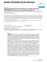
1a and 1b). In cultures pre-treated with Asc-p (400 µM),
the increase in iNOS expression was greatly attenuated
(figure 1c). Figure 1d is a quantitative assessment of the
amount of iNOS immunoreactivity in each condition
based on image analysis.
Pre-treatment with ascorbate attenuates neuronal
structural damage associated with gp120 exposure
Parallel aggregate cultures exposed to 1 nM gp120, 1 nM
gp120 following Asc-p supplementation, or media alone
for 24 hours were labelled for MAP2 or GFAP. Substantial
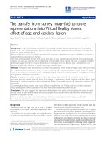
astrocytic hypertrophy (as measured by GFAP immunore-
activity) occurred following exposure to gp120 (figure 2a
and 2b), and this was averted by Asc-p pre-treatment (fig-
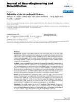
ure 2c). In addition, figure 3a and 3b demonstrate the
reduction in neuronal process complexity following
gp120 exposure (MAP2 staining). In the cultures pre-
treated with Asc-p prior to gp120 exposure, MAP2 expres-
sion was preserved (figure 3c). Figures 2d and 3d are
graphical representations of these trends based on image
analysis of ten random fields in each condition from one
culture subset. In contrast to astrocytes, microglia did not
change in number or size following exposure to gp120, as
detected by immunohistochemistry with CD68. Asc-p
alone did not induce any structural injury or iNOS upreg-
ulation in the cultures.
Specificity of iNOS antibody
Wildtype and iNOS knockout mouse monolayer astro-
cytes were treated with the bacterial endotoxin lipopoly-
saccharide (LPS, 25 ng/ml) and interferon gamma (IFN-γ,
100 U/ml), or with vehicle, for 24 hours. Treatment of
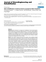
wild type astrocytes with LPS+IFN-γ resulted in a marked
upregulation of iNOS as detected by immunohistochem-
istry (figure 4e) compared to untreated wild type cultures
(figure 4c). However, iNOS was not detected in either the
untreated or treated iNOS knockout astrocytes (figure 4d
and 4f), further supporting the specificity of the antibody
[44,45].
iNOS co-localizes extensively with astrocytic GFAP and
rarely with MAP2
Confocal scanning laser microscopy was used to examine
monolayer cultures for the source of iNOS upregulation.
Almost all GFAP positive cells (astrocytes) co-expressed
iNOS (figure 5, upper 3 panels). However, there were only
rare examples of MAP2 positive cells (neurons) co-
expressing iNOS (figure 5, lower 3 panels).
Caspase-3 expression does not increase with gp120
exposure
Aggregate cultures were stained for caspase-3 expression
to identify the presence of apoptotic cells. Gp120, with or
without Asc-p pre-treatment, had no detectable effect on
caspase-3 expression (figure 6). This suggests that gp120
did not stimulate apoptosis during the 24-hour experi-
mental period.
Journal of Neuroinflammation 2004, 1 />Page 6 of 14
(page number not for citation purposes)
Discussion
Our in vitro observations of neuroglial injury following
gp120 exposure are reminiscent of post-mortem findings
of HAD [46,47]. This study is novel for the use of primary
human mixed CNS culture to demonstrate the upregula-
tion of iNOS in response to gp120 exposure and supports
the previous findings of iNOS upregulation in human
glial cultures exposed to gp120. [48].
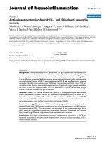
iNOS upregulation following gp120 exposure was attenuated by Asc-p supplementationFigure 1
iNOS upregulation following gp120 exposure was attenuated by Asc-p supplementation. Representative images of
control (a), gp120 exposed (b)and gp120 exposed primary mixed human CNS aggregate cultures at 4 weeks in vitro after Asc-p
supplementation (c) demonstrate that Asc-p supplementation reduced iNOS upregulation. The cultures were examined by
immunohistochemistry for iNOS expression (brown) with a hematoxylin counterstain (blue) for nuclei. (Bar= 40 µm). Quanti-
tative analysis of ten random fields taken from each of the three treatment groups from one culture (d) corroborated the qual-
itative trend and showed the means of control and Asc-p supplemented groups to be significantly different from cultures
treated with gp120. Control and gp120 treated group means were significantly different at p < 0.0001, the means of the gp120
and gp120+Asc-p groups were significantly different at p < 0.0001, and the means of control and gp120+Asc-p supplemented
groups were significantly different (p = 0.0001). Error bars: +/- 1 standard error.
d
0
1000
2000
3000
4000
5000
6000
7000
8000
Mean area of iNOS immunoreactivity (pixels)
c ontrol gp120 gp120+A s c -p
Treatment
ba
c
Journal of Neuroinflammation 2004, 1 />Page 7 of 14
(page number not for citation purposes)
The expression of iNOS by microglia and astrocytes is well
documented [49-51]. It has been demonstrated that astro-
cytic markers co-localize with iNOS in the setting of HIV-
1 [34,52]. Our co-localization studies using confocal
microscopy also identified astrocytes as a major source of
iNOS. The co-localization of MAP2 and iNOS, although
rare, also implies that iNOS may be produced in select
neurons. A recent study conducted by Hori et al.,
Astrocytic hypertrophy following gp120 exposure was prevented by Asc-p supplementationFigure 2
Astrocytic hypertrophy following gp120 exposure was prevented by Asc-p supplementation. Representative
images of control (a), gp120 exposed(b) and gp120 exposed primary mixed human CNS aggregate cultures at 4 weeks in vitro
after Asc-p supplementation (c) demonstrate that Asc-p supplementation prevented astrocytic hypertrophy. The cultures were
examined by immunohistochemistry for increased GFAP expression (brown) indicative of astrocytic hypertrophy (arrows)
with a hematoxylin counterstain (blue) for nuclei. (Bar= 20 µm). Quantitative analysis of ten random fields taken from each of
the three treatment groups from one culture (d) corroborated the qualitative trend. Control and gp120 treated group means
were significantly different at p < 0.0001, the means of the gp120 and gp120+Asc-p groups were significantly different at p =
0.0005 while the control and gp120+Asc-p treated group means were not significantly different (p = 0.0879). Error bars: +/- 1
standard error.
d
ba c
0
2500
5000
7500
10000
12500
15000
17500
20000
22500
25000
Mean area of GFA P immunoreactivity (pixels)
c ontrol gp120 gp120+A s c -p
Treatment
Journal of Neuroinflammation 2004, 1 />Page 8 of 14
(page number not for citation purposes)
suggested that astrocytes were responsible for the
dysregulated overproduction of NO from iNOS rather
than monocyte-derived macrophages [53], but did not
address neuronal iNOS production, which would pre-
sumably be additionally deleterious.
Not only has neuronal expression of iNOS been identified
in vitro and in animal models [54-56], but it has also been
associated recently with other human neurodegenerative
diseases. For instance, iNOS was upregulated in degener-
ated anterior horn neurons in the setting of amyotrophic
lateral sclerosis. [57]. In a study by Vodovotz et al.,
Neuronal dendritic injury following gp120 exposure was prevented by Asc-p supplementationFigure 3
Neuronal dendritic injury following gp120 exposure was prevented by Asc-p supplementation. Representative
images of control (a), gp120 exposed (b) and gp120 exposed primary mixed human CNS aggregate cultures at 4 weeks in vitro
after Asc-p supplementation (c) demonstrate that Asc-p supplementation protected neuronal MAP2 expression dendrites. The
cultures were examined by immunohistochemistry for decreased MAP2 expression (brown) indicating the loss of synaptic
complexity with a hematoxylin counterstain (blue) for nuclei. (Bar = 40 µm). Quantitative analysis of ten random fields taken
from each of the three treatment groups from one culture (d) corroborated the qualitative trend. Control and gp120 treated
group means were significantly different at p = 0.0002, the means of the gp120 and gp120+Asc-p groups were significantly dif-
ferent at p = 0.0012. However the control and gp120+Asc-p treated group means were not significantly different (p = 0.5052).
Error bars: +/- 1 standard error.
d
ba c
0
20000
40000
60000
80000
100000
120000
140000
Mean area of MAP2 immunoreactivity (pixels)
control gp120 gp120+A s c-p
Trea tment
Journal of Neuroinflammation 2004, 1 />Page 9 of 14
(page number not for citation purposes)
neurons with neurofibrillary tangles in affected brain
regions in patients with Alzheimer disease, expressed
iNOS [58]. Moreover, cytokines known to induce iNOS
have been shown to be elevated in the brains of patients
with Alzheimer disease, along with an increase in nitroty-
rosine staining indicative of the presence of excessive lev-
els of NO or peroxynitrite [59]. Although the presence of
iNOS immunoreactivity in neurons has been demon-
strated in Alzheimer disease [60], it has not been docu-
mented in the setting of HAD.
Nitrosative and oxidative stress have been implicated in
the pathogenesis of HAD and a number of other inflam-
matory and neurodegenerative conditions such as Alzhe-
imer disease, amyotrophic lateral sclerosis, and Parkinson
disease [61-63]. In such diseases, cellular damage can be
attributed to the nitrosation or oxidation of vital cellular
components such as lipids, proteins and DNA by reactive
nitrogen and oxygen species (RNS and ROS). Relevant
defence mechanisms include the scavenging of RNS and
ROS and their precursors, binding of metal ions needed
for catalytic formation of ROS, and up-regulation of
endogenous defences [64]. The role of the reductant
ascorbate includes the regeneration of vitamin E from its
radical and inhibition of the peroxidation of membrane
phospholipids [64]. Evidence has been presented to sug-
gest that brain ascorbate levels are decreased in the setting
of HAD [65]. In addition to causing cellular damage
directly, ROS have also been implicated as being key inter-
mediates in signalling cascades under both normal and
aberrant cellular conditions. Hydrogen peroxide has been
demonstrated to be involved in several signal transduc-
tion pathways, stimulate mitogenesis, endothelial migra-
tion and capillary tube formation, and has also been
demonstrated to enhance cellular survival at low concen-
trations as reviewed by Stone and Collins [66]. In addi-
tion, of relevance to this research, studies have shown that
Specificity of primary iNOS antibody confirmed by immunohistochemistryFigure 4
Specificity of primary iNOS antibody confirmed by
immunohistochemistry. GFAP immunoreactivity was
detected in both wild type (a) and iNOS knockout (b) mouse
astrocyte monolayer cultures confirming the presence of
astrocytes. iNOS immunoreactivity was not detected in wild
type (c) or iNOS knockout (d) cultures treated with vehicle
alone. In wild type cultures treated with LPS+IFN-γ, iNOS
immunoreactivity increased (e). However iNOS was not
detected in iNOS knockout cultures treated with LPS+IFN-γ
(f). Bar= 20 µm.
Astrocytes were found to be the major source of iNOS expressionFigure 5
Astrocytes were found to be the major source of
iNOS expression. Confocal scanning laser microscopy
studies of primary mixed human CNS monolayer cultures at
4 weeks in vitro revealed astrocytes to be the major sources
of iNOS with rare neurons expressing iNOS. Immunofluo-
rescence imaging by confocal microscopy was performed for
detection of GFAP, MAP2 and iNOS antigens. Each panel of
images shows individual fluorophores and merged fluoro-
phores with colocalization represented in yellow. Gp120
exposed cultures show increased iNOS expression to colo-
calize extensively with the astrocytic marker GFAP (upper 3
panels) and rarely with the neuronal marker MAP2 (lower 3
panels). Bar = 10 µm.
Journal of Neuroinflammation 2004, 1 />Page 10 of 14
(page number not for citation purposes)
endothelial NOS is activated by hydrogen peroxide
through defined pathways [67-69].
Inducible NOS upregulation occurs in a wide range of
neurological disorders [70,71] and conditions including
sepsis, Alzheimer dementia. [72], Parkinson disease [73],
and in response to traumatic brain injury. [74]. Both
iNOS mRNA and protein were increased in the brains of
AIDS patients that died with severe dementia compared to
those with less severe dementia or no dementia at all [75].
These in vivo observations correlate with our in vitro model
of HAD in which iNOS was upregulated in cultures
treated with gp120. Our understanding of the mechanism
of neuroglial injury in this setting and the factors involved
remains unfinished [76-81]. The present study implicates
a role for iNOS and ROS.
There have been no previous studies that have demon-
strated the protective capacity of ascorbate in the setting of
HIV-1 gp120 induced neurotoxicity and iNOS upregula-
tion. This study establishes the ability of intracellular
ascorbate to attenuate the upregulation of iNOS associ-
ated with gp120 exposure and the capacity of intracellular
ascorbate to prevent gp120 induced astrocytic
hypertrophy and neuronal dendritic injury. It is worth
noting that others have demonstrated that ascorbate
increased nitrite and nitrate production in a mouse mac-
rophage-like cell line activated with LPS and IFN-γ [82].
However, ascorbate alone exhibited no inductive activity
in the iNOS pathway. In our own experience with human
and murine CNS culture models of inflammation and
sepsis ascorbate supplementation has been consistently
associated with decreased iNOS expression [38,83].
The exact mechanism by which ascorbate is able to reduce
the upregulation of iNOS is unknown. A recent study by
Wu et al. using rat microvascular endothelial cell cultures
demonstrated that inhibition of iNOS induction by
intracellular ascorbate was attributable to the reduction of
intracellular oxidant stress, associated with attenuation of
interferon regulatory factor-1 (IRF-1) activation [83].
Interferon regulatory factor-1 and NFκB are transcription
factors with binding sites in the promotor region of the rat
iNOS gene. [84,85]. However, Wu et al. showed that
ascorbate had no effect of on LPS and LPS+IFN-γ induced
activation of NFκB [83]. Moreover, in cultured RAW 264.7
monocyte/macrophages, antioxidants were able to blunt
the DNA binding activity of IRF-1, which mediates iNOS
induction by LPS and IFN-γ [86]. Additional studies have
Caspase 3 expression was not increased 24 hours after 1 nM gp120 exposureFigure 6
Caspase 3 expression was not increased 24 hours after 1 nM gp120 exposure. There was no significant difference in
the average percentage of caspase-3 positive cells per field in control aggregate cultures and those treated with 1 nM gp120 or
400 µM Asc-p prior to gp120 exposure. Ten random fields of each of the three treatment groups from one culture were used
for quantitative analysis. Error bars: +/- 1 standard error.
0
.5
1
1.5
2
2.5
3
3.5
4
Mean % of caspase-3 positive cells per field
c ontrol gp120 gp120+A s c -p
Tr eatment
Journal of Neuroinflammation 2004, 1 />Page 11 of 14
(page number not for citation purposes)
demonstrated a strong association between NFκB activa-
tion and iNOS upregulation [87-93].
The neuroprotective capacity of antioxidant drugs in the
setting of neurodegenerative disease has been both
encouraging and variable. In the case of AIDS related
dementia, the antioxidant thiol thioctic acid (α-lipoic
acid) was not successful. However, deprenyl was effective
in improving cognitive function [94]. The exact mecha-
nism by which deprenyl protects cognitive function is not
known but this monoamine oxidase-B inhibitor has been
demonstrated to scavenge hydroxyl and peroxyl radicals
[68] and increase the activities of the antioxidant enzymes
superoxide dismutase and catalase [95-97]. No previous
studies have addressed the protective capacity of ascorbate
in a human in vitro model of HAD. We feel that it is impor-
tant to explore the capacity of inexpensive, non-toxic and
more accessible therapeutic agents for all patients with
HIV-1/AIDS but especially for the vast majority of patients
that cannot afford antiretroviral therapy. We have demon-
strated that intracellular ascorbate attenuates the upregu-
lation of iNOS, as well as the reduction in dendritic
complexity and astrocytic hypertrophy associated with
gp120 exposure. The effectiveness of ascorbate in the pre-
vention and treatment of HAD awaits further study in a
clinical setting.
Conclusions
The present studies demonstrate that ascorbate supple-
mentation is able to prevent the deleterious upregulation
of iNOS and associated neuronal and astrocytic protein
expression and structural changes caused by gp120 in pri-
mary human CNS cultures.
Ascorbate is a safe, readily available and inexpensive anti-
oxidant and is therefore potentially beneficial to all
patients, especially those in impoverished countries. We
recognize that our research is in vitro and that further stud-
ies are needed to confirm the findings and explore the
mechanisms underlying the phenomenon.
Abbreviations used
Asc-p; ascorbate-2-O-phosphate
C3BT; Class III beta tubulin
CSLM; confocal scanning laser microscopy
DAB; 3,3'-diaminobenzidine
DMEM; Dulbecco's Modified Eagle Medium
eNOS; endothelial NOS
FITC; fluorescein isothiocyanate
GFAP; glial fibrillary acidic protein
gp120; HIV-1 120 kDa envelope glycoprotein
HAART; highly active antiretroviral therapy
HAD; HIV-1 Associated Dementia (HAD)
HIV-1; Human Immunodeficiency Virus I
HPLC; high performance liquid chromatography
IFN-γ; interferon-gamma
iNOS; inducible nitric oxide synthase
LDH; Lactate dehydrogenase
L-NAME; N
G
-nitro-L-arginine methyl ester
LPS; lipopolysaccaride
MAP2; microtubule-associated protein 2
MEM; Modified Eagle Medium
nNOS; neuronal NOS
NO; nitric oxide
NOS; nitric oxide synthases
NPBM; Neural progenitor base media
PBS; phosphate buffered saline
RNS; reactive nitrogen species
ROS; reactive oxygen species
SYN; synaptophysin
TUNEL; terminal dUTP nick end labelling
Competing Interests
None declared.
Authors' contributions
RH conceived of the study. KW designed and carried out
the experiments and collected and analyzed the data in
the laboratory of RH. RH, JM and JXW aided in experi-
mental design and analysis of results and co-wrote the
manuscript with KW. JC performed preliminary
immunohistochemistry and assisted with confocal micro-
scopy. VEL established and provided the knockout mice
Journal of Neuroinflammation 2004, 1 />Page 12 of 14
(page number not for citation purposes)
and tissue used in these studies and provided advice. All
authors read and approved the final manuscript.
Acknowledgements
The authors wish to thank Ewa Jaworski, Kris Milne, Stephanie Totten, and
Dr. Fraser Fellows for their technical and clinical contributions. JXW was
supported by the Natural Sciences and Engineering Research Council of
Canada. RH was supported by the Ontario HIV Treatment Network.
References
1. Davies J, Everall IP, Weich S, Glass J, Sharer LR, Cho ES, Bell JE,
Majteny C, Gray F, Scaravilli F, Lantos PL: HIV-associated brain
pathology: a comparative international study. Neuropathol Appl
Neurobiol 1998, 24:118-124.
2. Hofman P, Saint-Paul MC, Battaglione V, Michiels JF, Loubiere R:
Autopsy findings in the acquired immunodeficiency syn-
drome (AIDS). A report of 395 cases from the south of
France. Pathol Res Pract 1999, 195:209-217.
3. Price RW, Brew B, Sidtis J, Rosenblum M, Scheck AC, Cleary P: The
brain in AIDS: central nervous system HIV-1 infection and
AIDS dementia complex. Science 1988, 239:586-592.
4. Masliah E, Achim CL, Ge N, DeTeresa R, Terry RD, Wiley CA: Spec-
trum of human immunodeficiency virus-associated neocorti-
cal damage. Ann Neurol 1992, 32:321-329.
5. Wiley CA, Achim C: Human immunodeficiency virus encepha-
litis is the pathological correlate of dementia in acquired
immunodeficiency syndrome. Ann Neurol 1994, 36:673-676.
6. Budka H: Neuropathology of human immunodeficiency virus
infection. Brain Pathol 1991, 1:163-175.
7. Kaul M, Garden GA, Lipton SA: Pathways to neuronal injury and
apoptosis in HIV-associated dementia. Nature 2001,
410:988-994.
8. Lipton SA: Neuronal injury associated with HIV-1: approaches
to treatment. Annu Rev Pharmacol Toxicol 1998, 38:159-177.
9. Masliah E, Heaton RK, Marcotte TD, Ellis RJ, Wiley CA, Mallory M,
Achim CL, McCutchan JA, Nelson JA, Atkinson JH, Grant I: Den-
dritic injury is a pathological substrate for human immuno-
deficiency virus-related cognitive disorders. HNRC Group.
The HIV Neurobehavioral Research Center. Ann Neurol 1997,
42:963-972.
10. Everall IP, Luthert PJ, Lantos PL: Neuronal loss in the frontal cor-
tex in HIV infection. Lancet 1999, 337:1119-1121.
11. Scorziello A, Florio T, Bajetto A, Schettini G: Intracellular signal-
ling mediating HIV-1 gp120 neurotoxicity. Cell Signal 1998,
10:75-84.
12. Corasaniti MT, Bagetta G, Rotiroti D, Nistico G: The HIV envelope
protein gp120 in the nervous system: interactions with nitric
oxide, interleukin-1beta and nerve growth factor signalling,
with pathological implications in vivo and in vitro. Biochem
Pharmacol 1998, 56:153-156.
13. Savio T, Levi G: Neurotoxicity of HIV coat protein gp120,
NMDA receptors, and protein kinase C: a study with rat cer-
ebellar granule cell cultures. J Neurosci Res 1993, 34:265-272.
14. Toggas SM, Masliah E, Rockenstein EM, Rall GF, Abraham CR, Mucke
L: Central nervous system damage produced by expression
of the HIV-1 coat protein gp120 in transgenic mice. Nature
1994, 367:188-193.
15. Shi B, De Girolami U, He J, Wang S, Lorenzo A, Busciglio J, Gabuzda
D: Apoptosis induced by HIV-1 infection of the central nerv-
ous system. J Clin Invest 1996, 98:1979-1990.
16. Giulian D, Wendt E, Vaca K, Noonan CA: The envelope glycopro-
tein of human immunodeficiency virus type 1 stimulates
release of neurotoxins from monocytes. Proc Natl Acad Sci U S
A 1993, 90:2769-2773.
17. Lipton SA, Sucher NJ, Kaiser PK, Dreyer EB: Synergistic effects of
HIV coat protein and NMDA receptor-mediated
neurotoxicity. Neuron 1991, 7:111-118.
18. Torre D, Pugliese A, Speranza F: Role of nitric oxide in HIV-1
infection: friend or foe? Lancet Infect Dis 2002, 2:273-280.
19. Wang Y, Marsden PA: Nitric oxide synthases: gene structure
and regulation. Adv Pharmacol 1995, 34:71-90.
20. Espey MG, Miranda KM, Thomas DD, Wink DA: Distinction
between nitrosating mechanisms within human cells and
aqueous solution. J Biol Chem 2001, 276:30085-30091.
21. Brown GC: Regulation of mitochondrial respiration by nitric
oxide inhibition of cytochrome c oxidase. Biochim Biophys Acta
2001, 1504:46-57.
22. Moncada S, Erusalimsky JD: Does nitric oxide modulate mito-
chondrial energy generation and apoptosis? Nat Rev Mol Cell
Biol 2002, 3:214-220.
23. Radi R, Cassina A, Hodara R: Nitric oxide and peroxynitrite
interactions with mitochondria. Biol Chem 2002, 383:401-409.
24. Murphy MP: Nitric oxide and cell death. Biochim Biophys Acta 1999,
1411:401-414.
25. Salgo MG, Stone K, Squadrito GL, Battista JR, Pryor WA: Peroxyni-
trite causes DNA nicks in plasmid pBR322. Biochem Biophys Res
Commun 1995, 210:1025-1030.
26. Wink DA, Kasprzak KS, Maragos CM, Elespuru RK, Misra M, Dunams
TM, Cebula TA, Koch WH, Andrews AW, Allen JS: DNA deami-
nating ability and genotoxicity of nitric oxide and its
progenitors. Science 1991, 254:1001-1003.
27. Zhang J, Dawson VL, Dawson TM, Snyder SH: Nitric oxide activa-
tion of poly(ADP-ribose) synthetase in neurotoxicity. Science
1994, 263:687-689.
28. Lautier D, Lagueux J, Thibodeau J, Menard L, Poirier GG: Molecular
and biochemical features of poly (ADP-ribose) metabolism.
Mol Cell Biochem 1993, 122:171-193.
29. Scorziello A, Florio T, Bajetto A, Schettini G: Intracellular signal-
ling mediating HIV-1 gp120 neurotoxicity. Cell Signal 1998,
10:75-84.
30. Masliah E, Achim CL, Ge N, DeTeresa R, Terry RD, Wiley CA: Spec-
trum of human immunodeficiency virus-associated neocorti-
cal damage. Ann Neurol 1992, 32:321-329.
31. Masliah E, Heaton RK, Marcotte TD, Ellis RJ, Wiley CA, Mallory M,
Achim CL, McCutchan JA, Nelson JA, Atkinson JH, Grant I: Den-
dritic injury is a pathological substrate for human immuno-
deficiency virus-related cognitive disorders. HNRC Group.
The HIV Neurobehavioral Research Center. Ann Neurol 1997,
42:963-972.
32. Rafaeli Y, Levy DN, Weiner DB: The glucocorticoid receptor
type II complex is a target of the HIV-1 vpr gene product. Proc
Natl Acad Sci U S A 1995, 92:3621-3625.
33. Pozzoli G, Tringali G, Dello RC, Vairano M, Preziosi P, Navarra P:
HIV-1 Gp120 protein modulates corticotropin releasing fac-
tor synthesis and release via the stimulation of its mRNA
from the rat hypothalamus in vitro: involvement of inducible
nitric oxide synthase. J Neuroimmunol 2001, 118:268-276.
34. Dugas N, Lacroix C, Kilchherr E, Delfraissy JF, Tardieu M: Role of
CD23 in astrocytes inflammatory reaction during HIV-1
related encephalitis. 2001, 15:96-107.
35. Dawson VL, Dawson TM, Uhl GR, Snyder SH: Human immunode-
ficiency virus type 1 coat protein neurotoxicity mediated by
nitric oxide in primary cortical cultures. Proc Natl Acad Sci U S A
1993, 90:3256-3259.
36. Sacktor N, McDermott MP, Marder K, Schifitto G, Selnes OA,
McArthur JC, Stern Y, Albert S, Palumbo D, Kieburtz K, De Marcaida
JA, Cohen B, Epstein L: HIV-associated cognitive impairment
before and after the advent of combination therapy. J
Neurovirol 2002, 8:136-142.
37. Everall IP, Hudson L, Kerwin RW: Decreased absolute levels of
ascorbic acid and unaltered vasoactive intestinal polypeptide
receptor binding in the frontal cortex in acquired immuno-
deficiency syndrome. Neurosci Lett 1997, 224:119-122.
38. Korcok J, Wu F, Tyml K, Hammond RR, Wilson JX: Sepsis inhibits
reduction of dehydroascorbic acid and accumulation of
ascorbate in astroglial cultures: intracellular ascorbate
depletion increases nitric oxide synthase induction and
glutamate uptake inhibition. J Neurochem 2002, 81:185-193.
39. Pulliam L, Berens ME, Rosenblum ML: A normal human brain cell
aggregate model for neurobiological studies. J Neurosci Res
1988, 21:521-530.
40. Hammond RR, Iskander S, Achim CL, Hearn S, Nassif J, Wiley CA: A
reliable primary human CNS culture protocol for morpho-
logical studies of dendritic and synaptic elements. J Neurosci
Methods 2002, 118:189-198.
41. Halliwell B: Oxidative stress in cell culture: an under-appreci-
ated problem? FEBS Lett 2003, 540:3-6.
Journal of Neuroinflammation 2004, 1 />Page 13 of 14
(page number not for citation purposes)
42. Laubach VE, Shesely EG, Smithies O, Sherman PA: Mice lacking
inducible nitric oxide synthase are not resistant to lipopoly-
saccharide-induced death. Proc Natl Acad Sci U S A 1995,
92:10688-10692.
43. Dixon SJ, Wilson JX: Fluorescence measurement of cytosolic
pH in cultured rodent astrocytes. 1995, 27:196-213.
44. Lowenstein CJ, Glatt CS, Bredt DS, Snyder SH: Cloned and
expressed macrophage nitric oxide synthase contrasts with
the brain enzyme. Proc Natl Acad Sci U S A 1992, 89:6711-6715.
45. Lyons CR, Orloff GJ, Cunningham JM: Molecular cloning and func-
tional expression of an inducible nitric oxide synthase from a
murine macrophage cell line. J Biol Chem 1992, 267:6370-6374.
46. Masliah E, Achim CL, Ge N, DeTeresa R, Terry RD, Wiley CA: Spec-
trum of human immunodeficiency virus-associated neocorti-
cal damage. Ann Neurol 1992, 32:321-329.
47. Wiley CA, Masliah E, Morey M, Lemere C, DeTeresa R, Grafe M,
Hansen L, Terry R: Neocortical damage during HIV infection.
Ann Neurol 1991, 29:651-657.
48. Koka P, He K, Zack JA, Kitchen S, Peacock W, Fried I, Tran T, Yashar
SS, Merrill JE: Human immunodeficiency virus 1 envelope pro-
teins induce interleukin 1, tumor necrosis factor alpha, and
nitric oxide in glial cultures derived from fetal, neonatal, and
adult human brain. J Exp Med 1995, 182:941-951.
49. Brosnan CF, Lee SC, Liu J: Regulation of inducible nitric oxide
synthase expression in human glia: implications for inflam-
matory central nervous system diseases. Biochem Soc Trans
1997, 25:679-683.
50. Minghetti L, Levi G: Microglia as effector cells in brain damage
and repair: focus on prostanoids and nitric oxide. Prog
Neurobiol 1998, 54:99-125.
51. Murphy S, Simmons ML, Agullo L, Garcia A, Feinstein DL, Galea E,
Reis DJ, Minc-Golomb D, Schwartz JP: Synthesis of nitric oxide in
CNS glial cells. Trends Neurosci 1993, 16:323-328.
52. Zhao ML, Kim MO, Morgello S, Lee SC: Expression of inducible
nitric oxide synthase, interleukin-1 and caspase-1 in HIV-1
encephalitis. J Neuroimmunol 2001, 115:182-191.
53. Hori K, Burd PR, Furuke K, Kutza J, Weih KA, Clouse KA: Human
immunodeficiency virus-1-infected macrophages induce
inducible nitric oxide synthase and nitric oxide (NO) produc-
tion in astrocytes: astrocytic NO as a possible mediator of
neural damage in acquired immunodeficiency syndrome.
Blood 1999, 93:1843-1850.
54. Heneka MT, Feinstein DL: Expression and function of inducible
nitric oxide synthase in neurons. J Neuroimmunol 2001, 114:8-18.
55. Minc-Golomb D, Tsarfaty I, Schwartz JP: Expression of inducible
nitric oxide synthase by neurones following exposure to
endotoxin and cytokine. Br J Pharmacol 1994, 112:720-722.
56. Sato I, Kim Y, Himi T, Murota S: Induction of calcium-independ-
ent nitric oxide synthase activity in cultured cerebellar gran-
ule neurons. Neurosci Lett 1995, 184:145-148.
57. Sasaki S, Shibata N, Komori T, Iwata M: iNOS and nitrotyrosine
immunoreactivity in amyotrophic lateral sclerosis. Neurosci
Lett 2000, 291:44-48.
58. Vodovotz Y, Lucia MS, Flanders KC, Chesler L, Xie QW, Smith TW,
Weidner J, Mumford R, Webber R, Nathan C, Roberts AB, Lippa CF,
Sporn MB: Inducible nitric oxide synthase in tangle-bearing
neurons of patients with Alzheimer's disease. J Exp Med 1996,
184:1425-1433.
59. Smith MA, Richey HP, Sayre LM, Beckman JS, Perry G: Widespread
peroxynitrite-mediated damage in Alzheimer's disease. J
Neurosci 1997, 17:2653-2657.
60. Vodovotz Y, Lucia MS, Flanders KC, Chesler L, Xie QW, Smith TW,
Weidner J, Mumford R, Webber R, Nathan C, Roberts AB, Lippa CF,
Sporn MB: Inducible nitric oxide synthase in tangle-bearing
neurons of patients with Alzheimer's disease. J Exp Med 1996,
184:1425-1433.
61. Maxwell SR: Prospects for the use of antioxidant therapies.
Drugs 1995, 49:345-361.
62. Pratico D, Delanty N: Oxidative injury in diseases of the central
nervous system: focus on Alzheimer's disease. Am J Med 2000,
109:577-585.
63. Shaw PJ, Ince PG, Falkous G, Mantle D: Oxidative damage to pro-
tein in sporadic motor neuron disease spinal cord. Ann Neurol
1995, 38:691-695.
64. Gilgun-Sherki Y, Melamed E, Offen D: Oxidative stress induced-
neurodegenerative diseases: the need for antioxidants that
penetrate the blood brain barrier. 2001, 40:959-975.
65. Everall IP, Hudson L, Kerwin RW: Decreased absolute levels of
ascorbic acid and unaltered vasoactive intestinal polypeptide
receptor binding in the frontal cortex in acquired immuno-
deficiency syndrome. Neurosci Lett 1997, 224:119-122.
66. Stone JR, Collins T: The role of hydrogen peroxide in endothe-
lial proliferative responses. Endothelium 2002, 9:231-238.
67. Cai H, Li Z, Davis ME, Kanner W, Harrison DG, Dudley SC Jr: Akt-
dependent phosphorylation of serine 1179 and mitogen-acti-
vated protein kinase kinase/extracellular signal-regulated
kinase 1/2 cooperatively mediate activation of the endothe-
lial nitric-oxide synthase by hydrogen peroxide. Mol Pharmacol
2003, 63:325-331.
68. Thomas CE, Huber EW, Ohlweiler DF: Hydroxyl and peroxyl rad-
ical trapping by the monoamine oxidase-B inhibitors depre-
nyl and MDL 72,974A: implications for protection of
biological substrates. Free Radic Biol Med 1997, 22:733-737.
69. Zou MH, Hou XY, Shi CM, Nagata D, Walsh K, Cohen RA: Modula-
tion by peroxynitrite of Akt- and AMP-activated kinase-
dependent Ser1179 phosphorylation of endothelial nitric
oxide synthase. J Biol Chem 2002, 277:32552-32557.
70. Adamson DC, Wildemann B, Sasaki M, Glass JD, McArthur JC, Chris-
tov VI, Dawson TM, Dawson VL: Immunologic NO synthase: ele-
vation in severe AIDS dementia and induction by HIV-1
gp41. Science 1996, 274:1917-1921.
71. Bo L, Dawson TM, Wesselingh S, Mork S, Choi S, Kong PA, Hanley
D, Trapp BD: Induction of nitric oxide synthase in demyelinat-
ing regions of multiple sclerosis brains. Ann Neurol 1994,
36:778-786.
72. Haas J, Storch-Hagenlocher B, Biessmann A, Wildemann B: Inducible
nitric oxide synthase and argininosuccinate synthetase: co-
induction in brain tissue of patients with Alzheimer's demen-
tia and following stimulation with beta-amyloid 1-42 in vitro.
Neurosci Lett 2002, 322:121-125.
73. Iravani MM, Kashefi K, Mander P, Rose S, Jenner P: Involvement of
inducible nitric oxide synthase in inflammation-induced
dopaminergic neurodegeneration. 2002, 110:49-58.
74. Orihara Y, Ikematsu K, Tsuda R, Nakasono I: Induction of nitric
oxide synthase by traumatic brain injury. Forensic Sci Int 2001,
123:142-149.
75. Adamson DC, Wildemann B, Sasaki M, Glass JD, McArthur JC, Chris-
tov VI, Dawson TM, Dawson VL: Immunologic NO synthase: ele-
vation in severe AIDS dementia and induction by HIV-1
gp41. Science 1996, 274:1917-1921.
76. Klegeris A, Walker DG, McGeer PL: Toxicity of human THP-1
monocytic cells towards neuron-like cells is reduced by non-
steroidal anti-inflammatory drugs (NSAIDs). 1999,
38:1017-1025.
77. Klegeris A, McGeer PL: Interaction of various intracellular sig-
naling mechanisms involved in mononuclear phagocyte tox-
icity toward neuronal cells. J Leukoc Biol 2000, 67:127-133.
78. Piani D, Spranger M, Frei K, Schaffner A, Fontana A: Macrophage-
induced cytotoxicity of N-methyl-D-aspartate receptor pos-
itive neurons involves excitatory amino acids rather than
reactive oxygen intermediates and cytokines. Eur J Immunol
1992, 22:2429-2436.
79. Piani D, Frei K, Pfister HW, Fontana A: Glutamate uptake by
astrocytes is inhibited by reactive oxygen intermediates but
not by other macrophage-derived molecules including
cytokines, leukotrienes or platelet-activating factor. J
Neuroimmunol 1993, 48:99-104.
80. Piani D, Fontana A: Involvement of the cystine transport sys-
tem xc- in the macrophage-induced glutamate-dependent
cytotoxicity to neurons. J Immunol 1994, 152:3578-3585.
81. Barger SW, Basile AS: Activation of microglia by secreted amy-
loid precursor protein evokes release of glutamate by cys-
tine exchange and attenuates synaptic function. J Neurochem
2001, 76:846-854.
82. Mizutani A, Maki H, Torii Y, Hitomi K, Tsukagoshi N: Ascorbate-
dependent enhancement of nitric oxide formation in acti-
vated macrophages. Nitric Oxide 1998, 2:235-241.
83. Wu F, Tyml K, Wilson JX: Ascorbate inhibits iNOS expression
in endotoxin- and IFN gamma-stimulated rat skeletal mus-
cle endothelial cells. FEBS Lett 2002, 520:122-126.
Publish with Bio Med Central and every
scientist can read your work free of charge
"BioMed Central will be the most significant development for
disseminating the results of biomedical researc h in our lifetime."
Sir Paul Nurse, Cancer Research UK
Your research papers will be:
available free of charge to the entire biomedical community
peer reviewed and published immediately upon acceptance
cited in PubMed and archived on PubMed Central
yours — you keep the copyright
Submit your manuscript here:
/>BioMedcentral
Journal of Neuroinflammation 2004, 1 />Page 14 of 14
(page number not for citation purposes)
84. Martin E, Nathan C, Xie QW: Role of interferon regulatory fac-
tor 1 in induction of nitric oxide synthase. J Exp Med 1994,
180:977-984.
85. Xie QW, Whisnant R, Nathan C: Promoter of the mouse gene
encoding calcium-independent nitric oxide synthase confers
inducibility by interferon gamma and bacterial
lipopolysaccharide. J Exp Med 1993, 177:1779-1784.
86. Hecker M, Preiss C, Klemm P, Busse R: Inhibition by antioxidants
of nitric oxide synthase expression in murine macrophages:
role of nuclear factor kappa B and interferon regulatory fac-
tor 1. Br J Pharmacol 1996, 118:2178-2184.
87. Adams V, Nehrhoff B, Spate U, Linke A, Schulze PC, Baur A, Gielen
S, Hambrecht R, Schuler G: Induction of iNOS expression in
skeletal muscle by IL-1beta and NFkappaB activation: an in
vitro and in vivo study. Cardiovasc Res 2002, 54:95-104.
88. Han YJ, Kwon YG, Chung HT, Lee SK, Simmons RL, Billiar TR, Kim
YM: Antioxidant enzymes suppress nitric oxide production
through the inhibition of NF-kappa B activation: role of
H(2)O(2) and nitric oxide in inducible nitric oxide synthase
expression in macrophages. Nitric Oxide 2001, 5:504-513.
89. Kim SH, Johnson VJ, Shin TY, Sharma RP: Selenium attenuates
lipopolysaccharide-induced oxidative stress responses
through modulation of p38 MAPK and NF-kappaB signaling
pathways. Exp Biol Med (Maywood) 2004, 229:203-213.
90. Liu X, Jana M, Dasgupta S, Koka S, He J, Wood C, Pahan K: Human
immunodeficiency virus type-1 (HIV-1) Tat induces nitric
oxide synthase in human astroglia. J Biol Chem 2002,
277:39312-39319.
91. Madrigal JL, Moro MA, Lizasoain I, Lorenzo P, Castrillo A, Bosca L,
Leza JC: Inducible nitric oxide synthase expression in brain
cortex after acute restraint stress is regulated by nuclear
factor kappaB-mediated mechanisms. J Neurochem 2001,
76:532-538.
92. Matsumura M, Kakishita H, Suzuki M, Banba N, Hattori Y: Dexame-
thasone suppresses iNOS gene expression by inhibiting NF-
kappaB in vascular smooth muscle cells. Life Sci 2001,
69:1067-1077.
93. Nomura Y: NF-kappaB activation and IkappaB alpha dyna-
mism involved in iNOS and chemokine induction in astro-
glial cells. Life Sci 2001, 68:1695-1701.
94. A randomized, double-blind, placebo-controlled trial of
deprenyl and thioctic acid in human immunodeficiency
virus-associated cognitive impairment. Dana Consortium on
the Therapy of HIV Dementia and Related Cognitive
Disorders. Neurology 1998, 50:645-651.
95. Carrillo MC, Kitani K, Kanai S, Sato Y, Miyasaka K, Ivy GO: The
effect of a long term (6 months) treatment with (-)deprenyl
on antioxidant enzyme activities in selective brain regions in
old female Fischer 344 rats. Biochem Pharmacol 1994,
47:1333-1338.
96. Carrillo MC, Ivy GO, Milgram NW, Head E, Wu P, Kitani K: (-
)Deprenyl increases activities of superoxide dismutase
(SOD) in striatum of dog brain. Life Sci 1994, 54:1483-1489.
97. Carrillo MC, Kitani K, Kanai S, Sato Y, Miyasaka K, Ivy GO: (-)depre-
nyl increases activities of superoxide dismutase and catalase
in certain brain regions in old male mice. Life Sci 1994,
54:975-981.