báo cáo hóa học: " Interleukin 1 receptor antagonist knockout mice show enhanced microglial activation and neuronal damage induced by intracerebroventricular infusion of human β-amyloid" pdf
Bạn đang xem bản rút gọn của tài liệu. Xem và tải ngay bản đầy đủ của tài liệu tại đây (375.85 KB, 9 trang )
BioMed Central
Page 1 of 9
(page number not for citation purposes)
Journal of Neuroinflammation
Open Access
Research
Interleukin 1 receptor antagonist knockout mice show enhanced
microglial activation and neuronal damage induced by
intracerebroventricular infusion of human β-amyloid
Jeffrey M Craft
1,2
, D Martin Watterson
1,3
, Emmet Hirsch
4,5
and Linda J Van
Eldik*
1,2
Address:
1
Center for Drug Discovery and Chemical Biology, Northwestern University, Chicago, IL, USA,
2
Cell and Molecular Biology,
Northwestern University Feinberg School of Medicine, Chicago, IL, USA,
3
Molecular Pharmacology and Biological Chemistry, Northwestern
University Feinberg School of Medicine, Chicago, IL, USA,
4
Obstetrics and Gynecology, Northwestern University Feinberg School of Medicine,
Chicago, IL, USA and
5
Department of Obstetrics and Gynecology, Evanston Northwestern Healthcare, Evanston, IL, USA
Email: Jeffrey M Craft - ; D Martin Watterson - ; Emmet Hirsch - e-
; Linda J Van Eldik* -
* Corresponding author
Alzheimer's diseaseamyloid betaanimal modelglial activationinterleukin-1microglia
Abstract
Background: Interleukin 1 (IL-1) is a key mediator of immune responses in health and disease. Although classically the
function of IL-1 has been studied in the systemic immune system, research in the past decade has revealed analogous
roles in the CNS where the cytokine can contribute to the neuroinflammation and neuropathology seen in a number of
neurodegenerative diseases. In Alzheimer's disease (AD), for example, pre-clinical and clinical studies have implicated IL-
1 in the progression of a pathologic, glia-mediated pro-inflammatory state in the CNS. The glia-driven neuroinflammation
can lead to neuronal damage, which, in turn, stimulates further glia activation, potentially propagating a detrimental cycle
that contributes to progression of pathology. A prediction of this neuroinflammation hypothesis is that increased IL-1
signaling in vivo would correlate with increased severity of AD-relevant neuroinflammation and neuronal damage.
Methods: To test the hypothesis that increased IL-1 signaling predisposes animals to beta-amyloid (Aβ)-induced damage,
we used IL-1 receptor antagonist Knock-Out (IL1raKO) and wild-type (WT) littermate mice in a model that involves
intracerebroventricular infusion of human oligomeric Aβ1–42. This model mimics many features of AD, including robust
neuroinflammation, Aβ plaques, synaptic damage and neuronal loss in the hippocampus. IL1raKO and WT mice were
infused with Aβ for 28 days, sacrificed at 42 days, and hippocampal endpoints analyzed.
Results: IL1raKO mice showed increased vulnerability to Aβ-induced neuropathology relative to their WT
counterparts. Specifically, IL1raKO mice exhibited increased mortality, enhanced microglial activation and
neuroinflammation, and more pronounced loss of synaptic markers. Interestingly, Aβ-induced astrocyte responses were
not significantly different between WT and IL1raKO mice, suggesting that enhanced IL-1 signaling predominately affects
microglia.
Conclusion: Our data are consistent with the neuroinflammation hypothesis whereby increased IL-1 signaling in AD
enhances glia activation and leads to an augmented neuroinflammatory process that increases the severity of
neuropathologic sequelae.
Published: 20 June 2005
Journal of Neuroinflammation 2005, 2:15 doi:10.1186/1742-2094-2-15
Received: 24 May 2005
Accepted: 20 June 2005
This article is available from: />© 2005 Craft et al; licensee BioMed Central Ltd.
This is an Open Access article distributed under the terms of the Creative Commons Attribution License ( />),
which permits unrestricted use, distribution, and reproduction in any medium, provided the original work is properly cited.
Journal of Neuroinflammation 2005, 2:15 />Page 2 of 9
(page number not for citation purposes)
Background
There is increasing evidence that CNS inflammation
(termed neuroinflammation) driven by abnormal or pro-
longed glia activation contributes to the pathogenesis and
progression of both acute and chronic disorders [1,2].
Normally, glia respond to stresses by a transient activation
that serves a homeostatic function. However, increased
levels of inflammatory and oxidative stress molecules pro-
duced by chronically activated glia can lead to neuron
damage or death, which can induce further glial activa-
tion, thus leading to a self-propagating, detrimental cycle
of neuroinflammation and neurodegeneration [3]. A large
body of evidence [4-8] suggests that targeting this glia-
neuron cycle represents an attractive potential strategy for
development of new therapeutic approaches to AD that
would alter disease progression. To this end, a more
detailed understanding of the proteins, pathways, and
inflammatory responses involved in neuroinflammation
relevant to AD progression is critical.
One of the biochemical responses of glia to both acute
and chronic conditions of brain damage is increased pro-
duction of the pro-inflammatory cytokine IL-1. An exten-
sive body of research strongly suggests that IL-1 has an
integral role in AD pathogenesis and progression. First,
analysis of AD brain tissue demonstrates IL-1 overproduc-
tion, primarily in the activated microglia that surround β-
amyloid (Aβ) plaques and neurons containing neurofi-
brillary tangles [9,10], the two neuropathological hall-
marks of AD. This finding is complemented by the
revelation that this overproduction of IL-1 closely corre-
sponds to the level of neuropathology found in a given
brain region [11]. Second, cell-based studies show that IL-
1 can elicit the production of a number of detrimental
molecules from microglia, astrocytes, and neurons. For
example, IL-1 can stimulate the production of α 1 anti-
chymotrypsin, IL-6, S100B, and inducible nitric oxide syn-
thase [12-15], which are themselves increased in the AD
brains [2]. These molecules, either by themselves or by
stimulating the production of other molecules, contribute
to a neuroinflammatory cascade that has been suggested
to result in cell injury, dysfunction, and death in AD[16].
This hypothesis is supported by the neuroprotection
observed following suppression of the neuroinflamma-
tory cascade in AD animal models [4,5]. Finally, multiple
studies examining IL-1 genetics have shown that polymor-
phisms in the IL-1gnd IL-1 receptor genes increase the risk
of AD by as much as three times in a homozygous carrier
[17,16].
All these studies to date are consistent with the hypothesis
that increased brain IL-1 levels or activity would correlate
with increased severity of AD-relevant neuroinflamma-
tion and neuronal damage. To test this hypothesis, we
used interleukin-1 receptor antagonist knockout
(IL1raKO) mice, which have enhanced IL-1 signaling
because of the loss of the IL-1 receptor's physiological
antagonist. We induced AD-relevant neuroinflammation
and neuronal damage by intracerebroventricular (ICV)
infusion of human Aβ1–42 in a mouse experimental
model previously developed by us [4,5], and determined
the degree of glia activation and neuroinflammation and
synaptic degeneration in the hippocampus. We report
here that IL1raKO mice are significantly more susceptible
than WT mice to the neuroinflammatory and neurodegen-
erative sequelae of Aβ infusion, supporting the concept
that elevated IL-1 signaling in the brain participates in AD
pathogenesis.
Methods
Interleukin-1 receptor antagonist knockout mice
(IL1raKO)
IL1raKO mice were derived as previously described [18]
and the colony maintained by mating of heterozygous lit-
termates. Homozygous IL1raKO mice and WT littermates
were selected following genotyping, and were allowed to
mature until 16 weeks of age before surgery. All mice were
kept at the Center for Comparative Medicine (CCM) at
Northwestern University Feinberg School of Medicine. All
animal procedures were approved by the Animal Care and
Use Committee at Northwestern University.
A
β
infusion
ICV Infusion of human oligomeric Aβ1–42 or vehicle into
IL1raKO and WT littermates was performed essentially as
described [4]. Briefly, four-month-old mice (n = 5–12 per
group) were anesthetized with 2% vaporized isoflurane,
and an Alzet micro-osmotic pump (Durect, Model #1002)
was attached to a pre-cut 2.5 mm long cannula (Plastics
One) stereotaxically implanted into the right lateral cere-
bral ventricle (at coordinates -1.0 mm mediolateral, -0.5
mm anterioposterior from Bregma; -2.0 mm dorsal-ven-
tral from skull). Pumps contained either vehicle (4 mM
Hepes + 250 µg/ml human high-density lipoprotein,
HDL) or oligomeric Aβ1–42 (45 µg; American Peptide)
[19] dissolved in vehicle. Since HDL normally carries Aβ
in serum, it was used in the pump to reduce Aβ aggrega-
tion and facilitate better delivery to the neuropil [20,21].
Osmotic pumps were partially coated with paraffin to a
point 5 mm above the distal end of the pump. This slows
the osmotic passage of water into the pumps' gel casings
and has been shown in ex vivo experiments to reduce the
infusion rate to ~1.6 µg/3.5 µl per day for ~28 days (data
not shown).
At 42 days after the start of Aβ infusion, mice were anes-
thetized with pentobarbital (50 mg/kg) and perfused with
a Hepes buffer containing a protease inhibitor cocktail.
The superior portion of the cranium was then incised, and
brains were removed and longitudinally bisected. In order
Journal of Neuroinflammation 2005, 2:15 />Page 3 of 9
(page number not for citation purposes)
to exclude the potential that one side of the brain may
possess more significant pathology following a unilateral
infusion and, therefore, confound the results and/or con-
clusions, only the right half of the brain was fixed in a
paraformaldehyde/ phosphate buffer solution and
embedded in paraffin for histological examination, while
the hippocampus was isolated from only the left hemi-
sphere and snap frozen for biochemical evaluation. In
addition, the Alzet pumps were examined to insure that
the paraffin coating was intact and the reservoir solution
was expelled.
Biochemical analysis of inflammatory and neural markers
Hippocampal soluble extracts were prepared by dounce
and sonication in a Hepes buffer containing a protease
inhibitor cocktail followed by centrifugation and collec-
tion of supernatant as described [4]. Levels of the pro-
inflammatory cytokines IL-1β, tumor necrosis factor
(TNF)α, S100B, and the presynaptic protein synapto-
physin in hippocampal supernatants were determined by
ELISA as previously described [4]. Western blots of hip-
pocampal supernatants (10 µg supernatant protein
loaded per lane) were done with an antibody to postsyn-
aptic density protein-95 kDa (PSD-95) (1:100,000 dilu-
tion; Upstate Biotechnology) as described [4]. Antibodies
against β-actin (1:500,000 dilution, Sigma) were used to
confirm equal protein loading among the samples. Densi-
tometry was done with ImageQuant software (Molecular
Dynamics).
Histology
Immunohistochemical detection of activated astrocytes
and microglia was performed on 10 µm sections with
anti-GFAP (1:1500 dilution; Sigma) and anti-F4/80
(1:100 dilution; Serotek) antibodies, respectively, as pre-
viously described [4]. Cell counts were determined by two
blinded observers and subsequently analyzed as follows.
For microglia and astrocyte analysis, all diaminobenzi-
dine (DAB)-stained cell bodies were manually counted in
the hippocampus (excluding the fimbria) of three F4/80-
and GFAP- labeled sections positioned at -1.8, -2.1, and -
2.3 mm from Bregma. In all studies, concordance between
observers was within 10% or the section was removed
from analysis.
Statistical analyses
Experimental and control groups were compared as four
independent groups using one-way ANOVA with SNK
post-hoc analysis using a statistical software package
(GraphPad Prism version 4.00, GraphPad Software, San
Diego CA,
). GraphPad Prism
was also used to construct Kaplan-Meier mortality curves
and assess for significance. Statistical significance was
assumed when p < 0.05.
Results
Increased mortality in A
β
-infused IL1raKO mice
In our previous studies utilizing the Aβ-infusion model
[4,5], we found that intra-, peri-, and post-operative ani-
mal mortality was approximately 1–2%. Mortality in
IL1raKO mice that received an Aβ infusion was much
higher, reaching 50% by the time of sacrifice at 42 days
(Fig 1). In sharp contrast, no animal mortality was experi-
enced in the IL1raKO mice that received a vehicle infu-
sion, or in WT littermates infused with either Aβ or
vehicle.
Enhanced microglial responses in A
β
-infused IL1raKO mice
Based on the high mortality seen in Aβ-infused IL1raKO
mice, the infusion experiment was repeated with addi-
tional mice to allow survival of enough KO mice for sub-
sequent analyses. At 42 days after the start of surgery, mice
were sacrificed and hippocampal tissue analyzed. Meas-
urement of microglia activation endpoints (Fig 2)
revealed no significant differences in the basal levels of the
pro-inflammatory cytokines IL-1β (Fig 2A) and TNFα (Fig
2B) in vehicle-infused IL1raKO and WT mice. There was a
slight increase in the numbers of F4/80 positive microglia
in vehicle-infused IL1raKO mice compared with the WT
counterparts (Fig 2C). However, in IL1raKO mice infused
with Aβ, the intensity of the microglial response, as meas-
ured by several biochemical and histological endpoints,
was much greater than in Aβ-infused WT mice. For exam-
ple, IL-1β levels were significantly greater in Aβ-infused
IL1raKO compared to Aβ-infused WT mice (Fig 2A). Like-
wise, TNFα levels were significantly higher following Aβ
infusion in IL1raKO mice versus WT littermates (Fig 2B).
Finally, the number of activated microglia as measured by
F4/80 immunostaining was greater in Aβ-infused
IL1raKO mice versus their WT counterparts (Fig 2C). Rep-
resentative photomicrographs from the hippocampus of
WT and IL1raKO mice infused with Aβ (Fig 2D and 2E,
respectively) demonstrate the extent of this microglial
activation.
Astrocyte activation in A
β
-infused IL1raKO mice
Unlike the findings above with microglia endpoints, we
observed no significant differences in astrocyte activation
state between IL1raKO and WT mice following Aβ infu-
sion. For example, levels of the astrocyte-derived cytokine
S100B were significantly upregulated following Aβ infu-
sion for both the WT and IL1raKO mice (Fig 3A); how-
ever, there was no significant difference in the magnitude
of this increase between the Aβ-infused WT and IL1raKO
mice. These results were also seen by glial fibrillary acidic
protein (GFAP) immunohistochemistry. As shown in Fig
3B, there were significant increases in GFAP staining in the
hippocampus of all mice following Aβ infusion, and the
numbers of GFAP-positive astrocytes were similar in WT
and IL1raKO mice.
Journal of Neuroinflammation 2005, 2:15 />Page 4 of 9
(page number not for citation purposes)
Increased synaptic degeneration in A
β
-infused IL1raKO
mice
Given the significant increase in microglial neuroinflam-
mation following Aβ infusion in IL1raKO mice, it was
important to investigate the effect of this enhanced neu-
roinflammation on neuronal responses. Therefore, two
different biochemical markers of synaptic degradation
were examined. Aβ infusion led to a reduction in levels of
the postsynaptic protein PSD-95 in both the IL1raKO and
WT mice compared to vehicle-infused mice; however, the
reduction was significantly greater in IL1raKO mice com-
pared to WT (Fig 4A). Similarly, levels of the presynaptic
protein synaptophysin were reduced in Aβ-infused mice
compared to vehicle-infused mice and, while not quite
reaching statistical significance (p = 0.07), the reduction
in synaptophysin levels was greater in Aβ-infused
IL1raKO mice compared to Aβ-infused WT (Fig 4B).
Discussion
The principle finding of this study is that enhanced IL-1
signaling results in increased mortality, microglial neu-
roinflammation, and neuronal damage following a
chronic infusion of human Aβ1–42 in a murine model.
These data provide further support for the idea that IL-1 is
an important component in the neuroinflammation cas-
cade that drives AD progression.
An extensive body of evidence indicates the importance of
IL-1 in regulating susceptibility to CNS injury. For exam-
ple, IL-1β levels in cerebrospinal fluid (CSF) are substan-
tially increased shortly after severe traumatic brain injury
in humans, and the magnitude of this increase is directly
proportional to intracranial pressure [22]. Animal studies
have also demonstrated the importance of IL-1 in mediat-
ing damage following both an acute insult, such as
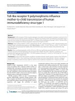
Increased mortality in IL1raKO mice during Aβ infusionFigure 1
Increased mortality in IL1raKO mice during Aβ infusion. Alzet pumps containing Aβ1–42 or vehicle were surgically implanted
in IL1raKO and WT littermate mice (n = 10–12 mice per Aβ-infused group; n = 5 mice per vehicle-infused group), and post-
operative survival was monitored for 42 days. Kaplan-Meier survival curves show that WT mice infused with vehicle or Aβ, and
IL1raKO mice infused with vehicle experienced no mortality during the time period. In contrast, Aβ-infused IL1raKO mice
experienced a 50% mortality rate (6 of the 12 animals died before 42 days). This mortality was significantly different from the
other experimental and control groups (error bars = SEM; p < 0.05).
Journal of Neuroinflammation 2005, 2:15 />Page 5 of 9
(page number not for citation purposes)
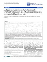
Microglia activation following Aβ infusionFigure 2
Microglia activation following Aβ infusion. WT and IL1raKO mice infused with Aβ or vehicle for 28 days were sacrificed on day
42 (n = 5–10 mice/group survived for analysis). Brains were bisected and the right side of the brain was processed for immuno-
histochemistry while the left hippocampus was dissected and used for biochemical analysis. A) Levels of the pro-inflammatory
cytokine IL-1β were significantly higher in IL1raKO mice infused with Aβ compared to WT mice infused with Aβ. B) TNFα lev-
els also showed a stronger upregulation in Aβ-infused IL1raKO mice compared to Aβ-infused WT mice. C) F4/80 immunos-
taining for activated microglia also revealed a significant increase in IL1raKO mice infused with Aβ versus WT mice infused
with Aβ. Representative photomicrographs of F4/80-positive microglia in the hippocampus of a D) WT mouse infused with Aβ,
and E) IL1raKO mouse infused with Aβ. Arrowheads point to microglia cell bodies. Bar in D-E = 50 µm (error bars = SEM; *
Significantly different, p < 0.01; ***Significantly different, p < 0.001).
C. M icroglia
WT WT KO KO
Veh. Aβ
ββ
β Ve h. Aβ
ββ
β
WT WT KO KO
Veh. Aβ
ββ
β Ve h. Aβ
ββ
β
WT WT KO KO
Veh. Aβ
ββ
β Ve h. Aβ
ββ
β
A. IL-1β
ββ
β B. TNFα
αα
α
D.
E.
C. M icroglia
WT WT KO KO
Veh. Aβ
ββ
β Ve h. Aβ
ββ
β
WT WT KO KO
Veh. Aβ
ββ
β Ve h. Aβ
ββ
β
WT WT KO KO
Veh. Aβ
ββ
β Ve h. Aβ
ββ
β
A. IL-1β
ββ
β B. TNFα
αα
α
D.
E.
C. M icroglia
WT WT KO KO
Veh. Aβ
ββ
β Ve h. Aβ
ββ
β
WT WT KO KO
Veh. Aβ
ββ
β Ve h. Aβ
ββ
β
WT WT KO KO
Veh. Aβ
ββ
β Ve h. Aβ
ββ
β
A. IL-1β
ββ
β B. TNFα
αα
α
D.
E.
C. M icroglia
WT WT KO KO
Veh. Aβ
ββ
β Ve h. Aβ
ββ
β
WT WT KO KO
Veh. Aβ
ββ
β Ve h. Aβ
ββ
β
WT WT KO KO
Veh. Aβ
ββ
β Ve h. Aβ
ββ
β
A. IL-1β
ββ
β B. TNFα
αα
α
D.
E.
Journal of Neuroinflammation 2005, 2:15 />Page 6 of 9
(page number not for citation purposes)
neonatal hypoxia-ischemia [23], and the progressive
neurodegeneration that follows mild acute insults in
rodents [24]. This is not unexpected given the array of
potentially detrimental molecules produced by the CNS
in response to increased production of IL-1. For example,
IL-1β and/or IL-1α have been implicated in the produc-
tion of other pro-inflammatory cytokines such as S100B
[14]. Furthermore, IL-1β can stimulate glial iNOS produc-
tion [15], which in turn can greatly increase the oxidative
stress experienced by the brain and potentially lead to
neuronal damage through protein nitration pathways
[25].
More relevant to the current study, there is increased IL-1
signaling in chronic neurodegenerative diseases. In addi-
tion to the IL-1 overexpression and disease-relevant distri-
bution in AD [26,10], IL-1 is also increased in other
chronic conditions that involve neurodegeneration. These
include Down's syndrome [26], which possesses many of
the neuropathological hallmarks of AD, Creutzfeldt-Jakob
disease [27], and HIV dementia [28]. In particular, in vivo
rodent models of AD have also revealed a correlation
between the extent of neuropathology and the level of IL-
1 production [4,5,21]. Most importantly, a number of dif-
ferent therapeutic interventions targeted towards decreas-
ing neuroinflammation have been shown to both
decrease IL-1 production and reduce the amount of syn-
aptic degeneration and neuron death [8,4,6]. These obser-
vations support the utility of measuring IL-1β levels, in
terms of demonstrating a linkage to disease progression
and monitoring response to therapeutic interventions that
result in attenuation of disease.
The results of the current study, in which a rodent model
that has increased IL-1 signaling due to loss of the IL-1
receptor's physiologic antagonist shows enhanced Aβ-
induced neuroinflammation and neuronal damage, are
consistent with previous work in the field. The increases in
TNFα levels and F4/80-positive cells document that
enhanced IL-1 signaling stimulates a robust and general-
ized microglia response following Aβ infusion. These
observations also illustrate the escalating, cyclical nature
of the Aβ-induced neuroinflammatory response, since
with enhanced IL-1 signaling there are also increased lev-
els of IL-1β itself. This is similar to findings with the
IL1raKO mouse in models of systemic inflammation [18].
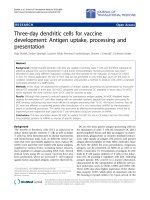
Astrocyte activation following Aβ infusionFigure 3
Astrocyte activation following Aβ infusion. WT and IL1raKO mice were infused with Aβ or vehicle, and brains prepared as in
Figure 2. A) Levels of the pro-inflammatory astrocyte-derived cytokine S100B showed a similar degree of upregulation in Aβ-
infused IL1raKO and WT mice. B) Numbers of GFAP-positive astrocytes were increased to a similar degree in both WT and
IL1raKO mice infused with Aβ. (error bars = SEM; p > 0.05 between Aβ-infused IL1raKO and WT mice).
WT WT KO KO
Veh. Aβ
ββ
β Ve h. Aβ
ββ
β
WT WT KO KO
Veh. Aβ
ββ
β Ve h. Aβ
ββ
β
A. S100B B. A s trocytes
Journal of Neuroinflammation 2005, 2:15 />Page 7 of 9
(page number not for citation purposes)
In addition, the resultant increased neuroinflammation in
the IL1raKO mice infused with Aβ was accompanied by an
exacerbation in the loss of synaptic markers, especially
PSD-95. This particular finding, in conjunction with our
similar findings in Aβ-infused S100B overexpressing
transgenic mice [29], strongly argues for the conclusion
that animals predisposed to neuroinflammation suffer
more severely from neurodegenerative sequelae following
Aβ infusion. Evidence from the epidemiological
assessment of AD risk factors also supports this conclu-
sion. Previous head injury, for example, is a significant
environmental risk factor for development of AD in which
it is hypothesized that IL-1-mediated neuroinflammation
plays a key role [30,31].
A somewhat surprising finding was that, unlike the
enhanced microglia and neuronal responses in the Aβ-
infused IL1raKO mice compared to WT mice, the astrocyte
responses to Aβ infusion were very similar in the two
mouse strains. Both IL1raKO and WT mice showed simi-
lar upregulation of S100B levels and GFAP immunoreac-
tivity after Aβ infusion. A possible explanation is that, at
the time point examined (42 days), astrocyte responses
had not yet reached their maximum following Aβ infu-
sion. This possibility indicates a need for future studies to
examine the temporal development of microglia, astro-
cyte, and neuronal responses after start of Aβ infusion.
The IL1raKO mice infused with Aβ experienced extensive
mortality during the course of the experiment, despite
minimal mortality of other strains of mice in our previous
studies [4,5,29]. At first inspection, this increased mortal-
ity could be explained by the pro-inflammatory status of
IL1raKO mice, which may predispose them to systemic
septic-like episodes at a higher frequency than their WT
littermates, especially following a major surgical opera-
tion to place an indwelling pump and ICV catheter. How-
ever, the lack of mortality in the IL1raKO mice that
received a vehicle infusion would argue against this con-
clusion. A more intriguing possibility is that these mice
died either directly or indirectly from a more severe neu-
roinflammatory response to Aβ than the mice that sur-
vived. The robust and consistent neuroinflammation,
which is one of the key hallmarks that characterizes the Aβ
infusion model, supports this conclusion as a distinct pos-
sibility. While quite interesting, especially in light of a
Loss of synaptic markers following Aβ infusionFigure 4
Loss of synaptic markers following Aβ infusion. WT and IL1raKO mice were infused with Aβ or vehicle, and brains prepared as
in Figure 2. A) Aβ-infused mice had reduced hippocampal PSD-95 levels compared to vehicle-infused mice, and there was a sig-
nificantly larger decrease in IL1raKO mice infused with Aβ versus their WT counterparts (error bars = SEM; * p < 0.05). B)
The presynaptic marker synaptophysin was reduced in Aβ-infused mice compared to the vehicle-infused mice. In addition, the
reduction in synaptophysin in Aβ-infused mice was greater in IL1raKO mice compared to WT mice, although the difference did
not quite reach statistical significance (error bars = SEM; p = 0.07).
WT K Oı
Veh . A
β
ββ
β
Veh . A
β
ββ
β
KO KO
Veh . A
β
ββ
β
Veh . A
β
ββ
β
A. PSD -95 B. Synaptophysin
WT WT WTKO
Journal of Neuroinflammation 2005, 2:15 />Page 8 of 9
(page number not for citation purposes)
similar syndrome afflicting a subset of individuals
enrolled in the now discontinued Aβ vaccine trials [32],
elucidation of the mechanisms underlying the increased
mortality will require additional research.
Conclusion
The major finding of this study is the demonstration that
IL1raKO mice show selective up-regulation of microglial
neuroinflammation and increased neuronal damage fol-
lowing Aβ infusion when compared to WT littermates.
The susceptibility of the IL1raKO mice to increased Aβ-
induced neuroinflammation was demonstrated by bio-
chemical and histological measurements of microglial
activation. This increase in microglial activation in the
IL1raKO mice is also associated with an increase in the
degree of synaptic degeneration observed following Aβ
infusion, suggesting that enhanced IL1 signaling leads to
deleterious neuroinflammation that either directly dam-
ages neurons and/or potentiates the neurotoxic effects of
Aβ. These data provide further support for the hypothesis
that increases in the level of IL1 signaling in the AD brain
can be detrimental through the cytokine's role as a key
component of the neuroinflammatory cascade that con-
tributes to progression of neuropathology. It also suggests
that manipulation of IL-1 signaling and other neuroin-
flammatory mediators and pathways could be utilized to
develop clinically meaningful, disease-modifying AD
therapies.
Competing interests
The authors declare that they have no competing interests
in the outcome, results, or conclusions of these studies.
Authors' contributions
JMC helped conceive the study and conducted animal sur-
geries, care, and biochemical/ histological assays. DMW
helped conceive the study, interpret the results, and assist
in the preparation of the manuscript. EH developed and
provided the IL1raKO mice and gave helpful advice for
handling and care of the animals. LVE helped conceive the
study, analyze data and assist in the preparation of the
manuscript.
Acknowledgements
We thank Sara Medgysi for assistance with mouse colony maintenance and
assays. These studies were supported in part by the Institute for the Study
of Aging (DMW) and by NIH grants R37 AG13939 (LVE), R01 AG20243
(LVE), and P01 AG21184 (LVE, DMW). JMC is a predoctoral fellow of the
Center for Drug Discovery and Chemical Biology training program, sup-
ported in part by NIH T32 AG00260, a predoctoral fellowship from the
Pharmaceutical Research and Manufacturers of America Foundation
(PhRMA), and a Ruth L. Kirschstein NRSA fellowship F30 NS46942.
References
1. Allan SM, Rothwell NJ: Inflammation in central nervous system
injury. Philos Trans R Soc Lond B Biol Sci 2003, 358:1669-1677.
2. Akiyama H, Barger S, Barnum S, Bradt B, Bauer J, Cole GM, Cooper
NR, Eikelenboom P, Emmerling M, Fiebich BL, Finch CE, Frautschy S,
Griffin WS, Hampel H, Hull M, Landreth G, Lue L, Mrak R, Mackenzie
IR, McGeer PL, O'Banion MK, Pachter J, Pasinetti G, Plata-Salaman C,
Rogers J, Rydel R, Shen Y, Streit W, Strohmeyer R, Tooyoma I, Van
Muiswinkel FL, Veerhuis R, Walker D, Webster S, Wegrzyniak B,
Wenk G, Wyss-Coray T: Inflammation and Alzheimer's
disease. Neurobiol Aging 2000, 21:383-421.
3. Griffin WST, Sheng JG, Royston MC, Gentleman SM, McKenzie JE,
Graham DI, Roberts GW, Mrak RE: Glial-Neuronal interactions
in Alzheimer's disease: the potential role of a 'cytokine cycle'
in disease progression. Brain Pathol 1998, 8:65-72.
4. Craft JM, Watterson DM, Frautschy SA, Van Eldik LJ: Aminopyri-
dazines inhibit beta-amyloid-induced glial activation and
neuronal damage in vivo. Neurobiol Aging 2004, 25:1283-1292.
5. Craft JM, Van Eldik LJ, Zasadzki M, Hu W, Watterson DM: Ami-
nopyridazines attenuate hippocampus-dependent behavio-
ral deficits induced by human beta-amyloid in a murine
model of neuroinflammation. J Mol Neurosci 2004, 24:115-122.
6. Hu W, Ranaivo HR, Craft JM, Van Eldik LJ, Watterson DM: Valida-
tion of the neuroinflammation cycle as a drug discovery tar-
get using integrative chemical biology and lead compound
development with an Alzheimer's disease-related mouse
model. Cur Alzheim Res 2005, 2:197-205.
7. Mirzoeva S, Sawkar A, Zasakzki M, Guo L, Velentza AV, Dunlap V,
Bourguignon JJ, Ramstrom H, Haiech J, Van Eldik LJ, Watterson DM:
Discovery of a 3-amino-6-phenyl-pyridazine derivative as a
new synthetic antineuroinflammatory compound. J Med
Chem 2002, 45:563-566.
8. Lim GP, Yang F, Chu T, Chen P, Beech W, Teter B, Tran T, Ubeda O,
Ashe KH, Frautschy SA, Cole GM: Ibuprofen suppresses plaque
pathology and inflammation in a mouse model for Alzhe-
imer's disease. J Neurosci 2000, 20:5709-5714.
9. Griffin WS, Sheng JG, Roberts GW, Mrak RE: Interleukin-1 expres-
sion in different plaque types in Alzheimer's disease: signifi-
cance in plaque evolution. J Neuropathol Exp Neurol 1995,
54:276-281.
10. Sheng JG, Mrak RE, Griffin WS: Glial-neuronal interactions in
Alzheimer disease: progressive association of IL-1alpha+
microglia and S100beta+ astrocytes with neurofibrillary tan-
gle stages. J Neuropathol Exp Neurol 1997, 56:285-290.
11. Sheng JG, Mrak RE, Griffin WS: Microglial interleukin-1 alpha
expression in brain regions in Alzheimer's disease: correla-
tion with neuritic plaque distribution. Neuropathol Appli
Neurobiol 1995, 21:290-301.
12. Das S, Potter H: Expression of the Alzheimer amyloid-promot-
ing factors α1-antichymotrypsin and apolipoprotein E is
induced in astrocytes by IL-1. Neuron 1995, 14:447-456.
13. Woiciechowsky C, Schoning B, Stoltenburg-Didinger G, Stockham-
mer F, Volk HD: Brain-IL-1 beta triggers astrogliosis through
induction of IL-6: inhibition by propranolol and IL-10. Med Sci
Monit 2004, 10:BR325-330.
14. Sheng JG, Ito K, Skinner RD, Mrak RE, Rovnaghi CR, Van Eldik LJ, Grif-
fin WS: In vivo and in vitro evidence supporting a role for the
inflammatory cytokine interleukin-1 as a driving force in
Alzheimer pathogenesis. Neurobiol Aging 1996, 17:761-766.
15. Akama KT, Van Eldik LJ: Beta-amyloid stimulation of inducible
nitric-oxide synthase in astrocytes is interleukin-1beta- and
tumor necrosis factor-alpha (TNFalpha)-dependent, and
involves a TNFalpha receptor-associated factor- and NFkap-
paB-inducing kinase-dependent signaling mechanism. J Biol
Chem 2000, 275:7918-7924.
16. Mrak RE, Griffin WS: Interleukin-1, neuroinflammation, and
Alzheimer's disease. Neurobiol Aging 2001, 22:903-908.
17. Nicoll JA, Mrak RE, Graham DI, Stewart J, Wilcock G, MacGowan S,
Esiri MM, Murray LS, Dewar D, Love S, Moss T, Griffin WS: Associ-
ation of interleukin-1 gene polymorphisms with Alzheimer's
disease. Ann Neurol 2000, 47:365-368.
18. Hirsch E, Irikura VM, Paul SM, Hirsh D: Functions of interleukin 1
receptor antagonist in gene knockout and overproducing
mice. Proc Natl Acad Sci U S A 1996, 93:11008-11013.
19. Dahlgren KN, Manelli AM, Stine WB Jr, Baker LK, Krafft GA, LaDu
MJ: Oligomeric and fibrillar species of amyloid-beta peptides
differentially affect neuronal viability. J Biol Chem 2002,
277:32046-32053.
Publish with Bio Med Central and every
scientist can read your work free of charge
"BioMed Central will be the most significant development for
disseminating the results of biomedical research in our lifetime."
Sir Paul Nurse, Cancer Research UK
Your research papers will be:
available free of charge to the entire biomedical community
peer reviewed and published immediately upon acceptance
cited in PubMed and archived on PubMed Central
yours — you keep the copyright
Submit your manuscript here:
/>BioMedcentral
Journal of Neuroinflammation 2005, 2:15 />Page 9 of 9
(page number not for citation purposes)
20. Frautschy SA, Yang F, Calderon L, Cole GM: Rodent models of
Alzheimer's disease: rat Aβ infusion approaches to amyloid
deposits. Neurobiol Aging 1996, 17:311-321.
21. Frautschy SA, Hu W, Kim P, Miller SA, Chu T, Harris-White ME, Cole
GM: Phenolic anti-inflammatory antioxidant reversal of Aβ-
induced cognitive deficits and neuropathology. Neurobiol Aging
2001, 22:993-1005.
22. Hayakata T, Shiozaki T, Tasaki O, Ikegawa H, Inoue Y, Toshiyuki F,
Hosotubo H, Kieko F, Yamashita T, Tanaka H, Shimazu T, Sugimoto
H: Changes in CSF S100B and cytokine concentrations in
early-phase severe traumatic brain injury. Shock 2004,
22:102-107.
23. Hu X, Nesic-Taylor O, Qiu J, Rea HC, Fabian R, Rassin DK, Perez-
Polo JR: Activation of nuclear factor-kappaB signaling path-
way by interleukin-1 after hypoxia/ischemia in neonatal rat
hippocampus and cortex. J Neurochem 2005, 93:26-37.
24. Basu A, Lazovic J, Krady JK, Mauger DT, Rothstein RP, Smith MB, Lev-
ison SW: Interleukin-1 and the interleukin-1 type 1 receptor
are essential for the progressive neurodegeneration that
ensues subsequent to a mild hypoxic/ischemic injury. J Cereb
Blood Flow Metab 2005, 25:17-29.
25. Sarchielli P, Galli F, Floridi A, Floridi A, Gallai V: Relevance of pro-
tein nitration in brain injury: a key pathophysiological mech-
anism in neurodegenerative, autoimmune, or inflammatory
CNS diseases and stroke. Amino Acids 2003, 25:427-36.
26. Griffin WS, Stanley LC, Ling C, White L, MacLeod V, Perrot LJ, White
CL 3rd, Araoz C: Brain interleukin 1 and S-100 immunoreac-
tivity are elevated in Down syndrome and Alzheimer
disease. Proc Natl Acad Sci U S A 1989, 86:7611-7615.
27. Van Everbroeck B, Dewulf E, Pals P, Lubke U, Martin JJ, Cras P: The
role of cytokines, astrocytes, microglia and apoptosis in
Creutzfeldt-Jakob disease. Neurobiol Aging 2002, 23:59-64.
28. Mrak RE, Griffin WS: The role of chronic self-propagating glial
responses in neurodegeneration: implications for long-lived
survivors of human immunodeficiency virus. J Neurovirol 1997,
3:241-246.
29. Craft JM, Watterson DM, Marks A, Van Eldik LJ: Enhanced suscep-
tibility of S-100B transgenic mice to neuroinflammation and
neuronal dysfunction induced by intracerebroventricular
infusion of human beta-amyloid. Glia 2005. available online Apr 4
30. Griffin WS, Sheng JG, Gentleman SM, Graham DI, Mrak RE, Roberts
GW: Microglial interleukin-1 alpha expression in human head
injury: correlations with neuronal and neuritic beta-amyloid
precursor protein expression. Neurosci Lett 1994, 176:133-136.
31. Mortimer JA, van Duijn CM, Chandra V, Fratiglioni L, Graves AB, Hey-
man A, Jorm AF, Kokmen E, Kondo K, Rocca WA, EURODEM Risk
Factors Research Group: Head trauma as a risk factor for
Alzheimer's disease: a collaborative re-analysis of case-con-
trol studies. Int J Epidemiol 1991, 20(Suppl 2):S28-35.
32. Orgogozo JM, Gilman S, Dartigues JF, Laurent B, Puel M, Kirby LC,
Jouanny P, Dubois B, Eisner L, Flitman S, Michel BF, Boada M, Frank
A, Hock C: Subacute meningoencephalitis in a subset of
patients with AD after Abeta42 immunization. Neurology
2003, 61:46-54.