báo cáo hóa học: " Quinolinic acid selectively induces apoptosis of human astrocytes: potential role in AIDS dementia complex" ppt
Bạn đang xem bản rút gọn của tài liệu. Xem và tải ngay bản đầy đủ của tài liệu tại đây (623.06 KB, 6 trang )
BioMed Central
Page 1 of 6
(page number not for citation purposes)
Journal of Neuroinflammation
Open Access
Short report
Quinolinic acid selectively induces apoptosis of human astrocytes:
potential role in AIDS dementia complex
Gilles J Guillemin*
1,3
, Lily Wang
1
and Bruce J Brew
1,2
Address:
1
Centre for Immunology, St Vincent's Hospital, Sydney, Australia,
2
Department of Neurology, St Vincent's Hospital, Sydney, Australia
and
3
University of New South Wales, Faculty of Medicine, Sydney, Australia
Email: Gilles J Guillemin* - ; Lily Wang - ; Bruce J Brew -
* Corresponding author
Humanastrocyteapoptosisquinolinic acidcaspase 3AIDS dementia complex
Abstract
There is evidence that the kynurenine pathway (KP) and particularly one of its end products,
quinolinic acid (QUIN) play a role in the pathogenesis of several major neuroinflammatory diseases,
and more particularly AIDS dementia complex (ADC). We hypothesized that QUIN may be
involved in astrocyte apoptosis because: 1) apoptotic astrocytes have been observed in the brains
of ADC patients, 2) ADC patients have elevated cerebrospinal fluid QUIN concentrations, and 3)
QUIN can induce astrocyte death. Primary cultures of human fetal astrocytes were treated with
three pathophysiological concentrations of QUIN. Numeration of apoptotic cells was assessed
using double immunocytochemistry for expression of active caspase 3 and for nucleus
condensation. We found that treatment of human astrocytes with QUIN induced morphological
(cell body shrinking) and biochemical changes (nucleus condensation and over-expression of active
caspase 3) of apoptosis. After 24 hours of treatment with QUIN 500 nM and 1200 nM respectively
10 and 14% of astrocytes were undergoing apoptosis. This would be expected to lead to a relative
lack of trophic support factors with consequent neuronal dysfunction and possibly death. Astroglial
apoptosis induced by QUIN provides another potential mechanism for the neurotoxicity of QUIN
during ADC.
Findings
The kynurenine pathway (KP) is a major route of L-tryp-
tophan catabolism, resulting in the production of nicoti-
namide adenine dinucleotide and other neuroactive
intermediates [1]. Of the metabolites, the N-methyl-D-
aspartate (NMDA) receptor agonist and neurotoxin quin-
olinic acid (QUIN) is likely to be one of the most impor-
tant. There is evidence that QUIN is involved in the
neurocytotoxicity associated with several major inflam-
matory brain diseases [2,3] such as AIDS dementia com-
plex (ADC) [4,5] and other viral brain infections [2]. In
the central nervous system (CNS), astrocytes play essential
roles including metabolic homeostasis especially provid-
ing neurotrophic support, detoxification, maintenance of
the blood brain barrier and immune response. During the
brain inflammation associated with ADC, many media-
tors are released and astrocytes are activated leading to
their cellular hypertrophy and/or proliferation [6]. For
some astrocytes, this prolonged activation may induce
apoptosis and the death of these reactive astrocytes can
Published: 26 July 2005
Journal of Neuroinflammation 2005, 2:16 doi:10.1186/1742-2094-2-16
Received: 02 March 2005
Accepted: 26 July 2005
This article is available from: />© 2005 Guillemin et al; licensee BioMed Central Ltd.
This is an Open Access article distributed under the terms of the Creative Commons Attribution License ( />),
which permits unrestricted use, distribution, and reproduction in any medium, provided the original work is properly cited.
Journal of Neuroinflammation 2005, 2:16 />Page 2 of 6
(page number not for citation purposes)
directly and/or indirectly affect functions and survival of
the neighbouring neurons and astrocytes [7]. The conse-
quences of astrocyte apoptosis could be either neuropro-
tective [8] or neurodamaging [7,9]. Apoptosis of
astrocytes has been described in the brains of patients
with ADC [10-12]. Furthermore, in ADC both brain
parenchyma and cerebrospinal fluid (CSF) concentrations
of QUIN are strongly elevated [4,5,13] respectively 300
and 100 fold compared to controls. The HIV-1 proteins
Nef and Tat induce macrophages to produce QUIN [14].
The association between brain cell apoptosis and
increased levels of QUIN have been found in various
other neurodegenerative diseases. We therefore hypothe-
sized that QUIN could be linked with astrocyte apoptosis.
We used primary cultures of human fetal astrocytes
treated with three pathophysiological concentrations of
QUIN respectively 350, 500 and 1200 nM (similar to
those in brain parenchyma of ADC patients [15]) and
assessed them for apoptosis with immunocytochemistry.
We found that 99% of the cells were GFAP positive (green
staining, Fig. 1, left column) demonstrating the high
purity of the primary cultures of human astrocytes. No
staining was detected for CD68, MAP2, factor VIII or 5B5
(data not included). Apoptotic cells started to be detected
after only 6 hours(data not presented). Apoptotic astro-
cytes displayed an atypical morphology with a shrunken
body and abnormal processes. Moreover, most of these
cells are in the process of detaching from the culture flask.
These apoptotic cells were easily spotted due to their con-
densed and very bright nucleus with the DAPI staining
(cyan staining, Fig. 1). All these cells also displayed a
strong cytoplasmic and perinuclear staining for the anti-
active caspase-3 (red staining, Fig. 1, right column). The
percentage of apoptotic cells was calculated after 24 hours
of treatment (as described in the methodological section
below) (Fig. 2). After 24 hours, the percentage of apop-
totic cells in cycloheximide-treated slides was increased 5-
fold whilst in QUIN 350, QUIN 500 and QUIN 1200 nM-
treated slides they were respectively increased by 2.2, 4.2
and 5.6-fold compared to baseline. The p-values between
the cycloheximide-treated slides and the QUIN 500 and
1200 nM-treated slides were not significant 24 hours but
were significant between control slides and treated slides,
with exception for QUIN 350 nM (see Fig. 2 legend).
This study demonstrated that treatments with QUIN in
pathophysiological concentrations induced morphologi-
cal and biochemical changes of apoptosis in a sub-group
of human astrocytes in a dose dependent manner. Some
issues can be raised in term of potential experimental lim-
itations of our in vitro results. Firstly, there are two known
subtypes of astrocytes [16] but only type1 astrocytes can
be grown in the primary cultures obtained by our method
[17]. Differences between these subtypes are not well
known and it is possible that they may have a different
sensitivity to QUIN. Secondly, it can be argued that fetal
and adult astrocytes may have a different sensitivity to
QUIN. This is unlikely because we previously showed the
high degree of similarity between immature and mature
astrocytes [18,19]. Moreover, in ADC brains high levels of
QUIN [15] are associated with a high rate of apoptosis of
adult astrocytes [10]. Finally, the limited number of apop-
totic astrocytes (<15%) implies that only a subset of astro-
cytes is susceptible to QUIN toxicity. The receptors and
signalling pathways that initiate astroglial apoptosis has
not been identify yet. However, two main mechanisms
may potentially be involved. The first is the activation of
NMDA receptors by QUIN [4], which is already known to
mediate neuronal apoptosis with caspase-3 activation
[20-23]. However, the presence of NMDA receptors on
astrocytes is very controversial [24,25]. A similar pathway
may be involved in neurons and astrocytes, but the pres-
ence of functional NMDA receptors on astrocytes still has
to be proved. The second possibility, which represent
another important aspect of QUIN toxicity, is lipid perox-
idation [26]. QUIN in concentration as low as 120 nM
induces lipid peroxidation and formation of free radicals
leading to neuronal death [27]. This second mechanism is
more likely to be involved astrocyte apoptosis induced by
QUIN.
Interestingly, our in vitro results on QUIN toxicity in
human astrocytes correlate well with previous in vivo stud-
ies using animal models. After QUIN injection into the rat
brain, a reduction in the density of GFAP-immunoreactiv-
ity of astrocytes occurs early (within 6 hours of injection)
suggesting astrocytic death; which could be associated
with necrosis rather than apoptosis [28,29]. Another in
vivo study using fetuses derived from ewes and damaged
by hypoxia and hypoglycemia showed a significant
increase in QUIN concentration associated with reduction
in GFAP in fetal brain [30]. However, whether QUIN is
able to induce astrogliosis and/or astrocytosis is still con-
troversial [28,29]. QUIN can increase local inflammation
by inducing production of cytokines and chemokines by
human astrocytes [18]. QUIN increases neuronal release
of glutamate and inhibits its uptake by astrocytes, leading
to an excessive glutamate concentration in the neuronal
microenvironment and secondly to neurotoxicity [9].
It is still debated whether astroglial activation is beneficial
or detrimental to neurons [7]. For some astrocytes, this
activation will lead to an early cellular death, as we
showed here with QUIN. This may be part of a selective
neuroprotective mechanism because on one hand reactive
astrocytes may contribute to a decline of neurological
function through various mechanisms [8]. On the other
hand, the loss of astrocytes compromises their beneficial
effects on neuronal function and survival [31]. The results
Journal of Neuroinflammation 2005, 2:16 />Page 3 of 6
(page number not for citation purposes)
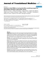
Detection of apoptotic astrocytes using immunocytochemistryFigure 1
Detection of apoptotic astrocytes using immunocytochemistry. Double staining for GFAP (Left column/green)) and active cas-
pase 3 (Right column/red). Nucleuses are stained in blue with DAPI. A) Untreated cells; B) treated with QUIN 350 nM; C)
treated with QUIN 500 nM; D) treated with QUIN 1200 nM; E) treated with cycloheximide 20 µg/ml for 24 hours. Arrows
point apoptotic astrocytes.
GFAP
Active Caspase 3
A
C
D
B
E
Untreated
QUIN
350 nM
QUIN
500 nM
QUIN
1200 nM
Cyclo
heximide
Journal of Neuroinflammation 2005, 2:16 />Page 4 of 6
(page number not for citation purposes)
of this study are biologically relevant because brain cell
apoptosis (including neurons and astrocytes) and
increased levels of QUIN have been found associated in
various neurodegenerative diseases and brain disorders
such as ADC [10], Alzheimer's disease [32,33], cerebral
malaria [34], and traumatic brain injury [35].
A large majority of the in vivo or ex vivo studies concerning
astrocyte apoptosis are related to HIV brain infection and
ADC [12]. Thompson et al. [10] found that there is a cor-
relation between an increased number of HIV DNA-posi-
tive astrocytes and an increased number of apoptotic
astrocyte and rapid progression of patients to dementia.
Furthermore, QUIN production is directly related to the
viral load in patients with ADC [36]. There is strong evi-
dence that ADC is associated with NMDA receptor activa-
tion [37,38]. We previously demonstrated that HIV-1
proteins Nef and Tat lead to production of high levels of
QUIN by human macrophages [14]; that inhibition of
QUIN production by HIV-1 infected macrophages
strongly reduces neurotoxicity [39]; and finally that QUIN
can amplify neuroinflammation and increase astroglial
expression of HIV-1 co-receptors [18]. Interestingly, viral
induction of IDO in human macrophages differs accord-
ing to particular HIV-1 isolates [40] and similarly diverse
HIV-1 primary isolates have different effects on astrocyte
apoptosis [11]. The present study provides a direct link
between the astrocyte apoptosis and high levels of QUIN
in ADC brains. These data provide a new mechanism by
which HIV-1 may be involved in astrocyte apoptosis
directly and indirectly via QUIN production. The charac-
terisation of molecular pathways leading to QUIN-
induced astrocyte apoptosis has the potential for develop-
ing agents that reduce glial and neuronal death related to
QUIN-toxicity. Development of pharmacological agents
targeting specific KP enzymes was reviewed recently [41].
All cell culture media and additives were from Invitrogen
(Melbourne, VIC, Australia) unless otherwise stated.
QUIN, DAPI, cycloheximide was obtained from Sigma-
Aldrich Chemical Co. (Sydney, NSW, Australia). anti-
active caspase-3 antibody (polyclonal) was from BD
Pharmingen. Mouse mAb anti-GFAP (clone GA-5) was
obtained from Novacostra (Newcastle, UK). Secondary
goat anti-mouse IgG and anti-rabbit Alexa 488 (green) or
Alexa 594 (red) conjugated antibodies were purchased
from Molecular Probes (Eugene, OR, USA). All commer-
cial antibodies were used at the concentrations recom-
mended by the manufacturer. Human fetal brains were
obtained from 16 to 19 week old fetuses collected follow-
ing informed consent. Astrocytes were prepared using a
protocol adapted from previously described methods
[17]. The experiments were carried out in triplicate
throughout. Initially, cells were incubated in serum-free
AIM-V. The negative control cells were incubated in AIM-
V only. The QUIN group was incubated in 350, 500 and
1200 nM QUIN in AIM-V. The positive control cells were
incubated in cycloheximide (20 µg/ml) [42] in AIM-V.
Dose-response and time course (3, 6, 12, 24, 48, 72
hours) have been done for QUIN and cycloheximide
(data not shown). The optimal time of incubation was 24
hours for the active caspase 3 detection. QUIN concentra-
tions between 350 nM to 1200 nM are known to be found
in patients with ADC [15]. The characterization of human
brain cell using immunocytochemistry was previously
described [17]. The following three controls were per-
formed for each labelling experiment: 1) isotypic anti-
body controls, 2) incubation with only the secondary
labelled antibodies, and 3) estimation of auto-fluores-
cence of unlabelled cells. The cell counting was performed
in a blinded manner. The whole controls and treated
chamber slides were counted. Enumeration for each slide
was rechecked by the experimenter and cells classified
according to the following scheme: DAPI staining for total
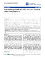
Numeration of apoptotic astrocytes using immunocytochemistryFigure 2
Numeration of apoptotic astrocytes using immunocytochem-
istry. Histogram showing the percentage of apoptotic astro-
cytes relative to the total number of astrocytes after 24
hours of treatment. Mean values and standard errors were
calculated for each treatment. Unpaired t tests were used to
analyse the significance of differences between pairs of the
three treatments. A p value of <0.05 was regarded as statisti-
cally significant. p value between controls and QUIN 350 nM,
QUIN 500 nM, 1200 nM treated slides were respectively
0.07 (NS), 0.04 (**) and 0.004 (***); and 0.002 (***) between
controls and cycloheximide treated slides. p values between
QUIN 500 nM, 1200 nM and cycloheximide treated slides
were not significantly different.
**
%
0
4
2
6
8
20
10
12
14
16
18
Control QUIN
1200 nM
Cyclo
heximide
QUIN
500 nM
QUIN
350 nM
NS
***
***
Journal of Neuroinflammation 2005, 2:16 />Page 5 of 6
(page number not for citation purposes)
cell number, GFAP immunoreactivity for astrocytes, and
active caspase 3 immunoreactivity together with DAPI for
apoptotic cells. Experiments were done in triplicates using
brain cells from three different brain tissues. Mean values
and standard errors were calculated for each treatment
and the results were plotted on a histogram (Fig. 2).
Unpaired t tests were performed on the results obtained at
24 hours. Student's t-test was used to analyse the signifi-
cance of differences between pairs of the three treatments.
A p value of <0.05 was regarded as statistically significant.
List of abbreviations
AIDS dementia complex (ADC), cerebrospinal fluid
(CSF), 4',6-diamidino-2-phenylindole (DAPI), Glial
fibrillary acid protein (GFAP), human immunodeficiency
virus (HIV), kynurenine pathway (KP), NMDA (N-
methyl-D-aspartate), Quinolinic acid (QUIN).
Competing interests
The author(s) declare that they have no competing
interests.
Authors' contributions
GG was responsible for conception and planning of the
experiments, for performing the immunocytochemical
study and for writing of the manuscript. LW was growing
the primary cultures of human astrocytes and did the var-
ious treatments. BJB contributed to the interpretation of
the results, discussion and writing of the manuscript.
Acknowledgements
The St Vincent's Clinic Foundation, The Australian Alzheimer Foundation,
the National Health and Medical Research Council, the NSW Health Dept
and the University of NSW have supported this work. We thank Ms Belinda
Creighton (UNSW) for the advice and aliquot of anti caspase 3 pAb.
References
1. Stone TW: Neuropharmacology of quinolinic and kynurenic
acids. Pharmacol Rev 1993, 45:309-379.
2. Heyes MP, Saito K, Crowley JS, Davis LE, Demitrack MA, Der M, Dill-
ing LA, Elia J, Kruesi MJ, Lackner A, et al.: Quinolinic acid and
kynurenine pathway metabolism in inflammatory and non-
inflammatory neurological disease. Brain 1992, 115 ( Pt
5):1249-1273.
3. Stone TW: Endogenous neurotoxins from tryptophan. Toxicon
2001, 39:61-73.
4. Guillemin GJ, Kerr SJ, Brew BJ: Involvement of quinolinic acid in
AIDS dementia complex. Neurotox Res 2004, 7:103-124.
5. Heyes MP, Brew BJ, Martin A, Price RW, Salazar AM, Sidtis JJ, Yergey
JA, Mouradian MM, Sadler AE, Keilp J, et al.: Quinolinic acid in cer-
ebrospinal fluid and serum in HIV-1 infection: relationship to
clinical and neurological status. Ann Neurol 1991, 29:202-209.
6. Eddelston M, Mucke L: Molecular profile of reactive astrocytes-
implications for their role in neurologic disease. Neuroscience
1993, 54:15-36.
7. Tacconi MT: Neuronal death: is there a role for astrocytes?
Neurochem Res 1998, 23:759-765.
8. Takuma K, Baba A, Matsuda T: Astrocyte apoptosis: implications
for neuroprotection. Prog Neurobiol 2004, 72:111-127.
9. Tavares RG, Tasca CI, Santos CE, Alves LB, Porciuncula LO, Emanuelli
T, Souza DO: Quinolinic acid stimulates synaptosomal gluta-
mate release and inhibits glutamate uptake into astrocytes.
Neurochem Int 2002, 40:621-627.
10. Thompson KA, McArthur JC, Wesselingh SL: Correlation between
neurological progression and astrocyte apoptosis in HIV-
associated dementia. Ann Neurol 2001, 49:745-752.
11. Ohagen A, Ghosh S, He J, Huang K, Chen Y, Yuan M, Osathanondh
R, Gartner S, Shi B, Shaw G, Gabuzda D: Apoptosis induced by
infection of primary brain cultures with diverse human
immunodeficiency virus type 1 isolates: evidence for a role of
the envelope. J Virol 1999, 73:897-906.
12. Sabri F, Titanji K, De Milito A, Chiodi F: Astrocyte activation and
apoptosis: their roles in the neuropathology of HIV infection.
Brain Pathol 2003, 13:84-94.
13. Heyes MP, Ellis RJ, Ryan L, Childers ME, Grant I, Wolfson T, Archibald
S, Jernigan TL: Elevated cerebrospinal fluid quinolinic acid lev-
els are associated with region-specific cerebral volume loss
in HIV infection. Brain 2001, 124:1033-1042.
14. Smith DG, Guillemin GJ, Pemberton L, Kerr S, Nath A, Smythe GA,
Brew BJ: Quinolinic acid is produced by macrophages stimu-
lated by platelet activating factor, Nef and Tat. J Neurovirol
2001, 7:56-60.
15. Heyes MP, Saito K, Lackner A, Wiley CA, Achim CL, Markey SP:
Sources of the neurotoxin quinolinic acid in the brain of HIV-
1- infected patients and retrovirus-infected macaques. Faseb
J 1998, 12:881-896.
16. Raff MC, Abney ER, Cohen J, Lindsay R, Noble M: Two types of
astrocytes in cultures of developing rat white matter: differ-
ences in morphology, surface gangliosides and growth
characteristics. J Neurosc 1983, 3:1289-1300.
17. Guillemin G, Boussin FD, Croitoru J, Franck-Duchenne M, Le Grand
R, Lazarini F, Dormont D: Obtention and characterization of
primary astrocyte and microglial cultures from adult mon-
key brains. J Neurosci Res 1997, 49:576-591.
18. Guillemin GJ, Croitoru-Lamoury J, Dormont D, Armati PJ, Brew BJ:
Quinolinic acid upregulates chemokine production and
chemokine receptor expression in astrocytes. Glia 2003,
41:371-381.
19. Croitoru-Lamoury J, Guillemin GJ, Boussin FD, Mognetti B, Gigout LI,
Cheret A, Vaslin B, Le Grand R, Brew BJ, Dormont D: Expression
of chemokines and their receptors in human and simian
astrocytes: Evidence for a central role of TNFalpha and IFN-
gamma in CXCR4 and CCR5 modulation. Glia 2003,
41:354-370.
20. Yu SP, Yeh CH, Gottron F, Wang X, Grabb MC, Choi DW: Role of
the outward delayed rectifier K+ current in ceramide-
induced caspase activation and apoptosis in cultured cortical
neurons. J Neurochem 1999, 73:933-941.
21. Tenneti L, Lipton SA: Involvement of activated caspase-3-like
proteases in N-methyl-D-aspartate-induced apoptosis in cer-
ebrocortical neurons. J Neurochem 2000, 74:134-142.
22. Kerr SJ, Armati PJ, Brew BJ: Neurocytotoxity of quinolinic acid
in human brain cultures. J Neurovirol 1995, 1:375-380.
23. Stone TW: Kynurenines in the CNS: from endogenous obscu-
rity to therapeutic importance. Prog Neurobiol 2001, 64:185-218.
24. Schipke CG, Ohlemeyer C, Matyash M, Nolte C, Kettenmann H,
Kirchhoff F: Astrocytes of the mouse neocortex express func-
tional N-methyl-D-aspartate receptors. FASEB J
2001:00-0439fje.
25. Conti F, Minelli A, DeBiasi S, Melone M: Neuronal and glial local-
ization of NMDA receptors in the cerebral cortex. Mol
Neurobiol 1997, 14:1-18.
26. Rios C, Santamaria A: Quinolinic acid is a potent lipid peroxi-
dant in rat brain homogenates. Neurochem Res 1991,
16:1139-1143.
27. Behan WM, McDonald M, Darlington LG, Stone TW: Oxidative
stress as a mechanism for quinolinic acid-induced hippocam-
pal damage: protection by melatonin and deprenyl. Br J
Pharmacol 1999, 128:1754-1760.
28. Bjorklund H, Olson L, Dahl D, Schwarcz R: Short- and long-term
consequences of intracranial injections of the excitotoxin,
quinolinic acid, as evidenced by GFA immunohistochemistry
of astrocytes. Brain Res 1986, 371:267-277.
29. Dihne M, Block F, Korr H, Topper R: Time course of glial prolif-
eration and glial apoptosis following excitotoxic CNS injury.
Brain Res 2001, 902:178-189.
30. Nicholls T, Lacey B, Nitsos I, Smythe G, Walker DW: Regional
changes in kynurenic acid, quinolinic acid, and glial fibrillary
acidic protein concentrations in the fetal sheep brain after
Publish with BioMed Central and every
scientist can read your work free of charge
"BioMed Central will be the most significant development for
disseminating the results of biomedical research in our lifetime."
Sir Paul Nurse, Cancer Research UK
Your research papers will be:
available free of charge to the entire biomedical community
peer reviewed and published immediately upon acceptance
cited in PubMed and archived on PubMed Central
yours — you keep the copyright
Submit your manuscript here:
/>BioMedcentral
Journal of Neuroinflammation 2005, 2:16 />Page 6 of 6
(page number not for citation purposes)
experimentally induced placental insufficiency. Am J Obstet
Gynecol 2001, 184:203-208.
31. Srebro Z, Dziobek K: Neuroprotection: the role of neuroglia.
Folia Med Cracov 2001, 42:113-121.
32. Barinaga M: Is apoptosis key in Alzheimer's disease? Science
1998, 281:1303-1304.
33. Guillemin GJ, Brew BJ, Noonan C, Cullen K: Indoleamine 2,3 diox-
ygenase and quinolinic acid in plaque and neurons in post-
mortem brain tissue from Alzheimer’s disease and control
cases. Neuropathol Appl Neurobiol 2005, in press:.
34. Sanni LA, Thomas SR, Tattam BN, Moore DE, Chaudhri G, Stocker R,
Hunt NH: Dramatic changes in oxidative tryptophan metab-
olism along the kynurenine pathway in experimental cere-
bral and noncerebral malaria. Am J Pathol 1998, 152:611-619.
35. Sinz EH, Kochanek PM, Heyes MP, Wisniewski SR, Bell MJ, Clark RS,
DeKosky ST, Blight AR, Marion DW: Quinolinic acid is increased
in CSF and associated with mortality after traumatic brain
injury in humans. J Cereb Blood Flow Metab 1998, 18:610-615.
36. Brew BJ, Rosenblum M, Cronin K, Price RW: AIDS dementia com-
plex and HIV-1 brain infection: clinical-virological
correlations. Ann Neurol 1995, 38:563-570.
37. Lipton SA: Neuronal injury associated with HIV-1: approaches
to treatment. Annu Rev Pharmacol Toxicol 1998, 38:159-177.
38. Chao CC, Hu S, Peterson PK: Glia: the not so innocent
bystanders. J Neurovirol 1996, 2:234-239.
39. Kerr SJ, Armati PJ, Pemberton LA, Smythe G, Tattam B, Brew BJ:
Kynurenine pathway inhibition reduces neurotoxicity of
HIV-1-infected macrophages. Neurology 1997, 49:1671-1681.
40. Grant RS, Naif H, Thuruthyil SJ, Nasr N, Littlejohn T, Takikawa O,
Kapoor V: Induction of indolamine 2,3-dioxygenase in pri-
mary human macrophages by human immunodeficiency
virus type 1 is strain dependent. J Virol 2000, 74:4110-4115.
41. Stone TW, Darlington LG: Endogenous kynurenines as targets
for drug discovery and development. Nat Rev Drug Discov 2002,
1:609-620.
42. Tsuchida T, Kato T, Yamada A, Kawamoto K: Cycloheximide
induces apoptosis of astrocytes. Pathol Int 2002, 52:181-185.