báo cáo hóa học: " Activation of microglial NADPH oxidase is synergistic with glial iNOS expression in inducing neuronal death: a dual-key mechanism of inflammatory neurodegeneration" docx
Bạn đang xem bản rút gọn của tài liệu. Xem và tải ngay bản đầy đủ của tài liệu tại đây (1.18 MB, 15 trang )
BioMed Central
Page 1 of 15
(page number not for citation purposes)
Journal of Neuroinflammation
Open Access
Research
Activation of microglial NADPH oxidase is synergistic with glial
iNOS expression in inducing neuronal death: a dual-key mechanism
of inflammatory neurodegeneration
Palwinder Mander and Guy C Brown*
Address: Biochemistry Department, University of Cambridge, Tennis Court Road, Cambridge, CB2 1QW, UK
Email: Palwinder Mander - ; Guy C Brown* -
* Corresponding author
microgliaperoxynitritenitric oxideprion proteininflammationcytokines
Abstract
Background: Inflammation-activated glia are seen in many CNS pathologies and may kill neurons
through the release of cytotoxic mediators, such as nitric oxide from inducible NO synthase
(iNOS), and possibly superoxide from NADPH oxidase (NOX). We set out to determine the
relative role of these species in inducing neuronal death, and to test the dual-key hypothesis that
the production of both species simultaneously is required for significant neuronal death.
Methods: Primary co-cultures of cerebellar granule neurons and glia from rats were used to
investigate the effect of NO (from iNOS, following lipopolysaccharide (LPS) and/or cytokine
addition) or superoxide/hydrogen peroxide (from NOX, following phorbol 12-myristate 13-
acetate (PMA), ATP analogue (BzATP), interleukin-1β (IL-1β) or arachidonic acid (AA) addition) on
neuronal survival.
Results: Induction of glial iNOS caused little neuronal death. Similarly, activation of NOX alone
resulted in little or no neuronal death. However, if NOX was activated (by PMA or BzATP) in the
presence of iNOS (induced by LPS and interferon-γ) then substantial delayed neuronal death
occurred over 48 hours, which was prevented by inhibitors of iNOS (1400W), NOX (apocynin)
or a peroxynitrite decomposer (FeTPPS). Neurons and glia were also found to stain positive for
nitrotyrosine (a putative marker of peroxynitrite) only when both iNOS and NOX were
simultaneously active. If NOX was activated by weak stimulators (IL-1β, AA or the fibrillogenic
prion peptide PrP106-126) in the presence of iNOS, it caused microglial proliferation and delayed
neurodegeneration over 6 days, which was prevented by iNOS or NOX inhibitors, a peroxynitrite
decomposer or a NMDA-receptor antagonist (MK-801).
Conclusion: These results suggest a dual-key mechanism, whereby glial iNOS or microglial NOX
activation alone is relatively benign, but if activated simultaneously are synergistic in killing neurons,
through generating peroxynitrite. This mechanism may mediate inflammatory neurodegeneration
in response to cytokines, bacteria, ATP, arachidonate and pathological prions, in which case
neurons may be protected by iNOS or NOX inhibitors, or scavengers of NO, superoxide or
peroxynitrite.
Published: 12 September 2005
Journal of Neuroinflammation 2005, 2:20 doi:10.1186/1742-2094-2-20
Received: 25 July 2005
Accepted: 12 September 2005
This article is available from: />© 2005 Mander and Brown; licensee BioMed Central Ltd.
This is an Open Access article distributed under the terms of the Creative Commons Attribution License ( />),
which permits unrestricted use, distribution, and reproduction in any medium, provided the original work is properly cited.
Journal of Neuroinflammation 2005, 2:20 />Page 2 of 15
(page number not for citation purposes)
Background
Glia (microglia and astrocytes) can become inflammation
activated in many CNS pathologies, including infectious,
ischaemic, inflammatory and neurodegenerative disor-
ders [1,2]. Glial activation may be protective to the host,
as it can lead to the removal of cell debris and killing of
pathogens [3]. However excessive or chronic glial activa-
tion can kill nearby neurons [4,5]. Thus inflammation
may contribute to many CNS pathologies including
Alzheimer's, Parkinson's and motor neuron diseases, mul-
tiple sclerosis, meningitis, AIDS dementia, strokes, trauma
and normal brain ageing [6,7]. It is therefore important to
understand the mechanisms by which inflammatory-acti-
vated glia kill neurons.
Astrocytes and microglia can become activated by a range
of factors, including pathogens and pro-inflammatory
cytokines, and can lead to the subsequent death of co-cul-
tured neurons [8,9]. Activated astrocytes and/or microglia
produce a variety of factors which can mediate neuronal
death, including reactive oxygen species (ROS) [10,11],
nitric oxide [8,9,12] and glutamate [8,13], as well as pro-
inflammatory cytokines that perpetuate glial activation,
such as interleukin-1β (IL-1β) and tumour necrosis factor-
α (TNF-α) [14].
The neuroprotective effects of anti-oxidants have been
established [15] and are thought to be due to the removal
of ROS (such as superoxide) and as well as more toxic
molecules (such as peroxynitrite) [16]. There is evidence
that NADPH oxidase is activated in Alzheimer's disease
and AIDS dementia [17-19]. The major source of ROS
during inflammation is NADPH oxidase [20,21],
although other sources may also contribute [22,23].
NADPH oxidase is expressed mainly by microglia in the
brain [21,24], and produces superoxide (O
2
-
) extracellu-
larly or within phagocytic vesicles, in order to kill patho-
gens. The oxidase can be acutely activated by PMA, ATP,
arachidonic acid, some chemokines and cytokines [25-
28]. Superoxide is then broken down mainly by extracel-
lular and intracellular superoxide dismutase to give
hydrogen peroxide (H
2
O
2
).
iNOS is not normally expressed in the brain, but is
induced in astrocytes and microglia by proinflammatory
cytokines and pathogen components, such as lipopolysac-
charide (LPS)/endotoxin of Gram-negative bacteria [29].
Once expressed iNOS produces high, sustained levels of
NO which can, in certain conditions, kill nearby neurons,
by mechanisms including inhibition of mitochondrial
respiration and the release of glutamate from neurons and
glia, resulting in excitotoxicity [8]. However, such mecha-
nisms may require a relatively high level of NO and/or a
relatively low level of oxygen [30,31]. An alternative
mechanism would be for NO to react with superoxide
(e.g. from the NADPH oxidase) to produce peroxynitrite
(ONOO-), which is potentially more neurotoxic to neu-
rons than NO or superoxide [32,33].
This suggests a dual-key hypothesis of inflammatory neu-
rodegeneration whereby iNOS expression or NADPH oxi-
dase activation alone is relatively benign, but when
combined together at the same time causes neurodegener-
ation via peroxynitrite. We have previously shown that
acute activation of the NADPH oxidase in isolated micro-
glia expressing iNOS results in the rapid disappearance of
NO and produces ONOO
-
[32]. In this paper we report
that activation of the microglial NADPH oxidase to pro-
duce superoxide is synergistic with NO from iNOS in
inducing death of co-cultured neurons, whereas activation
of either alone causes little or no death of co-cultured
neurons.
Materials & methods
Materials
The following materials were purchased from the indi-
cated sources: 1400W.dihydrochloride from Alexis (Not-
tingham, UK); MK-801 maleate, apocynin and FeTPPS
(5,10,15,20-Tetrakis(4-sulfonatophenyl)porphyrinato
Iron (III) chloride) from Calbiochem (Nottingham, UK).
All other reagents were ordered from Sigma (Poole, UK).
Neuronal-glial culture
Cerebellar granule cell (CGC) cultures were prepared
from 7-day-old Wistar rats, as described in Bal-Price &
Brown, 2001. Briefly, the pups were anaesthetised using
5% halothane in oxygen, followed by decapitation. Brains
were removed under sterile conditions and the cerebellum
dissected. Meninges were removed and the cerebella dis-
sociated in Versene solution (1:5000, Gibco BRL) and
plated at 0.25 × 10
6
cells/cm
2
in 24-well plates (in 500 µl
DMEM) coated with 0.001% poly-L-lysine. Cultures were
maintained in DMEM (Gibco BRL) supplemented with
5% horse serum, 5% foetal calf serum, 38 mM glucose, 5
mM HEPES, 2 mM glutamine, 25 mM KCl and 10 µg/ml
gentamicin. Cells were kept at 37°C in a humidified
atmosphere of 5% CO
2
/95% air and used for experiments
at 16–18 days in vitro (DIV). Cultures of CGC's contained
22 ± 4% astrocytes and 2 ± 1% microglia as assessed by
immunocytochemistry using antibodies against glial
fibrillary acidic protein (GFAP: a marker for astrocytes)
and complement receptor-3 (a marker for microglia),
CGC's were identified based on morphology and at 16–18
DIV 76 ± 5% of the cells in the culture were CGC's. All
experiments were undertaken in accordance with the UK
Animals (Scientific Procedures) Act 1986.
Activation of glia in neuronal-glial cultures
Lipopolysaccharide (LPS), a cell wall component of
Gram-negative bacteria and interferon-γ (IFN-γ), a pro-
Journal of Neuroinflammation 2005, 2:20 />Page 3 of 15
(page number not for citation purposes)
inflammatory cytokine, are potent activators of glia when
administered together. Neuronal-glial cultures were
treated with 100 ng/ml LPS (from Salmonella typhimurium)
and 10 ng/ml IFN-γ (rat recombinant, Sigma) for 48 hours
(or longer where indicated). The proinflammatory
cytokines tumour necrosis factor-α (TNF-α; 10 ng/ml, rat
recombinant, Sigma) and interleukin-1β (IL-1β; 10 ng/
ml, rat recombinant, Sigma) were also used in combina-
tion with IFN-γ to activate the glia in neuronal-glial cul-
tures (48 hours). Where present, inhibitors were added at
the same time as LPS/IFN-γ.
In some experiments IL-1β or arachidonic acid (AA, 30
µM) were added to the cultures as well as LPS/IFN-γ. In
these experiments, IL-1β or AA were added 24 hours after
LPS/IFN-γ addition, but inhibitors were added at the same
time as LPS/IFN-γ. Activators, inhibitors and IL-1β or AA
were added once only and neuronal death was assessed
144 hours after LPS/IFN-γ addition.
In some experiments, prion protein or a fragment of the
human prion protein were used (kindly provided by
David R. Brown, University of Bath). Recombinant mouse
prion protein was expressed in bacteria and purified using
a histidine-tagging method, as described previously [34].
The prion peptide (PrP106-126) with sequence KTNM-
KHMAGAAAAGAVVGGLG was derived from amino acid
residues 106–126 of the human prion protein sequence,
and a scrambled sequence of the peptide was used as a
control; sequence: NGAKALMGGHGATKVMVGAAA.
Prion protein was used at 5 µg/ml and the prion protein
peptides at 225 µg/ml.
To activate NADPH oxidase, phorbol 12-myristate 13-ace-
tate (PMA, 50 ng/ml) or benzoyl(benzoyl)-ATP (BzATP, 1
mM) are used and are added to neuronal-glial cultures
either alone or at the same time as LPS/IFN-γ.
Enrichment of microglia in neuronal-glial cultures
Primary rat microglia were obtained from mixed glial cul-
tures (astrocytes and microglia). Glial cultures were pre-
pared from the cerebral cortices of 7-day-old Wistar rats
(the same brains that were used to isolate cerebellar gran-
ule neurons). Meninges were removed from the cerebral
hemispheres and then dissociated using a solution of
EBSS containing 0.3% BSA, 0.004% DNase I and 0.025%
Trypsin. Cells were plated at 0.1 × 10
6
cells/cm
2
in 75 cm
2
cell culture flasks (Falcon) coated with 0.0005% poly-L-
lysine. Cultures were maintained in DMEM supple-
mented with 10% foetal calf serum and 1% Penicillin-
Streptomycin. Cells were kept at 37°C in a humidified
atmosphere of 5% CO
2
/95% air.
At confluency, glial cultures were used to isolate micro-
glial cells by gently shaking/tapping the mixed glial cul-
tures to dislodge microglia loosely attached to astrocytes.
Medium from the mixed glial cultures, containing micro-
glia was removed and centrifuged (135 g for 5 minutes).
Microglia were re-suspended in conditioned medium
from CGC cultures and added to neuronal-glial cultures
in some experiments (50, 000 microglia/cm
2
). Fifteen
minutes after the addition of microglia to some neuronal-
glial cultures, LPS/IFN-γ and inhibitors where appropriate
were added together. Neuronal death was assessed 48
hours after LPS/IFN-γ addition.
Assessment of glial activation
Activation of glia in the neuronal-glial culture was
assessed by NADPH diaphorase staining and measure-
ments of nitrite in the medium. Nitric oxide synthase
(NOS) is an NADPH diaphorase, using a chromogen
(nitroblue tetrazolium, NBT), and NADPH as the reduct-
ant, diaphorase staining was used to detect cells with NOS
activity. Following treatment (with cytokines or untreated
for control staining) the neuronal glial cultures were fixed
with 4% paraformaldehyde in phosphate buffer for 30
minutes at 4°C. After fixation, cells were incubated in
0.3% Triton X-100 (in phosphate buffer) for 5 minutes.
Cells were then incubated for 2 hours at 37°C in 0.3% Tri-
ton X-100 containing 1 mg/ml NADPH and 0.2 mg/ml
NBT. Cells were washed once with 0.3% Triton X-100 and
then viewed using an inverted light microscope (Leica).
Nitrite levels in the medium were measured using the
Griess reaction. Briefly, aliquots of medium following
treatments were taken and centrifuged (8000 g for 5 min-
utes). 6 mM HCl was added to the supernatant and then
1 mM sulfanilamide and 1 mM N-1 (1-naphthyl)ethylen-
ediamine (NEDA) were added. Absorbance at a wave-
length of 548 nm was measured by plate reader (BMG,
Fluostar Optima), before and after the addition of NEDA.
Nitrite concentrations in samples were calculated from a
standard curve of sodium nitrite in DMEM.
Assessment of cell viability
The viability of CGC's was assessed by propidium iodide
(PI, 2 µg/ml) and Hoechst 33342 (6 µg/ml) staining,
using a fluorescence microscope (Axiovert S-100) and fil-
ters for excitation at 365 nm and emission at 420 nm. The
cell-impermeable nuclear dye, PI stains the nuclei of cells
that have lost plasma membrane integrity and are consid-
ered to be necrotic. Using the cell-permeable DNA dye
Hoechst 33342, the nuclear morphology of the CGC's was
studied. Neuronal nuclei exhibiting irregular Hoechst
staining, nuclear shrinkage, chromatin condensation and/
or nuclear fragmentation but PI negative were classified as
showing chromatin condensation (CC). Individual cells
exhibiting both CC and PI staining were included in the
PI data. Cells were counted in three microscopic fields in
each well (3 wells per treatment) and expressed as a
Journal of Neuroinflammation 2005, 2:20 />Page 4 of 15
(page number not for citation purposes)
percentage of the total number of neurons. Each treat-
ment was repeated at least three times.
Assessment of microglia proliferation
Microglia cells were identified using Isolectin IB
4
(from
Griffonia simplicifolia), which has strong affinity for micro-
glia but not astrocytes. An Alexa Fluor 488 conjugate of
isolectin IB
4
(10 ng/ml) was added to cultures activated
with LPS/IFN-γ and treated with IL-1β, AA or prion pro-
tein/peptide and incubated for 15 minutes at 37°C.
Stained cells (microglia) were visualised and counted by
viewing under a fluorescence microscope (excitation 488
nm, emission 530 nm).
3-nitrotyrosine immunocytochemistry
Mixed neuronal-glial cultures were stained for the perox-
ynitrite marker, 3-nitrotyrosine (3-NT). Cultures were
untreated (control) or treated with LPS/IFN-γ, PMA, LPS/
IFN-γ/PMA or FeTPPS + LPS/IFN-γ/PMA. Cultures were
fixed with 4% paraformaldehyde and then incubated with
10 µg/ml of anti-nitrotyrosine monoclonal antibody
(Upstate). The primary antibody was detected using a
Cy3-conjugated secondary antibody (Jackson ImmunoRe-
search Laboratories). 3-NT -positive cells were visualised
using a fluorescence microscope (excitation 546 nm,
emission 590 nm).
Statistical analysis
Data are expressed as mean ± SEM and were analysed for
significance using ANOVA.
Results
Inflammatory activation of glia in neuronal-glial cultures
does not lead to substantial death of the co-cultured
neurons
A mature mixed culture (16–18 days in vitro) of cerebellar
granule neurons and glia (22% astrocytes and 2% micro-
glia) was used to investigate inflammation-activated glia-
induced neuronal death. The glia in the neuronal-glial
cultures were activated with a combination of endotoxin
(lipopolysaccharide, LPS) and a pro-inflammatory
cytokine (interferon-γ, IFN-γ) or different combinations
of pro-inflammatory cytokines including tumour necrosis
factor-α (TNF-α) and interleukin-1β (IL-1β). Neuronal
death was assessed 48 hours after treatment with the
inflammatory activators (LPS/IFN-γ, TNF-α/IFN-γ, IL-1β/
IFN-γ or TNF-α/IL-1β/IFN-γ). Two nuclear dyes were used
to stain the cultures and assess for necrosis and apoptosis:
cell-impermeable propidium iodide (PI) to stain necrotic
cells and the cell-permeable Hoechst 33342 used to char-
acterise any neuronal nuclei showing signs of chromatin
condensation or nuclear fragmentation (characteristic of
apoptosis). Although relatively small, significant levels of
neuronal death were induced following activation with
LPS/IFN-γ, TNF-α/IFN-γ or TNF-α/IL-1β/IFN-γ but not IL-
1β/IFN-γ (Table 1).
To confirm that the glia in the culture had actually been
activated to express iNOS, we used a simple stain for nitric
oxide synthase (NOS) activity (NADPH diaphorase stain-
ing) enabling us to visualise cells with NOS activity and
distinguish between microglia and astrocytes based on
morphology. Additionally we assessed nitrite levels in the
culture medium as a measure of NO production (Table:
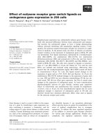
1). Non-activated cultures showed no NADPH diaphorase
staining in glia, but low-level staining was seen in neurons
(probably due to nNOS) and correlates with the low level
of nitrite present in the medium (Figure: 1a; Table: 1).
However, after treatment with LPS/IFN-γ, a high propor-
tion of glia (both microglia and astrocytes) stained
intensely for diaphorase activity (Figure: 1b). Treatment
with TNF-α/IFN-γ, IL-1β/IFN-γ or TNF-α/IL-1β/IFN-γ
resulted in much less diaphorase staining of glia, and little
or no nitrite elevation, indicating a requirement for LPS to
induce substantial iNOS expression.
Relatively pure neuronal cultures (CGC cultures isolated
as described in the methods section and then treated with
10 µM arabinoside cytosine at 18 hours to inhibit the
proliferation of glia) consisting of 97 ± 4% neurons, 2 ±
Table 1: Effects of inflammatory activated-glia in mixed neuronal-glial cultures on neuronal death. Neuronal death was assessed by
propidium iodide staining (PI, necrosis) or chromatin condensation of neuronal nuclei by Hoechst 33342 staining (CC, a marker of
apoptosis) 48 hours after treatment. Nitrite (the primary breakdown product of NO) levels were measured in the culture medium 48
hours following treatments. Statistical differences were established using ANOVA at *p < 0.05 and ***p < 0.001. Data expressed is
mean ± SEM, n = 3 or more.
TREATMENT PI (%) CC (%) NITRITE (µM)
UNTREATED 0.9 ± 0.9 0.5 ± 0.6 2.7 ± 3.0
LPS/IFN-γ 5.7 ± 3.4 * 3.6 ± 1.5 * 18.6 ± 8.4 ***
TNF-α/IFN-γ 5.6 ± 0.4 *** 4.3 ± 2.9 * 4.2 ± 2.8
IL-1β/IFN-γ 1.1 ± 1.1 0.6 ± 0.7 3.7 ± 2.1
TNF-α/IL-1β/IFN-γ 6.3 ± 4.1 * 5.5 ± 3.2 * 4.5 ± 1.4
Journal of Neuroinflammation 2005, 2:20 />Page 5 of 15
(page number not for citation purposes)
1% astrocytes and 1 ± 1% microglia were not affected by
the presence of cytokines alone (mean % of chromatin-
condensed (CC) and propidium iodide-positive (PI) neu-
rons ± SEM of 3 separate cultures; control: CC: 4 ± 3%, PI:
8 ± 4%; 10 ng/ml IL-1β: CC: 3 ± 2%, PI: 7 ± 3%; 10 ng/ml
TNF-α: CC: 3 ± 2%, PI: 9 ± 4%). Additionally, no signifi-
cant adverse effects were seen even if the concentrations of
IL-1β or TNF-α were increased 10-fold (mean % of neu-
rons ± SEM of 1 culture; control: CC: 4 ± 3%, PI: 8 ± 4%;
100 ng/ml IL-1β: CC: 4 ± 2%, PI: 5 ± 4%; 100 ng/ml TNF-
α: CC: 4 ± 2%, PI: 6 ± 4%) or if combined with 10 ng/ml
IFN-γ treatment (mean % of neurons ± SEM of 2 separate
cultures; control: CC: 4 ± 3%, PI: 8 ± 4%; 10 ng/ml IL-1β
+ IFN-γ: CC: 3 ± 2%, PI: 5 ± 3%; 10 ng/ml TNF-α + IFN-γ:
CC: 3 ± 3%, PI: 7 ± 2%).
These results suggest that the cytokines have no direct tox-
icity for neurons, and although nitric oxide (NO) is pro-
duced by iNOS expressed in glia following activation with
LPS/IFN-γ, it is not able to kill the co-cultured neurons
alone, or the quantities of NO produced are not sufficient
to induce widespread death of these mature neuronal
cultures.
Simultaneous activation of iNOS and NADPH oxidase
results in massive neuronal death, mediated by
peroxynitrite
As NO produced by inflammatory activated glia did not
induce substantial neuronal death, we investigated
whether simultaneous production of superoxide resulting
in peroxynitrite would be more toxic to neurons. Perox-
ynitrite is formed from the diffusion-limited reaction of
NO with superoxide. Under inflammatory conditions in
the brain, NADPH oxidase is the major source of superox-
ide, therefore we used phorbol 12-myristate 13-acetate
(PMA) to activate this enzyme and generate a source of
superoxide in the neuronal-glial culture. As the number of
NADPH diaphorase-positive glia was greatest following
treatment with LPS/IFN-γ, we used LPS/IFN-γ to induce
iNOS expression in the glia and provide a source of NO.
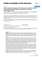
We found that treating neuronal-glial cultures with LPS/
IFN-γ/PMA for 48 hours induced extensive neuronal
death (Figure: 2a). Treatment of the cultures with PMA
alone induced only low levels of neuronal death, similar
to that seen with LPS/IFN-γ treatment alone. However,
activation of both NADPH oxidase and iNOS was syner-
gistic in inducing neuronal death. This neuronal death
was prevented by an iNOS inhibitor of (1400W), a
NADPH oxidase (apocynin) a peroxynitrite scavenger
(FeTPPS), but not by a blocker of the NMDA receptor
(MK-801).
As PMA activates the protein kinase C pathway, the effects
of PMA might be due to reasons other than stimulating
the microglial NADPH oxidase, such as increased iNOS
expression leading to more NO production and neuronal
death by NO and not peroxynitrite. However, the levels of
nitrite and nitrate in the culture medium of neuronal-glial
cultures treated with LPS/IFN-γ/PMA were not different to
those found in the absence of PMA (Figure: 2b).
NOS activity in mature neuronal-glial culturesFigure 1
NOS activity in mature neuronal-glial cultures.
NADPH diaphorase staining was used to assess for NOS
activity. Non-activated (control) cultures show weak staining
in neurons and along their processes (a), but following LPS/
IFN-γ treatment (b) dark staining is visible in glia (astrocytes
and microglia). The photographs shown are representative
and were taken 48 hours after treatment, n>3.
Journal of Neuroinflammation 2005, 2:20 />Page 6 of 15
(page number not for citation purposes)
Activation of NADPH oxidase in the presence of glial iNOS is synergistic in killing co-cultured neuronsFigure 2
Activation of NADPH oxidase in the presence of glial iNOS is synergistic in killing co-cultured neurons. Cul-
tures were stained with the cell-impermeable dye propidium iodide (PI) to count necrotic cells and the cell-permeable dye
Hoechst 33342 to count neuronal nuclei showing chromatin condensation/fragmentation (CC), 48 hours after treatment (a).
PMA stimulation of NADPH oxidase did not substantially affect neuronal survival, but in the presence of LPS/IFN-γ had syner-
gistic effects on neuronal death, which were blocked by inhibitors of iNOS (25 µM 1400W), NADPH oxidase (1 mM apocynin),
or a peroxynitrite scavenger (100 µM FeTPPS) but not by a blocker of the NMDA receptor (10 µM MK-801). Nitrite and
nitrate levels were not affected by the presence of PMA or apocynin but were significantly reduced by 1400W (b). Statistical
differences were established using ANOVA at *p <0.05, **p < 0.01 and ***p < 0.001, the symbol # replaces * when comparing
protection against LPS/IFN-γ/PMA induced neuronal death. The symbol ¶ is used to demonstrate a significant difference in
comparison to PMA or LPS/IFN-γ alone. Statistical significance refers to the total death (black + white parts of the bar). Data
expressed is mean ± SEM, n = 3 or more.
0
10
20
30
40
50
60
70
80
90
100
CELL DEATH (%)
PI
CC
a
***
¶¶¶
##
##
###
CONTROL LPS + IFN PMA LPS + IFN + PMA + 1400W + MK801 + APO + FeTPPS
Untreated LPS/IFN-
J
PMA Control 1400W MK-801 Apocynin FeTPPS
LPS/IFN-
J
+ PMA
b
0
10
20
30
40
50
60
70
80
90
12345
Nitrate
Nitrite
NO
X
CONCENTRATION (PM)
***
***
##
Untreated LPS/IFN-
J
LPS/IFN-
J
/PMA 1400W + Apocynin +
LPS/IFN-J/PMA LPS/IFN-J/PMA
Journal of Neuroinflammation 2005, 2:20 />Page 7 of 15
(page number not for citation purposes)
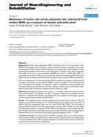
To determine whether peroxynitrite generated by glia
reaches the neurons, the neuronal-glial cultures were
tested for nitrotyrosine immunoreactivity. Positively
stained neurons (and glia) were only seen following treat-
ment with LPS/IFN-γ/PMA (Figure: 3) and not in the pres-
ence of the peroxynitrite scavenger FeTPPS or when
treated with LPS/IFN-γ (data not shown) or PMA alone
(data not shown). However, no PI-positive glia or changes
in glial nuclear morphology were observed in any of the
conditions, implying that although they were exposed to
peroxynitrite it did not induce glial death.
ATP is known to be released by neurons and glia in a vari-
ety of conditions, and has been reported to activate the
microglial NADPH oxidase via P2X7 receptors [26]. We
found that ATP rapidly stimulated superoxide/hydrogen
peroxide production by isolated microglia, which was
sensitive to diphenyleneiodonium (DPI), an inhibitor of
NADPH oxidase (ATP: 80 ± 7 picomoles H
2
O
2
/minute/1
× 10
5
microglia). However, ATP did not induce neuronal
death alone, or in synergy with LPS/IFN-γ treatment (data
not shown), probably because it is rapidly hydrolysed in
cell culture medium [35]. Therefore, we used a non-
hydrolysable ATP analogue, 2'-3'-O-(4- benzoylbenzoyl)-
ATP (BzATP), known to be a specific P2X7 receptor ago-
nist [36]. BzATP was also found to stimulate DPI-sensitive
hydrogen peroxide production by isolated microglia,
which was comparable to that produced by PMA (control:
12 ± 3; PMA: 204 ± 50; BzATP: 124 ± 15 picomoles H
2
O
2
/
minute/1 × 10
5
microglia). BzATP did not induce
neuronal death alone but had synergistic effects on neuro-
nal death in the presence iNOS expression (Figure: 4).
LPS/IFN-γ/BzATP induced neuronal death was blocked by
inhibitors of iNOS, NADPH oxidase and a peroxynitrite
scavenger, but not by the NMDA receptor blocker.
Activation of NADPH oxidase in the presence of iNOS expression leads to 3-nitrotyrosine immunoreactivity in neurons and gliaFigure 3
Activation of NADPH oxidase in the presence of iNOS expression leads to 3-nitrotyrosine immunoreactivity
in neurons and glia. Neuronal-glial cultures treated with LPS/IFN-γ/PMA for 48 hours showed extensive immunoreactivity
for 3-nitrotyrosine, which was absent in the presence of FeTPPS. Untreated cultures (control) showed no staining for 3-nitro-
tyrosine. The photographs shown are representative and were taken 48 hours after treatment, n>3.
FeTPPS +
LPS/IFN- J/PMA
CONTROL LPS/IFN- J/PMA
P
H
A
S
E
C
ON
T
R
A
S
T
3
-
N
I
T
R
OT
Y
R
OS
I
N
E
20 Pm
Journal of Neuroinflammation 2005, 2:20 />Page 8 of 15
(page number not for citation purposes)
Activation of glia in microglia-enriched neuronal-glial
cultures potently kills co-cultured neurons
We have found that IL-1β or arachidonic acid (AA) can
activate the microglial NADPH oxidase, although to lesser
extent than PMA (control: 12 ± 3; IL-1β: 37 ± 20; AA: 24 ±
4 picomoles H
2
O
2
/minute/1 × 10
5
microglia). We there-
fore tested whether IL-1β or AA could synergise with LPS/
IFN-γ to induce neuronal death. The addition of either IL-
1β or AA did not induce further neuronal death than that
induced by LPS/IFN-γ alone up to 48 hours after additions
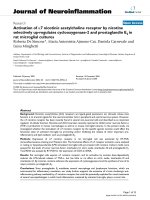
(data not shown). However if such cultures were main-
tained for 6 days, we found that widespread neuronal
death occurred (Figure: 5a, b) and was blocked by inhibi-
tors of iNOS, NADPH oxidase, a peroxynitrite scavenger
and a blocker of the NMDA receptor. Treatment with IL-
1β or AA alone did not have any effect on neuronal sur-
vival, but did increase the number of microglia in neuro-
nal-glial cultures (Figure: 5c). Treatment with LPS/IFN-γ
was found to inhibit microglia proliferation but in the
presence of IL-1β or AA this inhibition was overcome and
lead to a progressive increase in the number of microglia
and subsequent neuronal death. The mitogenic effects of
IL-1β or AA are probably mediated by hydrogen peroxide
following stimulation of NADPH oxidase (unpublished
data) and we found that the NADPH oxidase inhibitor,
apocynin, prevented this increase in the number of micro-
glia. Nitrite and nitrate (NO
X
) levels (Figure: 5d) were
higher in cultures treated with IL-1β or AA plus LPS/IFN-
γ, but not in the presence of apocynin, which blocked
NADPH oxidase stimulation by P2X7 receptor activation in the presence of glial iNOS kills co-cultured neuronsFigure 4
NADPH oxidase stimulation by P2X7 receptor activation in the presence of glial iNOS kills co-cultured neu-
rons. Neuronal death was assessed by propidium iodide staining (PI) and chromatin condensation of neuronal nuclei by
Hoechst 33342 staining (CC) 48 hours after treatment. Neuronal death induced by BzATP following LPS/IFN-γ activation, was
prevented by inhibitors of iNOS (25 µM 1400W), NADPH oxidase (1 mM apocynin) and a peroxynitrite scavenger (100 µM
FeTPPS) but not by a blocker of the NMDA receptor (10 µM MK-801). Statistical differences were established using ANOVA
at *p < 0.05 and ***p < 0.001, the symbol # replaces * when comparing protection against LPS/IFN-γ/BzATP induced neuronal
death. Statistical significance refers to the total death (black + white parts of the bar). Data expressed is mean ± SEM, n = 3 or
more.
0
20
40
60
80
100
UT LPS/IFN BzATP LPS + IFN + + 1400W + MK801
BzATP
+ APO + FeTPPS
CELL DEATH (%)
PI
CC
***
¶¶¶
### ###
###
Untreated LPS/IFN-
J
BzATP Control 1400W MK-801 Apocynin FeTPPS
LPS/IFN-
J
+ BzATP
Journal of Neuroinflammation 2005, 2:20 />Page 9 of 15
(page number not for citation purposes)
microglial proliferation, suggesting that microglia were
the predominant source of NO and/or peroxynitrite.
Since IL-1β and AA stimulated microglial proliferation (in
the presence or absence of LPS/IFN-γ), we wanted to test
whether increasing the microglial density would sensitise
to LPS/IFN-γ induced neuronal death. So we investigated
whether enriching the microglia population in the
neuronal-glial culture followed by inflammatory activa-
tion would result in widespread neuronal death. The neu-
ronal-glial culture used in the last section was enriched
with microglia by adding freshly isolated microglia. LPS/
IFN-γ activation of a microglia-rich (15% microglia as
opposed to 2%) neuronal-glial culture resulted in all neu-
rons rapidly losing their dendritic processes and shrinkage
of the cell body (Figure: 6b), in addition to chromatin
condensation or propidium iodide staining of the nuclei
at 48 hours of treatment (Figure: 6a). This neuronal death
was prevented by inhibitors of iNOS, NADPH oxidase, a
peroxynitrite decomposition catalyst and a blocker of the
NMDA receptor. The addition of microglia alone (non-
activated) did not affect neuronal survival (Figure: 6a,
Effects of IL-1β or arachidonic acid (AA) on neuronal survival in the presence of inflammation-activated glia in neuronal-glial culturesFigure 5
Effects of IL-1β or arachidonic acid (AA) on neuronal survival in the presence of inflammation-activated glia in
neuronal-glial cultures. Neuronal death was assessed by propidium iodide staining (PI; a) or chromatin condensation of neu-
ronal nuclei (CC; b) after 6 days of treatment. Neuronal death was prevented by inhibitors of iNOS (25 µM 1400W), NADPH
oxidase (1 mM apocynin), a blocker of the NMDA-receptor (10 µM MK-801) or a peroxynitrite scavenger (100 µM FeTPPS).
Neuronal death was accompanied by proliferation of microglia (c). Microglial proliferation was inhibited by LPS/IFN-γ treat-
ment alone but in the presence of IL-1β or AA it was stimulated and returned to basal levels. This stimulation of proliferation
by IL-1β or AA (in the presence of LPS/IFN-γ) was completely prevented by apocynin. Additionally, nitrite/nitrate (NO
X
) levels
correlated with the number of microglia present (d). Statistical differences were established using ANOVA at *p < 0.05, **p <
0.01 and ***p < 0.001, the symbol * is used when assessing prevention of neuronal death in comparison to LPS/IFN-γ with IL-
1β or AA. The symbol ¶ is used when comparing neuronal death to that induced by LPS/IFN-γ alone and # when comparing
neuronal death induced by IL-1β or AA treatment alone. In c & d, the differences are in comparison to IL-1β or AA alone (*),
LPS/IFN-γ (¶) or LPS/IFN-γ plus IL-1β or AA (#). Data expressed is mean ± SEM, n = 3 or more.
Journal of Neuroinflammation 2005, 2:20 />Page 10 of 15
(page number not for citation purposes)
Activation of microglia-enriched neuronal-glial cultures induces complete neurodegenerationFigure 6
Activation of microglia-enriched neuronal-glial cultures induces complete neurodegeneration. The microglia
population was enriched in neuronal-glial cultures by adding isolated microglia (50,000 microglia/cm
2
). Neuronal death was
assessed by propidium iodide staining (PI) or chromatin condensation of neuronal nuclei (CC) at 48 hours after treatment (a).
Neuronal death was prevented by inhibitors of iNOS (25 µM 1400W), NADPH oxidase (1 mM apocynin), a blocker of the
NMDA-receptor (10 µM MK-801), or a peroxynitrite scavenger (100 µM FeTPPS). LPS/IFN-γ activation of the microglia-
enriched neuronal-glial cultures led to complete disintegration of neuronal processes and severe shrinkage of neuronal cell
bodies (b). Statistical differences were established using ANOVA at *p < 0.05 and ***p < 0.001, in comparison to control
(added microglia) non-activated cultures, and the symbol # replaces * when comparing protection against neuronal death
induced by LPS/IFN-γ activated cultures. Statistical significance refers to the total death (black + white parts of the bar). Data
expressed is mean ± SEM, n = 3 or more. Photographs shown are representative and were taken 48 hours after the addition of
LPS/IFN-γ.
a
0
20
40
60
80
100
***
CC
PI
CELL DEATH (%)
###
### ###
###
control LPS + IFN 1400W + MK801 + APO + FeTPPS +
Untreated Control 1400W MK-801 Apocynin FeTPPS
LPS/IFN-J
b
20
P
m
CONTROL LPS/IFN-J
Journal of Neuroinflammation 2005, 2:20 />Page 11 of 15
(page number not for citation purposes)
untreated). In support of microglia as the key cell type in
inflammatory neurodegeneration, LPS/IFN-γ activation of
astrocyte-enriched neuronal-glial cultures did not lead to
widespread killing of co-cultured neurons (isolation of
neuronal-glial cultures as normal but plated onto a con-
fluent bed of astrocytes; data not shown).
Prion protein or PrP106-126 induce neuronal death in the
presence of inflammatory activation mediated by
microglia and NADPH oxidase activation
The prion peptide, PrP106-126, has previously been
shown to activate microglia, causing proliferation and
ROS production [37,38]. We have recently found that the
prion protein and peptide stimulate the NADPH oxidase
in isolated microglia (control: 12 ± 3; prion protein: 29 ±
3; PrP106-126: 38 ± 13 picomoles H
2
O
2
/minute/1 × 10
5
microglia). We decided to investigate whether the addi-
tion of PrP106-126 to iNOS-expressing glia in neuronal-
glial cultures would also lead to delayed
neurodegeneration, mediated by peroxynitrite and micro-
glia. The addition of prion protein or PrP106-126 alone
did not affect neuronal survival in these mature neuronal-
glial cultures, but did lead to microglial proliferation
(Table: 2). In the presence of glial iNOS (following LPS/
IFN-γ treatment), PrP106-126 or prion protein did not
exacerbate neuronal death over a period of 2 days, but
were synergistic in killing the co-cultured neurons at 6
days (Figure: 7a, b), while a scrambled peptide of the
PrP106-126 sequence had no effect. Neuronal death was
prevented by blocking NO production from iNOS
(1400W), or superoxide from NADPH oxidase
(apocynin), through the removal of peroxynitrite
(FeTPPS), or by inhibiting the NMDA receptor (MK-801).
Additionally, neuronal death was accompanied by micro-
glia proliferation, which was blocked by apocynin (Figure:
7c). Nitrite/nitrate levels were also suppressed in the pres-
ence of apocynin, as well as 1400W (Figure: 7d).
Discussion
We found that LPS/IFN-γ induced NOS activity within cul-
tured glia, but induced relatively little death of co-cultured
neurons. It has previously been reported that LPS/
cytokine-induced iNOS expression in glia is sufficient
[5,8,39,40] or insufficient [41-43] to induce death of co-
cultured neurons. Similarly, in vivo it has been reported
that iNOS expression is sufficient [44,45] or insufficient
[46-48] to induce neuronal death. This suggests that either
there is a threshold level for NO/iNOS induced neuronal
death [49], or NO/iNOS-induced neuronal death is con-
ditional upon some other factors. We have recently
reported one such conditional factor (hypoxia) that syner-
gises with NO/iNOS to induce neuronal death [31]. In
this report we have tested the hypothesis that NO/iNOS
induced neuronal death is conditional upon microglial
NADPH oxidase activation.
It has previously been shown that PMA stimulation of
microglia results in superoxide production through stim-
ulation of NADPH oxidase [50] and, in the presence of
LPS/IFN-γ activated glia (producing NO from iNOS), the
superoxide combines with NO to form peroxynitrite [32].
We found that if the NADPH oxidase was stimulated by
PMA in the presence of LPS/IFN-γ activated glia, it resulted
in extensive death of the co-cultured neurons, while PMA
alone induced very little neuronal death. In pathophysio-
logical conditions, extracellular levels of ATP can increase
[51], and ATP can activate purinergic receptors (more spe-
cifically P2X7 receptors), which can lead to the activation
of NADPH oxidase [26]. We used a specific P2X7 receptor
agonist (BzATP) to activate the NADPH oxidase in the
presence of iNOS expression (LPS/IFN-γ activated cul-
tures) and we found extensive neuronal death, compara-
ble to that induced by LPS/IFN-γ/PMA. Neuronal-glial
cultures activated with LPS/IFN-γ/PMA or LPS/IFN-γ/
BzATP induced delayed neuronal death that occurred over
2 days. This is partly due to the time taken for iNOS
expression, but it also implies that once peroxynitrite is
generated neuronal death is not immediate.
In both cases (LPS/IFN-γ/PMA or LPS/IFN-γ/BzATP),
inhibitors of iNOS or NADPH oxidase or a scavenger of
peroxynitrite prevented this neuronal death, implicating
Table 2: Prion protein or peptide (PrP106-126) does not affect neuronal survival. Neuronal-glial cultures treated once with either
prion protein (5 µg/ml) or PrP106-126 (225 µg/ml) did not induce neuronal death over a period of 7 days (assessed by Hoechst 33342 to
visualise chromatin condensation (CC) or propidium iodide (PI) to stain necrotic cells). However, prion protein or PrP106-126 did
stimulate the proliferation of microglia in neuronal-glial cultures over the same period of time. Statistical differences were established
using ANOVA at *p < 0.05, **p < 0.01 and ***p < 0.001 and are in comparison to untreated cultures (symbol *); data expressed is mean
± SEM, n = 3 or more.
Treatment PI (%) CC (%) Microglia per field
Untreated 2.0 ± 1.6 0.6 ± 0.3 22 ± 5
Prion protein 2.9 ± 0.5 0.7 ± 0.6 53 ± 8 ***
PrP106-126 1.0 ± 0.8 0.7 ± 0.4 51 ± 7 ***
Journal of Neuroinflammation 2005, 2:20 />Page 12 of 15
(page number not for citation purposes)
peroxynitrite as the potential mediator of neuronal death
and the source of peroxynitrite as NO from iNOS and
superoxide from NADPH oxidase. The putative
peroxynitrite marker nitrotyrosine, was found in both
neurons and some glia, implying that LPS/IFN-γ/PMA
treatment does result in peroxynitrite production that
reacts with neurons. Furthermore, the peroxynitrite
decomposition catalyst prevents the occurrence of nitroty-
rosine-positive neurons following LPS/IFN-γ/PMA treat-
ment. FeTPPS has been shown to rapidly react and
catalyse the decomposition of extracellular peroxynitrite
[52] and inhibit tyrosine nitration [53]. The presence of
nitrotyrosine immunoreactivity in glia did not appear to
induce glial death. It has been found that glia can up-reg-
ulate their antioxidant defences to become more resistant
to oxidative stress [54], which may explain the lack of
change in glial morphology.
Delayed neurodegeneration induced by prion protein or PrP106-126 in the presence of iNOS expression is microglia-depend-ent and mediated by peroxynitriteFigure 7
Delayed neurodegeneration induced by prion protein or PrP106-126 in the presence of iNOS expression is
microglia-dependent and mediated by peroxynitrite. The addition of prion protein (5 µg/ml) or PrP106-126 (225 µg/
ml) to LPS/IFN-γ treated neuronal-glial cultures induced delayed death of co-cultured neurons, over 6 days. Neuronal death,
assessed by Hoechst 33342 to visualise chromatin condensation (CC; b) or PI for necrosis (a) was prevented by inhibitors of
iNOS (25 µM 1400W) and NADPH oxidase (1 mM apocynin), a peroxynitrite scavenger (100 µM FeTPPS) or a blocker of the
NMDA receptor (10 µM MK-801). Neuronal death was accompanied by proliferation of microglia (c). Microglial proliferation
was inhibited by LPS/IFN-γ treatment alone but in the presence of prion protein or PrP106-126 it was stimulated and returned
to basal levels. This stimulation of proliferation by prion protein or PrP106-126 (in the presence of LPS/IFN-γ) was completely
prevented by apocynin. Additionally, nitrite/nitrate (NO
X
) levels correlated with the number of microglia present (d). Statistical
differences were established using ANOVA at *p < 0.05, **p < 0.01 and ***p < 0.001 are in comparison to untreated cultures
(symbol *) or LPS/IFN-γ treatment (symbol ¶) or LPS/IFN-γ plus prion protein or PrP106-126 (symbol #); data expressed is
mean ± SEM, n = 3 or more. In c & d, the differences are in comparison to prion protein or PrP106-126 alone (*), LPS/IFN-γ
(¶) or LPS/IFN-γ plus prion protein or PrP106-126 (#). Data expressed is mean ± SEM, n = 3 or more.
Journal of Neuroinflammation 2005, 2:20 />Page 13 of 15
(page number not for citation purposes)
The mechanism of peroxynitrite-induced neuronal death
is still unclear but has been proposed to involve DNA-
damage induced PARP activation [55], damage to the
mitochondrial respiratory chain [56], and lipid peroxida-
tion [57]. It is still controversial whether peroxynitrite-
induced neuronal death involves activation of the NMDA
receptor [58,59]. We found that a blocker of the NMDA-
receptor did not prevent the relatively acute neuronal
death induced by LPS/IFN-γ/PMA or LPS/IFN-γ/BzATP,
but did prevent the relatively slow neuronal death
induced by LPS/IFN-γ/IL-1β or LPS/IFN-γ/AA, although in
both cases death was prevented by a peroxynitrite decom-
poser. It is possible that low, sustained levels of peroxyni-
trite induce neuronal death via the NMDA receptor,
whereas high, acute levels induce death by other means,
but we have not directly tested this. We found that IL-1β
or AA activated NADPH oxidase hydrogen peroxide
production to a lesser extent than PMA but, like PMA,
either IL-1β or AA synergised with LPS/IFN-γ to induce
neuronal death mediated by peroxynitrite following acti-
vation of iNOS and NADPH oxidase. However the neuro-
nal death induced by LPS/IFN-γ/IL-1β or LPS/IFN-γ/AA
occurred over 6 days, rather than 2 days as with LPS/IFN-
γ/PMA or LPS/IFN-γ/BzATP. This relative delay might be
due to the lower level of NADPH oxidase activation and
thus peroxynitrite production. Additionally, Il-1β or AA
caused microglial proliferation during the 6-day cultures,
which may have contributed to the delayed neuronal
death. Recently we found that IL-1β or AA stimulated
microglial proliferation in microglia-astrocyte cultures via
hydrogen peroxide production from NADPH oxidase
(manuscript in preparation). Here we have shown that IL-
1β or AA stimulate the proliferation of microglia in neu-
ronal-glial cultures, even in the presence of LPS/IFN-γ
(which itself inhibits microglial proliferation). In order to
test whether an increase in microglia would potentiate
LPS/IFN-γ induced neuronal death, we added extra iso-
lated microglia to the neuronal-glial culture, increasing
the microglial population from 2% to 15% of cells in the
co-culture. In such microglia-enriched cultures, LPS/IFN-γ
induced neuronal death was greatly increased. These
observations suggest that microglia are essential for
inflammatory activated glia-induced neuronal death, and
one reason for this may be the expression of NADPH oxi-
dase, which is predominantly localised to microglia [24].
Transmissible spongiform encephalopathies (Prion dis-
eases) are lethal neurodegenerative disorders character-
ised by the progressive accumulation of a protease
resistant isoform (PrP
sc
) of the normal host prion protein
(PrP
c
), in amyloid plaques [60]. An inflammatory
response, predominantly mediated by microglia, is seen
in post-mortem brain tissue, in transgenic models of the
disease, and in culture [61]. A peptide, consisting of resi-
dues 106–126 (PrP106-126) of the human prion protein,
replicates many of the pathological mechanisms involved
in prion diseases and provides a good in vitro model.
Contrary to published data [38,62], we observed no neu-
rotoxicity following the addition of PrP106-126 alone to
this mature neuronal-glial culture. We found that both
prion protein and PrP106-126, but not a scrambled pep-
tide, stimulated microglial proliferation when added to
neuronal-glial cultures, and this proliferation was blocked
by a NADPH oxidase inhibitor. Both prion protein and
peptide were synergistic in killing neurons in the presence
of glial iNOS, in a peroxynitrite and microglia-dependent
mechanism. The addition of the cellular isoform of prion
protein to cell cultures has previously been shown to have
no toxic effects [34]. However, in the presence of glial
iNOS we found it to induce significant levels of neuronal
death, although significantly less than that induced by
PrP106-126.
Conclusion
We have shown that in a mature mixed culture of neurons
and glia, activation of iNOS or NADPH oxidase alone
does not result in substantial neuronal death, but that
simultaneous activation of both is synergistic in killing co-
cultured neurons. This neuronal death appears to be
dependent on microglia, and microglial proliferation is
itself stimulated by activating the NADPH oxidase. These
results suggest a dual-key hypothesis for inflammatory
neurodegeneration; i.e. that activation of glial iNOS or
NADPH oxidase alone may be relatively benign, but when
activated together they cause peroxynitrite-mediated neu-
ronal death. The conditionality of NO/iNOS-induced
neuronal death provides insight into the mechanisms of
inflammatory neurodegeneration and suggests that
microglial NADPH oxidase may be a key therapeutic
target.
Competing interests
The author(s) declare that they have no competing
interests.
Authors' contributions
PM participated in the design of this study, did the lab
work, data analysis and wrote major parts of the paper.
GCB conceived the study, participated in its design and
helped to draft the manuscript. Both authors read and
approved the final manuscript.
Acknowledgements
This work was supported by the BBSRC, MRC and Alzheimer's Research
Trust.
References
1. Eddleston M, Mucke L: Molecular profile of reactive astrocytes-
-implications for their role in neurologic disease. Neuroscience
1993, 54:15-36.
2. Kreutzberg GW: Microglia: a sensor for pathological events in
the CNS. Trends Neurosci 1996, 19:312-318.
Journal of Neuroinflammation 2005, 2:20 />Page 14 of 15
(page number not for citation purposes)
3. Polazzi E, Contestabile A: Reciprocal interactions between
microglia and neurons: from survival to neuropathology. Rev
Neurosci 2002, 13:221-242.
4. Banati RB, Gehrmann J, Schubert P, Kreutzberg GW: Cytotoxicity
of microglia. Glia 1993, 7:111-118.
5. Boje KM, Arora PK: Microglial-produced nitric oxide and reac-
tive nitrogen oxides mediate neuronal cell death. Brain Res
1992, 587:250-256.
6. Brown GC, Bal-Price A: Inflammatory neurodegeneration
mediated by nitric oxide, glutamate, and mitochondria. Mol
Neurobiol 2003, 27:325-355.
7. McGeer PL, McGeer EG: The inflammatory response system of
brain: implications for therapy of Alzheimer and other neu-
rodegenerative diseases. Brain Res Brain Res Rev 1995,
21:195-218.
8. Bal-Price A, Brown GC: Inflammatory neurodegeneration
mediated by nitric oxide from activated glia-inhibiting neu-
ronal respiration, causing glutamate release and
excitotoxicity. J Neurosci 2001, 21:6480-6491.
9. Chao CC, Hu S, Sheng WS, Bu D, Bukrinsky MI, Peterson PK:
Cytokine-stimulated astrocytes damage human neurons via
a nitric oxide mechanism. Glia 1996, 16:276-284.
10. Beckman JS, Chen J, Crow JP, Ye YZ: Reactions of nitric oxide,
superoxide and peroxynitrite with superoxide dismutase in
neurodegeneration. Prog Brain Res 1994, 103:371-380.
11. Chao CC, Hu S, Peterson PK: Modulation of human microglial
cell superoxide production by cytokines. J Leukoc Biol 1995,
58:65-70.
12. Bolanos JP, Almeida A, Stewart V, Peuchen S, Land JM, Clark JB,
Heales SJ: Nitric oxide-mediated mitochondrial damage in the
brain: mechanisms and implications for neurodegenerative
diseases. J Neurochem 1997, 68:2227-2240.
13. Barger SW, Basile AS: Activation of microglia by secreted amy-
loid precursor protein evokes release of glutamate by cys-
tine exchange and attenuates synaptic function. J Neurochem
2001, 76:846-854.
14. Chao CC, Hu S, Ehrlich L, Peterson PK: Interleukin-1 and tumor
necrosis factor-alpha synergistically mediate neurotoxicity:
involvement of nitric oxide and of N-methyl-D-aspartate
receptors. Brain Behav Immun 1995, 9:355-365.
15. Floyd RA: Antioxidants, oxidative stress, and degenerative
neurological disorders. Proc Soc Exp Biol Med 1999, 222:236-245.
16. Metodiewa D, Koska C: Reactive oxygen species and reactive
nitrogen species: relevance to cyto(neuro)toxic events and
neurologic disorders. An overview. Neurotox Res 2000,
1:197-233.
17. Zekry D, Epperson TK, Krause KH: A role for NOX NADPH oxi-
dases in Alzheimer's disease and other types of dementia?
IUBMB Life 2003, 55:307-313.
18. Shimohama S, Tanino H, Kawakami N, Okamura N, Kodama H,
Yamaguchi T, Hayakawa T, Nunomura A, Chiba S, Perry G, Smith MA,
Fujimoto S: Activation of NADPH oxidase in Alzheimer's dis-
ease brains. Biochem Biophys Res Commun 2000, 273:5-9.
19. Boven LA, Gomes L, Hery C, Gray F, Verhoef J, Portegies P, Tardieu
M, Nottet HS: Increased peroxynitrite activity in AIDS
dementia complex: implications for the neuropathogenesis
of HIV-1 infection. J Immunol 1999, 162:4319-4327.
20. Qin L, Liu Y, Wang T, Wei SJ, Block ML, Wilson B, Liu B, Hong JS:
NADPH oxidase mediates lipopolysaccharide-induced neu-
rotoxicity and proinflammatory gene expression in activated
microglia. J Biol Chem 2004, 279:1415-1421.
21. Green SP, Cairns B, Rae J, Errett-Baroncini C, Hongo JA, Erickson
RW, Curnutte JT: Induction of gp91-phox, a component of the
phagocyte NADPH oxidase, in microglial cells during central
nervous system inflammation. J Cereb Blood Flow Metab 2001,
21:374-384.
22. Sastre J, Pallardo FV, Vina J: The role of mitochondrial oxidative
stress in aging. Free Radic Biol Med 2003, 35:1-8.
23. Dugan LL, Sensi SL, Canzoniero LM, Handran SD, Rothman SM, Lin
TS, Goldberg MP, Choi DW: Mitochondrial production of reac-
tive oxygen species in cortical neurons following exposure to
N-methyl-D-aspartate. J Neurosci 1995, 15:6377-6388.
24. Klegeris A, McGeer PL: Rat brain microglia and peritoneal mac-
rophages show similar responses to respiratory burst
stimulants. J Neuroimmunol 1994, 53:83-90.
25. Benna JE, Dang PM, Gaudry M, Fay M, Morel F, Hakim J, Gougerot-
Pocidalo MA: Phosphorylation of the respiratory burst oxidase
subunit p67(phox) during human neutrophil activation. Reg-
ulation by protein kinase C-dependent and independent
pathways. J Biol Chem 1997, 272:17204-17208.
26. Parvathenani LK, Tertyshnikova S, Greco CR, Roberts SB, Robertson
B, Posmantur R: P2X7 mediates superoxide production in pri-
mary microglia and is up-regulated in a transgenic mouse
model of Alzheimer's disease. J Biol Chem 2003,
278:13309-13317.
27. Shiose A, Sumimoto H: Arachidonic acid and phosphorylation
synergistically induce a conformational change of p47phox
to activate the phagocyte NADPH oxidase. J Biol Chem 2000,
275:13793-13801.
28. Babior BM: NADPH oxidase. Curr Opin Immunol 2004, 16:42-47.
29. Murphy S, Simmons ML, Agullo L, Garcia A, Feinstein DL, Galea E,
Reis DJ, Minc-Golomb D, Schwartz JP: Synthesis of nitric oxide in
CNS glial cells. Trends Neurosci 1993, 16:323-328.
30. Bonfoco E, Krainc D, Ankarcrona M, Nicotera P, Lipton SA: Apop-
tosis and necrosis: two distinct events induced, respectively,
by mild and intense insults with N-methyl-D-aspartate or
nitric oxide/superoxide in cortical cell cultures. Proc Natl Acad
Sci U S A 1995, 92:7162-7166.
31. Mander P, Borutaite V, Moncada S, Brown GC: Nitric oxide from
inflammatory-activated glia synergizes with hypoxia to
induce neuronal death. J Neurosci Res 2005, 79:208-215.
32. Bal-Price A, Matthias A, Brown GC: Stimulation of the NADPH
oxidase in activated rat microglia removes nitric oxide but
induces peroxynitrite production. J Neurochem 2002, 80:73-80.
33. Beckman JS, Crow JP: Pathological implications of nitric oxide,
superoxide and peroxynitrite formation. Biochem Soc Trans
1993, 21:330-334.
34. Brown DR, Wong BS, Hafiz F, Clive C, Haswell SJ, Jones IM: Normal
prion protein has an activity like that of superoxide
dismutase. Biochem J 1999, 344 Pt 1:1-5.
35. Mietkiewski K, Domka F, Malendowicz L, Malendowicz J: Studies on
ATP hydrolysis in medium for histochemical demonstration
of ATPase activity. Histochemie 1970, 24:343-353.
36. Michel AD, Xing M, Humphrey PP: Serum constituents can affect
2'-& 3'-O-(4-benzoylbenzoyl)-ATP potency at P2X(7)
receptors. Br J Pharmacol 2001, 132:1501-1508.
37. Brown DR, Schmidt B, Kretzschmar HA: A neurotoxic prion pro-
tein fragment enhances proliferation of microglia but not
astrocytes in culture. Glia 1996, 18:59-67.
38. Brown DR, Schmidt B, Kretzschmar HA: Role of microglia and
host prion protein in neurotoxicity of a prion protein
fragment. Nature 1996, 380:345-347.
39. Chao CC, Hu S, Molitor TW, Shaskan EG, Peterson PK: Activated
microglia mediate neuronal cell injury via a nitric oxide
mechanism. J Immunol 1992, 149:2736-2741.
40. Dawson VL, Brahmbhatt HP, Mong JA, Dawson TM: Expression of
inducible nitric oxide synthase causes delayed neurotoxicity
in primary mixed neuronal-glial cortical cultures. Neurophar-
macology 1994, 33:1425-1430.
41. Demerle-Pallardy C, Lonchampt MO, Chabrier PE, Braquet P: Nitric
oxide synthase induction in glial cells: effect on neuronal
survival. Life Sci 1993, 52:1883-1890.
42. Hewett SJ, Csernansky CA, Choi DW: Selective potentiation of
NMDA-induced neuronal injury following induction of astro-
cytic iNOS. Neuron 1994, 13:487-494.
43. Stone R, Stewart VC, Hurst RD, Clark JB, Heales SJ: Astrocyte
nitric oxide causes neuronal mitochondrial damage, but
antioxidant release limits neuronal cell death. Ann N Y Acad Sci
1999, 893:400-403.
44. Iravani MM, Kashefi K, Mander P, Rose S, Jenner P: Involvement of
inducible nitric oxide synthase in inflammation-induced
dopaminergic neurodegeneration. Neuroscience 2002,
110:49-58.
45. Ding M, Zhang M, Wong JL, Rogers NE, Ignarro LJ, Voskuhl RR: Anti-
sense knockdown of inducible nitric oxide synthase inhibits
induction of experimental autoimmune encephalomyelitis in
SJL/J mice. J Immunol 1998, 160:2560-2564.
46. David JP, Ghozali F, Fallet-Bianco C, Wattez A, Delaine S, Boniface B,
Di Menza C, Delacourte A: Glial reaction in the hippocampal
formation is highly correlated with aging in human brain.
Neurosci Lett 1997, 235:53-56.
Publish with Bio Med Central and every
scientist can read your work free of charge
"BioMed Central will be the most significant development for
disseminating the results of biomedical research in our lifetime."
Sir Paul Nurse, Cancer Research UK
Your research papers will be:
available free of charge to the entire biomedical community
peer reviewed and published immediately upon acceptance
cited in PubMed and archived on PubMed Central
yours — you keep the copyright
Submit your manuscript here:
/>BioMedcentral
Journal of Neuroinflammation 2005, 2:20 />Page 15 of 15
(page number not for citation purposes)
47. Ma SX, Holley AT, Sandra A, Cassell MD, Abboud FM: Increased
expression of nitric oxide synthase in the gracile nucleus of
aged rats. Neuroscience 1997, 76:659-663.
48. Vernet D, Bonavera JJ, Swerdloff RS, Gonzalez-Cadavid NF, Wang C:
Spontaneous expression of inducible nitric oxide synthase in
the hypothalamus and other brain regions of aging rats.
Endocrinology 1998, 139:3254-3261.
49. Golde S, Chandran S, Brown GC, Compston A: Different pathways
for iNOS-mediated toxicity in vitro dependent on neuronal
maturation and NMDA receptor expression. J Neurochem
2002, 82:269-282.
50. Sumimoto H, Kage Y, Nunoi H, Sasaki H, Nose T, Fukumaki Y, Ohno
M, Minakami S, Takeshige K: Role of Src homology 3 domains in
assembly and activation of the phagocyte NADPH oxidase.
Proc Natl Acad Sci U S A 1994, 91:5345-5349.
51. Le Feuvre R, Brough D, Rothwell N: Extracellular ATP and P2X7
receptors in neurodegeneration. Eur J Pharmacol 2002,
447:261-269.
52. Crow JP: Manganese and iron porphyrins catalyze peroxyni-
trite decomposition and simultaneously increase nitration
and oxidant yield: implications for their use as peroxynitrite
scavengers in vivo. Arch Biochem Biophys 1999, 371:41-52.
53. Misko TP, Highkin MK, Veenhuizen AW, Manning PT, Stern MK, Cur-
rie MG, Salvemini D: Characterization of the cytoprotective
action of peroxynitrite decomposition catalysts. J Biol Chem
1998, 273:15646-15653.
54. Acarin L, Peluffo H, Barbeito L, Castellano B, Gonzalez B: Astroglial
nitration after postnatal excitotoxic damage: correlation
with nitric oxide sources, cytoskeletal, apoptotic and antioxi-
dant proteins. J Neurotrauma 2005, 22:189-200.
55. Nguyen T, Brunson D, Crespi CL, Penman BW, Wishnok JS, Tannen-
baum SR: DNA damage and mutation in human cells exposed
to nitric oxide in vitro. Proc Natl Acad Sci U S A 1992,
89:3030-3034.
56. Bolanos JP, Heales SJ, Land JM, Clark JB: Effect of peroxynitrite on
the mitochondrial respiratory chain: differential susceptibil-
ity of neurones and astrocytes in primary culture. J Neurochem
1995, 64:1965-1972.
57. Beckman JS, Koppenol WH: Nitric oxide, superoxide, and per-
oxynitrite: the good, the bad, and ugly. Am J Physiol 1996,
271:C1424-37.
58. Leist M, Fava E, Montecucco C, Nicotera P: Peroxynitrite and
nitric oxide donors induce neuronal apoptosis by eliciting
autocrine excitotoxicity. Eur J Neurosci 1997, 9:1488-1498.
59. Trackey JL, Uliasz TF, Hewett SJ: SIN-1-induced cytotoxicity in
mixed cortical cell culture: peroxynitrite-dependent and -
independent induction of excitotoxic cell death. J Neurochem
2001, 79:445-455.
60. Prusiner SB: Novel proteinaceous infectious particles cause
scrapie. Science 1982, 216:136-144.
61. Brown DR, Kretzschmar HA: Microglia and prion disease: a
review. Histol Histopathol 1997, 12:883-892.
62. Forloni G, Angeretti N, Chiesa R, Monzani E, Salmona M, Bugiani O,
Tagliavini F: Neurotoxicity of a prion protein fragment. Nature
1993, 362:543-546.