báo cáo hóa học: " Interferon-γ increases neuronal death in response to amyloid-β1-42" pdf
Bạn đang xem bản rút gọn của tài liệu. Xem và tải ngay bản đầy đủ của tài liệu tại đây (318.83 KB, 7 trang )
BioMed Central
Page 1 of 7
(page number not for citation purposes)
Journal of Neuroinflammation
Open Access
Research
Interferon-γ increases neuronal death in response to amyloid-β
1-42
Clive Bate*, Sarah Kempster, Victoria Last and Alun Williams
Address: Department of Pathology and Infectious Diseases, Royal Veterinary College, Hawkshead Lane, North Mymms, Herts, AL9 7TA, UK
Email: Clive Bate* - ; Sarah Kempster - ; Victoria Last - ;
Alun Williams -
* Corresponding author
Abstract
Background: Alzheimer's disease is a neurodegenerative disorder characterized by a progressive
cognitive impairment, the consequence of neuronal dysfunction and ultimately the death of
neurons. The amyloid hypothesis proposes that neuronal damage results from the accumulation of
insoluble, hydrophobic, fibrillar peptides such as amyloid-β
1-42
. These peptides activate enzymes
resulting in a cascade of second messengers including prostaglandins and platelet-activating factor.
Apoptosis of neurons is thought to follow as a consequence of the uncontrolled release of second
messengers. Biochemical, histopathological and genetic studies suggest that pro-inflammatory
cytokines play a role in neurodegeneration during Alzheimer's disease. In the current study we
examined the effects of interferon (IFN)-γ, tumour necrosis factor (TNF)α, interleukin (IL)-1β and
IL-6 on neurons.
Methods: Primary murine cortical or cerebellar neurons, or human SH-SY5Y neuroblastoma cells,
were grown in vitro. Neurons were treated with cytokines prior to incubation with different
neuronal insults. Cell survival, caspase-3 activity (a measure of apoptosis) and prostaglandin
production were measured. Immunoblots were used to determine the effects of cytokines on the
levels of cytoplasmic phospholipase A
2
or phospholipase C γ-1.
Results: While none of the cytokines tested were directly neurotoxic, pre-treatment with IFN-γ
sensitised neurons to the toxic effects of amyloid-β
1-42
or HuPrP82-146 (a neurotoxic peptide
found in prion diseases). The effects of IFN-γ were seen on cortical and cerebellar neurons, and on
SH-SY5Y neuroblastoma cells. However, pre-treatment with IFN-γ did not affect the sensitivity to
neurons treated with staurosporine or hydrogen peroxide. Pre-treatment with IFN-γ increased the
levels of cytoplasmic phospholipase A
2
in SH-SY5Y cells and increased prostaglandin E
2
production
in response to amyloid-β
1-42
.
Conclusion: Treatment of neuronal cells with IFN-γ increased neuronal death in response to
amyloid-β
1-42
or HuPrP82-146. IFN-γ increased the levels of cytoplasmic phospholipase A
2
in
cultured neuronal cells and increased expression of cytoplasmic phospholipase A
2
was associated
with increased production of prostaglandin E
2
in response to amyloid-β
1-42
or HuPrP82-146. Such
observations suggest that IFN-γ produced within the brain may increase neuronal loss in
Alzheimer's disease.
Published: 28 March 2006
Journal of Neuroinflammation2006, 3:7 doi:10.1186/1742-2094-3-7
Received: 31 January 2006
Accepted: 28 March 2006
This article is available from: />© 2006Bate et al; licensee BioMed Central Ltd.
This is an Open Access article distributed under the terms of the Creative Commons Attribution License ( />),
which permits unrestricted use, distribution, and reproduction in any medium, provided the original work is properly cited.
Journal of Neuroinflammation 2006, 3:7 />Page 2 of 7
(page number not for citation purposes)
Background
Alzheimer's disease (AD) is a neurodegenerative disorder
characterized by progressive cognitive impairment as a
consequence of neuronal dysfunction and loss. The amy-
loid hypothesis maintains that the neuronal dysfunction
and death that give rise to the clinical symptoms of AD are
caused by the accumulation of fibrils consisting of amy-
loid-β peptides [1]. These peptides are formed following
the cleavage of the amyloid precursor protein by γ-secre-
tases [2], and depositions of amyloid-β peptides are a
component of the senile plaques found in diseased brains
[3]. The neuronal loss that occurs in AD has been mod-
elled in vitro by incubating neurons with specific peptides
derived from the amyloid-β protein [4]. The neuronal
injury induced by these peptides includes characteristics
of apoptosis such as chromatin condensation and DNA
fragmentation [5].
In AD, amyloid deposits containing fibrillar amyloid-β
peptides frequently co-localise with inflammatory cells
strongly suggesting that the deposits of amyloid-β stimu-
late a chronic inflammatory process [6]. Genetic studies
have identified polymorphisms in the genes of some
inflammatory cytokines as risk factors for AD [7] suggest-
ing that cytokine production within the brain may influ-
ence neuropathogenesis. While the effects of cytokines on
astroglial cells within the brain are well reported, less is
known about the direct effects of individual cytokines on
neurons. In the current study we report that pre-treatment
with interferon (IFN)-γ significantly increased the sensi-
tivity of neurons to the toxic effects of amyloid-β
1-42
. The
increased sensitivity of IFN-γ treated neurons to amyloid-
β
1-42
correlated with increased expression of cytoplasmic
phospholipase A
2
(cPLA
2
) in neuroblastoma cells and
increased prostaglandin production in response to exoge-
nous amyloid-β
1-42
. These results are consistent with prior
observations that uncontrolled activation the cPLA
2
/
cyclo-oxygenase (COX) pathway by amyloid-β
1-42
leads to
neuronal death [8].
Methods
Cell lines
The human neuroblastoma cell line SH-SY5Y was grown
in RPMI-1640 medium supplemented with 2 mM
glutamine, standard antibiotics (100 U/ml Penicillin, 100
µg/ml Streptomycin) and 2% fetal calf serum (FCS). For
toxicity studies cells were seeded at 3 × 10
4
cells per well
in 48 well plates, treated with cytokines and allowed to
adhere overnight before use. After 24 hours, different con-
centrations of peptides, staurosporine or hydrogen perox-
ide were added. Cell viability and/or prostaglandin E
2
content were determined after a further 24 hours.
Primary neuronal cultures
Primary cortical neurons were prepared from embryonic
day 15.5 mice as previously described [9]. Neuronal pro-
genitors were seeded at 500,000 cells per well in 48 well
plates in RPMI-1640 supplemented with 2 mM
glutamine, standard antibiotics and 10% FCS. After 2
hours, cultures were washed and subsequently grown in
neurobasal medium containing 2 mM glutamine and B27
components (Invitrogen, Paisley, UK). Primary cerebellar
neurons were prepared from the brains from newborn
mice pups following dissection of the cerebellum,
removal of the meninges and cell dissociation as previ-
ously described [9]. Neuronal progenitors were plated in
10% FCS for 2 hours, and then grown in neurobasal
medium containing glutamine and B27. In both these
neuronal cultures, medium was supplemented with 5 mM
L-leucine methyl ester to reduce the numbers of contami-
nating microglial cells. After 7 days, cultures were treated
with cytokines for 24 hours before the addition of neuro-
toxins/peptides. Caspase-3 activity was measured 24
hours after the addition of neurotoxins using a flouromet-
ric immunosorbent enzyme assay kit as per the manufac-
turer's instructions (Roche Diagnostics, Lewes, UK).
Results are expressed as fluorescent units which are pro-
portional to caspase-3 activity. For toxicity assays medium
was replaced 48 hours after the addition of neurotoxins/
peptides and cell viability was determined after another
48 hours (4 days after the addition of neurotoxins/pep-
tides).
Peptides
A peptide corresponding to amino acids 1 to 42 of the
amyloid-β protein (amyloid-β
1-42
) and a control peptide
(amyloid-β
42-1
) were obtained from Bachem (St Helens,
UK). Peptides containing amino acid residues 82 to 146
of the human PrP protein (HuPrP82-146) corresponding
to a PrP fragment found in certain prion-infected human
brains [10], a control peptide containing the same amino
acids in a scrambled order (HuPrP82-146scrambled) were
a gift from Professor Mario Salmona (Mario Negri Insti-
tute, Milan).
Cell viability assays
To determine cell survival, cultures were treated with
WST-1 (Roche Diagnostics Ltd, Lewes, UK) for 3 hours
and optical density was read on a spectrophotometer at a
wavelength of 450 nm. WST-1 is cleaved to formazan by
mitochondrial dehydrogenases and the amount of dye
formed correlates to the number of metabolically active
cells. Percentage cell survival in cultures was calculated by
reference to untreated cells incubated with WST-1
(100%).
Journal of Neuroinflammation 2006, 3:7 />Page 3 of 7
(page number not for citation purposes)
Cellular lysates
SH-SY5Y neuroblastoma cells were lysed in an extraction
buffer containing 10 mM Tris-HCl, pH 7.8, 100 mM
sodium chloride, 10 mM EDTA, 0.5% Nonidet P-40, 0.5%
sodium deoxycholate and 2 mM phenylmethylsulphonyl-
flouride at 1 × 10
6
cells per ml. Protein content was deter-
mined using a BCA kit (Pierce, Cramlington UK) and
protein concentrations standardised. 20 µl samples were
analysed via PAGE or blotted onto a PVDF membrane.
Where appropriate, dilutions of lysates were made prior to
blotting. Blots were probed with monoclonal antibodies
(mabs) to cPLA
2
or phospholipase C (PLC)γ-1 (Upstate,
Milton Keynes, UK) and developed with an anti-mouse
IgG-alkaline phosphatase conjugate followed by BCIP/
NBT (Sigma).
Prostaglandin E
2
assay
Analysis of total prostaglandin E
2
levels was performed
using an enzyme-immunoassay kit Amersham Biotech
(Amersham, UK).
Drugs
Recombinant murine TNFα, IL-6, IL-1β, IFN-γ were sup-
plied from (R&D systems, Abingdon, UK). Human IFN-
was obtained from (Sigma, Poole, UK).
Statistical analysis
Comparison of treatment effects were carried out using
one and two way analysis of variance techniques as appro-
priate. Post hoc comparisons of means were performed as
necessary.
Results
Pre-treatment with IFN-
γ
reduces the survival of cortical
neurons incubated with amyloid-
β
1-42
Preliminary studies examined the effects of varying con-
centrations of murine cytokines (0.01 to 10 ng/ml) on the
survival of primary murine cortical neurons. We were una-
ble to detect any significant reduction in the survival of
neurons following culture with any of the following
recombinant murine cytokines; TNF-α, IL-1β, IL-6, or
IFN-γ. Similarly, none of the recombinant cytokines
affected the survival of cerebellar neurons, or the survival
of the SH-SY5Y neuroblastoma cells. Amyloid-β
1-42
caused a dose-dependent reduction in the survival of neu-
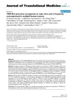
Pre-treatment with IFN-γ reduces the survival of neurons incubated with amyloid-β
1-42
Figure 1
Pre-treatment with IFN-γ reduces the survival of
neurons incubated with amyloid-β
1-42
. The survival of
primary cortical neurons pre-treated with 1 ng/ml TNFα, IL-
1β, IL-6 or IFN-γ prior to the addition of 10 µm amyloid-β
1-42
(shaded bars) or 10 µm amyloid-β
42-1
(open bars). Values
shown are the mean percentage cell survival from triplicate
experiments repeated 3 times (n = 9), ± standard deviation
(SD). ** = Neuronal survival significantly less than untreated
neurons incubated with amyloid-β
1-42
(p < 0.05).
The IFN-γ-induced sensitization of neurons to amyloid-β
1-42
is dose-dependentFigure 2
The IFN-γ-induced sensitization of neurons to amy-
loid-β
1-42
is dose-dependent. The survival of primary cor-
tical neurons pre-treated with different concentrations of
IFN-γ prior to the addition of 10 µm amyloid-β
1-42
(❍) or 10
µm amyloid-β
42-1
(●). Values shown are the mean percent-
age cell survival from triplicate experiments repeated 3 times
(n = 9), ± SD.
Journal of Neuroinflammation 2006, 3:7 />Page 4 of 7
(page number not for citation purposes)
rons that was not observed after the addition of a control
peptide (amyloid-β
42-1
). To determine if cytokines could
modify the effects of amyloid-β
1-42
, primary cortical neu-
rons were pre-treated with 1 ng/ml individual cytokines,
before the addition of 10 µM amyloid-β
1-42
. There was no
significant difference between the survival of neurons pre-
treated in control medium and those pre-treated in
medium containing TNF-α, IL-1β or IL-6 prior to the addi-
tion of amyloid-β
1-42
. In contrast, the survival of neurons
pre-treated with IFN-γ and amyloid-β
1-42
was significantly
less than neurons treated with amyloid-β
1-42
alone (Figure
1). Further studies demonstrated that this effect of IFN-γ
was dose-dependent; and a significant reduction in neuro-
nal survival was still observed when cells were treated with
40 pg per ml of IFN-γ (Figure 2).
The effects of IFN-γ were tested on both primary cortical
and cerebellar neuronal cultures. Pre-treatment with IFN-
γ (100 pg/ml) resulted in reduced survival of both primary
cerebellar and cortical neurons following the addition of
10 µM amyloid-β
1-42
. Since it is possible that the effects of
IFN-γ in these neuronal cultures were via effects on con-
taminating astroglial cells, we also tested the effects of
IFN-γ on the SH-SY5Y neuroblastoma cell line. Pre-treat-
ment with IFN-γ reduced the survival of SH-SY5Y neurob-
lastoma cells following the addition of 10 µM amyloid-β
1-
42
indicating that IFN-γ had a direct effect on neuroblast-
oma cells (Table 1).
To determine if IFN-γ treated neurons show increased sen-
sitivity to other neurotoxins, cortical neurons were treated
with 100 pg/ml of IFN-γ prior to exposure to HuPrP82-
146, a synthetic correlate of a neurotoxic peptide found in
the brains of patients with prion disease [10], stau-
rosporine or hydrogen peroxide. The survival of neurons
pre-treated with IFN-γ was significantly less than that of
untreated neurons, when incubated with HuPrP82-146.
However, there were no significant differences between
the survival of neurons treated with IFN-γ and untreated
neurons that were exposed to hydrogen peroxide, or to
staurosporine, a drug that caused programmed cell death
in neurons via activation of the ceramide pathway [11]
(Table 2).
Table 1: Treatment with IFN-γ reduces neuronal survival following incubation with amyloid-β
1-42
. SH-SY5Y cells or primary neuronal
cell cultures were pre-treated with IFN-γ (1 ng/ml) for 24 hours prior to the addition of amyloid-β peptides as shown. Cell survival was
determined using the WST-1 test after 24 hours (cell lines) or 4 days (primary neuronal cultures). Each value is the mean percentage
cell survival ± SD from triplicate experiments repeated 3 times (9 observations). ** = Neuronal survival significantly less than
untreated neurons incubated with amyloid-β
1-42
(p < 0.05).
% Neuronal Survival
Cell Type Amyloid-β
1-42
Amyloid-β
42-1
Control IFN-γ Control IFN-γ
SH-SY5Y cells 62 ± 4 33 ± 9** 101 ± 4 98 ± 7
Cortical neurons 68 ± 7 36 ± 7** 98 ± 3 96 ± 5
Cerebellar neurons 79 ± 4 58 ± 11** 101 ± 8 102 ± 8
Table 2: IFN-γ treated neurons show increased sensitivity to HuPrP82-146. Neurons treated with 1 ng/ml IFN-γ for 24 hours prior to
the addition of neurotoxins as shown. Cell survival was determined 24 hours later using the WST-1 test. Each value is the mean
percentage cell survival ± SD from triplicate experiments repeated 3 times (9 observations). ** = Neuronal survival significantly less
than untreated neurons incubated with HuPrP82-146 (p < 0.05).
Neuronal survival (%)
conc Control IFN-γ
HuPrP82-146 (µM) 40 48 ± 6 12 ± 7**
10 64 ± 5 22 ± 8**
2.5 92 ± 9 41 ± 9**
Staurosporine (ng/ml) 20 37 ± 4 40 ± 6
5 78 ± 6 72 ± 8
1.25 92 ± 3 95 ± 4
Hydrogen peroxide (µM) 1 22 ± 5 18 ± 7
0.25 58 ± 7 55 ± 6
0.06 87 ± 6 89 ± 9
Journal of Neuroinflammation 2006, 3:7 />Page 5 of 7
(page number not for citation purposes)
Caspase-3 activity
Caspase-3 is an enzyme that is increased during apoptosis
[12] and was measured as an alternative indicator of neu-
ronal injury. Caspase-3 activity was increased in primary
cortical neurons treated with amyloid-β
1-42
or HuPrP82-
146, but not in primary cortical neurones treated with
control peptides (amyloid-β
42-1
or HuPrP82-
146scrambled) or with IFN-γ (100 pg/ml) alone. Follow-
ing pre-treatment with 100 pg/ml IFN-γ caspase-3 activity
in cortical neurons treated with either amyloid-β
1-42
or
HuPrP82-146 was significantly higher than in untreated
cells incubated with amyloid-β
1-42
or HuPrP82-146 (Fig-
ures 3 &4).
IFN-
γ
raises cytoplasmic PLA
2
levels in neurons
Since recent studies demonstrated that cPLA
2
is involved
in amyloid-β
1-42
induced neuronal injury [13] we com-
pared levels of cPLA
2
and another enzyme involved in cell
signalling (PLCγ-1) in IFN-γ-treated and untreated SH-
SY5Y cells. Treatment with IFN-γ did not significantly alter
the total protein content of cells (data not shown). When
lysed cells were diluted and analysed in a dot blot we
found that SH-SY5Y cells treated with IFN-γ (100 pg/ml)
had higher levels of cPLA
2
than did untreated cells (Figure
5). However, pre-treatment with IFN-γ did not affect the
levels of PLCγ-1 indicating that IFN-γ up-regulates specific
pathways in these neurons. We next determined if pre-
treatment with cytokines affected prostaglandin produc-
tion. Levels of prostaglandin E
2
were not altered by any of
the cytokines tested. Prostaglandin E
2
levels were signifi-
cantly raised after the addition of either amyloid-β
1-42
or
HuPrP82-146, but not after the addition of control pep-
tides (amyloid-β
42-1
or HuPrP82-146scrambled). Pre-
treatment of neurons of 100 pg/ml IFN-γ resulted in
increased prostaglandin E
2
production following the addi-
tion of 10 µM amyloid-β
1-42
or 10 µM HuPrP82-146. Pros-
taglandin E
2
levels in neurons incubated with 10 µM
amyloid-β
1-42
or 10 µM HuPrP82-146 was not affected by
pre-treatment with TNF-α, IL-1β, IL-6 (Table 3).
Pre-treatment with IFN-γ increases amyloid-β
1-42
-induced caspase-3 activityFigure 3
Pre-treatment with IFN-γ increases amyloid-β
1-42
-
induced caspase-3 activity. Levels of caspase-3 activity in
cortical neurons pre-treated with control medium (❍), or
with 100 ng/ml IFN-γ (●) prior to the addition of varying
concentrations of amyloid-β
1-42
. Also shown is the caspase-3
activity from neurons pre-treated with control medium (ᮀ),
or with 100 ng/ml IFN-γ (■) and incubated with varying con-
centrations of amyloid-β
42-1
. Values shown are the mean per-
centage fluorescence units from quadruplicate experiments
repeated twice (n = 8), ± SD. ** = Caspase 3 activity signifi-
cantly greater than untreated neurons incubated with amy-
loid-β
1-42
(p < 0.05).
Pre-treatment with IFN-γ increases HuPrP82-146-induced caspase-3 activityFigure 4
Pre-treatment with IFN-γ increases HuPrP82-146-
induced caspase-3 activity. Levels of caspase-3 activity in
cortical neurons pre-treated with control medium (❍), or
with 100 ng/ml IFN-γ (●) prior to the addition of varying
concentrations of HuPrP82-146. Also shown is the caspase-3
activity from neurons pre-treated with control medium (ᮀ),
or with 100 ng/ml IFN-γ (■) and incubated with varying con-
centrations of HuPrP82-146scrambled. Values shown are the
mean percentage fluorescence units from quadruplicate
experiments repeated twice (n = 8), ± SD. ** = Caspase 3
activity significantly greater than untreated neurons incu-
bated with HuPrP82-146 (p < 0.05).
Journal of Neuroinflammation 2006, 3:7 />Page 6 of 7
(page number not for citation purposes)
Discussion
Reports that activated microglial cells are found in close
association with damaged neurons in AD raise the possi-
bility that glial-derived cytokines are involved in neu-
ropathogenesis. In the current studies the survival of
either primary neuronal cultures (cortical or cerebellar
neurons) or SH-SY5Y neuroblastoma cells was not
affected by incubation with high concentrations of recom-
binant cytokines (up to 10 ng/ml). However, while none
of the cytokines were directly neurotoxic, pre-treatment
with IFN-γ significantly reduced the survival of neurons
that incubated with amyloid-β
1-42
. This effect of IFN-γ was
dose-dependent and was observed at concentrations pre-
viously reported in the cerebral cortex of APP(SWE) trans-
genic mice [14].
Pre-treatment with IFN-γ also increased the sensitivity of
neurons to HuPrP82-146, a neurotoxic peptide found in
prion diseases [10]. However, neurons pre-treated with
IFN-γ did not demonstrate increased sensitivity to all neu-
rotoxins: there was no change in the neurotoxicity of stau-
rosporine, a drug that causes programmed cell death in
neurons via activation of the ceramide pathway [11], or of
hydrogen peroxide which causes oxidation of cellular
membranes. These observations strengthen the hypothe-
sis that IFN-γ treatment selectively increases the expres-
sion of proteins involved in specific apoptotic pathways.
Previous reports showed that amyloid-β peptides activate
PLA
2
[15], that PLA
2
inhibitors protect against the amy-
loid-β
1-42
induced neurotoxicity [16], and more specifi-
cally, that the cPLA
2
isoform is required for induced
neurotoxicity [13]. The current study showed that IFN-γ
increased expression of cPLA
2
in neurons, a result consist-
ent with previous observations that IFN-γ increases gene
expression of cPLA
2
in epithelial cells [17]. The activation
of cPLA
2
results in the release of arachidonic acid which is
subsequently metabolised by the COXs to prostaglandins
and in the present study the increased expression of cPLA
2
in IFN-γ treated neurons was associated with significantly
greater amounts of prostaglandin E
2
produced following
the addition of amyloid-β
1-42
or HuPrP82-146. IFN-γ
treatment increased cPLA
2
levels without affecting levels
of PLCγ-1, further evidence that IFN-γ selectively increases
expression of specific pathways.
In AD and prion diseases much of the neuronal death
occurs though apoptosis [3]. Although neurons incubated
with fibrillar PrP/amyloid-β peptides in vitro show signs of
apoptosis, the precise mechanisms that activate neuronal
apoptosis remain unknown. In the present study both
Table 3: Pre-treatment with IFN-γ increases prostaglandin E
2
production in response to amyloid-β or HuPrP82-146. SH-SY5Y
neuroblastoma cells were pre-treated for 24 hours with 1 ng/ml cytokines as shown prior to the addition of amyloid-β or PrP peptides.
Cells were lysed 24 hours later and total prostaglandin E
2
levels were measured. Each value given represents the mean ± SD from
triplicate experiments repeated twice (6 observations). ** = Prostglandin E
2
levels significantly higher than untreated neurons
incubated with amyloid-β
1-42
(p < 0.05).
Prostaglandin E
2
(pg/ml)
Amyloid-β
1-42
Amyloid-β
42-1
HuPrP82-146 HuPrP82-146sc
Control medium 241 ± 62 < 50 187 ± 40 < 50
IFN-γ 443 ± 112** 66 ± 38 303 ± 55** < 50
IL-1β 228 ± 54 < 50 202 ± 46 < 50
IL-6 218 ± 66 < 50 210 ± 48 55 ± 35
TNFα 255 ± 82 < 50 214 ± 33 < 50
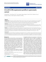
IFN-γ increases levels of cPLA
2
in SH-SY5Y neuroblastoma cellsFigure 5
IFN-γ increases levels of cPLA
2
in SH-SY5Y neurob-
lastoma cells. Immunoblot showing levels of cPLA
2
and
phospholipase C in lysates made from untreated cells and
cells that had been treated with 100 pg/ml of IFN-γ for 3
hours. The fractions of the original samples that were added
to membrane are shown.
Journal of Neuroinflammation 2006, 3:7 />Page 7 of 7
(page number not for citation purposes)
amyloid-β
1-42
and HuPrP82-146 increased neuronal cas-
pase-3 activity, a marker of apoptosis that is increased in
AD [18]. IFN-γ has been implicated in the pathogenesis of
AD and IFN-responsive mRNAs have been found in Creut-
zfeldt-Jakob disease [19]. IFN-γ can be produced in the
brain by glial cells and IFN-γ immunoreactivity and IFN-
γ-gene expression have been detected in human sensory
neurons [20]. Thus, these results indicate that IFN-γ has
the potential to increase neuronal loss in AD or prion dis-
eases, consistent with a previous report that the induction
of IFNs hastens the progression of experimental prion dis-
eases in mice [21].
Conclusion
We report that pre-treatment with IFN-γ increased the lev-
els of cPLA
2
in SH-SY5Y neuroblastoma cells without
affecting total cellular protein concentrations, or the levels
of PLCγ-1. The increased levels of cPLA
2
were associated
with increased prostaglandin E
2
production in response to
amyloid-β
1-42
or HuPrP82-146. More importantly, pre-
treatment with IFN-γ resulted in reduced neuronal sur-
vival following the addition of amyloid-β
1-42
or HuPrP82-
146. Such results are consistent with previous observa-
tions that cPLA
2
is involved in neurodegeneration in AD
or prion diseases and indicate that IFN-γ may hasten neu-
ronal loss in these diseases.
Abbreviations
Alzheimer's disease (AD), interferon (IFN), cytoplasmic
phospholipase A
2
(cPLA
2
), phospholipase C (PLC), cyclo-
oxygenases (COX), flourometric immunosorbent enzyme
assay (FIENA), tumour necrosis factor (TNF), interleukin
(IL).
Competing interests
The author(s) declare that they have no competing inter-
ests.
Authors' contributions
CB was responsible for the conception, planning and per-
formance of experiments, and for writing this manuscript.
Both SK and VL prepared western and dot blot analysis.
AW contributed to the planning of experiments, interpre-
tation of results and the writing of the manuscript.
Acknowledgements
This work was supported by a grant from the European Commission
(QLK3-CT-2001-00283) and the EU FP6 – Network of Excellence "Neuro-
prion".
References
1. Hardy J, Selkoe DJ: The amyloid hypothesis of Alzheimer's dis-
ease: progress and problems on the road to therapeutics. Sci-
ence 2002, 297:353-356.
2. Esler WP, Wolfe MS: A portrait of Alzheimer secretases – new
features and familiar faces. Science 2001, 293:1449-1454.
3. Selkoe DJ: Translating cell biology into therapeutic advances
in Alzheimer's disease. Nature 1999, 399:A23-A31.
4. Yankner BA, Dawes LR, Fisher S, Villa-Komaroff L, Oster-Granite ML,
Neve RL: Neurotoxicity of a fragment of the amyloid precur-
sor associated with Alzheimer's disease. Science 1989,
245:417-420.
5. Forloni G, Chiesa R, Smiroldo S, Verga L, Salmona M, Tagliavini F,
Angeretti N: Apoptosis mediated neurotoxicity induced by
chronic application of beta amyloid fragment 25–35. Neurore-
port 1993, 4:523-526.
6. Akiyama H, Arai T, Kondo H, Tanno E, Haga C, Ikeda K: Cell medi-
ators of inflammation in the Alzheimer disease brain. Alzhe-
imer Dis Assoc Disord 2000, 14(Suppl 1):S47-S53.
7. Papassotiropoulos A, Bagli M, Jessen F, Bayer TA, Maier W, Rao ML,
Heun RA: A genetic variation of the inflammatory cytokine
interleukin-6 delays the initial onset and reduces the risk for
sporadic Alzheimer's disease. Ann Neurol 1999, 45:666-668.
8. Bate C, Salmona M, Williams A: The role of platelet activating
factor in prion and amyloid-β neurotoxicity. Neuroreport 2004,
15:509-513.
9. Bate C, Reid S, Williams A: Killing of prion-damaged neurones
by microglia. Neuroreport 2001, 12:2589-2594.
10. Salmona M, Morbin M, Massignan T, Colombo L, Mazzoleni G, Capo-
bianco R, Diomede L, Thaler F, Mollica L, Musco G, Kourie JJ, Bugiani
O, Sharma D, Inouye H, Kirschner DA, Forloni G, Tagliavini F: Struc-
tural properties of Gerstmann-Straussler-Scheinker disease
amyloid protein. J Biol Chem 2003, 278:48146-48153.
11. Wiesner DA, Dawson G: Staurosporine induces programmed
cell death in embryonic neurons and activation of the cera-
mide pathway. J Neurochem 1996, 66:1418-1425.
12. Cohen GM: Caspases: the executioners of apoptosis. Biochem J
1997, 326(Pt 1):1-16.
13. Kriem B, Sponne I, Fifre A, Malaplate-Armand C, Lozac'h-Pillot K,
Koziel V, Yen-Potin FT, Bihain B, Oster T, Olivier JL, Pillot T:
Cytosolic phospholipase A2 mediates neuronal apoptosis
induced by soluble oligomers of the amyloid-β peptide. The
FASEB Journal 2004:04-1807fje.
14. Abbas N, Bednar I, Mix E, Marie S, Paterson D, Ljungberg A, Morris
C, Winblad B, Nordberg A, Zhu J: Up-regulation of the inflam-
matory cytokines IFN-gamma and IL-12 and down-regula-
tion of IL-4 in cerebral cortex regions of APP(SWE)
transgenic mice. J Neuroimmunol 2002, 126:50-57.
15. Lehtonen JY, Holopainen JM, Kinnunen PK: Activation of phos-
pholipase A2 by amyloid beta-peptides in vitro. Biochemistry
1996, 35:9407-9414.
16. Bate C, Reid S, Williams A: Phospholipase A2 inhibitors or plate-
let activating factor antagonists prevent prion replication. J
Biol Chem 2004, 279:36405-36411.
17. Lindbom J, Ljungman AG, Lindahl M, Tagesson C: Increased gene
expression of novel cytosolic and secretory phospholipase
A(2) types in human airway epithelial cells induced by tumor
necrosis factor-alpha and IFN-gamma. J Interferon Cytokine Res
2002, 22:947-955.
18. Blasko I, Stampfer-Kountchev M, Robatscher P, Veerhuis R, Eikelen-
boom P, Grubeck-Loebenstein B: How chronic inflammation can
affect the brain and support the development of Alzheimer's
disease in old age: the role of microglia and astrocytes. Aging
Cell 2004, 3:169-176.
19. Baker CA, Lu ZY, Manuelidis L: Early induction of interferon-
responsive mRNAs in Creutzfeldt-Jakob disease. J Neurovirol
2004, 10:29-40.
20. Neumann H, Schmidt H, Wilharm E, Behrens L, Wekerle H: Inter-
feron gamma gene expression in sensory neurons: evidence
for autocrine gene regulation. J Exp Med 1997, 186:2023-2031.
21. Allen LB, Cochran KW: Acceleration of scrapie in mice by tar-
get-organ treatment with interferon inducers. Ann N Y Acad
Sci 1977, 284:676-680.