báo cáo hóa học: " Expression of innate immune complement regulators on brain epithelial cells during human bacterial meningitis" docx
Bạn đang xem bản rút gọn của tài liệu. Xem và tải ngay bản đầy đủ của tài liệu tại đây (1.6 MB, 9 trang )
BioMed Central
Page 1 of 9
(page number not for citation purposes)
Journal of Neuroinflammation
Open Access
Research
Expression of innate immune complement regulators on brain
epithelial cells during human bacterial meningitis
Cecile Canova
1
, Jim W Neal
2
and Philippe Gasque*
1,3
Address:
1
Brain Inflammation and Immunity Group, Department of Medical Biochemistry, Cardiff University, Heath Park, Cardiff, CF14 4XN, UK,
2
Department of Pathology, Neuropathology Laboratory; Cardiff University, Heath Park, Cardiff, CF14 4XN, UK and
3
LBGM, Faculty of Sciences
and Technologies, University of la Reunion, 15 Avenue René Cassin, BP7151, 97715, Saint Denis, Reunion
Email: Cecile Canova - ; Jim W Neal - ; Philippe Gasque* -
* Corresponding author
Abstract
Background: In meningitis, the cerebrospinal fluid contains high levels of innate immune
molecules (e.g. complement) which are essential to ward off the infectious challenge and to
promote the infiltration of phagocytes (neutrophils, monocytes). However, epithelial cells of either
the ependymal layer, one of the established niche for adult neural stem cells, or of the choroid
plexus may be extremely vulnerable to bystander attack by cytotoxic and cytolytic complement
components.
Methods: In this study, we assessed the capacity of brain epithelial cells to express membrane-
bound complement regulators (ie, CD35, CD46, CD55 and CD59) in vitro and in situ by
immunostaining of control and meningitis human brain tissue sections.
Results: Double immunofluorescence experiments for ependymal cell markers (GFAP, S100, ZO-
1, E-cadherin) and complement regulators indicated that the human ependymal cell line model was
strongly positive for CD55, CD59 compared to weak stainings for CD46 and CD35. In tissues, we
found that CD55 was weakly expressed in control choroid plexus and ependyma but was
abundantly expressed in meningitis. Anti-CD59 stained both epithelia in apical location while
increased CD59 staining was solely demonstrated in inflamed choroid plexus. CD46 and CD35
were not detected in control tissue sections. Conversely, in meningitis, the ependyma,
subependyma and choroid plexus epithelia were strongly stained for CD46 and CD35.
Conclusion: This study delineates for the first time the capacity of brain ependymal and epithelial
cells to respond to and possibly sustain the innate complement-mediated inflammatory insult.
Background
The activation of complement is an important component
of the innate immune response providing the capacity to
detect and to clear pathogens (for review [1]). The main
source of complement proteins is the liver, but many cell
types including fibroblasts, epithelial and endothelial
cells as well as glia and neurons also synthesise most of
the complement components [2]. The activation of one of
three different complement pathways, the classical, alter-
native or mannan binding lectin pathways, leads to the
formation of C3 and C5 convertases [1]. Some of the
resulting compounds, called opsonins, bind to pathogens
allowing the formation of the membranolytic attack com-
plex (MAC) [3,4]. During the acute phase of inflamma-
Published: 02 September 2006
Journal of Neuroinflammation 2006, 3:22 doi:10.1186/1742-2094-3-22
Received: 19 April 2006
Accepted: 02 September 2006
This article is available from: />© 2006 Canova et al; licensee BioMed Central Ltd.
This is an Open Access article distributed under the terms of the Creative Commons Attribution License ( />),
which permits unrestricted use, distribution, and reproduction in any medium, provided the original work is properly cited.
Journal of Neuroinflammation 2006, 3:22 />Page 2 of 9
(page number not for citation purposes)
tion, complement fragments may also bind to the surface
of resident host cells and promote bystander effects with
the formation of cytotoxic and cytolytic MAC. Under
these conditions, many cell types facing complement
attack express several key complement regulators (CRegs)
on their membranes to avoid damages by inhibiting
either the C3 convertases (complement receptor type 1,
CR1/CD35; membrane cofactor protein, MCP/CD46;
decay accelerating factor, DAF/CD55) or by avoiding the
formation of MAC (CD59). In the human brain, astro-
cytes, microglia and oligodendrocytes express CRegs (for
review [5,6]) but neurons are extremely susceptible to
complement mediated lysis as they express low levels of
CRegs [7,8].
The expression of CRegs by the different brain epithelial
cells remains poorly characterized. Very high levels of
complement proteins are present in the cerebrospinal
fluid (CSF) particularly in infection or inflammatory con-
ditions of the brain [9] and presumably as a consequence
of plasma transudation or intrathecal synthesis by infil-
trating leukocytes and resident activated epithelial cells
[10,11]. The epithelium lining brain ventricles (epend-
yma), spinal cord and choroid plexus consists of special-
ized glial cells (for review [12]) and ependymal cells of the
ventricles express the glial fibrillary acidic protein (GFAP)
and S100 markers [13]. In adult, neural stem cells reside
within the ependyma and/or subependyma (also known
as the subventricular zone, SVZ) and, from these prolifer-
ative zones, cells migrate to their destiny in the injured
brain where they differentiate into neurons and glia [13-
15]. Remarkably, ependymal cells contribute to a unique
and heterogeneous epithelial layer providing a critical
physical barrier against pathogen infiltration and cell-
mediated cytotoxicity (e.g. neutrophil compounds). In
bacterial meningitis, the levels of complement anaphyla-
toxins C3a and C5a are dramatically increased in the CSF
enhancing the influx of inflammatory cells into the ventri-
cle and increasing complement biosynthesis and promot-
ing activation [11,16,17]. Critically, the epithelial cells
exposed to the CSF have to withstand the activities of
strong and sustained complement activation.
Histopathological assessment together with electron
microscopy studies (this study) have revealed that the
ependymal cells in meningitis appear resistant to the
potential toxic effects of microbial and neutrophil prod-
ucts. In contrast, it has been reported that in severe bacte-
rial and fungal ependymitis, the layer of epithelial cells is
highly destructed [18]. The ependyma is vulnerable to
injury throughout both fetal and adult life and particu-
larly in diseased conditions but the cellular and molecular
nature of the intrinsic mechanisms conferring resistance
(or not) to tissue damage remains poorly characterised.
To explore the capacity of brain epithelial cells (i.e.
choroid plexus as well as ependymal layer lining the ven-
tricle) to protect themselves from severe complement
attack in disease conditions we have investigated and
compared the expression of CRegs between control and
several meningitis cases. Moreover, a human ependy-
moma primary culture model was established to provide
additional information about Cregs expression by epend-
ymal cells in culture.
Methods
Source of tissues
Small blocks of paraffin wax embedded temporal lobe,
containing the hippocampus and the choroid plexus from
the lateral ventricles were available for examination from
cases of meningococcal meningitis. From the same cases,
small tissue blocks from the caudate nucleus, lined by
ependymal cells, were also available. In all cases, there
was significant numbers of neutrophils within the cere-
brospinal fluid in contact with the lining ependyma and
within the ventricle system (see Figure 1Bb). Blocks of
hippocampus with choroid plexus, together with blocks
of caudate nucleus lined by ependymal cells were availa-
ble from control cases without evidence of systemic infec-
tion, cerebral ischemia or neurodegeneration. Control
cases did not present astrogliosis and microgliosis as indi-
cated by the GFAP and HLA-II stainings, respectively. Tan-
gles and βA4-plaques were not present in control cases. All
cases were available from the Neuropathology laboratory
(JWN, Cardiff University, Heath Hospital, Cardiff) and
used under the guidelines approved by the Bro Taf Health
Authority local ethical approval (reference 98/2773).
Tissues from each case had been fixed in 2% neutral buff-
ered formalin for two weeks, subsequently processed in
paraffin wax and sections 6 µm-thick cut and stained for
further light microscopy and immunocytochemical inves-
tigations.
Primary cell cultures of ependymoma cell line (clone 9945)
A primary culture of ependymal cells was prepared from a
fragment of a biopsy from a spinal cord ependymoma.
Briefly, tissue was minced in MEM-medium with iridec-
tomy scissors. Subsequent trypsinization (0.025% trypsin
in calcium and magnesium free phosphate-buffered
saline) was performed for 15 min at 37°C. After removing
trypsin by centrifugation the cells were resuspended in
RPMI medium (Gibco) supplemented with 10% foetal
calf serum/L-glutamine 1X/streptomycin (100 µg/ml)/
penicillin (62.5 µg/ml) and dissociated by mechanical
trituration using a Pasteur pipette in order to obtain a sus-
pension of single cells. Cells were plated and cultured at
37°C in a humidified 5% CO
2
incubator (Heraeus,
Hanau, Germany).
Journal of Neuroinflammation 2006, 3:22 />Page 3 of 9
(page number not for citation purposes)
Electron microscopy of ependymoma cells
A small fragment of tissue from the ependymoma sample
was dissected and placed in a solution of 2.5% glutamine/
3% osmium/Araldite resin. Further processing for
ultrastructural electron microscopy was carried out as pre-
viously described [19].
Source of antibodies
Mouse anti-HLA Class II (clone CR3/43, M0775), rabbit
anti-GFAP (Z0334) and the FITC- and Rhodamine-cou-
pled secondary antibodies, goat anti-mouse IgG and goat
anti-rabbit IgG, were obtained from DAKO Ltd. (High
Wycombe, Bucks, United Kingdom). Rabbit antibodies
against CD59, CD35, CD55, and CD46 were all raised in
house after immunization using purified human CRegs
[20]. Mouse monoclonal anti-CD59 (clone BRIC 229)
and anti-CD55 (clone BRIC 216) were from the Interna-
tional Blood Group Reference Laboratory (IBGRL, Elstree,
Herts, UK). Mouse anti-CD35 was from Dako and mouse
anti-CD46 was from Serotec (Oxford, UK) The peroxi-
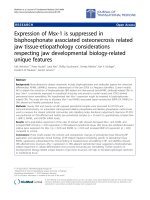
Structural and ultrastructural (electron microscopy) analyses of human brain tissue sections obtained from the ependymoma (case 9945) and meningitis casesFigure 1
Structural and ultrastructural (electron microscopy) analyses of human brain tissue sections obtained from the ependymoma
(case 9945) and meningitis cases. Aa. H&E staining of paraffin embedded wax tissue sections; original magnification, ×200. The
presence of perivascular rosette (Ro) formation is typical of an ependymoma. Ab. Electron micrographs taken of the araldite-
enhanced ependymoma cells. Original magnification, ×13500; inset: original magnification, × 23000. The white arrows indicate
junction complexes between cells and the black arrows indicate microvilli (see inset). These structures reveal key characteris-
tics of ependymal cells. Ba, Bb, Meningitis cases. Original magnification, ×100. Choroid plexus (a) and ependyma (Ep) (b) show
a continuous layer of intact epithelial cells despite the presence of neutrophils (PMN) inside the ventricle (particularly in panel
Bb, see inset, magnification ×1000). V, vessel; Cp, choroid plexus; Ep, ependymal layer.
Journal of Neuroinflammation 2006, 3:22 />Page 4 of 9
(page number not for citation purposes)
dase-conjugated secondary antibodies goat anti-mouse
IgG and goat anti-rabbit IgG were from Bio-Rad (Hermel
Hempstead, Hertfordshire, United Kingdom).
Immunohistochemistry
6 µm thick tissue sections were mounted on super-frost
glass slides (Surgipath Europe Ltd., Peterborough, United
Kingdom). Antigen retrieval was required for all anti-
CRegs antibody staining protocols. To this aim, sections
were heated in freshly prepared 0.2% citric acid buffer at
pH 6.0 for 30 min in a microwave at full power (750
watts). Sections were left 30 min at room temperature and
then rinsed in tap water.
Sections were immunostained by the indirect immu-
noperoxidase/3'3' diaminobenzidine HCl method. All
sections were incubated overnight with their appropriate
antibodies diluted in 1% BSA prepared in phosphate
buffer. The secondary antibodies were similarly prepared
and used at 1:200 (1 to 2.5 ug/ml final). Sections were
washed 3 times in PBS 1× after which they were developed
for 5 minutes in a freshly made solution of 0.05% diami-
nobenzidine (DAB) and 0.005% (v/v) hydrogen peroxide
diluted in PBS 1×. After a wash in tap water, sections were
counterstained using hematoxylin. After a full dehydra-
tion in ethanol, the sections were cleared in xylene and
mounted.
Immunocytochemistry
Ependymoma cells were cultured in RPMI medium sup-
plemented with 10% foetal calf serum on poly-D-lysine-
coated coverslips for 2 days. Then, after 5 washes in NaCl
0.9%, cells were fixed in cold acetone. The phenotype of
the cells was further assessed by staining the coverslips
with polyclonal antibody against GFAP (Dako Ltd, High
Wycombe, Bucks, United Kingdom) and monoclonal
antibodies against S-100 protein (Sigma, Saint Louis, Mis-
souri, USA) ZO-1 (zonula occludens 1) and E-cadherin
(Becton Dickinson, Oxford UK) [12,21]. The coverslips
were also stained with primary rabbit and mouse antibod-
ies against human CRegs 1 h at room temperature. After
three washes in PBS 1×, they were incubated for 1 h at
room temperature with 4'-6-diamino-2-puenylindole-2
HCl (DAPI; 1:1000, nuclear staining) and FITC-coupled
goat anti-mouse IgG or rhodamine-coupled goat anti-rab-
bit IgG (1:100). Coverslips were washed 3 times in PBS 1×
and then mounted with Vectashield medium (Vector lab-
oratories, Peterborough, UK) on a glass slide.
Results
Histopathological assessment of the human ependymoma
(case 9945) and meningitis cases
All clinical samples used in the study were first thoroughly
analysed for histopathology hallmarks. Histological fea-
tures of the ependymoma revealed the presence of
perivascular rosette formation with acellular areas (Figure
1Aa, Ro) and peripheral individual cells with vesicular
nuclei. There was no evidence of either necrosis, mitosis
or endothelial proliferation. On this basis, the tumour
was classified as WHO grade II [22]. Further immunohis-
tological examinations of paraffin embedded tissue sec-
tions indicated that the ependymoma was strongly
stained for GFAP and S100 (data not shown). Electron
micrographs were taken of the Araldite-enhanced speci-
men on a Joel Electron micrograph, at 27000 and 13500
magnifications (Figure 1Ab). The ultrastructural features
were of a tumour with junctural complexes between adja-
cent cells (white arrows). A small rosette-like structure
containing cells with apical microvilli was also present
(black arrow, inset). The overall ultrastructural findings
were characteristic of an ependymoma [23]. These tumors
are extremely rare and we were able to establish primary
cultures of ependymal cells isolated from the same biopsy
used for histology (see below).
We also performed histopathological assessments of the
meningitis cases. Figure 1Ba–b depicts the level of poly-
morphonuclear (PMN) infiltration within the brain ven-
tricles closed to remarkably well preserved epithelia of the
choroid plexus and the ependymal layer. Although con-
trol brains were free of any infiltrating leukocytes, we
found robust PMN infiltration in all bacterial meningitis
cases. The local inflammation was associated with a
strong GFAP staining of ependymal cells (Table 1). Inter-
estingly, Kolmer cells (macrophage-like cells) but not the
epithelial cells were found to express high levels of HLA-
class II (Table 1).
Tumour-derived ependymal cells (clone 9945) express
several key complement regulators
Ependymal primary cultures were analysed for specific
cell markers and CRegs by double immunofluorescence
staining experiments. The large majority of cells presented
a typical ependymal cell phenotype and were strongly
stained for GFAP and S100 (data not shown). CD11b+,
CD14+ contaminating Kolmer cells were not identified in
our cultures. However, epithelial-like cells demonstrated
strong membrane staining with the anti-CD55 and CD59.
In contrast, they were weakly stained for CD46 and CD35
(Figure 2). These data were confirmed using different cell
culture passages (1–5). FACS analyses could not be per-
formed given the limited number of cells isolated from
the ependymoma.
The expression of several key regulators of the
complement system is dramatically upregulated in choroid
plexus epithelium in meningitis
We next analysed the capacity of epndymal cells and the
epithelial cells of the choroid plexus to express Cregs in
healthy and inflamed brains. Of important note, we
Journal of Neuroinflammation 2006, 3:22 />Page 5 of 9
(page number not for citation purposes)
observed that the epithelium of the choroid plexus and
ependymal cells lining the ventricles in meningitis was
largely preserved despite the presence of large number of
neutrophils in the ventricles (Figure 1B and Figure 3).
Control and meningitis cases were immunostained for all
membrane-bound CRegs and the level of staining was
scored by three independent examiners blinded to the
individual treatment groups (Table 1 and Figure 3A). Kol-
mer cells in choroid plexus stroma were strongly stained
using an antibody to HLA Class II (clone CR3/43) but no
differences in the intensity or pattern of staining were
noticed between control and meningitis cases (Figure 3A,
a/b).
Epithelial cells of the choroid plexus were clearly negative
for GFAP. Affinity purified polyclonal antibodies against
CRegs were used on paraffin-embedded tissue sections.
First, the staining with anti-CD55 antibody was weak in
the choroid plexus in control cases but was more promi-
nent in one case of meningitis (Figure 3Ae/f,; Table 1).
Anti-CD46 staining showed a significant increase between
normals compared to all meningitis case (Figure 3Ag/h;
Table 1). Interestingly, CD35 was solely expressed by Kol-
mer cells in normal choroid plexus epithelium (Fig. 3Ai).
In contrast, during meningitis, ependymal and Kolmer
cells were strongly stained for CD35 (Figure 3Ai/j; Table
1). The apical CD59 staining was much pronounced on
epithelial cells of the choroid plexus in pathological con-
ditions (Figure 3Ak/l; Table 1).
Overexpression of CD46 and CD35 in ependymal cells
from meningitis cases
Ependymal cells from ventricle lining showed staining
differences compared to the choroid plexus from the same
cases. Some anti-GFAP staining was detected in normal
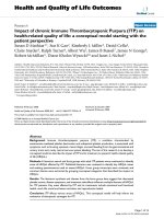
Immunofluorescence analyses of the human tumour-derived ependymoma primary cultures (clone 9945) stained for com-plement regulatory proteins and cell markersFigure 2
Immunofluorescence analyses of the human tumour-derived
ependymoma primary cultures (clone 9945) stained for com-
plement regulatory proteins and cell markers. Cells on cov-
erslips were fixed with acetone and stained with antibodies
against ependymal cells specific markers (GFAP, ZO-1 and
S100, not shown) and complement regulators proteins
(CD55, CD59, CD46 and CD35). Original magnification
×400. Background staining was observed using irrelevant
antibodies (inset). Nuclei were counterstained with DAPI
(blue).
Table 1: History, pathology and immunostaining data of control and meningitis cases for complement regulatory proteins and cell
markers.
Gender Age (yr) PM interval
(hours)
Cases Immunodetection of complement regulatory proteins Inflammatory index
CD55 CD59 CD46 CD35 HLA-II GFAP
CP Ep CP Ep CP Ep CP Ep CP Ep Ep
M 52 48 normal + + + + + 0 0 0 ++ (K) 0 +
M 86 48 normal + + + + 0 0 0 0 + (K) 0 +
M 23 12 bacterial meningitis ++ + ++ + ++ 0 + + ++ (K) 0 +++
F 68 48 bacterial meningitis + ++ ++ ++ ++ ++ ++ + ++ (K) 0 +++
F 55 48 bacterial meningitis +++ na ++ na +++ na ++ na ++ (K) 0 na
Abbreviations used: CP, choroid plexus epithelial cells; Ep, ependymal cell layer; M, male; F, female; K, Kolmer cells; 0: negative; +: very few cells
positive; ++: positive; +++: strongly positive. (PM, Postmortem); na: not applicable
Journal of Neuroinflammation 2006, 3:22 />Page 6 of 9
(page number not for citation purposes)
conditions but the immunostaining was highly increased
in meningitis cases (Figure 3B, Table 1). Ependymal cells
did not show any HLA Class II staining either in normal
or pathological cases (Figure 3B, Table 1). Ependymal
cells express CD55, CD59 and CD46 antigens in normal
cases and the stainings were increased in meningitis cases
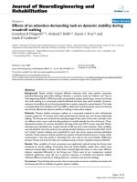
Immunoperoxidase histochemistry analyses of paraffin-wax sections to assess the expression of complement regulatory pro-teins (CRegs) in control and meningitis casesFigure 3
Immunoperoxidase histochemistry analyses of paraffin-wax sections to assess the expression of complement regulatory pro-
teins (CRegs) in control and meningitis cases. Rehydrated paraffin wax sections of human choroid plexus in normal and menin-
gitis cases immunostained with antisera to inflammatory cells and to membrane regulators proteins. Original magnification,
×400. Panel A: Choroid plexus staining data: a and b, Rabbit anti-GFAP. c and d, Rabbit anti-HLA II staining only Kolmer cells
(arrow). No differences are noticed between normal and meningitis cases. e and f, Rabbit anti-CD55. Choroid plexus epithe-
lium in normal cases is weakly stained (e) but more strongly in meningitis cases (f). Erythrocytes in f are stained for CD55
(arrow). g and h, Rabbit anti-CD46. Epithelium is weakly stained in normal cases (g) but strongly in meningitis epithelia, while
infiltrating PMN (arrow) are strongly CD46+ (h). i and j, Rabbit anti-CD35. Weak staining is detected on Kolmer cells in nor-
mal sections (i) but a strong staining is noticed in meningitis epithelia and infiltrating PMNs (j). k and l, Rabbit anti-CD59. Nor-
mal choroid plexus epithelia are stained for CD59 (k) and with a stronger staining in meningitis (l). Panel B: same as above but
assessing the expression of CRegs on ependymal cells. Note that erythrocytes (Er) within blood vessels are strongly stained for
CD55 (f).
Journal of Neuroinflammation 2006, 3:22 />Page 7 of 9
(page number not for citation purposes)
(Figure 3B; Table 1). Interestingly CD35 expression was
not detected on ependymal cells of control brains but was
expressed in meningitis cases (Figure 3B, Table 1).
Discussion
The organisation of brain epithelia is very similar to most
other epithelial membranes as they form a cellular net-
work tightly interconnected by gap junctions [24].
Despite the presence of these physical barriers, it is now
well established that several micro-organisms can infil-
trate the meninges and choroid plexus and gain access to
the brain parenchyma [25]. Moreover, these infectious
challenges will promote a sustained cellular and molecu-
lar innate immune responses, the local production of sev-
eral key cytotoxic and cytolytic proteins (complement,
TNFα, defensins) and with the potential to harm the sur-
rounding neural cells [26-30]. Several elegant studies have
demonstrated a pro-inflammatory reaction of ependyma
and choroid plexus epithelia in response to bacterial
infection including the expression of tumour necrosis fac-
tor-α [31,32] and ICAM-1 to facilitate neutrophil invasion
[22,33]. Whether the ependyma and choroid plexus are
able to control these inflammatory insults may be impor-
tant to the plasticity and homeostasis of the inflamed
brain. Remarkably, comprehensive structural and ultras-
tuctural analyses of meningitis cases (illustrated in Figure
1Bb and JWN's unpublished data) indicated that the epi-
thelial cells remained largely unaffected. This study was
undertaken to decipher some of the intrinsic pathways
expressed by reactive brain epithelial cells to control
bystander cytotoxic properties of the local innate immune
response.
Our data indicate that the integrity of the ependymal and
choroid plexus layers was preserved despite the large
number of neutrophils in the CSF while the expression of
CRegs on both epithelia was dramatically increased dur-
ing meningitis. We found that the level of all membrane-
bound regulators was dramatically upregulated on
choroid plexus epithelial cells and ependyma in all men-
ingitis cases. The regulation of CD55 expression by epithe-
lial cells of the choroid plexus demonstrated minor
changes between control and meningitis cases. In con-
trast, levels of CD55 and CD46 were strongly elevated on
ependymal cells in disease conditions. These data argue
for a regional specificity and independent regulation of
Cregs between epithelial cells of the choroid plexus and
the ependyma of the ventricle which may be due to the
local inflamed microenvironment.
The mechanisms controlling the expression of Cregs on
brain epithelial cells are largely ill-characterised. The
expression of Cregs has been studied on several cell types
including human vascular endothelial cells and was
shown to be regulated by a plethora of proinflammatory
cytokines (e.g. TNF, IL1) and LPS [34]. In meningitis, both
epithelia are exposed to inflammatory cytokines (TNF-α),
to complement-derived products (e.g. C3a, C5a and sub-
lytic doses of C5b9) as well as bacterial products such as
lipopolysaccharide (LPS) and peptidoglycans (PGs).
Together these compounds may profoundly affect the
plasticity of the brain epithelial cells and potentially driv-
ing robust expression of regulatory proteins to protect
from bystander complement attack. In human meningitis,
we found that epithelial cells and glial cells of the sub-
ependyma failed to express MHC class II antigens but in
contrast, were strongly stained for GFAP, a classical
marker of ependymal cell activation. Interestingly, the
expression of TLR4 mRNA in choroid plexus epithelium
has been reported in normal rat brain [35] and correlated
with CD14 mRNA expression [36]. Our preliminary
unpublished observations confirmed that human brain
epithelial cells of the choroid plexus are strongly stained
for TLR4 and CD14 while ependymal cells were solely
CD14+. The presence of these two key pattern recognition
receptors of the innate immune system raises the possibil-
ity that brain epithelial cells are capable of sensing carbo-
hydrate structures released from bacterial cell wall (LPS,
PGs) [37,38]. It remains to be ascertained whether the
treatment of brain epithelial cells with LPS and/or PGs can
control the level of Cregs expression and experiments
along these lines are now highly warranted.
It is important to emphasise that a delicate balance prob-
ably exits between the anti-inflammatory/protective
responses to protect the brain against the rather pro-
inflammatory/toxic response in severe bacterial meningi-
tis. The severity of the lesioned microenvironment may
determine ependymal cell survival and ultimately, the
clinical outcome with associated sequelae. The final
response will involve the proliferation and differentiation
of the neural stem cells which again could be affected by
inflammatory mediators.
New data emphasise the key role of brain epithelial cells
to integrate and further orchestrate the local innate
immune response with the production of innate compo-
nents of the complement system while preventing second-
ary tissue damage. Of note, the increased expression of
CRegs by brain epithelial cells may also contribute to a
double-edged sword scenario. On the one hand, high lev-
els of membrane-bound and soluble Cregs from brain
epithelial cells would certainly confer increased protec-
tion from complement-mediated attack but on the other,
bacteria and viruses (e.g Measles) are known to bind to
several Cregs and so evading the host innate immune
defense mechanisms [29,39-41].
A better understanding of the cellular and molecular
innate immune responses in the CNS and deciphering the
Journal of Neuroinflammation 2006, 3:22 />Page 8 of 9
(page number not for citation purposes)
pathways involved in the cross-talk between brain epithe-
lial cells and infectious agents will help enormously to
develop novel therapeutic strategies against brain infec-
tion [42].
Conclusion
Our findings underscore the remarkable capacity of brain
epithelial cells to withstand complement activation and
to survive within an inflammatory site. The Cregs on brain
epithelial cells may on one hand help to protect from
bystander complement attack but on the other provide a
niche for bacterial infection and contributing to meningi-
tis pathology.
Abbreviations
MAC: Membrane attack complex
CRegs: Complement regulators
CSF: Cerebro spinal fluid
GFAP: Glial fibrillary acidic protein
CR: complement receptor
MCP: membrane cofactor protein
DAF: Decay accelerating factor
Competing interests
The author(s) declare that they have no competing inter-
ests.
Authors' contributions
Dr Canova was involved with the day-to-day experimental
approach and the analyses of the data. Histopathological
assessment was performed by Dr Jim Neal, Neuropathol-
ogist at Cardiff University, Medical School. Prof. Philippe
Gasque was involved with the design and the supervision
of the work; preparation of the manuscript was done by
Dr Canova/Prof. Gasque.
Acknowledgements
This work was supported with funds from the Wales Office for Research
and Development for Health and Social Care (CC/PG) and the Medical
Research Council (PG). We thank Dr Karen Francis for critical reading of
the manuscript.
References
1. Morgan BP: The complement system: an overview. Methods
Mol Biol 2000, 150:1-13.
2. Morgan BP, Gasque P, Singhrao S, Piddlesden SJ: The role of com-
plement in disorders of the nervous system. Immunopharmacol-
ogy 1997, 38(1-2):43-50.
3. Medicus RG, Gotze O, Muller-Eberhard HJ: Alternative pathway
of complement: recruitment of precursor properdin by the
labile C3/C5 convertase and the potentiation of the pathway.
J Exp Med 1976, 144(4):1076-1093.
4. Taylor P, Botto M, Walport M: The complement system. Curr Biol
1998, 8(8):R259-61.
5. Morgan BP, Gasque P: Expression of complement in the brain:
role in health and disease. Immunol Today 1996, 17(10):461-466.
6. Zajicek J, Wing M, Skepper J, Compston A: Human oligodendro-
cytes are not sensitive to complement. A study of CD59
expression in the human central nervous system. Lab Invest
1995, 73(1):128-138.
7. Singhrao SK, Neal JW, Rushmere NK, Morgan BP, Gasque P: Spon-
taneous classical pathway activation and deficiency of mem-
brane regulators render human neurons susceptible to
complement lysis. Am J Pathol 2000, 157(3):905-918.
8. Chen S, Caragine T, Cheung NK, Tomlinson S: Surface antigen
expression and complement susceptibility of differentiated
neuroblastoma clones. Am J Pathol 2000, 156(3):1085-1091.
9. Aldred AR, Brack CM, Schreiber G: The cerebral expression of
plasma protein genes in different species. Comp Biochem Physiol
B Biochem Mol Biol 1995, 111(1):1-15.
10. Stahel PF, Barnum SR: Bacterial meningitis: complement gene
expression in the central nervous system. Immunopharmacology
1997, 38(1-2):65-72.
11. Stahel PF, Frei K, Fontana A, Eugster HP, Ault BH, Barnum SR: Evi-
dence for intrathecal synthesis of alternative pathway com-
plement activation proteins in experimental meningitis. Am
J Pathol 1997, 151(4):897-904.
12. Sarnat HB: Histochemistry and immunocytochemistry of the
developing ependyma and choroid plexus. Microsc Res Tech
1998, 41(1):14-28.
13. Chiasson BJ, Tropepe V, Morshead CM, van der Kooy D: Adult
mammalian forebrain ependymal and subependymal cells
demonstrate proliferative potential, but only subependymal
cells have neural stem cell characteristics. J Neurosci 1999,
19(11):4462-4471.
14. Johansson CB, Momma S, Clarke DL, Risling M, Lendahl U, Frisen J:
Identification of a neural stem cell in the adult mammalian
central nervous system. Cell 1999, 96(1):25-34.
15. Doetsch F, Caille I, Lim DA, Garcia-Verdugo JM, Alvarez-Buylla A:
Subventricular zone astrocytes are neural stem cells in the
adult mammalian brain. Cell 1999, 97(6):703-716.
16. Williams BJ, Morlin G, Valentine N, Smith AL: Serum resistance in
an invasive, nontypeable Haemophilus influenzae strain.
Infect Immun 2001, 69(2):695-705.
17. Rasmussen JM, Brandslund I, Teisner B, Isager H, Svehag SE, Maarup
L, Willumsen L, Ronne-Rasmussen JO, Permin H, Andersen PL, et al.:
Screening for complement deficiencies in unselected
patients with meningitis. Clin Exp Immunol 1987, 68(2):437-445.
18. Sarnat HB: Ependymal reactions to injury. A review. J Neu-
ropathol Exp Neurol 1995, 54(1):1-15.
19. Singhrao S, Cole G, Henderson WJ, Newman GR: LR White
embedding allows a multi-method approach to the analysis
of brain tissue from patients with Alzheimer's disease. Histo-
chem J 1990, 22(5):257-268.
20. Singhrao SK, Neal JW, Rushmere NK, Morgan BP, Gasque P: Differ-
ential expression of individual complement regulators in the
brain and choroid plexus. Lab Invest 1999, 79(10):1247-1259.
21. Bruni JE: Ependymal development, proliferation, and func-
tions: a review. Microsc Res Tech 1998, 41(1):2-13.
22. Kleihues PCWK: Pathology and genetics of tumors of the nerv-
ous system. Lyon , International Agency for Research on Cancer
(IARC); 1997.
23. Moss TH: Tumours of the nervous system. An ultrastructural
Atlas. London Berlin Heildelberg New York Paris Tokyo , Springer
Verlag; 1986.
24. Jarvis CR, Andrew RD: Correlated electrophysiology and mor-
phology of the ependyma in rat hypothalamus. J Neurosci 1988,
8(10):3691-3702.
25. Nassif X, Bourdoulous S, Eugene E, Couraud PO: How do extracel-
lular pathogens cross the blood-brain barrier? Trends Microbiol
2002, 10(5):227-232.
26. Virji M: Meningococcal disease: epidemiology and pathogene-
sis. Trends Microbiol 1996, 4(12):466-9; discussion 469-70
27. Pollard AJ, Galassini R, van der Voort EM, Booy R, Langford P, Nadel
S, Ison C, Kroll JS, Poolman J, Levin M: Humoral immune
responses to Neisseria meningitidis in children. Infect Immun
1999, 67(5):2441-2451.
Publish with Bio Med Central and every
scientist can read your work free of charge
"BioMed Central will be the most significant development for
disseminating the results of biomedical research in our lifetime."
Sir Paul Nurse, Cancer Research UK
Your research papers will be:
available free of charge to the entire biomedical community
peer reviewed and published immediately upon acceptance
cited in PubMed and archived on PubMed Central
yours — you keep the copyright
Submit your manuscript here:
/>BioMedcentral
Journal of Neuroinflammation 2006, 3:22 />Page 9 of 9
(page number not for citation purposes)
28. Jack DL, Read RC, Tenner AJ, Frosch M, Turner MW, Klein NJ: Man-
nose-binding lectin regulates the inflammatory response of
human professional phagocytes to Neisseria meningitidis
serogroup B. J Infect Dis 2001, 184(9):1152-1162.
29. Jarva H, Janulczyk R, Hellwage J, Zipfel PF, Bjorck L, Meri S: Strepto-
coccus pneumoniae evades complement attack and
opsonophagocytosis by expressing the pspC locus-encoded
Hic protein that binds to short consensus repeats 8-11 of fac-
tor H. J Immunol 2002, 168(4):1886-1894.
30. Casarsa C, De Luigi A, Pausa M, De Simoni MG, Tedesco F: Intrac-
erebroventricular injection of the terminal complement
complex causes inflammatory reaction in the rat brain. Eur J
Immunol 2003, 33(5):1260-1270.
31. Liu L, Kita T, Tanaka N, Kinoshita Y: The expression of tumour
necrosis factor in the hypothalamus after treatment with
lipopolysaccharide. Int J Exp Pathol 1996, 77(1):37-44.
32. Tarlow MJ, Jenkins R, Comis SD, Osborne MP, Stephens S, Stanley P,
Crocker J: Ependymal cells of the choroid plexus express
tumour necrosis factor-alpha. Neuropathol Appl Neurobiol 1993,
19(4):324-328.
33. Isaksson J, Farooque M, Holtz A, Hillered L, Olsson Y: Expression
of ICAM-1 and CD11b after experimental spinal cord injury
in rats. J Neurotrauma 1999, 16(2):165-173.
34. Moutabarrik A, Nakanishi I, Namiki M, Hara T, Matsumoto M, Ishi-
bashi M, Okuyama A, Zaid D, Seya T: Cytokine-mediated regula-
tion of the surface expression of complement regulatory
proteins, CD46(MCP), CD55(DAF), and CD59 on human
vascular endothelial cells. Lymphokine Cytokine Res 1993,
12(3):167-172.
35. Laflamme N, Rivest S: Toll-like receptor 4: the missing link of
the cerebral innate immune response triggered by circulat-
ing gram-negative bacterial cell wall components. Faseb J
2001, 15(1):155-163.
36. Lacroix S, Feinstein D, Rivest S: The bacterial endotoxin lipopol-
ysaccharide has the ability to target the brain in upregulating
its membrane CD14 receptor within specific cellular popula-
tions. Brain Pathol 1998, 8(4):625-640.
37. Gregory CD: CD14-dependent clearance of apoptotic cells:
relevance to the immune system. Curr Opin Immunol 2000,
12(1):27-34.
38. Devitt A, Moffatt OD, Raykundalia C, Capra JD, Simmons DL, Gre-
gory CD: Human CD14 mediates recognition and phagocyto-
sis of apoptotic cells. Nature 1998, 392(6675):505-509.
39. Smith GL: Virus strategies for evasion of the host response to
infection. Trends Microbiol 1994, 2(3):81-88.
40. Ram S, Mackinnon FG, Gulati S, McQuillen DP, Vogel U, Frosch M,
Elkins C, Guttormsen HK, Wetzler LM, Oppermann M, Pangburn MK,
Rice PA: The contrasting mechanisms of serum resistance of
Neisseria gonorrhoeae and group B Neisseria meningitidis.
Mol Immunol 1999, 36(13-14):915-928.
41. Johansson L, Rytkonen A, Bergman P, Albiger B, Kallstrom H, Hokfelt
T, Agerberth B, Cattaneo R, Jonsson AB: CD46 in meningococcal
disease. Science 2003, 301(5631):373-375.
42. Martino G, Furlan R, Comi G, Adorini L: The ependymal route to
the CNS: an emerging gene-therapy approach for MS. Trends
Immunol 2001, 22(9):483-490.