báo cáo hóa học: " Anandamide inhibits Theiler’s virus induced VCAM-1 in brain endothelial cells and reduces leukocyte transmigration in a model of blood brain barrier by activation of CB1 receptors" pdf
Bạn đang xem bản rút gọn của tài liệu. Xem và tải ngay bản đầy đủ của tài liệu tại đây (2.36 MB, 13 trang )
RESEARC H Open Access
Anandamide inhibits Theiler’s virus induced
VCAM-1 in brain endothelial cells and reduces
leukocyte transmigration in a model of blood
brain barrier by activation of CB
1
receptors
Leyre Mestre
1
, Paula M Iñigo
1
, Miriam Mecha
1
, Fernando G Correa
1,2
, Miriam Hernangómez-Herrero
1
, Frida Loría
1
,
Fabian Docagne
1,3
, José Borrell
1
and Carmen Guaza
1*
Abstract
Background: VCAM-1 represents one of the most important adhesion molecule involved in the transmigration of
blood leukocytes acr oss the blood-brain barrier (BBB) that is an essential step in the pathogenesis of MS. Several
evidences have suggested the potential therapeutic value of cannabinoids (CBs) in the treatment of MS and their
experimental models. However, the effects of endocannabinoids on VCAM-1 regulation are poorly understood. In
the prese nt study we in vestigated the effects of anandamide (AEA) in the regulation of VCAM-1 expression
induced by Theiler’s virus (TMEV) infection of brain endothelial cells using in vitro and in vivo approaches.
Methods: i) in vitro: VCAM-1 was measured by ELISA in supernatants of brain endothelial cells infected with TMEV
and subjected to AEA and/or cannabinoid receptors antagon ist treatment. To evaluate the functional effect of
VCAM-1 modulation we developed a blood brain barrier model based on a system of astrocytes and brain
endothelial cells co-culture. ii) in vivo:CB
1
receptor deficient mice (Cnr1
-/-
) infected with TMEV were treated with
the AEA uptake inhibitor UCM-707 for three days. VCAM-1 expres sion and microglial reactivity were evaluated by
immunohistochemistry.
Results: Anandamide-induced inhibition of VCAM-1 expression in brain endothelial cell cultures was mediated by
activation of CB
1
receptors. The study of leukocyte transmigration confirmed the function al relevance of VCAM-1
inhibition by AEA. In vivo approaches also showed that the inhibition of AEA uptake reduced the expression of
brain VCAM-1 in response to TMEV infection. Although a decreased expression of VCAM-1 by UCM-707 was
observed in both, wild type and CB
1
receptor deficient mice (Cnr1
-/-
), the magnitude of VCAM-1 inhibition was
significantly higher in the wild type mice. Interestingly, Cnr1
-/-
mice showed enhanced microglial reactivity and
VCAM-1 expression following TMEV infection, indicating that the lack of CB
1
receptor exacerbated
neuroinflammation.
Conclusions: Our results sugg est that CB
1
receptor dependent VCAM-1 inhibition is a novel mechanism for AEA-
reduced leukocyte transmigration and contribute to a better understanding of the mechanisms underlying the
beneficial role of endocannabinoid system in the Theiler’s virus model of MS.
Keywords: Endocannabinoids, VCAM-1, Blood brain ba rrier, TMEV, Multiple Sclerosis
* Correspondence:
1
Neuroimmunology Group, Functional and Systems Neurobiology
Department, Cajal Institute, CSIC, 28002 Madrid, Spain
Full list of author information is available at the end of the article
Mestre et al . Journal of Neuroinflammation 2011, 8:102
/>JOURNAL OF
NEUROINFLAMMATION
© 2011 Mestre et al; licensee BioMed Central Ltd. This is an Open Access article distributed under the terms of the Creative Commons
Attribution License ( /by/2.0), w hich permits unrestricted use, distribution, and reproduction in
any medium, provided the original work is properly cited.
Background
Vascular cell adhesion molecule-1 (VCAM-1), an
endothelial receptor belonging to the immunoglobulin
superfamily is a key player in leukocyte extravasation in
multiple sclerosis (MS) [[1]; rev [ 2]]. High levels of this
molecule have been found i n chronic active lesions as
well as in blood and CSF from MS patients [3] whereas it
was hardly detectable in normal brain tissue [4]. Blockade
of the interaction of VCAM-1 with its ligand, the very
late antigen-4 (VLA-4), has been tested in animal models
and also in clinical trials in relapsing remitting MS
patients showing a significant reduction of relapse rates
and MRI activity which led to the development of a new
drug for MS treatment (natalizumab) [5-7]. Theiler’ s
murine encephalomyelitis virus-induced demyelinating
disease (TMEV-IDD) is a well characterized murine
model of human MS, which closely resembles the
chronic and progressive clinical form of the disease [8].
The endocannabinoid system (ECS), consists of endo-
genous ligands (AEA and 2-AG) and congeners, target
receptors, synthesis (NAPE-PLD; DAG lipase), and
degradation enzymes (FAAH, MAGL) and proteins
involved in their transport, and intracellular trafficking
[9]. Increasing evidence suggests the involvement of the
ECS in both the inflammatory and the neurodegenerative
processes associated to MS and other neurodegenerative
diseases [rev [10,11]]. Both AEA and 2-AG possess anti-
inflammatory and neuroprotective properties against
harmful insults [12-16]. Controversial changes in the
levels of endocannabinoids have been reported in MS
and in animal models of the disease [11]. It has been sug-
gested that the increased endocannabinoid tone might
respond to an attempt to limit brain damage thus having
a neuroprotective effect [13,15] whereas its decrease
would be related to pathogenic processes [17]. The thera-
peutic potential of exogenous CBs, but also the pharma-
cological modulation of the ECS in animal models of
multiple sclerosis has been related to their neuroprotec-
tive and anti-inflammatory activity [18-22]. A diminished
number of leukocyte infiltrates into the CNS has been
shown to occur in the EAE model by administering the
synthetic cannabinoid WIN 5,212-2 [23]. In the TMEV-
IDD model we showed that WIN 5,212-2 at the time of
virus infection inhibited brain VCAM-1 expression and
interfered with later disease onset [24]. However, there is
still little information ab out the effects of endocannabi-
noids, and in particular of AEA, on the mechanisms
involved in the control of leukocyte trafficking. Advance
in the knowledge of VCAM-1 regulation by endocannabi-
noids may be useful to clarify the mechanisms underlying
the efficacy of endocannabinoid-bases therapies. In this
report, we hav e addressed the role of AEA in regulating
1) VCAM-1 expression in brain endothelial cells infected
with TMEV and the possible receptors involved by using
antagonists of the classical cannabinoid receptors, CB1
and CB2, antagonists of the vanilloid receptor TRPV1
and inhibitors of PPAR-g receptors; 2) leukocyte transmi-
grat ion in a model of BBB; and 3) in vivo brain VCAM-1
expression and microglial reactivity in TMEV-infected
mice.
Methods
Animal and Theiler’s virus inoculation
We used female Biozzi ABH and ABH mice lacking the
CB
1
receptor (Cnr1) gene, susceptible to TMEV-IDD
development, gently gifted by Dr. Baker (University Col-
lege London). Mice were maintained on food and water
ad libitum in a 12 hours dark-light cycle. Four-to si x
week-old mice were inoculated intracerebrally in the right
cerebral hemisphere with 10
6
plaque forming units (PFU)
of Daniel’ s(DA)TMEVstrain,in30μl of Dulbecco’ s
modified Eagle’s medium supplemented with 10% of fetal
calf serum (FCS) as previously described [21,25]. Handling
of animals was performed in compliance with the guide-
lines of animal care set by the Euro pean Union (86/609/
EEC) and the Spanish regulations (BOE67/8509-12;
BOE1201/2005) on the use and care of laboratory animals,
and approved by the local Animal Care and Ethics Com-
mittee of the CSIC.
Experimental procedure
AtthetimeofTMEVinfection,themiceweretreated
with UCM-707 (3 mg/kg, injected i.p.) twice a day (morn-
ing and afternoon) for 3 consecutive days or appropriate
vehicl e (5% BSA and 0.2% DMSO in phosphate-buffered
saline). This dose was chosen on the basis of previous stu-
dies in our laboratory [18].
Tissue processing and immunohistochemistry
Animal tissue was processed as previously described [24].
Briefly, mice were perfused transcardially with saline.
Brains were fixed in 4% paraformaldehyde in 0.1 M PB,
washed in 0.1 M PB, cryoprotected with a 7%, 15% and
later 30% solution of sucrose in 0.1 M PB and frozen at
-80°C until used. Free-floating coronal brain sections
(30 μm thick) were processed as described previously
[24] to visualize the adhesion molecule VCAM-1 (anti-
VCAM -1 antibody; BD Pharmingen, San Diego, CA) and
microglia (Iba-1 antibody; Wako Chemical Pure Industry,
GmbH). Immunostaining was visualized with the corre-
sponding secondary antibodies conjugated with avidin-
peroxidase (Dako, Barcelona, Spain) and revealed with
the choromogen 3.3’ diaminobenzidine t etrahydrochlor-
ide (DAB; Sigma-Aldrich Inc, St. Louis, MO, USA) fol-
lowed by countersta ining with toluidine blue. In all cases
specificity of staining was confirmed by omitting the
Mestre et al . Journal of Neuroinflammation 2011, 8:102
/>Page 2 of 13
primary antibody. To quantify VCAM-1 expression fluor-
escence secondary antibody was used and six confocal
immunofluorescence microphotograps per level were
analyzed using the Image J software designed by National
Institutes of Health. Results are presented as intensity o f
staining per vessel in case of VCAM-1 study or percen-
tage of area occupied by CD11b
+
staining per field in
case of microglial analysis.
Cell cultures
b.End5: Murine brain endothelial cells (b.End5) which
are recognized to present brain endothelium like prop-
erties were obtained from European Collection of Cell
Cultures (UK). This cell model is an appropriate choice
to study blood-brain barrier function [26-28]. The cells
were grown in Dulbecco s’ s Modified Eagle’sMedium
supplemented with 10% heat inactivated fetal bovine
serum (FBS); 1% n onessential aminoacid, 1% sodium
pyruvate and 1% antibiotic penicillin and streptomycin
(all from Gibco, Scotland, UK) and were maintained
under standard ce ll culture con ditions at 37°C and 5%
CO
2
. On e hour be fore experiments, cells were subjected
to restricted condi tions (1% FBS). In order to assess the
possible receptors involved in the effects of AEA, one
hour before the treatment with AEA (10 μM) and
TMEV (2 × 10
5
pfu), cells were pre-treated with the
cannabinoid receptors antagonists SR141716A (CB
1
,1
μM); AM630 (CB
2
,1μM); capsazepine (TRPV1, 10 μM)
or GW9662 (PPARg, 100 nM, 1 μM).
Astrocytes: cell cul tures were obtaine d as previously
described [29]. Forebrains were dissociated mechanically,
filtered through a 150 μm nylon mesh, resuspended in
DMEM containing 10% heat-inactivated FCS, 10% heat-
inactivated FBS and 1% penicillin/streptomycin and plated
on poly-L-lysin-coated (5 μg/ml) 75 cm
2
flasks (Nunc,
Wiesbaden, Germany). Af ter 7 days in culture the flasks
were shaken at 260 rpm at 37°C overnight to remove
microglia and oligodendrocytes.
Leukocytes: Lymphatic nodes were homogenized in cold
PBS with the plunger of a syringe, filtered through a
70 μm cell strainer to obtain a single cell suspension, cen-
trifuged for 5 min at 1200 rpm and resuspended in RPMI
supplemented with 10 mM HEPES (pH 7.4), 2 mM gluta-
mine and 10% FCS, b-mercaptoethanol (50 μM).
Adhesion assay
Confluent brain endothelial cell monol ayer infected with
TMEV (2 × 10
5
pfu) and treated with AEA (10 μM) was
subjected or not to the cannabinoid receptors antagonist
by pre-treatment for 1 hour with the CB
1
or CB
2
selective
receptor antagonist, SR141716A (SR1, 1 μM) or AM630
(1 μM), respectively. After 6 hours, 2.5 × 10
5
leukocytes
stained with calcein acetoxymethyl ester (AM) (5 μM)
(Sigma-Aldrich Inc, St. Louis, MO, USA) were allowed to
adhere to endothelial monolayer for 20 hours. Lapsed
this time non bound leukocytes were removed, five
microphotographs /field, fluorescence and phase contrast,
were used for counting adhered leukocytes by Meta-
morph software. The assay was performed in triplicate
for each value and was repeated 3 times. Calcein acetoxy-
methyl ester is a vital dye what is membrane permeable
but becomes membrane impermeable and fluorescent
when cleaved by intracellular sterases.
Blood brain barrier model
Blood brain barrier model was performed as described
previously [30] with modifications. Briefly, transwell filters
(surface area 6.4 mm; pore size, 8 μm; BD Falcon™
Cell Culture Inserts) were coated with colagen type I
(50 μg/ml; BD Falcon) and fibronectine (50 μg/ml; Invitro-
gen, Barcelona, Spain). Astrocytes (5 × 10
4
cell/well) were
all owed to adhere to the bott om of the filter for 10 min-
utes in DMEM with 10% FBS, 10% FCS and 1% penicillin/
streptomycin. Contamination of adherent astrocytes on
the bottom of the well was avoided. After 24 hours, brain
endothelial cells (b.End5) were seeded on the top of the fil-
ter at a density of 5 × 10
4
cell/well in DMEM with 10%
FBS, 1% non-essential aminoacid, 1% sodium pyruvate and
1% penicillin/streptomycin. We consider that BBB was
established when transendothelial electrical resistance was
close to 200Ω/cm
2
[30]. Once confluent, endothelial cells
were infected with TMEV (2 × 10
5
pfu) and treated with
AEA (10 μM). To study the involvement of cannabinoid
receptor, cells were pre-treated with the CB
1
or CB
2
recep-
tors antagonist, SR1 (1 μM) or AM630 (1 μM) respec-
tively. Following stimulation for 6 hours, 2.5 × 10
5
leukocytes were added on the top of the insert for
20 hours. The entire transmigrating cell populations pre-
sent in the bottom chamber were collected and counted
by using a hemocytometer. For schematic illustration of
the BBB model see additional file 1A and additional file 2.
Permeability assay
Permeability assay was performed as described [31].
Briefly, after rinsed in phenol-red-free DMEM the top
and the bottom of the filter, 400 μl of 10% FBS/phenol-
red-free DMEM and 200 μl of 0.45% albumin conju-
gated to Evan’s blue dye were added to the bottom and
the t op of the well, respecti vely and incubated at 37°C
for 30 min. Absorbance of the bottom medium was read
at 620 nm [see Additional file 1B].
Immunocytochemistry
To visualize the tight junction zonula occludens-1 (ZO-1)
in the blood brain barrier model, cells were fixed with 4%
paraformaldehyde, washed with PBS and incubated over-
night at 4°C with the primary antibody (ZO-1, Zymed
Laboratorioes, Carlsbad, CA) in PBS containing 5% NGS
Mestre et al . Journal of Neuroinflammation 2011, 8:102
/>Page 3 of 13
and 0,1 Triton X-100. After washing with PBS, cells were
incubated for 1 h at RT with secondary anti-rabbit anti-
body IgGs, conjugated with Alexa 488 (Molecular Probes,
Eugene, OR, USA) washed with PBS and mounted on
glass slides with fluorescent m ounting medium. In all
cases, specificity of staining w as confirmed by omitting
the primary antibody [see Additional file 1C].
ELISA
Soluble fraction VCAM-1 (sVCAM-1) content in
endothelial cells supernatants was measured by solid
phase sandwich ELISA, using a monoclonal antibody
specific for mouse sVCAM-1 (R & D Systems Inc., MN,
USA), according to the manufacturer’s instructions. The
assay sensitivity was 30 pg/ml.
Statistical analysis
All results are presented as mean ± SEM. For in vitro
experiments the n value corresponds at least to three
independent experiments; with triplicate determinations
in each experiment. One-way ANOVA, followed by a
post hoc Tukey’s multiple comparison tests was used to
examine the statistical significance of in vitro assays.
Repeated measure test and post hoc Duncan test was
used to analyze the statistical significance of VCAM-1
and CD11b studies. p values < 0.05 were considered
significant.
Results
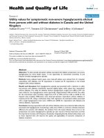
Anandamide inhibits VCAM-1 induced by TMEV in brain
endothelial cells by CB1 receptors
The endothelial blood brain barrier protects the CNS from
the changing environment in both physiologic and patho-
logic conditions. Previous work in our lab has demon-
strated that sVCAM-1 is constitutively expressed on b.
End5 cells and increased by TMEV infection [24]. We first
analyzed the effect of AEA on the production of VCAM-1
by TMEV-infected brain endothelial cells. Dose response
studies of AEA on sVCAM-1 production showed that
10 μM was the most effective dose to prevent the expres-
sion of VCAM-1 induced by TMEV at 20 hours postinfec-
tion (Figure 1A). AEA also inhibited VCAM-1 expression
in resting cells (data not shown). Down-regulation of
VCAM-1 induced by AEA (10 μM) was partially reversed
by the addition of the CB
1
receptor antagonist,
SR141716A (SR1) but not by the CB
2
receptor antagonist
AM630 (Figure 1B). The doses used for CB antagonists
were 1 μM on the basis of their capability for antagonizing
CB effects in our previous work. To examine if vanilloid
receptors expressed in brain endothelial cells [32] were
involved in AEA inhibition of VCAM-1 we pretreated the
cells with capsazepine (10 μM). As shown in Figure 1C,
the blockade of vanilloid receptors did not modify
the inhibitory effect of AEA on VCAM-1 expression. In
addition we explored the role of PPARg receptors as it has
been described to mediate some of the actions of AEA
[reviewed by [33]]. In our study the treatment with the
inhibitor of PPARg, GW9662 (at nanomolar and micro-
molar doses) did not prevent AEA-induced VCAM-1 inhi-
bition (Figure 1D). In conclusion, AEA-induced inhibition
of VCAM-1 in brain endothelial cells implies the activa-
tion of CB
1
receptors.
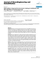
Anandamide limits leukocyte migration through a blood
brain barrier model by a mechanism involving CB1
receptors
VCAM-1 is critically involved in leukocyte transmigration
into the CNS. T herefore, our next step was to assess the
functional relevance of AEA-induced VCAM-1 inhibition
in leukocyte transmigration. First, we show ed that leuko-
cyte adhesion to TMEV-infected endothelium was signifi-
cantly increased (p < 0.01) in comparison to cell adhering
to resting cell monolayer. Importantly, the treatment with
AEA (10 μM), at the time of virus infection diminished
leukocyte adhesion (p < 0.01) (Figure 2A). In agreement
with the involvement of CB
1
receptors in AEA-induced
VCAM-1 inhibition, the pretreatment with the CB
1
recep-
torantagonist(SR1),butnotwiththeCB
2
antagonist
(AM630), reversed the inhibitory effect of AEA on leuko-
cyte adhesion (Figure 2A). Quantification is presented as a
ratio of number of leukocyte adhered to the endothelial
cell monolayer in each group normalized to control group
(Figure 2B).
Next, we analyzed whether the effect of AEA on the
adhesion of leukocytes interferes on leukocyte transmigra-
tion through the BBB model. As expected TMEV-infection
of brain endothelial cells increased the number of leuko-
cytes crossed the BBB model referred to control (Figure
2C). Accordingly to our results in the experiments of leu-
kocytes adhesion the treatment of endothelial cells with
AEA (10 μM) diminished leukocyte crossing by a mechan-
ismthatinvolvesCB
1
receptors. Figure 2D shows the
quantification data on the number of leukocytes that cross
the BBB model.
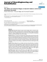
The increased anandamide tone inhibits VCAM-1
expression in Theiler’s virus-infected mice
On the basis of our in vitro results, we next analyze the
effect of the pharmacological modulation of the AEA
tone on VCAM-1 response against TMEV infectio n in
vivo, using wi ld type and CB
1
knockout mice (Cnr1
-/-
).
Accordingly to other studies [24], VCAM-1 expression
was not detected, by immunohistochemistry, in the
brains of sham animals in both type of mice, Cnr1
+/+
or
Cnr1
-/-
. The intracranial injection of TMEV induced the
expression of VCAM-1 in the ipsilateral cerebral cortex
surrounding blood vessels close to the site of injection
in both type of mice, Cnr1
+/+
as well as Cnr1
-/-
mice
Mestre et al . Journal of Neuroinflammation 2011, 8:102
/>Page 4 of 13
(Figure 3A). Corroborating our in vitro findi ngs, the
treatment with the inhibitor of AEA uptake UCM-707
induced a significant reduction of VCAM-1 expression
in TMEV-infected mice (Figure 3A). Although, UCM-
707 decreased VCAM-1 expression in Cnr1
+/+
and
Cnr1
-/-
mice, quantification analysis (Figure 3B) revealed
that the degree of VCAM-1 reduction in the ipsilateral
cerebral cortex of Cnr1
+/+
mice was significantly higher
than that observed in Cnr1
-/-
(p < 0.05). This observa-
tion suggests the participation of CB
1
receptors in the
effects of UCM-707 treatment. Additionally, when we
analysed the contralateral hemisphere (Figure 3C) we
found that only mice lacking CB
1
receptors showed
increased VCAM-1 expression in the vasculature in
response to TMEV that was significantly inhibited by
the treatmen t with UCM-707 as revealed the quantifica-
tion of staining intensity (Figure 3D).
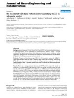
The increased anandamide tone limits microglial
activation in mice infected with Theiler’s virus
The intracranial injection of Theiler’s virus induced an
increase of microglia with reactive morphology in the
cerebral cortex at the level of infection (medium level)
but only in the ipsilateral infected hemisphere (Figure
4A). Interestingly, the microglial response was exacer-
bated in Cnr1
-/-
mice (Figure 4B), now extending from
prefrontal cortex (rostral level) to hippocampal level
(caudal level). When we analyzed the contralateral hemi-
sphere we found that microglial cells did not show reac-
tive morphology at the three brain levels examined in
3000
3500
s
ion
100
120
BA
**
#
** **
**
0
500
1000
1500
2000
2500
3000
% VCAM-1 expres
s
referred to TMEV
0
20
40
60
80
100
TMEV (2x10
5
pfu)
AEA (10
P
M)
+
-
+
+
+
+
+
+
+
+
##
++
##
++
##
##
10PM
##
100nM 500nM 1PM5PM
AEA
pg/ml
#
**
vehicle vehicle
100
120
p
ression
E
V
D
80
100
120
ression
E
V
C
AEA (10
P
M)
SR1 (1PM)
AM630 (1PM)
-
-
-
+
-
-
+
+
-
+
-
+
+
+
+
TMEV
(2x10
5
pfu)
AEA
GW9662
(100nM)
GW9662
(1PM)
vehiclevehicle
0
20
40
60
80
AEA
(10
M)
% VCAM-1 ex
p
referred to TM
E
##
##
##
0
20
40
60
80
vehicle CPZ
(10PM)
##
##
% VCAM-1 exp
referred to TM
E
AEA
vehicle
(10
P
M)
TMEV
(2x10
5
pfu)
TMEV
(
2x10
5
pfu
)
AEA
(10PM)
Figure 1 Ananda mide inhibits VCAM-1 production induced by TMEV through a mechanism that invol ves CB
1
receptor.(A)sVCAM-1
levels were measured by ELISA in supernatants of cell cultures 20 h after AEA treatment (100 nM, 500 nM, 1 μM, 5 μM, 10 μM). Confluent TMEV-
infected brain endothelial cell monolayers were pretreated for 1 hour before AEA treatment with (B) the cannabinoid receptor antagonist SR1 (1
μM) or AM630 (1 μM); (C) the vanilloid receptor antagonist capsazepine (10 μM); (D) the PPARg receptor antagonist GW9662 (100 nM and 1 μM).
Results show the means ± SEM from three independent experiments done in triplicate. (**p < 0,01 vs. vehicle; ##p < 0.01 vs. TMEV+vehicle; ++p
< 0.01 vs. TMEV+AEA, ANOVA followed by Tuckey’s test).
Mestre et al . Journal of Neuroinflammation 2011, 8:102
/>Page 5 of 13
Control TMEV TMEV+AEA
TMEV+AEA
+SR1
TMEV+AEA
+AM630
A
2
3
4
5
6
B **
**
##
Fold
+
0
1
SR1
1PM
AM630
1PM
AEA (10PM)
vehiclevehicle
TMEV (2x10
5
pfu)
vehicle
##
Control TMEV TMEV+AEA
TMEV+AEA
+SR1
TMEV+AEA
+AM630
C
+SR1
+AM630
40000
**
++
D
0
10000
20000
30000
leukocytes
SR1
AM630
vehicle
vehicle
vehicle
++
##
D
&
SR1
1PM
AM630
1PM
AEA (10PM)
vehicle
vehicle
TMEV
(
2x10
5
pfu
)
vehicle
Figure 2 AEA limits leukocyte adhesion to TMEV stimulated brain endothelial cells and leukocyte transmigration through in vitro BBB
by CB
1
involvement. Brain endothelial cell monolayer were stimulated with a combination of TMEV (2 × 10
5
pfu), AEA (10 μM), SR1 (1 μM) or
AM630 (1 μM) for 6 hours. After that, 2.5 × 10
5
leukocytes stained with AM-calcein (5 μM) were added to the endothelial culture for 20 hours.
(A) Representative immunofluorescence microphotographs of the leukocytes stained with AM-calcein adhered to the brain endothelial cell
monolayer in each case and phase contrast microphotographs of brain endothelial monolayer merged with immunofluorescence
microphotographs of AM-calcein stained leukocytes bring out with arrows. Scale bar 100 μm. (B) Quantification of leukocytes adhered to brain
endothelial monolayer in each case normalized to control group (n = 6). (**p < 0.01 vs. vehicle; ##p < 0.01 vs. TMEV; +p < 0.05 vs. TMEV+AEA,
ANOVA followed by Tuckey’s tests). (C) TMEV (2 × 10
5
pfu), plus AEA (10 μM), or plus SR1 (1 μM) or AM630 (1 μM) were added to the upper side
of the insert (endothelial culture) and IL1-b (10 ng/ml) was added to the bottom side (astrocyte culture) for 6 hours. 2.5 × 10
5
leukocytes were
added to the upper side of the insert for 20 hours and representative phase contrast microphotographs of leukocytes crossed to bottom side of
the insert were taken. (D) Quantification of leukocytes in the bottom side of the insert after 20 hours of experiment. (**p < 0.01 vs. vehicle; ##p
< 0.01 vs. TMEV+vehicle; ++p < 0.01 vs. TMEV+AEA; &p < 0.05 vs. TMEV+AEA+SR1, ANOVA followed by Tuckey’s test; n = 6).
Mestre et al . Journal of Neuroinflammation 2011, 8:102
/>Page 6 of 13
the wild type mice as well as in Cnr1
-/-
mice. Therefore,
in response to TMEV infection, activation of microglial
cells only occurred in the ipsilateral hemisphere. Quan-
tification analysis of percentage of area occupied by
microglia per field was summarized in Figure 4C.
The treatment with UCM-707 significantly (p < 0.01)
reduced the presence of microglia with reactive mor-
phology in Cnr1
+/+
mice (Figure 5B) at the medium
level close to the site of injection ( Figure 5A). Cerebral
cortex sections from Cnr1
-/-
mice showed a tendency
ipsilateral contralateral
AC
Cnr1
+/+
Cnr1
-/-
Sham
Cnr1
+/+
Cnr1
-/-
TMEV
+
vehicle
TMEV
+
40
50
60
+
UCM 707
40
50
60
**
##
s
taining
##
staining
BD
x
0
10
20
30
Cnr1
-/-
Cnr1
+/+
Sh
TMEV hi l
TMEV UCM 707
0
10
20
30
ND
Cnr1
+/+
Cnr1
-/-
++
&
Intensity of
s
##
&
ND
ND
Intensity of
Sh
am
TMEV
+ve
hi
c
l
e
TMEV
+
UCM 707
Figure 3 The treatment with UCM-707 inhibits VCAM-1 expression in TMEV-infected mice. Study with Cnr1
+/+
and Cnr1
-/-
mice. Both,
TMEV-infected and Sham mice were treated with UCM-707 (3 mg/kg) or the corresponding vehicle (n = 3 for each group) immediately after
virus infection for three consecutive days. Analysis were performed using representative microphotographs of coronal brain sections (30 μm) of
ipsilateral (A) or contralateral (C) brain tissue close to the virus side of injection, immunostained for VCAM-1. Arrows indicate VCAM-1
immunostaining. Scale bar is 50 μm. (B, D) Quantification of intensity of VCAM-1 staining as described in Material and methods in the ipsilateral
or contralateral hemispheres, respectively. ND, non detected; **p < 0.01 vs. Sham (Cnr1
+/+
); ##p < 0.01 vs. Sham (Cnr1
-/-
); ++p < 0.01 vs. TMEV
+vehicle (Cnr1
+/+
); &p < 0.05 vs. TMEV+vehicle (Cnr1
-/-
); Xp < 0.05 vs. TMEV+UCM-707 (Cnr1
+/+
).
Mestre et al . Journal of Neuroinflammation 2011, 8:102
/>Page 7 of 13
toward diminishing microglia reactivity but without
reaching statistical signif icance (p = 0,07; Figure 5C).
The analysis of the contralateral hemispheres didn’ t
reveal the presence of microglia with reactive morphol-
ogy (data not shown).
Discussion
The ECS has been suggested to contribute to the main-
tenance of homeostasis between the immune and the
nervous systems [34,35]. Besides, the pharmacological
activation of ECS is emerging as a potential therapeutic
strategy for neurodegenerative diseases including multi-
ple sclerosis [rev [10,36]]. Mechanisms underlying the
beneficial effects of CBs on MS are not fully clarified;
however, anti-inflammatory and/or neuroprotective
actions seem to be involved [37]. Leukocyte migration
into the CNS is widely recognized as a pivotal event in
the development of MS in which adhesion molecules
like VCAM-1 are critically involved and em erge as mar-
ker of endothelial activity [rev [2,7]].
The notion that rest riction of immune cells traffic into
the CNS by CBs could represent a novel mechanism to
suppress brain immune reactivity was first suggested by
two laboratories in both, TMEV-IDD and EAE models by
using the synthetic agonist WIN 55,212-2 [21,23]. In the
present study we show that the endocannabinoid, AEA
inhibits the expression of VCAM-1 in TMEV-infected
brain endothelial cells resulting in reduced leukocyte adhe-
sion and crossing through an in vitro model of BBB. In the
TMEV-IDD model, cumulative evidence suggests that
TMEV may enter the CNS by infection of cerebrovascular
endothelial cells. Thus, infection of endothelial cells might
represent one of the first events in the pathogenesis of
TMEV-induced demyelination. The persistence of TMEV
in cloned mouse cerebrovascular endothelial cells appears
to support this concept [38]. Pioneering studies on
TMEV-IDD showed that adhesion molecules play a criti-
cal role in leukocyte extravasation [39] pointing out the
interest of a reduction of VCAM-1 expression by AEA.
CB
1
and CB
2
rec eptors were expres sed in b.End5 as well
Cnr1
+
/
+
Cnr1
-
/
-
A
B
Cnr1
Cnr1
ipsilateral contralateral ipsilateral contralateral
A
B
TMEV
Cnr1
+/+
Cnr1
-/-
ipsilateral contralateral Ipsilateral contralateral
Rostrallevel 13,773± 0,592
++
12,013± 0,489 20,028± 1,257
##
16,956± 0,913
Mediallevel 25,277± 1,403** 13,223± 0,788 21,341± 0,958
##
17,151± 1,007
Caudal
level
16 621
±
1 465
++
15 909
±
2 327
19 657
±
1 120
#
16 320
±
0 796
C
Caudal
level
16
,
621
±
1
,
465
15
,
909
±
2
,
327
19
,
657
±
1
,
120
#
16
,
320
±
0
,
796
Figure 4 CB
1
deletion exacerbates microglial response against TMEV infection. Coronal brain sections (30 μm) were obtained from Cnr1
+/+
TMEV-infected mice (A) or Cnr1
-/-
TMEV-infected mice (B), stained for CD11b with Iba-1 antibody and counterstained with toluidine blue (n = 3
for each group). To perform the analysis of microglia phenotype morphology brain tissue was studied in both hemispheres and at rostral, medial
and caudal levels. (C) Quantification of percentage of area occupied by microglia per field is represented. Scale bar is 50 μm. **p < 0.01 vs.
contralateral (Cnr1
+/+
); #p < 0.05 vs. contralateral (Cnr1
-/-
); ##p < 0.01 vs. contralateral (Cnr1
-/-
); ++p < 0.01 vs. medial level (Cnr1
+/+
).
Mestre et al . Journal of Neuroinflammation 2011, 8:102
/>Page 8 of 13
Cnr1
+/+
Cnr1
-/-
A
B
Sham
TMEV+vehicle
TMEV+UCM 707
TMEV+UCM
707
25
30
**
##
d
by
d
C
&
0
5
10
15
20
Cnr1
+/+
Cnr1
/
++
% area occupie
d
microglia/fiel
d
&
Cnr1
+/+
Cnr1
-
/
-
Sham TMEV+vehicle TMEV+UCM 707
Figure 5 The treatment with UCM-707 decreases microglia reactivity in TME V-infected mice. Study with Cnr1
+/+
and Cnr1
-/-
mice. Both
TMEV-infected and Sham mice from both strains (Cnr1
+/+
and Cnr1
-/-
) were treated with UCM-707 (3 mg/kg) or the corresponding vehicle (n =
3 for each group) immediately after the virus infection for three consecutive days. (A) Coronal brain section level for the analysis of CD11b
+
expression. (B) Representative micrographs of ipsilateral cerebral cortex in sham, TMEV-infected plus vehicle or TMEV infected plus UCM-707 from
Cnr1
+/+
or Cnr1
-/-
mice. (C) Quantification of percentage of area occupied by microglia per field is represented. Scale bar is 50 μm. **p < 0.01 vs
Sham (Cnr1
+/+
); ++p < 0.01 vs. TMEV+vehicle (Cnr1
+/+
); ##p < 0.01 vs Sham (Cnr1
-/-
); &p = 0.07 vs. TMEV+vehicle (Cnr1
-/-
).
Mestre et al . Journal of Neuroinflammation 2011, 8:102
/>Page 9 of 13
as in primary cultures of murine brain endothelial cells
[40]. Most of the effects of CBs are mediated by their spe-
cific receptors CB
1
and CB
2
that are asymmetrically dis-
tributed in the BBB. CB
1
receptor is mainly located at the
luminal side while CB
2
receptors are on the abluminal side
of the endothelium [41,19]. In our study, VCAM-1 sup-
pression by AEA in brain endothelial cells was mainly
mediated by the activation of CB
1
receptors. Most impor-
tantly, AEA-induced inhibition of leukocyte adhesion and
crossing through the BBB also involved CB
1
receptors
accordingly to the specific distribution of this type of
receptors in the BBB. In agreement with our observations,
studies on HIV-1 Gp120-effects in brain microvascular
endothelial cells have shown that CB
1
based synthetic CBs
prevented monocyte transmigration across a human
model of BBB [42]. Although CB
1
,CB
2
[40] and TRPV1
[32] receptors are expressed in murine brain endothelial
cells, our results ruled out the involvement of CB
2
and
TRPV1 receptors in AEA-induced inhibition of VCAM-1.
Differential expression of CB
2
receptors and NAPE-PLD
(the major enzyme associated with synthesis of AEA) in
cerebral endothelium at different stages of MS has been
recently reported [43]. In the abov e study, increased CB
2
receptor staining was associated with BBB disruption in
active plaques from MS tissue samples, suggesting a role
for endothelial CB
2
in the protection and/or repair of BBB
injury. However, previous studies of MS brain t issue did
not find endothelial expression of CB
2
[44,45]. In TMEV-
infected brain endothelial cells the possibility that AEA
activates PPAR-g receptors [33] to suppress VCAM-1 can
be also discharged despite the fact that PPARs agonists
prevent the interaction of leukocytes with stimulated
endothelium [ 46].
The majority of studies on AEA actions in endothelial
cells have focused on its vasodilator and hypotensive
activity and there were discrepancies on the type of
receptor implicated, probably due to differences between
peripheral and brain endothelial cells [47]. Using mouse
cerebral endothelial cells and consistent with our results,
AEA-induced increased COX-2 expression involves the
activation of CB
1
receptors [48].
Although alterations in the ECS during the course of MS
have been suggested to represent a protective physiological
strategy [13,18,49,50] the role of endocannabinoids in MS
remains uncertain. While most of studies on ECS and MS
focused on established disease, understanding the role of
endocannabinoids during the induction phase would be an
important point as exacerbated leukocyte trafficking into
the CNS represents a key stage in the disease. Therefore,
here, we investigated the role of CB
1
receptors and the
effects of the inhibitor of AEA uptake, UCM-707, on
VCAM-1 expression in wild type and CB
1
knockout mice
(Cnr1
-/-
) during the early phases of TMEV-IDD. Intracra-
nial infection with TMEV induced the expression of
VCAM-1 in surrounding blood vessels close to the site of
injectioninCnr1
+/+
as well as in Cnr1
-/-
mice whereas
VCAM-1 was not detected in brains of sham animal in
both type of mice accordingly to other studies [4,24]. The
treatment with UCM-707 resulted in down-regulat ion of
VCAM-1 expression in both type of mice. However, the
degree of inhibition of VCAM-1 in the ipsilateral cerebral
cortex of Cnr1
+/+
mice was signifi cantly higher than that
observed in Cnr1
-/-
mice supporting the involvement of
CB
1
receptors and corroborating our in vitro results. In
addition, the analysis of the contralateral hemisphere
showed increased VCAM-1 expression only in the vascula-
ture of Cnr1
-/-
mice that was inhibited by UCM-707.
Thus, our in vivo data confirm the importance of CB1
receptors but, suggest that besides CB1 receptors, addi-
tional mechanisms are contributing to the effects of
UCM-707 on VCAM-1 inhibition. It is difficult to have
the overall picture of what is happening as consequence of
increasing AEA tone under the conditions of our s tudy
due to the multiple cellular targets for AEA actions on the
responses to TMEV infection. Nevertheless, we have
shown here that AEA by targeting brain endothelial cells
may interfere with leukocyte recruitment across the BBB
through the inhibition of VCAM-1.
As suggested in the cardiovascular endothelium [51,52]
in the brain endothelium AEA and other endocannabi-
noids like 2-AG, would be synthetized and released from a
nearby source such as astrocytes [53], microglia [54] and
even from the own endothelia l cells to regulate the
response of b rain endothelium to different stimuli as we
observed in the case of TMEV. The observation that
NAPE-PLD expression is elevated on blood vessels and in
reactive astrocytes distributed closely around them sug-
gests the synthesis of AEA by brain endothelium in MS
[43]. In other models of brain injury 2-AG has b een
shown to be released and to reduce BBB damage [14,55].
Additionally, endocannabinoids may control brain
innate immunity in MS by ac ting in different CNS cell
types such as astrocytes and microglial besides immune
cells [rev [56]]. Activating or inhibiting the innate immune
response influences the development of TMEV-IDD [57].
In this line, AEA enhances IL- 6 production in astro cytes
infected with TMEV by a CB
1
receptor-mediated pathway
[58] and in a more recent work AEA modulates TMEV-
induced IL-12, IL-23 and IL-10 in microglia by activating
CB
2
receptors [ 59].
An important finding of the present study is that the
lack of CB
1
receptor leads to an exacerbation of microglial
response to TMEV infection in the ipsilateral hemisphere.
Thus, microglial activation was observed from prefrontal
cortex to hippocampal levels instead of maintaining it
exclusively in the area close to the injection site. Currently,
we unknown the meaning of the extensive microglial acti-
vation in Cnr1
-/-
mice, but it is likely to be associated with
Mestre et al . Journal of Neuroinflammation 2011, 8:102
/>Page 10 of 13
the facilitation of spreading viral antigens as microglia/
macrophages are an important virus reservoir [60]. In line
withaprotectiveroleofCB
1
receptors previous studies
have reported that CB
1
-knockout mice develop more
severe CREAE [61,62]. Moreover, recent studies reveal
tha t rep eat polymorphism of the Cnr1 gene could repre-
sent a genetic risk factor for both the primary progressive
[63] and relapsing-remitting form of MS [64].
Conclusions
In summary, mechanisms underlying the decreased cel-
lular infiltration on the CNS by CBs in animal models
of MS are not yet clear but the present study showed
that anandamide was effective in reducing endothelial
VCAM-1 expression and BBB permeability via CB
1
receptors. More relevant is that the in hibitor of ananda-
mide uptake, UCM-707 reduced VCAM expression in
TMEV infected mice with the participation of CB
1
receptors. Available data from MS patients subjected to
success natalizumab therapy showed downregulation of
sVCAM-1 which is considered a good biomarker of
endothelial activity [7]. The inhibition of VCAM-1
expression in cerebral vasculature by anandamide pro-
vides a new mechanism that may explain the therapeutic
action of increased anandamide tone in neuroinfl amma-
tory diseases like MS.
Additional material
Additional file 1: Schematic drawing of the in vitro BBB model
performance and experimental design. Schematic drawing of the BBB
model, experimental design and confirmation of BBB characteristic by
Evan’s blue permeability assay and Zonula occludens 1
immunocytochemistry.
Additional file 2: Schematic drawing of the in vitro BBB model
performance and experimental design. Schematic drawing of the BBB
model, experimental design and confirmation of BBB characteristic by
Evan’s blue permeability assay and Zonula occludens 1
immunocytochemistry.
List of abbreviations
2-AG: 2-Arachidonoylglycerol; AEA: N-arachidonoylethanolamine or
anandamide; BBB: blood brain barrier; CBs: cannabinoids; CNS: central
nervous system; CREAE: chronic experimental autoimmune
encephalomyelitis; DAG lipase: Diacylglycerol lipase; EAE: experimental
autoimmune encephalomyelitis; ECS: endocannabinoid system; FAAH: Fatty
acid amide hydrolase; FBS: Fetal bovine serum; FCS: Fetal calf serum; MAGL:
monoacylglycerol lipase: MS: multiple sclerosis; NAPE-PLD: N-acyl
phosphatidylethanolamine phospholipase D; NGS: normal goat serum; PFU:
plaque forming units; PPARs: peroxisome proliferator-activating receptors; RT:
room temperature; SR1: SR141716A; TMEV: Theiler’s murine
encephalomyelitis virus; TMEV-IDD: TMEV-induced demyelinating disease;
TRPV1: transient receptor potential cation channel: subfamily V: member 1;
VCAM-1: vascular cell adhesion molecule-1; VLA-4: very late antigen-4.
Acknowledgements
The authors are grateful to Dr. Moses Rodriguez (Department of
Immunology and Neurology, Mayo Clinic/Foundation, Rochester, Minnesota,
USA) for gentile gift of Theiler’s virus DA strain. We gratefully appreciated Dr.
María L. de Ceballos (Neurodegeneration Group, Cajal Institute (CSIC),
Madrid, Spain) for their microscopy technical assistance. We are also grateful
to Dr. Mª Luz López Rodríguez (Organic chemistry Department, Chemistry
Faculty, UCM, Madrid, Spain), for the kind gift of UCM-707 and to Joaquín
Sancho, Elisa Baides and Ana J. Hernández for their excelent technical
assistance. This project was supported by grants from the Spanish Ministerio
de Ciencia, e Innovación (SAF 2007/60038 and SAF 2010/17501), from the
Comunidad Autónoma de Madrid (S-SAL/0261/2006) and by RETICS, Instituto
de Salud Carlos III (Red Española de Esclerosis Múltiple REEM; RD 07/0060/
0010).
Author details
1
Neuroimmunology Group, Functional and Systems Neurobiology
Department, Cajal Institute, CSIC, 28002 Madrid, Spain.
2
Department of
Medical Biochemistry and Cellular Biology, Institute for Biomedicine,
Sahlgrenska Academy, University of Gothenburg, Sweden.
3
INSERM, INSERM
U919 ‘Serine Proteases and Pathophysiology of the Neurovascular Unit’, GIP
Cyceron, Caen Cedex, France.
Authors’ contributions
LM performed the majority of all experiments, participated in the design of
the study, participated in the statistical analysis and drafted the manuscript.
PMI participated in the adhesion experiments and revising manuscript draft.
MM helped to immunohistochemistry studies and participated in the
interpretation of data and revising manuscript draft. FC, FL and FD
participated in the design of the study, interpretation of data and revision of
manuscript draft. MH helped to performed in vitro experiments. JB
participated in the design of the study, in the statistical analysis and revising
manuscript draft. CG conceived of the study, and participated in its design
and coordination and helped to draft the manuscript. All authors read and
approved the final manuscript.
Competing interests
The authors declare that they have no competing interests.
Received: 11 May 2011 Accepted: 18 August 2011
Published: 18 August 2011
References
1. Engelhardt B, Ransohoff RM: The ins and outs of T-lymphocyte trafficking
to the CNS: anatomical sites and molecular mechanisms. Trends Immunol
2005, 26:485-495.
2. Greenwood J, Heasman SJ, Alvarez JI, Prat A, Lyck R, Engelhardt B:
Leucocyte-endothelial cell crosstalk at the blood-brain barrier: A
prerequisite for successful immune cell entry to the brain. Neuropathol
Appl Neurobiol 2011, 37:24-39.
3. Ukkonen M, Wu K, Reipert B, Dastidar P, Elovaara I: Cell surface adhesion
molecules and cytokine profiles in primary progressive multiple
sclerosis. Mult Scler 2007, 13:701-707.
4. Brosnan CF, Cannella B, Battistini L, Raine CS: Cytokine localization in
multiple sclerosis lesions: correlation with adhesion molecule expression
and reactive nitrogen species. Neurology 1995, 45:S16-S21.
5. Yednock TA, Cannon C, Fritz LC, Sanchez-Madrid F, Steinman L, Karin N:
Prevention of experimental autoimmune encephalomyelitis by
antibodies against alpha 4 beta 1 integrin. Nature 1992, 356:63-66.
6. Polman CH, O’Connor PW, Havrdova E, Hutchinson M, Kappos L, Miller DH,
Phillips JT, Lublin FD, Giovannoni G, Wajgt A, Toal M, Lynn F, Panzara MA,
Sandrock AW, AFFIRM Investigators: A randomized, placebo-controlled
trial of natalizumab for relapsing multiple sclerosis. N Engl J Med 2006,
354:899-910.
7. Millonig A, Hegen H, Di Pauli F, Ehling R, Gneiss C, Hoelzl M, Künz B,
Lutterotti A, Rudzki D, Berger T, Reindl M, Deisenhammer : Natalizumab
treatment reduces endothelial activity in MS patients. J Neuroimmunol
2010, 227:190-194.
8. Lipton HL, Dal Canto MC: Chronic neurologic disease in Theiler’s virus
infection of SJL/J mice. J Neurol Sci 1976, 30:201-207.
9. Maccarrone M, Dainese E, Oddi S: Intracellular trafficking of anandamide:
new concepts for signaling. Trends Biochem Sci 2010, 35:601-608.
10. Scotter EL, Abood ME, Glass M: The endocannabinoid system as a target
for the treatment of neurodegenerative disease. Br J Pharmacol 2010,
160:480-498.
Mestre et al . Journal of Neuroinflammation 2011, 8:102
/>Page 11 of 13
11. Rossi S, Bernardi G, Centonze D: The endocannabinoid system in the
inflammatory and neurodegenerative processes of multiple sclerosis and
of amyotrophic lateral sclerosis. Exp Neurol 2010, 224:92-102.
12. Marsicano G, Goodenough S, Monory K, Hermann H, Eder M, Cannich A,
Azad SC, Cascio MG, Gutiérrez SO, van der Stelt M, López-Rodriguez ML,
Casanova E, Schütz G, Zieglgänsberger W, Di Marzo V, Behl C, Lutz B: CB1
cannabinoid receptors and on-demand defense against excitotoxicity.
Science 2003, 302:84-88.
13. Eljaschewitsch E, Witting A, Mawrin C, Lee T, Schmidt PM, Wolf S,
Hoertnagl H, Raine CS, Schneider-Stock R, Nitsch R, Ullrich O: The
endocannabinoid anandamide protects neurons during CNS
inflammation by induction of MKP-1 in microglial cells. Neuron 2006,
49:67-79.
14. Panikashvili D, Shein NA, Mechoulam R, Trembovler V, Kohen R,
Alexandrovich A, Shohami E: The endocannabinoid 2-AG protects the
blood-brain barrier after closed head injury and inhibits mRNA
expression of proinflammatory cytokines. Neurobiol Dis 2006, 22:257-264.
15. Cent onze D, Bari M, Rossi S, Prosperetti C, Furlan R, Fezza F, De Chiara V,
Battistini L, Bernardi G, Bernardini S, Martino G, Maccarrone M: The
endocannabinoid system is dysregulated in multiple sclerosis and in
experim ental autoimmune encephalomyelitis. Br ain 2007,
130:2543-2553.
16. Fowler CJ, Rojo ML, Rodriguez-Gaztelumendi A: Modulation of the
endocannabinoid system: neuroprotection or neurotoxicity? Exp Neurol
2010, 224:37-47.
17. Cabranes A, Venderova K, de Lago E, Fezza F, Sánchez A, Mestre L,
Valenti M, García-Merino A, Ramos JA, Di Marzo V, Fernández-Ruiz J:
Decreased endocannabinoid levels in the brain and beneficial effects of
agents activating cannabinoid and/or vanilloid receptors in a rat model
of multiple sclerosis. Neurobiol Dis 2005, 20:207-217.
18. Ortega-Gutiérrez S, Molina-Holgado E, Arévalo-Martín A, Correa F, Viso A,
López-Rodríguez ML, Di Marzo V, Guaza C: Activation of the
endocannabinoid system as therapeutic approach in a murine model of
multiple sclerosis. FASEB J 2005, 19:1338-1340.
19. Centonze D, Battistini L, Maccarrone M: The endocannabinoid system in
peripheral lymphocytes as a mirror of neuroinflammatory diseases. Curr
Pharm Des 2008, 14:2370-2342.
20. Baker D, Pryce G, Croxford JL, Brown P, Pertwee RG, Makriyannis A,
Khanolkar A, Layward L, Fezza F, Bisogno T, Di Marzo V: Endocannabinoids
control spasticity in a multiple sclerosis model. FASEB J 2001, 15:300-302.
21. Arévalo-Martín A, Vela JM, Molina-Holgado E, Borrell J, Guaza C:
Therapeutic action of cannabinoids in a murine model of multiple
sclerosis. J Neurosci 2003, 23:2511-2516.
22. Zajicek J, Fox P, Sanders H, Wright D, Vickery J, Nunn A, Thompson A, UK
MS Research Group: Cannabinoids for treatment of spasticity and other
symptoms related to multiple sclerosis (CAMS study): multicentre
randomised placebo-controlled trial. Lancet 2003, 362:1517-1526.
23. Ni X, Geller EB, Eppihimer MJ, Eisenstein TK, Adler MW, Tuma RF: Win
55212-2, a cannabinoid receptor agonist, attenuates leukocyte/
endothelial interactions in an experimental autoimmune
encephalomyelitis model. Mult Scler 2004, 10:158-164.
24. Mestre L, Docagne F, Correa F, Loría F, Hernangómez M, Borrell J, Guaza C:
A cannabinoid agonist interferes with the progression of a chronic
model of multiple sclerosis by downregulating adhesion molecules. Mol
Cell
Neurosci 2009, 40:258-266.
25. Lledó A, Borrell J, Guaza C: Dexamethasone regulation of interleukin-1-
receptors in the hippocampus of Theiler’s virus-infected mice: effects on
virus-mediated demyelination. Eur J Pharmacol 1999, 372:75-83.
26. Laschinger M, Engelhardt B: Interaction of alpha4-integrin with VCAM-1 is
involved in adhesion of encephalitogenic T cell blasts to brain
endothelium but not in their transendothelial migration in vitro. J
Neuroimmunol 2000, 102:32-43.
27. Omidi Y, Campbell L, Barar J, Connell D, Akhtar S, Gumbleton M: Evaluation
of the immortalised mouse brain capillary endothelial cell line, b.End3,
as an in vitro blood-brain barrier model for drug uptake and transport
studies. Brain Res 2003, 990:95-112.
28. Yang T, Roder KE, Abbruscato TJ: Evaluation of bEnd5 cell line as an in
vitro model for the blood-brain barrier under normal and hypoxic/
aglycemic conditions. J Pharm Sci 2007, 96:3196-3213.
29. Molina-Holgado F, Lledó A, Guaza C: Evidence for cyclooxygenase
activation by nitric oxide in astrocytes. Glia 1995, 15:167-172.
30. Gaillard PJ, Voorwinden LH, Nielsen JL, Ivanov A, Atsumi R, Engman H,
Ringbom C, de Boer AG, Breimer DD: Establishment and functional
characterization of an in vitro model of the blood-brain barrier,
comprising a co-culture of brain capillary endothelial cells and
astrocytes. Eur J Pharm Sci 2001, 12:215-222.
31. Eugenin EA, Berman JW: Chemokine-dependent mechanisms of
leukocyte trafficking across a model of the blood-brain barrier. Methods
2003, 29:351-361.
32. Golech SA, McCarron RM, Chen Y, Bembry J, Lenz F, Mechoulam R,
Shohami E, Spatz M: Human brain endothelium: coexpression and
function of vanilloid and endocannabinoid receptors. Brain Res Mol Brain
Res 2004, 132:87-92.
33. O’Sullivan SE: Cannabinoids go nuclear: evidence for activation of
peroxisome proliferator-activated receptors. Br J Pharmacol 2007,
152:576-582.
34. Wolf SA, Tauber S, Ullrich O: CNS immune surveillance and
neuroinflammation: endocannabinoids keep control. Curr Pharm Des
2008, 14:2266-2278.
35. Molina-Holgado E, Molina-Holgado F: Mending the broken brain:
neuroimmune interactions in neurogenesis. J Neurochem 2010,
114:1277-1290.
36. Pertwee RG: Emerging strategies for exploiting cannabinoid receptor
agonists as medicines. Br J Pharmacol 2009, 156:397-411.
37. Croxford JL, Pryce G, Jackson SJ, Ledent C, Giovannoni G, Pertwee RG,
Yamamura T, Baker D: Cannabinoid-mediated
neuroprotection, not
immunosuppression, may be more relevant to multiple sclerosis. J
Neuroimmunol 2008, 193:120-129.
38. Sapatino BV, Petrescu AD, Rosenbaum BA, Smith R, Piedrahita JA, Welsh CJ:
Characteristics of cloned cerebrovascular endothelial cells following
infection with Theiler’s virus. II. Persistent infection. J Neuroimmunol 1995,
62:127-135.
39. Inoue A, Koh CS, Yamazaki M, Ichikawa M, Isobe M, Ishihara Y, Yagita H,
Kim BS: Anti-adhesion molecule therapy in Theiler’s murine
encephalomyelitis virus-induced demyelinating disease. Int Immunol
1997, 9:1837-1847.
40. Mestre L, Correa F, Docagne F, Clemente D, Guaza C: The synthetic
cannabinoid WIN 55,212-2 increases COX-2 expression and PGE2 release
in murine brain-derived endothelial cells following Theiler’s virus
infection. Biochem Pharmacol 2006, 72:869-880.
41. Maccarrone M, Fiori A, Bari M, Granata F, Gasperi V, De Stefano ME, Finazzi-
Agrò A, Strom R: Regulation by cannabinoid receptors of anandamide
transport across the blood-brain barrier and through other endothelial
cells. Thromb Haemost 2006, 95:117-127.
42. Lu TS, Avraham HK, Seng S, Tachado SD, Koziel H, Makriyannis A,
Avraham S: Cannabinoids inhibit HIV-1 Gp120-mediated insults in brain
microvascular endothelial cells. J Immunol 2008, 181:6406-6416.
43. Zhang H, Hilton DA, Hanemann CO, Zajicek J: Cannabinoid Receptor and
N-acyl Phosphatidylethanolamine Phospholipase D-Evidence for Altered
Expression in Multiple Sclerosis. Brain Pathol 2011.
44. Benito C, Romero JP, Tolón RM, Clemente D, Docagne F, Hillard CJ,
Guaza C, Romero J: Cannabinoid CB1 and CB2 receptors and fatty acid
amide hydrolase are specific markers of plaque cell subtypes in human
multiple sclerosis. J Neurosci 2007, 27:2396-402.
45. Yiangou Y, Facer P, Durrenberger P, Chessell IP, Naylor A, Bountra C,
Banati RR, Anand P: COX-2, CB2 and P2X7-immunoreactivities are
increased in activated microglial cells/macrophages of multiple sclerosis
and amyotrophic lateral sclerosis spinal cord. BMC Neurol 2006, 6:12.
46. Ramirez SH, Heilman D, Morsey B, Potula R, Haorah J, Persidsky Y:
Activation of peroxisome proliferator-activated receptor gamma
(PPARgamma) suppresses Rho GTPases in human brain microvascular
endothelial cells and inhibits adhesion and transendothelial migration of
HIV-1 infected monocytes. J Immunol 2008, 180:1854-1865.
47. Craig LE, Spelman JP, Strandberg JD, Zink MC: Endothelial cells from
diverse tissues exhibit differences in growth and morphology. Microvasc
Res 1998, 55:65-76.
48. Chen P, Hu S, Yao J, Moore SA, Spector AA, Fang X: Induction of
cyclooxygenase-2 by anandamide in cerebral microvascular
endothelium. Microvasc Res 2005, 69:28-35.
49. Loría F, Petrosino S, Mestre L, Spagnolo A, Correa F, Hernangómez M,
Guaza C, Di Marzo V, Docagne F: Study of the regulation of the
endocannabinoid system in a virus model of multiple sclerosis reveals a
Mestre et al . Journal of Neuroinflammation 2011, 8:102
/>Page 12 of 13
therapeutic effect of palmitoylethanolamide. Eur J Neurosci 2008,
28:633-641.
50. Mestre L, Correa F, Arévalo-Martín A, Molina-Holgado E, Valenti M, Ortar G,
Di Marzo V, Guaza C: Pharmacological modulation of the
endocannabinoid system in a viral model of multiple sclerosis.
J Neurochem 2005, 92:1327-1339.
51. Wagner JA, Hu K, Karcher J, Bauersachs J, Schäfer A, Laser M, Han H, Ertl G:
CB(1) cannabinoid receptor antagonism promotes remodeling and
cannabinoid treatment prevents endothelial dysfunction and
hypotension in rats with myocardial infarction. Br J Pharmacol 2003,
138:1251-1258.
52. Battista N, Fezza F, Maccarrone M: Endocannabinoids and their
involvement in the neurovascular system. Curr Neurovasc Res 2004,
1:129-140.
53. Walter L, Stella N: Endothelin-1 increases 2-arachidonoyl glycerol (2-AG)
production in astrocytes. Glia 2003, 44:85-90.
54. Stella N: Endocannabinoid signaling in microglial cells.
Neuropharmacology 2009, 56:244-253.
55. Mechoulam R, Shohami E: Endocannabinoids and traumatic brain injury.
Mol Neurobiol 2007, 36:68-74.
56. Ullrich O, Merker K, Timm J, Tauber S: Immune control by
endocannabinoids - new mechanisms of neuroprotection? J
Neuroimmunol 2007, 184:127-135.
57. Olson JK, Miller SD: The innate immune response affects the
development of the autoimmune response in Theiler’s virus-induced
demyelinating disease. J Immunol 2009, 182:5712-5722.
58. Molina-Holgado F, Molina-Holgado E, Guaza C: The endogenous
cannabinoid anandamide potentiates interleukin-6 production by
astrocytes infected with Theiler’s murine encephalomyelitis virus by a
receptor-mediated pathway. FEBS Lett 1998, 433:139-142.
59. Correa F, Hernangómez-Herrero M, Mestre L, Loría F, Docagne F, Guaza C:
The endocannabinoid anandamide downregulates IL-23 and IL-12
subunits in a viral model of multiple sclerosis: Evidence for a cross-talk
between IL-12p70/IL-23 axis and IL-10 in microglial cells. Brain Behav
Immun 2011, 25:736-749.
60. Jin YH, Mohindru M, Kang MH, Fuller AC, Kang B, Gallo D, Kim BS:
Differential virus replication, cytokine production, and antigen-
presenting function by microglia from susceptible and resistant mice
infected with Theiler’s virus. J Virol 2007, 81:11690-11702.
61. Pryce G, Ahmed Z, Hankey DJ, Jackson SJ, Croxford JL, Pocock JM,
Ledent C, Petzold A, Thompson AJ, Giovannoni G, Cuzner ML, Baker D:
Cannabinoids inhibit neurodegeneration in models of multiple sclerosis.
Brain 2003, 126:2191-2202.
62. Jackson SJ, Pryce G, Diemel LT, Cuzner ML, Baker D: Cannabinoid-receptor
1 null mice are susceptible to neurofilament damage and caspase 3
activation. Neuroscience 2005, 134
:, 261-268.
63. Ramil E, Sánchez AJ, González-Pérez P, Rodríguez-Antigüedad A, Gómez-
Lozano N, Ortiz P, Arroyo R, De las Heras V, Vilches C, García-Merino A: The
cannabinoid receptor 1 gene (CNR1) and multiple sclerosis: an
association study in two case-control groups from Spain. Mult Scler 2010,
16:139-146.
64. Rossi S, Buttari F, Studer V, Motta C, Gravina P, Castelli M, Mantovani V, De
Chiara V, Musella A, Fiore S, Masini S, Bernardi G, Maccarrone M,
Bernardini S, Centonze D: The (AAT)n repeat of the cannabinoid CB1
receptor gene influences disease progression in relapsing multiple
sclerosis. Mult Scler 2011, 7:281-288.
doi:10.1186/1742-2094-8-102
Cite this article as: Mestre et al.: Anandamide inhibits Theiler’s virus
induced VCAM-1 in brain endothelial cells and reduces leukocyte
transmigration in a model of blood brain barrier by activation of CB
1
receptors. Journal of Neuroinflammation 2011 8:102.
Submit your next manuscript to BioMed Central
and take full advantage of:
• Convenient online submission
• Thorough peer review
• No space constraints or color figure charges
• Immediate publication on acceptance
• Inclusion in PubMed, CAS, Scopus and Google Scholar
• Research which is freely available for redistribution
Submit your manuscript at
www.biomedcentral.com/submit
Mestre et al . Journal of Neuroinflammation 2011, 8:102
/>Page 13 of 13