báo cáo hóa học: " Reduction of neutrophil activity decreases early microvascular injury after subarachnoid haemorrhage" potx
Bạn đang xem bản rút gọn của tài liệu. Xem và tải ngay bản đầy đủ của tài liệu tại đây (2.46 MB, 12 trang )
RESEARCH Open Access
Reduction of neutrophil activity decreases early
microvascular injury after subarachnoid
haemorrhage
Victor Friedrich
1
, Rowena Flores
2
, Artur Muller
2
, Weina Bi
2
, Ellinor IB Peerschke
3
and Fatima A Sehba
1,2*
Abstract
Background: Subarachnoid haemorrhage (SAH) elicits rapid pathological changes in the structure and function of
parenchymal vessels (≤ 100 μm). The role of neutrophils in these changes has not been determined. This study
investigates the role of neutrophils in early microvascular changes after SAH
Method: Rats were either untreated, treated with vinblastine or anti-polymorphonuclear (PMN) serum, which
depletes neutrophils, or treated with pyrrolidine dithiocarbamate (PDTC), which limits neutrophil activity. SAH was
induced by endovascular perforation. Neutrophil infiltration and the integrity of vascular endothelium and
basement membrane were assessed immunoh istochemically. Vascular collagenase activity was assessed by in situ
zymography.
Results: Vinblastine and anti-PMN serum reduced post-SAH accumulation of neutrophils in cerebral vessels and in
brain parenchyma. PDTC increased the neutrophil accumulation in cerebral vessels and decreased accumulation in
brain parenchyma. In addition, each of the three agents decreased vascular collagenase activity and post-SAH loss
of vascular endothelial and basement membrane immunostaining.
Conclusions: Our results implicate neutrophils in early microvascular injury after SAH and indicate that treatments
which reduce neutrophil activity can be beneficial in limiting microvascular injury and increasing survival after SAH.
Background
Subarachnoid haemorrhage (SAH) is followed by patho-
logical alterations in cerebral microvasculature (≤10 0
μm) [1-6]. These alterations develop rapidly (< 24
hours) and affect vascular structure and function. The
structural alterations include corrugation and in some
cases physical detachment of endothelium from the
basal lamina, loss of endothelial antigens, accumulation
of platelet aggregates in the vessel lumen, and degrada-
tion of collagen IV, the major protein of basal lamina
[4,5,7,8]. Functional changes closely follow the structural
alterations and include endothelial dysfunction, constric-
tion, perfusion deficits, and permeability increases [4-7].
Previous studies have implicated luminal platelets in
early microvas cular pathology after SAH [5,6]. The con-
tribution of platelets to microvascular injury may
represent an inflammatory response to the rupture of
the arterial wall, promoted by an initial reduction in cer-
ebral blood flow. Neutr ophils are another key compo-
nent of the inflammatory cascade, and have the ability
to generate pathologic changes in blood vessels. Overt
activation of neutrophils is implicated in vessel wall
pathology and in the progression of a variety of diseases
and disorders including cardiovascular diseases, haemo-
lyticuremicsyndromeandstroke[9-12].Markedneu-
trophil infiltration is also reported 3 days after SAH and
is associated with an increased risk of developing vasos-
pasm [13,14]. Recently, Provencio et al, [15,16] reported
that prior depletion of circulating myeloid cells amelio-
rates SAH-induced reduction in the calibre of middle
cerebral artery and, further, that neutrophils have accu-
mulated in parietal lobe parenchyma at one day post-
lesion. We have previously reported changes as early as
10 minutes post-haemorrhage in brain parenchymal
microvessels, including platelet accumulations, increased
microvascular collagenase activity, and destruction of
* Correspondence:
1
Department of Neuroscience Mount Sinai School of Medicine, New York,
NY 10029, USA
Full list of author information is available at the end of the article
Friedrich et al. Journal of Neuroinflammation 2011, 8:103
/>JOURNAL OF
NEUROINFLAMMATION
© 2011 Friedrich et al; licensee BioMed Central Ltd. This is an Open Ac cess article distributed under the terms of the Creativ e
Commons Attribution License ( which permits unrestricted use, distribution, and
reproduction in any medium, provided the origi nal work is properly cited.
microvascular basement membrane and blood-brain bar-
rier [3,7,8]. We here address the possible role of neutro-
phils in the very early development of these
microvascular pathologies. We report that pronounced
neutrophil accumulation is present in brain microvessels
and in brain parenchyma at 10 minutes post-haemor-
rhage. Furthermore inhibition of neutrophil-mediated
effects by two different pharmacological strategies par-
tially protected microvessels. These observations suggest
that neutrophils may play a pivotal role in microvascular
pathology following SAH and suggest neutrophils as
potential targets in SAH therapies.
Methods
All experimental procedures and protocols were
approved by the Institutional Animal Care and Use
Committee of the Mount Sinai Medical Center.
Induction of subarachnoid haemorrhage
Male Sprague-Dawley rats (325-350 g) underwent
experimental SAH using the endovascular suture model
developed in this laboratory [17,18]. Briefly, rats were
anesthetized with ketamine-xylazine (80 mg/kg+10 mg/
kg; i.p.), transorally intubated, ventilated, and maintained
on inspired isoflourane (1% to 2% in oxygen-supplemen-
ted room air). Rats were placed on a homeothermic
blanket Harvard Apparatus, MA, USA) attached to a
rectal temperature probe set to maintain body tempera-
ture at 37°C and positioned in a stereotactic frame. The
femoral artery was exposed and cannulated for blood
gas and blood pressure monitoring (ABL5, Radiometer
America Inc. Ohio, USA). For measurement of intracra-
nial pressure (ICP), the atlanto-occipital membrane was
exposed and cannulated, and the cannula was affixed
with methymethacrylate cement to a stainless steel
screw implanted in the occipital bone. Cerebral blood
flow (CBF) was measured by laser- Doppler flowmetry,
using a 0.8 mm diameter needle probe (Vasamedics,
Inc., St. Paul, MN, USA) placed over the skull away
from large pial vessels in the distribution of the middle
cerebral artery.
SAH was induced by advancing a suture retrogradely
through the ligated right external carotid artery (ECA),
and distally through the internal carotid artery (ICA)
until the suture perforated the intracranial bifurcation of
the ICA. This event was detected by a rapid rise in ICP
and fall in CBF. Physiological parameters (see below)
were recorded from 20 minutes prior to SAH to 10
minutes or 3 hours after SAH. As animals regained con-
sciousness and w ere able to breathe spontaneously they
were returned to their cages and sacrificed at 10 min-
utes, 1, 3, 6 hours, or 24 hours after SAH.
Sham-operated animals were used as controls in this
study. As described previously, sham s urgery included
all steps carried out in the surgery for SAH induction,
except for internal carotid artery perforation [6]. Sham
animals were matched in post-operati ve survival time to
the SAH animals.
SAH Physiological Parameters
Animals were assigned randomly to survival interval and
treatment groups (N = 7 for SAH and 5 for sham sur-
gery per time interval). ICP, CBF, and BP were recorded
in real time. The average ICP rise at SA H from baseline
was 5.4 ± 0.4 mmHg, with a peak of 60.0 ± 3.6 mmHg.
CBF fell to 12.9 ± 1.4% of baseline at SAH and recov-
ered to 47.7 ± 7.7% after 60 minutes. BP increased at
SAH and returned to the baseline within five minutes.
The ICP and CBF values indicated that rats experienced
moderate SAH (Figure 1) [19]. The mortality 24 hours
post SAH and sham surgeries in our laboratory on aver-
age are 29% and 10%, respectively.
Drug treatment
Three groups of animals were used. The first group was
treat ed with vinblastine to deplete neutrophils (see table
1). This method of neutrophil depletion has frequently
been used to study the role of neutrophil in cardiac,
lung, traumatic brain and stroke injuries [20-23]. T o
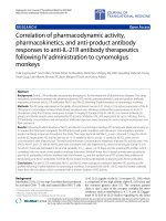
Figure 1 Neutrophil infiltration after SAH. A: Neut rophil staining
in representative brain sections. Note that a large number of
neutrophils are evident in the brain at 10 min and a smaller
number at 24 hours after SAH. B: Number of neutrophils per whole
coronal brain sections. Data are mean ± sem, N = 7 animals per
time point. *p < 0.05.
Friedrich et al. Journal of Neuroinflammation 2011, 8:103
/>Page 2 of 12
deplete neutrophils, animals received vinblastine sul-
phate (cat. No: V1377, Sigma Aldrich MO, USA), along
with Bicillin (cat. No: 3000979-A; King Pharmaceutical
Inc. Bristol, TN, USA) and gentamicin (cat. No: G1272,
Sigma Aldrich MO, USA) to prevent infection, 4 days
before surgery (adapted from [21,22]). For vinblastine
injection (N = 5 for SAH and 4 for Sham surgery) ani-
mals were anesthetized with ketamine-xylazine (80 mg/
kg + 10 mg/kg; i.p), femoral venous catheters were
inserted, and 0.5 mg/kg vinblastine sulphate, dissolved
in saline, was administered intravenously. Bicillin (100
000 U) and gentamicin (10 mg/kg) were a dministered
intramuscularly to prevent infection. Catheters were
removed, and as the rats recovered from anaesthesia
they were returned to their cages. All animals survived
for 1 hour after SAH.
The second group of animals was treated with rabbit
anti rat PMN serum to deplete neutrophils (see table 1).
Animals received daily intraperito neal injection of 1 mL
of saline-diluted (1:10) rabbit anti-rat PMN polyclonal
antibody (cat. No: AIA51140, Accurate Chemical and
Scientific NY, USA) for 3 days before SAH induction
[24]. Controls for this group received daily IP injection
of rabbit serum. The number of animals for anti PMN
treatment is 6 and 2 fo r rabbit serum treatment. One
anti PMN treated animal died within 1 hour after SAH.
The third group of animals was treated with pyrroli-
dine dithiocarbamate (PDTC) to reduce neutrophil
activity (cat. No: P 8765, Sigma Aldrich, MO, USA). The
dose and the route of administrati on used were adapted
from [25,26]. PDTC was dissolved in saline injected
twice, 100 mg/kg i.p. at 12 hours and 50 mg/kg, one
hour before surgery. The number of animals is 5 for
immunostaining, 5 for permeability studies; see below.
All animals survived for 1 hour after SAH.
Histology
Brain preparation
Rats were perfused transcardially with saline and brains
were rapidly removed, embedded in Tissue-Tek OCT
compound(Miles,Elkhart,IN),andfrozenin2-
methylbutane cooled in dry ice. 8 μm thick coronal
brain sections were cut on a cryostat and thaw-mounted
onto gelatin-coated slides. For neutrophil accumulation
analysis 12 sections each 1 mm apa rt, from bregma
+3.70 to - 8.7 mm [27 ] were use d. For immu nofluores-
cence, permeability, and zymography studies, sections
located at bregma +0.2 and - 3.6 mm [27] were used.
Measurement of subarachnoid blood volume
The volume of blood surrounding t he circle of Willis
was estimated as described previously [18] by measu ring
blood areas in the interhemispheric region and basal
subarachnoid space as seen in coronal brain sections
(IPLab v3.0, Signal Analytics).
Microvascular permeability: FITC-albumin Extravasation
Animals were either untreated or PDTC treated and
sacrificed 1 hour after SAH induction. Microvascular
permeability was studied as previously reported [6].
Briefly, rats were sedated and the femoral artery was
cannulated. FITC-albumin (Sigma, St. Louis, MO) was
injected 15 minutes befo re sacrifice (bolus injection; 0.5
ml of 20 mg/ml preparation, N = 3 for untreated SAH
control and 5 for PDTC treatment). Animals were killed
by transcardiac perfusion with chilled saline followed by
1% chilled formaldehyde prepared freshly from parafor-
maldehyde (PFA). The brains were isolated and fixed in
1% PFA followed by solutions that contained 10%, 20%
or 30% sucrose in 1% PFA. Fixation in each solution
was carried out overnight at 4°C. Finally, the brains
were embedded in Tissue-Tek OCT compound (Miles,
Elkhart, IN), and frozen in 2-methylbutane cooled with
dry ice and stored at -70°C until use.
Immunofluorescence and Zymography
Reagents
1. Primary antibodies: goat monoclonal anti-collagen IV
(Southern Biotechnology Associates Inc., Birmingham,
AL; cat. no. 1340-01), rabbit polyclonal anti-co llagen IV
(Abcam, Inc, Cambridge, MA; cat. no AB6586), mouse
monoclonal anti-rat endothelial cell antigen (RECA-1;
MCA970R; Serotec Inc., Raleigh, NC; cat. no.
MCA970R), mouse anti-neutrophil elastase (Senta Cruz
Biotech, Santa Cruz, CA; cat. no.sc-55549) and rabbit
polyclonal anti-neutrophil serum HB-199 (gift from Dr.
D. Anthony, Oxford UK[28]). 2. Secondary antibodies:
species-specific donkey anti-goat Alexa 350 (Invitrogen
Corp. Carlsbad, CA; cat. no. A-21081), donkey anti-
mouse Alexa 488 (Invitrogen Corp. cat. no. A-21202),
and donkey anti-rabbit Rhodamine Red X (Jackson
Immuno. Research; West Grove, PA; cat. no. 711-295-
152). 3. DQ-gelatin solution (EnzCheck collagenase kit,
Molecular Probes, Eugene, OR, USA; cat. no. E-12055).
Immunofluorescence
8 μm frozen brain sections were thawed and fixed for 15
minutes in 4% PFA. Sections were washed in
Table 1 Blood cell counts upon pharmacological
treatments
anti PMN anti PMN Vinblastine PDTC
Total WBC (10
3
/ul) 6.8 2 6 6
Neutrophils % 16 2 0.6 15
Platelets (10
3
/ul) 798 643 711 694
Animals were either untreated or were treated with vinblastine, anti PMN
serum, or PDTC (see methods). Blood (200 ul) was drawn before SAH
induction and analyzed for total white blood cells, neutrophil, and platelet
counts using an LH-755 automated analyzer (Beckman Coulter, Brea, CA; n = 2
per treatment group). Shown are counts from a single animal. Normal rat
white blood cell (WBC) counts are 6-18 10
3
/ul, neutrophils are 14-20%, and
platelet counts are 500-1000 10
3
/ul [54].
Friedrich et al. Journal of Neuroinflammation 2011, 8:103
/>Page 3 of 12
physiological salt solution (PBS), and blocked in a solu-
tion of 3% normal donkey serum in PBS (DB). The sec-
tions were then incubated overnight at 4°C in a
combination of anti-collagen IV, anti-RECA-1 and HB-
199 or in a combination of anti-collagen IV and anti-
neutrophil elastase (1:200 in DB) antibodies. Sections
were washed and then incubated overnight at 4°C with
species-specific secondary antibodies. Finally, sections
were washed with PBS and coverslipped. Neutrophil
elastase staining confirmed the specificity of HB-199 for
neutrophils.
In Situ zymography and Immunofluorescence combination
8 μm frozen brain sections from untreated, vinblastine
treated, anti PMN treated or PDTC treated animals
sacrificed 1 hour after surgery were used (N = 5 per
group). Unfixed brains were thawed and c oated with a
thin layer of FITC-labeled DQ-gelatin solution [3] con-
taining collagen IV antibodies. The coated sections were
incubated overnight at 37°C in a humid chamber, and
then incubated overnight at 4°C with species-specific
secondary antibodies. Finally, sections were fixed with
chilled 4% PFA and coverslipped.
Immunostaining of FITC-albumin injected brains
8 μm frozen brain sections from untreated or PDTC
treated animals sacrificed 1 hour after surgery were used
(N = 5 per group). Sections were thawed and fixed in
4% PFA for 15 minutes. Sections were washed in PBS,
and blocked in a solution of 5% normal donkey serum
in PBS. The sections were then incubated overnight at
with either rabbit anti-collagen IV, washed in PBS, incu-
bated overnight at 4°C with donkey anti-rabbit Rhoda-
mine Red-X, washed in PBS, and coverslipped with
Vectashield (Vector labs, Burlingame, CA, USA).
Data Acquisition
Physiology
CBF, ICP, and mean arterial blood pressure (MAP) were
continuously recorded starting 20 minutes before SAH
and ending 10 minutes, 1 hour, or 3 hour af ter SAH
(PolyView software; Grass Instruments; MS, USA). CBF
data were normalized to the b aseline value averaged
over 20 minutes prior to SAH, and subsequent values
were expressed as a percentage of baseline [29].
Morphometry
All evaluations wer e performed by an observer blinded
to specimen identity. Vessels studied were 100 μmor
less in diameter and included pre- and post capillary
arteries and venules. No distinction between capillaries
and venules was made. Quantitative analysis was per-
formed by manual counting or with IPLab (IPLab soft-
ware v 3.63; Scanalytic Inc.; USA).
Neutrophil count
Composite montage images of whole coronal brain sec-
tions were acquired with a Leica DM-600 microscope (5
× objective, NA: 0.15) equipped with automated stage
and montage acquisition software and assembled using
MetaMorph (Molecular Devices, CA, USA). The number
of neutrophils per section (both hemispheres, all brain
regions) was manually counted in the whole section
images.
Collagen IV and RECA-1 positive profile area fraction
10-12 fields per brain section we re selected at random
and analyzed f or the number and area fraction of col-
lagen IV and RECA-1 positive profiles and their coloca-
lization. Stained profiles were isolated by intensity
threshold segmentation with particle size gati ng. The IP
lab was used to compute the area fraction as the
summed area of segmented profiles in a field divided by
the total area of the field.
Neutrophil-collagen IV or RECA-1 colocalization and
parenchymal extravasation
HB-199 positive neutrophils were selected via threshold
segmentation and gating. Collagen IV and RECA-1 posi-
tive profiles were selected as above. The total number of
each labelled profile and the nu mber of co llagen IV and
RECA-1 profiles that colocalized with neutrophil was
determined using IP lab. Parenchymal extravasation of
neutroph ils was calculated by subtracting the number of
collagen IV and HB-199 colocalized profiles from the
total HB-199 image count.
In situ zymography-immunofluorescence combination
Four brain regions (basal, frontal and convexity cerebral
cortex as well as caudoputamen), separated into right
and left hemispheres, were analyzed by fluorescence
microscopy (Axiophot; Carl Zeiss, USA). For quantitative
analysis fluorescence images (2-3 fields per region and
hemisphere) were recorded under constant illumination
and exposure settings using a 20× objective (field area =
8×10
4
μm
2
), and were then studied for the number of
collagen IV profiles positive for collagenase activity.
FITC-albumin extravasation
Collagen IV immunosta ining was used to differentiate
between vascular and parenchymal FITC-albumin
deposits. Confocal images Z stacks were generated (see
above). The number and area fraction of vascular and
parenchymal FITC-albumin deposits in micrographs
from basal, frontal and convexity cortex as well as in
caudoputamen was determined using IP lab.
Statistical analysis
All data points are presented as average ± standard
error of mean (SEM). Each parameter (ICP, CBF, num-
ber and area fraction of collagen IV, RECA 1 or ne utro-
phil immunostaining, zymograph y, and permeability
data) was analyzed by two-way ANOVA (StatView v.
5.0.1, SAS Institute Inc. USA) with time and treatment
query (control, SAH). Pairwise comparison used F isher’s
PLSD post-hoc tests.
Friedrich et al. Journal of Neuroinflammation 2011, 8:103
/>Page 4 of 12
Results
Histology
Neutrophil infiltration
A large number of HB-199 stained neutrophils accumu-
lated in brain as early as 10 min after SAH (Figure 1).
Many of these neutrophils adhered to the endothelium
of parenchymal vessels while others had entered the
brain parenchyma. In addition, a small number of neu-
trophils were scattered within the b lood which had
accumulated in the subarachnoid space at the base of
brain. The neutrophil count remained elevated at 1 hour
and thereafter decreased with time (Figure 1B). In com-
parison to SAH animals, neut rophil numbers remained
low in sham operated cohorts throughout the interval
studied (P < 0.05).
Rostro-caudal differences in neutrophil invasion were
assessed by counting HB-199 positive neutrophils in 12
brain sections each located 1 m m apart, using animals
sacrificed 10 minutes after SAH. The results showed
no significant rostro-caudal gradient in neutrophil
numbers, confirming the global nature of ischemic
injury after SAH (Table 2). Similarly, neutrophil num-
ber in different brain regions (basal, frontal and con-
vexity cortex and c audoputamen) and between the two
hemispheres was compared. The only significant regio-
nal difference was a decreased number of infiltrating
neutrophils in the basal cortex. There were also signifi-
cant interhemispheric differences in neutrophil count,
with a larger count in the ipsilateral hemisphere (Table
3). This difference was present at 10 min, 3 hour, and
24 hours, while a trend towards significance was found
at 1 hour (p = 0.07) and no significant difference was
found at 6 hours (p = 0.31) after SAH. Interhemi-
spheric difference in neutrophil count was also present
in sham operated animals sacrificed at 10 minutes
after the surgery but not thereafter. Neutrophils were
not confined to vessels and in many cases had entered
into the brain parenchyma near collagen IV stained
vessels (see below). T his brain parenchyma neutrophil
infiltration was present at all examined time intervals
after SAH. The number of parenchymal neutrophils
after SAH was constant at approximately 40% of total
neutrophils at all times (data not shown).
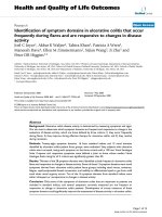
Colocalization of neutrophil, collagen IV and RECA-1
immunostaining
Animals were sacrificed at 10 minutes, 1 hour, 3 hour, or
24 hours after SAH. RECA-1 stained the endothelium and
collagen IV stained the basal lam ina of parenchymal ves-
sels. Both vascular stains were reduced after SAH. RECA-
1 staining was absent from most vascular sites that con-
tained neutrophil (HB-199) staining (Figure 2A). Collagen
IV staining was present in many but not all neutrophil
positive vascular sites. This trend was observed at all time
intervals in SAH animals but not in sham cohorts. Quanti-
tative analysis showed that the area fractions of RECA-1
and collagen IV immunostaining were decreased at 10
minu tes aft er SAH and remained decreased for 24 hours
(Figure 2B). Qualitative examination of specimens revealed
that, at any given time, more neutrophils colocalized with
collagen IV than with RECA-1 (Figure 2C).
Drug treatment The above studies find that a substan-
tial rise in vascular and parenchymal neutrophils, as well
as loss of RECA-1 and collagen IV immunostaining are
present at 1 hour after SAH. Hence, in the drug study,
the effect of reduction of neutrophil activity on micro-
vascular injury was evaluated at 1 hour after SAH.
Physiological Parameters
ICP peak following hemorrhage was higher in anti PMN
treated anima ls (77 ± 10 mmHg) than the rest of the
treated or untreated animals (65.5 ± 5.2 mmHg) but did
not reach significance (F = 0.9, p = 0.4; Figure 3). The
decline and 60 minute plateau of ICP, however, was sig-
nificantly higher in anti PMN treated animals as com-
pared to untreated and vinblastine or PDTC treated
animals(Controls:13±1,PDTC:25±8mmHg;P=
0.05). This data suggests that although initial bleed at
artery ru pture was similar a cross treatment gr oups,
bleeding continued for a longer duration in anti PMN
animals (see blood quantitation). CBF fall at SAH (13.4
± 1.1%) and 60 minute recovery (46 ± 6% of baseline)
was similar in all animals groups (F = 1.4, p = 0.2).
Subarachnoid blood volume
The volume of extravasated subarachnoid blood is
another indicator of SAH intensity. We measured the
Table 2 Rostro-caudal differences in neutrophil
infiltration 10 min after SAH
Effect Degrees of Freedom F p
Section 11 1.295 0.240
Brain region 1 12.02 0.0007
Section × Brain region 11 0.509 0.892
12 brain sections each located 1 mm apart, from bregma +3.70 to - 8.7 mm
[27] were used. No significant difference in the neutrophil numbers among
these brain sections was found. Data are mean ± sem, N = 5 animals
Table 3 Hemispheric and regional differences in
neutrophil infiltration 10 min after SAH
Effect Degrees of Freedom F p
Hemisphere 1 7.868 0.0054
Brain area 3 6.406 0.0003
Hemisphere × Brain area 3 0.264 0.8512
Four brain regions (basal, frontal and convexity cortex and caudoputamen)
were examined. A significant hemispheric and regional difference in
neutrophil infiltration was found (ANOVA). Moreover no interaction between
the hemispheres and brain regions was present, indicating global nature of
ischemic brain injury after SAH. Similar hemispheric and regional differences
in neutrophil count were present at 3, 6 and 24 hours after SAH (data not
shown; see text for explanation). Data are mean ± sem, N = 5 animals.
Friedrich et al. Journal of Neuroinflammation 2011, 8:103
/>Page 5 of 12
volume of blood after SAH to determine if anti PMN
treatment created a greater bleed. Quantitative analysis
showed 2.5 times more subarachnoid blood in anti
PMN treated animals as compared to untreated controls
(P = 0.05, Figure 4). No difference in the subarachnoid
blood volume among untreated and vinblastine or
PDTC treated animals was found (P > 0.05; Figure 4).
Neutrophil immunostaining
Animals were sacrificed 1 hour after SAH and brain sec-
tions were studied for neutrophil numbers. Neutrophil
(HB-199) immunostaining revealed only a few neutro-
phils in the vinblastine treated specimens and a large
Figure 2 Neutrophils in microvascular injury after SAH.A:
Representative image showing neutrophils in a brain section from
an animal sacrificed 10 min after SAH. Note that some neutrophils
(red) are within the collagen IV stained vessels (green; arrow heads)
and others are present in the brain parenchyma (arrows). B: Area
fractions of collagen IV and RECA-1 immunostaining in SAH and
sham animals. Note that the area fraction of collagen IV and RECA-1
staining in SAH animals remained lower than the sham operated
animals at all times examined. C: Numbers of neutrophils which are
colocalized with collagen IV and RECA-1 after SAH. Filled circles: all
neutrophils; filled squares: neutrophils that colocalized with collagen
IV only; filled triangles: neutrophils that colocalized RECA-1 only.
Open circles show all neutrophils in sham operated animals. Note
that a greater number of neutrophils colocalized with collagen IV
than with RECA-1 during the first 3 hours after SAH. Data are mean
± sem, N = 5 animals per time point and represent totals per whole
coronal brain section * significantly different from sham operated
animals (B) or from RECA-1 (C) at p < 0.05.
Figure 3 Early physiological changes after SAH: Animals were
either untreated or were treated with vinblastine, anti PMN
serum, or PDTC. ICP, CBF and BP were measured in real time from
20 minutes prior and 60 minutes post SAH (see methods). In A:
note that ICP peak is similar in all groups but ICP decline in anti
PMN group is significantly higher (25 ± 8 mmHg) than the
untreated SAH animals (13 ± 1 mmHg). In B note that CAF fall and
60 minutes recovery is similar among animal groups. In C note that
baseline BF and the transient increase in BP at SAH was similar
among groups. There after BP decreased to lower levels in
vinblastine and PDTC treated but not in anti PMN treated animals.
Data are mean ± sem, N is 5-7 animals per treatment group. *
significantly different at p < 0.05 from time matched untreated SAH
animals.
Friedrich et al. Journal of Neuroinflammation 2011, 8:103
/>Page 6 of 12
number in the PDTC treated brains (Figure 5A). Quan-
titative analysis showed that vinblastine treatment
reduced neutrophil count to less than 6%, and anti
PMN treatment to approximately 60% of the untreated
SAH animals (Figure 5B). In contrast, PDTC treatment
increased neutrophil count by 14% compared to the
untreated SAH animals (Figure 5B).
Neutrophil, collagen IV and RECA-1immunostaining
Animals were sacrificed 1 hour after SAH or sham
surgery and the area fractions of collagen IV and
RECA-1 positive profiles of treated animals was c om-
pared to untreated SAH and sham operated controls.
Since vinblastine treatment itself reduces collagen IV
immunostaining (data not shown), vinblastine treated
shams were used as controls for that group. After
SAH, significant reductions in the area fraction of col-
lagen IV and RECA-1 positive profiles occurred in
vinblastine-treated SAH animals as compared to vin-
blastine-treated shams (Figure 5C, p < 0.05). The
SAH-induced reduction in collagen IV area fraction i s
significantly less in vinblastine treated SAH animals
than in untreated SAH animals (untreated: 25% reduc-
tion, treated: 18% reduction; p = 0.02). A similar ame-
lioration in RECA-1 loss after SAH was also observed,
with marginal significance (untreated SAH 33% reduc-
tion, treated SAH 24% reduction; p = 0.09) (Figure
5C).
Anti PMN treatment Rabbit serum treated animals,
used as controls had similar reductions in RECA-1 and
collagen IV staining as untreated SAH animals (P >
0.05). Consequently, untreated animals were used to
compare the effect of anti PMN on RECA-1 and col-
lagen IV staining. As in untreated and vinblastine
treated animals, RECA-1 and collagen IV staining
decreased following SAH in animals treated with the
anti PMN serum. The extent of the reductions in
RECA-1 and collagen IV staining, however, was signifi-
cantly less in anti PMN compared to untreated animals
(Figure 5C. RECA-1: 12% reduction [treated] vs 25%
[untreated]; collagen IV: 18% reduction [treated] vs 33%
[untreated]; P = 0.001, P = 0.003 respectively).
PDTC treatment In contrast to vinblastine, anti PMN
and untreated SAH animals, RECA-1 staining was pre-
served in PDTC treated animals: the majority of vascular
profiles that were positive for neutrophils had retained
endothelium staining. Quantitative analysis showed a
significantly greater area fraction of RECA-1 positive
profiles as compared to untreated shams and untreated
SAH animals (108%) and a small but significant decrease
(17%) in the area fraction of colla gen IV positive vascu-
lar profiles in PDTC treated animals (Figure 5C, p <
0.05). Moreover, whereas in untreated animals over 35%
of overall brain neutrophils had entered the parenchyma
1 hour after SAH, in PDTC treated animals this number
was reduced to 20% (Figure 5D).
In situ zymography and collagen IV immunofluorescence
Untreated animals, as well as animals treated with vin-
blastine, anti PMN, or P DTC were sacrificed 1 hour after
SAH. A large number of collagen IV immunostained vas-
cular profiles that were positive for active collagenase
were observed in zymograms of untreated animals. In
comparison, fewer collagenase containing collagen IV
profiles could be seen in treated animals (Figure 5G).
The number of collagen IV immunos tained profiles that
were positive for collagenase activity was determined
(Figure 5E). In untreated animals, 48% of collagen IV
positive vessels had collagenase activity. Vinblastine, anti
PMN and PDTC treatments reduced this number to 15 %
(75% reduction), to 72% (28% reduction) and 23% (68%
reduction), respectively (Figure 5E, p = 0.0001).
Microvascular Permeability was assessed using intra-
vascular albumin-FITC. This study was performed in
PDTC pretreated animals, which showed the largest
sparing of RECA-1 immunostaining following SAH.
FITC-albumin deposits were numerous in brains of ani -
mals sacrificed 1 h after SAH. These deposits were scat-
tered in b oth hemispheres and all brain regions (frontal,
basal and convexity cortex as well as caudoputamen).
Collagen IV staining distinguished between vascular
(may indicate albumin incorporation in the growing pla-
telet clot) and parenchymal (indicate extra vasation)
FITC-albumin deposits. In untreated animals, signifi-
cantly more (p = 0.03) FITC-albumin deposits were pre-
sent in th e vessels (69% of total deposits) as compared
to brain parenchyma (31% of total deposits). PDTC
treatment did not affect the amount or distribution of
FITC-albumin deposits (Figure 5F).
Figure 4 Subarachnoid blood volume. Animals were either
untreated or were treated with vinblastine, anti PMN serum, or
PDTC and sacrificed one hour after SAH induction. The volume of
blood surrounding circle of Willis was measured (see methods).
Subarachnoid hemorrhage blood volume in anti PMN but not
vinblastine and PDTC treated animals was significantly greater than
the untreated SAH animals. Data are mean ± sem, N is 5 animals
per treatment group. * significantly different at p < 0.05 from time
matched untreated SAH animals.
Friedrich et al. Journal of Neuroinflammation 2011, 8:103
/>Page 7 of 12
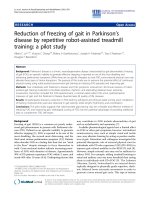
Figure 5 Pharmacological reduction of Neutrophils and their activity. A: Neutrophil staining in representative brain sections from untreated
or vinblastine, anti PMN or PDTC treated animals sacrificed 1 hour after SAH. Note that fewer neutrophils are present in vinblastine and anti
PMN treated animals and a large number are present in PDTC treated brains. B: Number of neutrophils in whole coronal brain sections. Values
are % of untreated SAH animals. Neutrophils are decreased by vinblastin and anti-PMN treated and increased by PDTC treatment. C: RECA-1
(filled bars) and collagen IV (open bars) immunostaining following SAH. Values are area fractions in SAH animals as % of area fractions in sham-
operated animals; both paramaters show trend or significant improvements in treated animals. D: Effect of PDTC treatment on the number of
extravasated (parenchymal) neutrophils in SAH animals. Neutrophil extravasation is reduced by PDTC. E: Number of collagenase-positive profiles
in treated SAH animals, given as % of values in untreated SAH animals. All three treatments reduce the extent of vascular collagenase activity. F:
Effect of PDTC treatment on post-SAH intravascular tracer leakage. Values are area fractions of intravascular (closed bars) and extravascular (open
bars) FITC-albumin deposits. G: Representative images of striatum showing vascular collagenase activity in untreated and anti PMN treated
animals sacrificed at 1 hour after SAH. Arrows: collagen IV stained vessels (red) positive for collagenase activity (green). Data are mean ± sem. N
= 5 animals per treatment group. * Significantly different at p < 0.05 from untreated SAH animals.
Friedrich et al. Journal of Neuroinflammation 2011, 8:103
/>Page 8 of 12
Discussion
The present study investigated if pharmacological reduc-
tion of neutrophil activity reduc es microvascular in jury
after SAH. The results demonstrate that depleting neu-
trophils or decreasing their activity prevents the loss of
endothelium and collagen IV, and decreas es collagenase
activity after SAH.
Neutrophil infiltration after SAH
Although animal and clinical studies indicate that a
marked infiltration of neutrophil occurs 1-3 days after
SAH [13-15], it has been unclear how soon after the
initial bleed this process begins. Furthermor e, most stu-
dies examined neutrophil accumulation in the subarach-
noid space (animal studies) or in CSF (human studies)
and did not provide information on neutrophils in brain
microvasculature or parenchyma. Hence, we began this
study by establishing the temporal profile of neutrophil
accumulation in cerebral microvessels and in the brain
parenchyma during the first 24 hours after SAH. Triple
immunostaining for collagen IV, endothelium (RECA-1),
and neutrophils (HB-199) allowed differentiation
between vascular and parenchymal neutrophils. More-
over, saline perfusion at the time of animal sacrifice
ensured that neutrophils floating in blood were removed
and only those adhering to the vessel wall were counted
as vascular neutrophils. This strategy revealed a massive
time dependent accumulation of neutrophils in cerebral
vessels and in brain parenchyma after SAH. As early as
10 minutes after SAH, a large number of neutrophils
adhered to the vascular endothelium and had begun to
infiltrate the brain parenchyma. The specific stimulus
leading to neutrophil activation after SAH is still to be
determined, but it is likely that platelet-derived cyto-
kines play a role. A growing body of evidence establishes
interplay between platelets and neutrophils in which
activation of one promotes activation of the other
[30-32]. Incidentally, it is impo rtant to note that plate-
lets are activated within 10 minutes after SAH [8], and
their interaction with vascular leukocytes is observed 2
hours later [33].
The increase in neutrophils 3 days after SAH, as
observed in previous studies, may indicate that the neu-
trophil infiltration observed in this study persists for an
extended period of time. This later phase of neutrophil
infiltration is implicated in the development of delayed
vasospasm [15,34]. The present study finds that the
early phase of neutrophil infiltration is associated with
early microvascular injury after SAH.
Neutrophils and microvascular injury after SAH
If, when, and to what extent neutrophils contribute to
early m icrovascular injury after SAH is not determined.
An interactio n of neutrophils with the vascular
endothelium is essential in their recruitment to the
injured area. The vascular consequences of this interac-
tion include opening of interendothelial cell junctions
and increased permeability, which facilitates neutrophil
migration to the point of injury. Under pathological
conditions, uncontrolled adhesion of neutrophils to the
vascular endothelium occurs and results in acute
endothelial injury [11,12]. In addition, vascular neutro-
phils plug and obstruct the vessel lumen to limit flow,
thus exacerbating brain injury and creating lo cal ische-
mia [35].
In the present study immunostaining of RECA-1
decreased after SAH, indicating d amage to the vascular
endothelium. Of note, RECA-1 was often missing from
vascular sites that contained neutrophils. Further more,
with time collagen IV also disappeared from most
RECA-1 deficient sites. This finding implies a contribu-
tion of neutrophils in endothelial and co llagen IV loss
after SAH. A similar combination of vascular neutrophil
accumulation, blood-brain barri er destruction, and col-
lagen IV degradation is observed upon hemorrhagic
transformation in humans and animals receiving tissue
plasminogen activator following occlusive ischemic
stroke, but this result develops over 24 ho urs [36,37].
The presence of these phenomena at 10 minutes in our
studies indicates that the nature of vascular injury after
SAH and ischemic stroke may be similar, but that injury
develops at a much faster pace after SAH as compared
to occlusive ischemic stroke.
Early alteration in the structure and function of cere-
bral vasculature is documented after SAH. This includes
loss of endothelial antigens, detachment of endothelium
from the basal lamina, degradation of collagen IV,
increase in permeability and decr ease in perfusion [1-6].
It is interesting to note that all of these events have a
similar temporal profile as the appearance of vascular
and parenchymal neutrophils; all are present at 10 min-
utes and persist for at least24hoursafterSAH.This
implies a role for neutrophil s in early vascular injury
after SAH.
Neutrophils can cause and promote vascular injury by
a number of mechanisms: (1) they can injure endothe-
lium by reactive oxidant species (such as hydrogen per-
oxide and superoxide) released during respiratory burst,
and by elastases and protease s released during degranu-
lation [12,38,39]. (2) They can degrade basal lamina by
releasing proteolytic enzymes, including collagenase and
MMP-9, which are known to digest collagen IV [36,37].
(3) The neutrophil-derived enzyme myeloperoxidase can
catalytically consume nitric oxide (NO) as a substrate,
which promotes endothelial dysfunction and constric-
tion [40,41]. An early decrease in cerebral NO, endothe-
lial dysfunction, and constri ction is established after
SAH [4,42,43]. Incidentally, decrease in cerebral NO
Friedrich et al. Journal of Neuroinflammation 2011, 8:103
/>Page 9 of 12
occurs around 10 min after the initial bleed, just as the
number of vascular neutrophils reaches its peak. A role
of myeloperoxidase in NO depletion remains to be
evaluated.
The effect of depleting or limiting neutrophil activity on
early microvascular injury after SAH
Most strategies for decreasing the activity of neutrophils
are aimed towards reducing their vascular accumulation
or activation [25,44,45]. We examined if neutrophil
depletion by vinblastine and anti PMN serum, or redu-
cing neutrophil induced oxidative stress by PDTC, could
prevent vascular injury after SAH. We found that neu-
trophil depletion reduces vascular collagenase activation
and protects against loss of collagen IV and endothe-
lium after SAH. However, side effects associated with
vinblastine and anti PMN treatmen ts make them unsui-
table as therapies. Anti PMN creates long lasting bleeds
from the ruptured artery, generating a larger hemor-
rhage. As the platelet count remains unchanged by anti
PMN, the long lasting bleeds may indicate a disturbance
in the coagulation pathway, delaying clot formation at
the site of arterial rupture. Vinblastine, on the other
hand significantly weakens the vascular cytoskeleton.
This side effect, resulting from disruption of microtu-
bules and inhibition of collagen synthesis and secretion,
is well documented [46].
PDTC, the third pharmacological agent examined in
this study, is an antioxidant and an inhibitor of tran-
scription factor nuclear factor kappa B (NF-B). As an
antioxidant, PDTC scavenges neutrophil-derived oxi-
dants, especially hypochlorous acid (HOCl). HOCl inac-
tivates plasma proteinase inhibitors and thereby
prolongs neutrophil elastase activity; in addition, it acti-
vates neutrophil-derived collagenase and gelatinase
(MMP-9). Together, these enzymes promote the degra-
dation of the extracellular matrix [47,48]. Thus, b y
scavenging HOCl, PDTC limits elastase and collagenase
activity, and decreases the de leteriouseffectstheyhave
on vascular tissue. NF-B activation i s a central event in
the basal and inducible expression of various inflamma-
tory cytokines in human neutrophils [49]. Hence, PDTC
represents a double edge sword that could prevent or
reduce the entire chain of inflammatory events induced
by neutrophils. Indeed, PDTC treatment has been used
to reduce ischemia/reperfusion injury and infarct size
after experimental stroke [25,26].
In the present study PDTC treatment significantly
increased the number of vascular neutrophils while
reducing the number that escaped into the parenchyma.
Recently, Langereis et al., have found that inh ibition of
NF-B activation in neutrophils increases their survival
[50]. Increased vascular neutrophil accumulation in
PDTC treated animals may indicate an inhibition of NF-
Bactivationinneutrophils.NF-Binhibitionmayalso
be the mechanism underlying the protective effects
PDTC exerts on post SAH vascular collagen IV and
RECA-1 immunostaining and on the reduction in post
SAH vascular collagenase we find following PCTD treat-
ment. Another known effect of PDTC, not related to
neutrophil activity, is the inhibition of endothelial cell
apoptosis [51]. This effect occurs 24 hours after SAH
[52]; it is likely n ot involved in the phenomena we
describe here at 1 hour after SAH.
That cerebral microvessels are only partially spared by
the treatments tested here most likely reflects the con-
tribution of elements other than neutrophils to micro-
vascular damage following SAH. Activated platelets and
alterations in the nitric oxide pathway represent two
other important aspects of this complex and multifa-
ceted process [5,42,53].
Conclusions
In conclusion, we have found that pharmacological
reduction of the activity of neutrophils reduces micro-
vascular injury after SAH. This finding suggests that
neutrophil-targeted interventions may prove beneficial
in ameliorating brain injury after SAH.
Acknowledgements
We thank Simon Buttrick for careful editing and proof reading of the
manuscript. This project was funded by the American Heart Association
grant number GRNT4570012 (FAS) and the National Institutes of Health,
grant numbers RO1 NS050576 (FAS).
Author details
1
Department of Neuroscience Mount Sinai School of Medicine, New York,
NY 10029, USA.
2
Department of Neurosurgery Mount Sinai School of
Medicine, New York, NY 10029, USA.
3
Department of Pathology Mount Sinai
School of Medicine, New York, NY 10029, USA.
Authors’ contributions
RF, AM and WB carried out animal studies and immunostaining and were
responsible for data collection. EP participated in blood cell analysis and
neutrophil depletion protocols. VF participated in the study design, data
analysis and interpretation and in the writing of the manuscript. FAS
conceived the study and the design, coordinated the work and the writing
of the manuscript. All authors have approved the final manuscript.
Competing interests
The authors declare that they have no competing interests.
Received: 5 January 2011 Accepted: 19 August 2011
Published: 19 August 2011
References
1. Yatsushige H, Ostrowski RP, Tsubokawa T, Colohan A, Zhang JH: Role of c-
Jun N-terminal kinase in early brain injury after subarachnoid
hemorrhage. J Neurosci Res 2007, 85:1436-1448.
2. Scholler K, Trinkl A, Klopotowski M, Thal SC, Plesnila N, Trabold R,
Hamann GF, Schmid-Elsaesser R, Zausinger S: Characterization of
microvascular basal lamina damage and blood-brain barrier dysfunction
following subarachnoid hemorrhage in rats. Brain Res 2007, 1142:237-246.
3. Sehba FA, Mostafa G, Knopman J, Friedrich V Jr, Bederson JB: Acute
alterations in microvascular basal lamina after subarachnoid
hemorrhage. J Neurosurg 2004, 101:633-640.
Friedrich et al. Journal of Neuroinflammation 2011, 8:103
/>Page 10 of 12
4. Park KW, Metais C, Dai HB, Comunale ME, Sellke FW: Microvascular
endothelial dysfunction and its mechanism in a rat model of
subarachnoid hemorrhage. Anesth Analg 2001, 92:990-996, pp. 990-996.;.
5. Friedrich V, Flores R, Muller A, Sehba FA: Escape of intraluminal platelets
into brain parenchyma after subarachnoid hemorrhage. Neuroscience
2010, 165:968-975.
6. Friedrich V, Flores R, Muller A, Sehba FA: Luminal platelet aggregates in
functional deficits in parenchymal vessels after subarachnoid
hemorrhage. Brain Res 2010, 1354:179-187.
7. Sehba FA, Makonnen G, Friedrich V, Bederson JB: Acute cerebral vascular
injury occurs after subarachnoid hemorrhage and can be prevented by
administration of a Nitric Oxide donor. J Neurosurg 2007, 106:321-329.
8. Sehba FA, Mustafa G, Friedrich V, Bederson JB: Acute microvascular
platelet aggregation after Subarachnoid hemorrhage. J Neurosurg 2005,
102:1094-1100.
9. Brennan ML, Hazen SL: Emerging role of myeloperoxidase and oxidant
stress markers in cardiovascular risk assessment. Curr Opin Lipidol 2003,
14:353-359.
10. Hallevi H, Hazan-Halevy I, Paran E: Modification of neutrophil adhesion to
human endothelial cell line in acute ischemic stroke by dipyridamole
and candesartan. Eur J Neurol 2007, 14:1002-1007.
11. Forsyth KD, Simpson AC, Fitzpatrick MM, Barratt TM, Levinsky RJ:
Neutrophil-mediated endothelial injury in haemolytic uraemic
syndrome. Lancet 1989, 2:411-414.
12. Smedly LA, Tonnesen MG, Sandhaus RA, Haslett C, Guthrie LA, Johnston RB
Jr, Henson PM, Worthen GS: Neutrophil-mediated injury to endothelial
cells. Enhancement by endotoxin and essential role of neutrophil
elastase. J Clin Invest 1986, 77:1233-1243.
13. Neil-Dwyer G, Cruickshank J: The blood leucocyte count and its
prognostic significance in subarachnoid haemorrhage. Brain 1974,
97:79-86.
14. Satoh S, Yamamoto Y, Toshima Y, Ikegaki II, Asano T, Suzuki Y, Shibuya M:
Fasudil, a protein kinase inhibitor, prevents the development of
endothelial injury and neutrophil infiltration in a two-haemorrhage
canine subarachnoid model. J Clin Neurosci 1999, 6:394-399.
15. Provencio JJ, Fu X, Siu A, Rasmussen PA, Hazen SL, Ransohoff RM: CSF
neutrophils are implicated in the development of vasospasm in
subarachnoid hemorrhage. Neurocrit Care 2010, 12:244-251.
16. Provencio JJ, Altay T, Smithason S, Moore SK, Ransohoff RM: Depletion of
Ly6G/C(+) cells ameliorates delayed cerebral vasospasm in subarachnoid
hemorrhage. J Neuroimmunol 2011, 232:94-100.
17. Bederson JB, Germano IM, Guarino L: Cortical blood flow and cerebral
perfusion pressure in a new noncraniotomy model of subarachnoid
hemorrhage in the rat. Stroke 1995, 26:1086-1091.
18. Schwartz AY, Masago A, Sehba FA, Bederson JB:
Experimental models of
subarachnoid
hemorrhage in the rat: A refinement of the endovascular
filament model. J Neurosci Methods 2000, 96:161-167.
19. Bederson JB, Levy AL, Ding WH, Kahn R, DiPerna CA, Jenkins ALr,
Vallabhajosyula P: Acute vasoconstriction after subarachnoid hemorrhage.
Neurosurgery 1998, 42:352-360.
20. Brown JM, Anderson BO, Repine JE, Shanley PF, White CW, Grosso MA,
Banerjee A, Bensard DD, Harken AH: Neutrophils contribute to TNF
induced myocardial tolerance to ischaemia. J Mol Cell Cardiol 1992,
24:485-495.
21. Biagas KV, Uhl MW, Schiding JK, Nemoto EM, Kochanek PM: Assessment of
posttraumatic polymorphonuclear leukocyte accumulation in rat brain
using tissue myeloperoxidase assay and vinblastine treatment. J
Neurotrauma 1992, 9:363-371.
22. Justicia C, Panes J, Sole S, Cervera A, Deulofeu R, Chamorro A, Planas AM:
Neutrophil infiltration increases matrix metalloproteinase-9 in the
ischemic brain after occlusion/reperfusion of the middle cerebral artery
in rats. J Cereb Blood Flow Metab 2003, 23:1430-1440.
23. Kotani M, Kotani T, Ishizaka A, Fujishima S, Koh H, Tasaka S, Sawafuji M,
Ikeda E, Moriyama K, Kotake Y, et al: Neutrophil depletion attenuates
interleukin-8 production in mild-overstretch ventilated normal rabbit
lung. Crit Care Med 2004, 32:514-519.
24. Hao Q, Chen Y, Zhu Y, Fan Y, Palmer D, Su H, Young WL, Yang GY:
Neutrophil depletion decreases VEGF-induced focal angiogenesis in the
mature mouse brain. J Cereb Blood Flow Metab 2007, 27:1853-1860.
25. Altura BM, Gebrewold A: Pyrrolidine dithiocarbamate attenuates alcohol-
induced leukocyte-endothelial cell interaction and cerebral vascular
damage in rats: possible role of activation of transcription factor NF-
kappaB in alcohol brain pathology. Alcohol 1998, 16:25-28.
26. Nurmi A, Lindsberg PJ, Koistinaho M, Zhang W, Juettler E, Karjalainen-
Lindsberg ML, Weih F, Frank N, Schwaninger M, Koistinaho J: Nuclear
factor-kappaB contributes to infarction after permanent focal ischemia.
Stroke 2004, 35:987-991.
27. Paxinos G, Watson C: The Rat Brain in Stereotaxic Coordinates. 2 edition. San
Diego, California: Academic Press Inc; 1986.
28. Anthony DC, Miller KM, Fearn S, Townsend MJ, Opdenakker G, Wells GM,
Clements JM, Chandler S, Gearing AJ, Perry VH: Matrix metalloproteinase
expression in an experimentally-induced DTH model of multiple
sclerosis in the rat CNS. J Neuroimmunol 1998, 87:62-72.
29. Sehba FA, Ding WH, Chereshnev I, Bederson JB: Effects of S-
nitrosoglutathione on acute vasoconstriction and glutamate release
after subarachnoid hemorrhage. Stroke 1999, 30:1955-1961.
30. Ruf A, Patscheke H: Platelet-induced neutrophil activation: platelet-
expressed fibrinogen induces the oxidative burst in neutrophils by an
interaction with CD11C/CD18. Br J Haematol 1995, 90:791-796.
31. Coeffier E, Joseph D, Prevost MC, Vargaftig BB: Platelet-leukocyte
interaction: activation of rabbit platelets by FMLP-stimulated
neutrophils. Br J Pharmacol
1987, 92:393-406.
32.
Yilmaz G, Granger DN: Leukocyte recruitment and ischemic brain injury.
Neuromolecular Med 2010, 12:193-204.
33. Ishikawa M, Kusaka G, Yamaguchi N, Sekizuka E, Nakadate H, Minamitani H,
Shinoda S, Watanabe E: Platelet and leukocyte adhesion in the
microvasculature at the cerebral surface immediately after subarachnoid
hemorrhage. Neurosurgery 2009, 64:546-553, discussion 553-544.
34. Dumont AS, Dumont RJ, Chow MM, Lin CL, Calisaneller T, Ley KF, Kassell NF,
Lee KS: Cerebral vasospasm after subarachnoid hemorrhage: putative role
of inflammation. Neurosurgery 2003, 53:123-133, discussion 133-125.
35. Xue M, Del Bigio MR: Intracortical hemorrhage injury in rats: relationship
between blood fractions and brain cell death. Stroke 2000, 31:1721-1727.
36. Rosell A, Cuadrado E, Ortega-Aznar A, Hernandez-Guillamon M, Lo EH,
Montaner J: MMP-9-positive neutrophil infiltration is associated to blood-
brain barrier breakdown and basal lamina type IV collagen degradation
during hemorrhagic transformation after human ischemic stroke. Stroke
2008, 39:1121-1126.
37. Kano T, Katayama Y, Tejima E, Lo EH: Hemorrhagic transformation after
fibrinolytic therapy with tissue plasminogen activator in a rat
thromboembolic model of stroke. Brain Res 2000, 854:245-248.
38. Sacks T, Moldow CF, Craddock PR, Bowers TK, Jacob HS: Oxygen radicals
mediate endothelial cell damage by complement-stimulated
granulocytes. An in vitro model of immune vascular damage. J Clin
Invest 1978, 61:1161-1167.
39. Harlan JM, Killen PD, Harker LA, Striker GE, Wright DG: Neutrophil-mediated
endothelial injury in vitro mechanisms of cell detachment. J Clin Invest
1981, 68:1394-1403.
40. Eiserich JP, Baldus S, Brennan ML, Ma W, Zhang C, Tousson A, Castro L,
Lusis AJ, Nauseef WM, White CR, Freeman BA: Myeloperoxidase, a
leukocyte-derived vascular NO oxidase. Science 2002, 296:2391-2394.
41. Vita JA, Brennan ML, Gokce N, Mann SA, Goormastic M, Shishehbor MH,
Penn MS, Keaney JF Jr, Hazen SL: Serum myeloperoxidase levels
independently predict endothelial dysfunction in humans. Circulation
2004, 110:1134-1139.
42. Sehba FA, Schwartz AY, Chereshnev I, Bederson JB: Acute decrease in
cerebral nitric oxide levels after subarachnoid hemorrhage. J Cereb Blood
Flow Metab 2000, 20:604-611.
43. Sehba FA, Flores R, Muller A, Friedrich V, Chen JF, Britz GW, Winn HR,
Bederson JB: Adenosine A(2A) receptors in early ischemic vascular injury
after subarachnoid hemorrhage. Laboratory investigation. J Neurosurg
2010, 113:826-834.
44. Romson JL, Hook BG, Kunkel SL, Abrams GD, Schork MA, Lucchesi BR:
Reduction of the extent of ischemic myocardial injury by neutrophil
depletion in the dog. Circulation 1983, 67:1016-1023.
45. Jaeschke H, Farhood A, Bautista AP, Spolarics Z, Spitzer JJ, Smith CW:
Functional
inactivation of neutrophils with a Mac-1 (CD11b/CD18)
monoclonal antibody protects against ischemia-reperfusion injury in rat
liver. Hepatology 1993, 17:915-923.
46. Diegelmann RF, Peterkofsky B: Inhibition of collagen secretion from bone
and cultured fibroblasts by microtubular disruptive drugs. Proc Natl Acad
Sci USA 1972, 69:892-896.
Friedrich et al. Journal of Neuroinflammation 2011, 8:103
/>Page 11 of 12
47. Weiss SJ: Tissue destruction by neutrophils. N Engl J Med 1989,
320:365-376.
48. Capodici C, Berg RA: Neutrophil collagenase activation: the role of
oxidants and cathepsin G. Agents Actions 1991, 34:8-10.
49. Cloutier A, Ear T, Blais-Charron E, Dubois CM, McDonald PP: Differential
involvement of NF-kappaB and MAP kinase pathways in the generation
of inflammatory cytokines by human neutrophils. J Leukoc Biol 2007,
81:567-577.
50. Langereis JD, Raaijmakers HA, Ulfman LH, Koenderman L: Abrogation of
NF-kappaB signaling in human neutrophils induces neutrophil survival
through sustained p38-MAPK activation. J Leukoc Biol 2010, 88:655-664.
51. Pfeilschifter W, Czech B, Hoffmann BP, Sujak M, Kahles T, Steinmetz H,
Neumann-Haefelin T, Pfeilschifter J: Pyrrolidine Dithiocarbamate Activates
p38 MAPK and Protects Brain Endothelial Cells From Apoptosis: A
Mechanism for the Protective Effect in Stroke? Neurochem Res 2010,
35:1391-1401.
52. Park S, Yamaguchi M, Zhou C, Calvert JW, Tang J, Zhang JH: Neurovascular
protection reduces early brain injury after subarachnoid hemorrhage.
Stroke 2004, 35:2412-2417.
53. Sehba FA, Chereshnev I, Maayani S, Friedrich V Jr, Bederson JB: Nitric Oxide
synthase in acute alteration of Nitric Oxide levels after subarachnoid
hemorrhage. Neurosurgery 2004, 55:671-677, discussion 677-678
54. Pass D, Freeth G: The rat. ANZCCART News 1993, 6:1-4.
doi:10.1186/1742-2094-8-103
Cite this article as: Friedrich et al .: Reduction of neutrophil activity
decreases early microvascular injury after subarachnoid haemorrhage.
Journal of Neuroinflammation 2011 8:103.
Submit your next manuscript to BioMed Central
and take full advantage of:
• Convenient online submission
• Thorough peer review
• No space constraints or color figure charges
• Immediate publication on acceptance
• Inclusion in PubMed, CAS, Scopus and Google Scholar
• Research which is freely available for redistribution
Submit your manuscript at
www.biomedcentral.com/submit
Friedrich et al. Journal of Neuroinflammation 2011, 8:103
/>Page 12 of 12