báo cáo hóa học: " Concurrent hippocampal induction of MHC II pathway components and glial activation with advanced aging is not correlated with cognitive impairment" ppt
Bạn đang xem bản rút gọn của tài liệu. Xem và tải ngay bản đầy đủ của tài liệu tại đây (3.3 MB, 21 trang )
RESEARCH Open Access
Concurrent hippocampal induction of MHC II
pathway components and glial activation with
advanced aging is not correlated with cognitive
impairment
Heather D VanGuilder
1
, Georgina V Bixler
1
, Robert M Brucklacher
1
, Julie A Farley
2
, Han Yan
2
, Junie P Warrington
2
,
William E Sonntag
2
and Willard M Freeman
1*
Abstract
Background: Age-related cognitive dysfunction, including impairment of hippocampus-dependent spatial learning
and memory, affects approximately half of the aged population. Induction of a variety of neuroinflammatory
measures has been reported with brain aging but the relationship between neuroinflammation and cognitive
decline with non-neurodegenerative, normative aging remains largely unexplored. This study sought to
comprehensively investigate expression of the MHC II immune response pathway and glial activation in the
hippocampus in the context of both aging and age-related cognitive decline.
Methods: Three independent cohorts of adult (12-13 months) and aged (26-28 months) F344xBN rats were
behaviorally characterized by Morris water maze testing. Expression of MHC II pathway-associated genes identified
by transcriptomic analysis as upregulated with advanced aging was quantified by qPCR in synaptosomal fractions
derived from whole hippocampus and in hippocampal subregion dissections (CA1, CA3, and DG). Activation of
astrocytes and microglia was assessed by GFAP and Iba1 protein expression, and by immunohistochemical
visualization of GFAP and both CD74 (Ox6) and Iba1.
Results: We report a marked age-related induction of neuroinflammatory signaling transcripts (i.e., MHC II
components, toll-like receptors, complement, and downstream signaling factors) throughout the hippocampus in
all aged rats regardless of cognitive status. Astrocyte and microglial activation was evident in CA1, CA3 and DG of
intact and impaired aged rat groups, in the absence of differences in total numbers of GFAP
+
astrocytes or Iba1
+
microglia. Both mild and moderate microglial activation was significantly increased in all three hippocampal
subregions in aged cognitively intact and cognitively impaired rats compared to adults. Neither induction of MHCII
pathway gene expression nor glial activation correlated to cognitive performance.
Conclusions: These data demonstrate a novel, coordinated age-related induction of the MHC II immune response
pathway and glial activation in the hippocampus, indicating an allostatic shift toward a para-inflammatory
phenotype with advancing age. Our findings demonstrate that age-related induction of these aspects of
hippocampal neuroinflammation, while a potential contributing factor, is not sufficient by itself to elicit impairment
of spatial learning and memory in models of normative aging. Future efforts are needed to understand how
neuroinflammation may act synergistically with cognitive-decline specific alterations to cause cognitive impairment.
Keywords: hippocampus, cogn itive decline, para-inflammation, neuroinflammation, aging, Morris water maze
* Correspondence:
1
Department of Pharmacology, Pennsylvania State Univ ersity College of
Medicine, 500 University Drive, Hershey, Pennsylvania, 17057, USA
Full list of author information is available at the end of the article
VanGuilder et al. Journal of Neuroinflammation 2011, 8:138
/>JOURNAL OF
NEUROINFLAMMATION
© 2011 VanGuilder et al; licensee BioMed Central Ltd. This is an Open Access article distributed under the term s of the Creative
Commons Attribution License ( whic h permits unrestricted use, distribution, and
reproduction in any medium, provided the original work is properly cited.
Background
Cognitive aging, characterized by a decline in a range of
cognitive functions central to independence and quality of
life, affects more than half of the population over 60 years
of ag e [1]. Spatial learnin g and memory is o ne of the
domains of cognitive function m ost frequently and
severely impacted with aging [2]. Spatial cognitive function
is mediated, to a large extent, by the hippocampus, which
undergoes numerous molecular and physiological changes
with aging. These alterations include vascular rarefaction,
decreased trophic support, d ecreased glucose utilization
and bioenergetic metabolism, and impaired protein synth-
esis and quality control (reviewed in [3]). Additionally,
with advancing age, hippocampal v olume decreases and
neurotransmission and synaptic integrity decline, all in the
absence of gross neuronal loss or overt neuropathology
[4-9]. The molecular and cellular basis of these changes
may include misfolded proteins and protein aggreg ates
[10], synaptic pruning [11], decreased synaptic protein
expression [12], and increased oxidative stress [8], which
together suggest that the neural microenvironment
becomes dysregulated in the aged hippocampus. This
dysregulation may indicate a declining ability of glial cells
to perform their roles in debris clearance, nutritional sup-
port, and even neurotransmission, which are vital f or
maintenance of hippocampal function and hippocampus-
dependent spatial learning and memory [13-16].
The glial shift toward an activated phenotype with nor-
mal aging likely reflects increased inflammatory signaling,
which has been implicated in damage- and disease-related
cognitive impairment as discussed below. Pathological
gliosis and inflammation are associated with severe cogni-
tive dysfunction in neurodegenerative/advanced disease
states (e.g., Alzheimer’s disease, vascular dementia), trau-
matic brain injury, chronic stress and direct inflammatory
stimulation (e.g., lipopolysaccharide injection, transgenic
manipulation) [17-24]. Deficits of hippocampus-dependent
cognitive function with healthy aging are less severe and
more heterogeneous, affecting a subset of the aging popu-
lation while others retain normal cognitive capabilities.
Rodent models of normative human aging reflect this
behavioral heterogeneity, which en ables segregation of
aged animals into cognitively intact and cognitively
impaired groups and assessment of both age-related and
cognitive impairment-specific phenomena [25-27]. Glial
activation and induction of inflammatory response factors
are recognized components of normal brain aging, but
characterization of hippocampal cellular and molecular
mediators of immune/inflammatory signaling in cogni-
tively stratified subjects remains incomplete. Studies of
severe neurodegenerative conditions characterized by sig-
nificant neuronal loss suggest that neuroinflammation is a
causative factor in cognitive impairment [28,29]. The
relationship between neuroinflammatory signaling and
non-neurodegenerative age-related cognitive decline, how-
ever, is not understood, and is likely less stra ightforward
than that observed with neurodegenerative conditions.
Here, we demonstrate the age-related induction of 21
inflammation-response genes including MHC II antigen
processing components, antigen-recognizing receptor
pathways, immune cell activating factors, and downstream
inflammatory signaling molecules in whole-hippocampus
synaptosomal fractions and in discrete hippocampal subre-
gions (CA1, CA3, DG) derived from independent cohorts
of rats behaviorally asse ssed for hippocampus-dependen t
learning and memory by Morris water maze testing. MHC
II signaling has a pivotal role in immune responses and
inflammation, responding to both exogenous (e.g., bacter-
ial) and endogenous (e.g., protein aggregates, necrotic cell
debris) antigenic proteins. These antigenic proteins bind
to molecules including toll-like receptors (Tlr2, Tlr4, Tlr7)
and complement components (C1s, C3, C4a, Serping1
[C1inh]), which recognize potentially threatening peptide
sequences classified as PAMPs (pathogen-associated mole-
cular patterns) and DAMPs (danger-associated molecular
patterns). Recognition of these sequences stimulates
immune cell-activating factors (Erbb3, Ccr5, Fcgr2a,
Fcgr2b) and internalization of the protein, processing, and
subsequent presentation by MHC II (for review see [30]
and [31]). The MHC II complex consists of alpha and beta
chains (Hla-Dra and Hla-Drb), which heterodimerize to
form an antigen binding pocket. This pocket is typically
blocked by a cathepsin (Ctse)-cleaved peptide product of
Cd74, called CLIP (i.e., the MHC II invariant chain),
which prevents spontaneous binding of self-derived pep-
tides. An MHC II cofactor consisting of its own alpha and
beta chain subunits (Hla-Dm) facilitates removal of CLIP
and loading of the antigenic peptide prior to MHC II traf-
ficking to the membrane for presentation to immune-
response cells.
In the central nervou s system, microglia constitute the
primary line of immunity and defense, and as such, are the
primary mediators of MHC II antigen processing and pre-
sentation, whereas MHC II is typically expressed only at
nominal levels in astrocytes in vivo [32,33]. Toll-like recep-
tors and complement components, as well as downstream
inflammatory signaling factors (Hsbp1, Lgals3, Cp, Icam1,
S100a6), on the other hand, are more broadly expressed
by astrocytes, microglia and neurons In the CNS, both
microglia and astrocytes play regulatory and supportive
roles in neuronal function by metabolizing glutamate, pro-
viding
nutritional support, and removing potentially toxic
cell debris [15,34,35] In the aged hippocampus, cellular
debris detected by activated microglia and astrocytes may
include degenerating synaptic terminals [5,11,36], myelin
fragments [37], and misfolded proteins [10].
VanGuilder et al. Journal of Neuroinflammation 2011, 8:138
/>Page 2 of 21
The goal of this study was to examine MHC II pathway
gene expression and glial activation measures in adult and
aged, cognitively stratified animals to determine the rela-
tionship of these neuroinflammatory measures to cognitive
decline. Despite the widely accepted concept of increased
glial activation and MHC II induction with aging, quanti-
tative assessments of hippocampal microglial activat ion
(percentage of total microglial activated) and comprehen-
sive assessment of MHC II gene expression have not been
performed in behaviorally characterized adult and aged
animals. Additionally, the distribution these neuroinflam-
matory measures across hippocampal subregions has not
been examined. This work demonstrates for the first time
that induction of the MHC II antigen processing and pre-
sentation pathway with aging occurs concomitantly with
glial activation, and involves upregulation of complement
and toll-like receptors as well as downstream inflamma-
tion response factors. Our findings also indicate that
induction of hippocampal neuroinflammation, while a
potential contributing factor to cognitive decline, does not
in itself manifest in age-related impairment of spatial
learning and memory.
Methods
Animals
Male Fischer 344 × Brown Norway (F1) hybrid rats (see
Table 1 for cohort information) were purchased from
the Harlan Industries (Indianapolis, IN) National Insti-
tute on Aging colony as previously described [27] and
quarantined/acclimatized for two weeks upon arrival.
Rats were singly housed in laminar flow cages with free
access to food (Purina Mills, Richmond, IN) and water
and maintained on a 12-hour light/dark cycl e with con-
stant temperature and humidity in the OUHSC specific
pathogen-free Barrier Fac ility. One week after comple-
tion of behavioral testing, rats were sacrificed by decapi-
tation without anesthesia, and the hippocampi rapidly
dissected for synaptosome preparation (set 1) or hippo-
campal subregion dissection (set 2). Alternatively, rats
were pe rfusion-fixed, and their brains extracted for
immunohistochemistry (set 3). At sacrifice, animals were
examined for exclusionary criteria (e.g., kidney disease,
cardiac hypertrophy, peripheral tumors, pituitary
tumors, cortical atrophy). The OUHSC animal facilities
are fully accredited by the Association for Assessment
and Accreditation of Laboratory Animal Care, and all
animal procedures were approved by the Institutional
Animal Care and Use Committee in compliance with
thePublicHealthServicePolicyonHumaneCareand
Use of Laboratory Animals and the National Research
Council’ s Guide for the Care and Use of Laboratory
Animals. The three independent animal cohorts used in
this study are summarized in Table 1.
Morris water maze testing
Rats were acclimatized to the OUHSC Barrier Facility for
two weeks prior to hippocampus-dependent spatial learn-
ing and memory assessment conducted as previously
described [27,38]. Briefly, a water maze (1.7 m × 0.6 m)
was filled to a depth of 25 cm with water made opaque
with non-toxic, water-based white food coloring, and a
retractable 12 cm
2
escape platform was fixed 2 cm beneath
the water’s surface. A curtain with fixed-position visual
cues, serving as reference cues for the location of the
escape platform, surrounded the maze pool. A center-
mounted camera provided visual input to an automated
tracking system (Noldus Ethovisio n XT, Wageningen,
Netherlands) for evaluation of maze performance. Task
acquisition was conducted over eight days, in two-day
blocks consisting of five 60 s trials each. The submer ged
escape platform position was fixed throughout acquisition.
Path length to find the platform was the dependent mea-
sure, with shorter path lengths indicating better perfor-
mance. After completion of each acquisition block (i.e., on
Table 1 Animal cohort information
Cohort Age
(months)
Group n Sample Type Analyses Performed
1* 12 Adult 5
28 Aged Intact 8 synaptosomes
(whole hippocampus)
Morris water maze, transcriptomic analysis, qPCR
28 Aged Impaired 7
2 12 Adult 7
26 Aged Intact 7 Hippocampal subregions
(CA1, CA3, DG)
Morris water maze, qPCR, immunoblotting
26 Aged Impaired 10
3* 13 Adult 3
26 Aged Intact 3 Perfusion-fixed sagittal brain sections Morris water maze, immunohistochemistry
26 Aged Impaired 3
* Described in reference 27
VanGuilder et al. Journal of Neuroinflammation 2011, 8:138
/>Page 3 of 21
days 2, 4, 6 and 8), a 30 s probe trial was performed with
the escape platform removed. Rats were placed into the
maze and the mean proximity to the platform location,
duration in the annulus-40 (the area 40 cm around the
platform location), cumulative distance, and mean swim
velocity were recorded. To avoid extinguishing memory of
the platform location, the platform was then replaced and
rats were given an additional 60 s to locate it using the
surrounding cues. Two days following conclusion of water
maze testing, visual performance was assessed over four
consecutive swim trials with the escape platform visible to
ensure that maze performance was not affected by visual
deficits.
Probe trial data were used to segregate aged animals into
cognitively intact and impaired groups relative to the per-
formance of adult rats, allowing retrospective analysis of
acquisit ion phase data by grou p. As previously described
[27], mean proximity to the escape platform location was
used as the primary measure of cognitive performance on
probe trials based on demonstration of its superior sens i-
tivity compared to alternative measures [39]. The number
of cumulative platform location crossings was used as a
secondary measure of cognitive performance [40]. For
descriptive purposes mean probe trial proximity-to-plat-
form values of retrospectively stratified groups were
assessed by one-way ANOVA with Student Newman
Keuls (SNK ) post hoc testing to confirm that the perfor-
mance of the aged, cognitively impaired group was indeed
inferior to the adult and aged, cognitively intact groups.
To ascertain successful learning of the task by probe per-
formance-stratified groups, acquisiti on data were statisti-
cally analyzed across blocks by one-way repeated
measures ANOVA with Holm-Sidak post hoc testing. Sig-
nificance of group differences for individual acquisition
blocks and probe trials was assessed by one-way ANOVA
with Student Newman Keuls post hoc testing.
Synaptosome isolation
Hippocampal synaptosomes were prepared as previously
described [12,27]. Briefly, hippocampi were rapidly dis-
sected into ice-cold HEPES-buffered sucrose (320 mM
sucrose, 4 mM HEPES, 1 mM, Na
3
VO
4
, pH 7.4) and incu-
bated o n ice for 30 min with buffer replaced twice at
10 minute intervals. Hippocampi were homogenized in
8 mL buffered sucrose with a mechanically-driven dounce
and nuclear, cytoskeletal, and synaptosomal fractions were
separated by differential centrifugation. Synaptosome sam-
ples were then resuspended in Tri-Reagent for subsequent
RNA extraction.
Hippocampal subregion dissection
For dissection of hippocampal subregions of interest (CA1,
CA3, DG), left and right hippocampi were individually
hemisected and the dorsomedial portion was further
dissected into four blocks perpendicular to the longitudi-
nal axis. From these blocks, the CA3 was dissected by cut-
ting along the edge of the DG and the CA1 and DG were
dissected by cutting along the hippocampal fissure as
described previously [41].
Perfusion fixation and embedding
Rats used for immunohistochemical assessment were
anesthetized with ketamine/xylazine and transcardially
perfused with 6U/mL heparin (sodium salt) in PBS fol-
lowed by phosphate-buffered 4% paraformaldehyde (pH
7.4). Brains were extracted and hemisected sagittally,
immersion-fixed in 4% paraformaldehyde (pH 7.4) over-
night at 4°C, rinsed twice in PBS, and impregnated with
30% sucrose as previously described [27]. Tissue samples
were embedded in Tissue-Tek optimal cutting tempera-
ture compound (Sakura Finetek, Torrance, CA, USA), fro-
zen in isopentane on dry ice, and stored at -80°C.
RNA isolation
Hippocampal synapto some and dissected subregion sam-
ples were homogenized in 300 μL TriReagent (Molecular
Research Center, Cincinnati, OH) by bead mill (Retsch
TissueLyzer II, Qiagen, Valencia CA, USA ) as previously
described [27]. RNA was isolated from synaptosomal
homogenates by addition of 10% BCP and standard phase
separation, followed by overnight isopropanol precipita-
tion at -20°C. RNA was purified using the Qiagen RNeasy
Mini kit (Qiagen), and quality and quantity were assessed
by microfluidics chip (Agilent 2100 Expert Bioanalyzer
Nano Chip, Agilent, Palo Alto, CA) and spectrometry
(NanoDrop ND1000; Thermo Scientific, Wilmington, DE),
respectively, with RNA integrity numbers > 8 used as
exclusion criteria.
Microarray analysis
Transcriptomic analysis of hippoca mpal synaptosomes
derived from adult, aged intact, and aged impaired rats
(n = 5-7/group) was performed using Illumina RatRef-12
microarrays (Illumina, San Diego, CA) according to stan-
dard methods and as previously described [42,43]. Briefly,
first-strand cDNA was synthesized from 500 ng input
RNA by two-hour incubation at 42°C with T7 Oligo(dT)
primer, 10 × First Strand buffer, dNTPs, RNase inhibitor,
and ArrayScript. Second-strand cDN A was synthesized
from first-strand cDNA by two hour incubation at 16°C
with 10 × Second Strand buffer, dNTPs, DNA polymerase,
and RNase H, purified using t he Illumina TotalPrep
kit (Ambio n, Foster City, CA) according to the manufac-
turer’s protocols and eluted in 19 μL 55°C nuclease-free
water. cRNA was synthesized from second-strand cDNA
using the MEGAscript kit (Ambion), and labelled by incu-
bation for 14 hours at 37°C with T7 10× Reaction Buffer,
T7 Enzyme mix, and Biotin-NTP mix. Following
VanGuilder et al. Journal of Neuroinflammation 2011, 8:138
/>Page 4 of 21
purification with the Illumina TotalPrep RNA Amplifica-
tion kit (Ambion) according to manufacturer’sinstruc-
tions, cRNA yields were quantitated using a NanoDrop
ND1000 spectrometer. Biotinylated cRNA (750 ng) was
hybridized to RatRef-12 BeadChips by incubating f or
20 hours at 58°C at a rocker speed of 5. After incubation,
BeadChips were washed and streptavidin-Cy3 stained,
dried by centrif ugation at 275 × g for 4 min and scanned
and digitized using a Bead Station Bead Array Reader.
Arrays were quality control checked, and initial data
analysis using average normalization with background
subtraction was perfor med in GenomeStudio software
(Illumina). The full microarray dataset has been deposited
in the Gene Expression Omnibus, accession# (GSE29511).
Using detection p-values generated by G enomeStudio,
probes were filtered for only those with present or mar-
ginal calls in 100% of the samples in at least one of the
three experimental gr oups. T his ensured that transcripts
not reliably detected in the experiment were excluded
from statistical analysis, and that genes potentially
expressed in only one exper imental animal group (i.e., in
adult, aged intact or aged impaired rats only) were
retained. Statistically significant differential gene expres-
sion was determined using a c ombination of pair-wise
absolute value fold-change cutoff of 1.2 and one-
way ANOVA with Student Newman Keuls post hoc test-
ing p < 0.05 [43].
Bioinformatic analysis and visualization
ArraydatawereimportedintoIngenuityPathwayAna-
lysis software (Ingenuity Systems, Redwood City, CA)
for determination of significantly regulated pathways/
networks (Fisher’s Exact Test, p < 0.001) represented by
genes differentially expressed between groups. The dis-
tribution of gene expression for the regulated pathway
of interest was visualized by a heatmap generated in
GeneSpring GX11 (Agilent) wit h hierarchical clustering
by individual samples and ge nes using ave rage distance
and complete linkage. Gene expression levels repre-
sented on the heatmap were log-scaled to the adult
mean expression per gene, with green < 1.0, black = 1.0,
and red > 1.0, with color hue indicative of degree of
down- or up-regulation.
RT-qPCR
Confirmation of gene expression levels was performed
as previously described using whole-hippocampus
synaptosomes (n = 5-8/group) with assessments
expanded to individual hippocampal subregions (n = 7-
10/group). cDNA was synthesized from purified RNA
with the ABI High-capacity cDNA Reverse Transcrip-
tion kit (Applied Biosystems, Foster City, CA). For each
sample, 1 μg RNA was reacted with random primers,
dNTPs, and MultiScribe Reverse Transcriptase enzyme
using a GeneAmp PCR 9700 System (Applied Biosys-
tems), as previously described [27,43,44]. qPCR analysis
of targets of interest was pe rformed using standard
laboratory methods and TaqMan Assay-On-Demand
(Applied Bio systems, Foster City, CA, USA) gene-speci-
fic primers/probe assays (Table 2) and a 7900HT
Sequence Detection System (Applied Bio systems)
[27,43,44]. Relative gene expression was calculated with
SDS 2.2.2 software using the 2
-ΔΔCt
analysis method
with b-acti n as an endogenous control. Statistical analy-
sis of age-related (i.e. , adult vs. aged) gene expression
changes in microarray-confirmation qPCR experiments
was performed b y two-tailed t-testing with Benjamini-
Hochberg multiple testing correction (BHMTC). To
assess the potential regulation of mRNA expression with
cognitive status (adult, aged cognitively intact, and aged
cognitively impaired), qPCR data were analyzed by one-
way ANOVA with BHMTC, followed by SNK post hoc
testing of pair-wise comparisons. Potential relationships
between gene expression and behavioral performance
(mean proximity to platform) were assessed by Pearson
product moment correlation analyses with BHMTC.
Protein Extraction
Soluble protein was isolated by homogenizing samples
in a detergent-based protein lysis buffe r containing pro-
tease and phosphatase inhibitors (100 mM NaCl, 20
mM HEPES, 1 mM EDTA, 1 mM dithiothreitol, 1.0%
Tween20, 1 mM Na
3
VO
4
, 1 Complete Mini EDTA-Free
Protease Inhibitor Cocktail Tablet (Roche Applied
Science, Indianapolis, IN) for every 10 mL lysis buffer)
using a bead mill. Homogenates were incubated for
15 min at 4°C with gentle rocking, insoluble protein was
pelleted by centrifugation (12 min, 10, 000 × g, 4°C),
and the soluble protein-containing supernatant was c ol-
lected. Protein yields were determined by bicinchoninic
acid quantitation (Pierce, Rockford, IL) in technical tri-
plicates, and samples were adjusted to a concentration
of 4 μg/μL in protein lysis buffer and LDS sample buffer
(Invitrogen, Carlsbad, CA).
Immunoblotting
Immunoblot analysis of GFAP and Iba1 expression was
conducted using standard laboratory methods [12,27].
10 μg of each prepared protein sample was denatured by
heating to 95°C for 5 min prior to separation by SDS-
PAGE using Criterion Tris-HCl precast gels (4-20% acry-
lamide gradient, 1 mm thick, 26 wells; BioRad, Hercules,
CA, USA). To ensure equal protein content between
samples, one gel c ontaining all study samples was fixed
with 10% ethanol/1% citric acid, stained with Deep Pur-
ple total protein stain according to manufacturer’ s
instructions (GE LifeSciences), and quantitated by whole-
lane digital densitometry (ImageQuant TL, Molecular
VanGuilder et al. Journal of Neuroinflammation 2011, 8:138
/>Page 5 of 21
Dynamics, Synnyvale, CA) as previously described [12].
For immunoblotting, SDS-PAGE separated proteins were
transferred to PVDF membranes (HyBond, GE Health-
care) and blocked with 5% nonfat milk in PBS with 1.0%
Tween-2 0 (PBST) prior to overnight incubation with pri-
mary antibodies to GFAP (1:1000) and Iba1 (1:2000)
(Table 3) in blocking solution at 4°C with gentle rocki ng.
Membranes were washed with PBST, incubated with sec-
ondary antibody (horseradish peroxidase-conugated don-
key-anti-rabbit IgG, 1:2500), and developed with
enhanced chemiluminescence substrate (GE Healthcare).
Immunoreactive bands were imaged on film, digitized at
a resolution of 800 dpi, and quantitated usin g automated
digital densitometry s oftware (ImageQuant TL). Immu-
noblot data were normalized to corresponding whole-
lane densitometric volumes of the total protein stained
gel. Pairwise comparisons (i.e., adult vs. aged groups)
were assessed by two-tailed t-tests.
Immunohistochemistry
Three rats from each group (adult, aged intact, aged
impaired) were included in this analysis. Tissues were
cryosectioned (12 μm) in the sagittal plane (HN 505E,
Microm International, Walld orf, Germany) at -19°C, and
sections were collected on glass slides (FisherBrand
SuperFrost Plus, Fisher Scientifi c, Pittsburg, PA). As pre-
viously described [27] sections were postfixed with 2.0%
paraformaldehyde, pH 7.4, and blocked with 10% donkey
serum (Jackson ImmunoResearch, WestGrove, PA) in
0.1% Triton X-100 in PBS. Sections were incubated over-
night at 4° C in blocking buffer with the addition of either
antibodies to Iba1 (microglia-specific marker) and CD74
(MHC II invariant chain; activation-specific microglial
marker) to visualize total and activated microglia, or to
GFAP to visualize astrocytes (Table 3). Negative control
slides with primary antibody were included to identify
potential non-specific, background immunofluorescence
of tissue and se condary antibodies. Sections were washed
with 0.1% Triton X-100 in PBS, incubated with affinity-
purified, species-appropriate fluorescence-conjugated
secondary antibodies (donkey-anti-rabbit DyLight 649,
1:200, or donkey-anti -mouse DyLight 488, 1:200; Jackson
ImmunoResearch, West Grove, PA) diluted in blocking
solution, and counterstain ed with Hoechst 33258 (5 μg/
mL, Invitrogen, Carlsbad, CA). After washing, slides were
coverslipped with Aqua Poly/mount (Polysciences, War-
rington, PA, USA) and imaged by confocal microscopy.
Images were acquired using a confocal laser scanning
microscope (Leica TCS SP2 AOBS, Exton, PA) equipped
with UV-diode (Hoechst, 405 nm), argon (488 nm), and
helium-neon (546 nm and 633 nm) lasers. Subregions
were imaged using a 20× objective, as 8 μmseriesof24
optical sections (0.3 μm step size, 1024 × 1024 pixel reso-
lution) and presented as average projections of z -stacks.
Table 2 Primer/probe information
Gene Symbol Gene ID Gene Name TaqMan AOD #
C1s 192262 complement component 1, subcomponent s Rn00594278_m1
C3 24232 complement component 3 Rn00566466_m1
C4a 24233 complement component 4a Rn00709527_m1
Ccr5 117029 chemokine (C-C motif) receptor 5 Rn00588629_m1
Cd74 25599 CD74 molecule, major histocompatibility complex, class II invariant chain Rn00565062_m1
Cp 24268 ceruloplasmin Rn00561049_m1
Ctse 25424 cathepsin E Rn00564036_m1
Erbb3 29496 v-erb-b2 erythroblastic leukemia viral oncogene homolog 3 Rn00568107_m1
Fcgr2a 116591 Fc fragment of IgG, low affinity IIa, receptor (CD32) Rn00821543_g1
Fcgr2b 289211 Fc fragment of IgG, low affinity IIb, receptor (CD32) Rn00598391_m1
Hla-dmb 3109 major histocompatibility complex, class II, DM beta Rn01429041_m1
Hla-dra 294269 major histocompatibility complex, class II, DR alpha Rn01427980_m1
Hla-drb1 294270 major histocompatibility complex, class II, DR beta 1 Rn01429350_m1
Hsbp1 286899 heat shock factor binding protein 1 Rn00583001_g1
Icam1 25464 intercellular adhesion molecule 1 Rn00564227_m1
Lgals3 83781 lectin, galactoside-binding, soluble, 3 Rn00582910_m1
S100a6 85247 S100 calcium binding protein A6 Rn00821474_g1
Serping1 295703 serpin peptidase inhibitor, clade G (C1 inhibitor), member 1 Rn01485600_m1
Tlr2 310553 toll-like receptor 2 Rn02133647_s1
Tlr4 29260 toll-like receptor 4 Rn00569848_m1
Tlr7 317468 toll-like receptor 7 Rn01771083_s1
VanGuilder et al. Journal of Neuroinflammation 2011, 8:138
/>Page 6 of 21
Detailed images were obtained using a 63× objective 4×
digital zoom with 2 × 2 line and frame averaging. Noise
reduction and background subtraction were performed
using Adobe Photoshop CS4 software, with equal adjust-
ments appl ied to all images of the same antibody (Adobe
Systems, San Jose, CA, USA). All images were assessed to
ensure that there was no signal saturation in any channel.
Quantitation of astrocyte and microglia populations
Hippocampal subregions of interest (CA1, CA3, DG) were
imaged as 8 μm stacks for quantitation in three tissue sec-
tions per animal per group (adult, aged cognitively intact,
aged cognitively impaired, n = 3/group. To identify poten-
tial proliferation of astrocytes, the number of GFAP
+
cells
per subregion of interest was quantified using ImageJ soft-
ware with the cell counter plug-in (NIH, Bethesda, MD).
Quantitation of total microglia (Iba1
+
, CD74
-
, red signal),
mildly activated microglia (Iba1
+
, weakly CD74
+
, orange
signal ), and moderately activated microglia (Iba1
+
, highly
CD74
+
, yellow signal) was performed using the cell coun-
ter plug-in for ImageJ. For each subregion and tissue sec-
tion, the number of mildly- and moderately-activated
microglia was calculated as a percentage of total microglia
present in that subregion. For all quantitation experiments,
the three sections per animal were treated as technical tri-
plicates and, for each subregion, were averaged to yield
either 1) the density of astrocytes per subregion (GFAP
+
cells per 100 μm
2
), 2) the density of microglia per subre-
gion (Iba1
+
cells per 100 μm
2
), or 3) the perce ntage of
mildly- and moderately-activated microglia. These data
were analyzed for statistical significance by one-way
ANOVA with SNK post hoc testing.
Results
Behavioral stratification of adult and aged rats by Morris
water maze performance
Hippocampus-dependent cognitive performance of Fischer
344 × Brown Norway hybrid (F1) mal e rats was assessed
by Morris water maze testing, and aged animals were
assigned to cognitively intact or cognitively impaired
groups based on mean proximity to the escape platform
during probe trials as previously described [25,27]. In
all three animal cohorts used in this study, aged rats
performing within the range of the adult group were clas-
sified as cognitive intact, while those with mean proximity
values greater than two standard deviations above the
adu lt group mean were classified as cognit ively impaired
(Figure 1). The mean probe trial performance of both
adult and aged cognitively intact groups was superior to
the aged impaired group, as verified by one-way ANOVA
with SNK post hoc testing (p < 0.001). Retrospective ana-
lysis of acquisition phase performance demonstrated that
all groups successfully learned the task, as demonst rated
by decreasing path length across acquisition blocks
(repeated measures ANOVA, p < 0.001). Characterization
of these animals has been described, in part, elsewhere
[27] and is summarized in Table 1 and Figure 1.
Age-related upregulation of the MHC II antigen
presentation pathway
Transcriptomic data obtained by microarray analysis of
hippocampal synaptosomes derived from adult, aged
intact, and aged impaired rats (cohort 1) were subjected
to bioinformatic analysis to identify ontologies and path-
ways over-represented in the differentially-expressed
genes. Gene Ontology (GO) analysis identified the anti-
gen processing and presentation of exogenous antigen via
MHC II (GO:019886) as over-represented among
changes (Fisher’s Exact Test, p < 0.002). Pat hway analysis
(Ingenuity) also identified the MHC II antigen presenta-
tion pathway as significantly upregulated with aging
(Fisher’s Exact test, p < 0.0001). Genes encoding 21 com-
ponents of t he MHC II machinery, antigen recognition
receptors, and downstream inflammatory signaling fac-
tors (Figure 2) were significantly increased in both aged
intact and aged impaired rats compared to adults. Differ-
ences in expression of these genes were not d etected
between aged intact and aged impaired groups. The full
transcriptomic dataset is pub lically available [Gene
Expression Omnibus, accession # (GSE29511)].
Table 3 Antibody information
Target Supplier Catalog # Host Use Method*
Glial fibrillary acidic protein (GFAP) Abcam 7620 rabbit primary IB/IHC
Ionized calcium binding adaptor molecule 1 (Iba1) Wako Pure Chemical Industries 1620001 rabbit primary IB
Ionized calcium binding adaptor molecule 1 (Iba1) Wako Pure Chemical Industries 1919741 rabbit primary IHC
MHC2 invariant chain (CD74; Ox6) Abcam 23990 mouse primary IHC
Rabbit IgG
(HRP-conjugated)
GE Healthcare NA934V donkey secondary IB
Rabbit [F(ab’)
2
] Jackson ImmunoResearch 711496152 donkey secondary IHC
Mouse [F(ab’)
2
] Jackson ImmunoResearch 715486150 goat secondary IHC
* IB: immunoblotting; IHC: immunohistochemistry
VanGuilder et al. Journal of Neuroinflammation 2011, 8:138
/>Page 7 of 21
Differential MHC II and related gene expression with
aging occurs throughout the hippocampus
To confirm t he transcriptomic finding that the MHC II
immune pathway is upregulated with aging in hippocam-
pal synaptosome samples and expand quantitation to
individual hippocampal subregions, qPCR analysis of 21
MHC II-associated genes diff erentially expressed in the
microarray analysis w as performed . This quanti tative
analysis was performed using the same synaptosomal
fractions assessed by microarray profiling, as well as
unfractionated CA1, CA3 and DG diss ections derived
from an independent, behaviorally characterized animal
cohort. qPCR data was first examined for age-related
changes (all aged vs. adult, t-test, BHMTC) to confirm
the microarray findings. qPCR data were also compared
to individual animal performance data for potential sig-
nificant correlations to behavioral performance (Pearson
correlation, BHMTC). Additionally, a three-group com-
parison with the aged group split into intact and
impaired groups was performed to examine potential
cognition specific-effects (ANOVA, with SNK pairwise
comparisons, BHMTC). Statistical results are summar-
ized in Table 4, and group means (± S.E.M.) are pre-
sented in Table 5. All 21 genes selected from the
transcriptomic data for confirmation were significantly
induced with aging in the synaptosomal samples. In sub-
region samples the age-related induction of gene expres-
sion was most evident in CA3 and CA1, with fewer genes
significantly upregulated in the DG (Table 4). MHC II
component genes were the most highly induced genes,
with age-related increases in expression as high as 10-
fold. While direct statistical comparisons between subre-
gions are not possible as these data were in collected
independent qPCR experiments, the magnitude o f
changes was generally highest in synaptosomal samples,
followed by CA3 and CA1. In no case did expression of
these 21 age-re gulated genes significantly correlate with
cognitive performance. In only two instances were
expression differences evident between aged impaired
and aged intact groups (DG; Hla-dmb, C3) and these
were of a small magnitude.
Hippocampal expression of astrocytic and microglial
markers is upregulated with aging
Upregulation of MHC II-associated genes with aging sug-
gests a heightened immune response [45] in the aged hip-
pocampus. To determine whether increased expression of
these genes is associated with age- or cognitive status-
related increases in glial activation, and whether potential
glial activation occurs throughout the hippocampus or in
Adult Aged
Intact
Aged
Impaired
Adult Aged
Intact
Aged
Impaired
Adult Aged
Intact
Aged
Impaired
90
80
70
60
50
40
30
20
10
0
90
80
70
60
50
40
30
20
10
0
90
80
70
60
50
40
30
20
10
0
mean proximity to platform (cm)
mean proximity to platform (cm)
mean proximity to platform (cm)
ABC
Synaptosome Preparation Subregion Dissection Immunohistochemistry
***
***
***
***
***
***
Figure 1 Stratification of aged rats by cognitive performance. Three independent cohorts of adult (12-13 months) and aged (26-28 months)
male Fischer × Brown Norway hybrid (F1) rats were assessed for hippocampus-dependent spatial learning and memory using the Morris water
maze. Using mean proximity to the escape platform location as the dependent variable, aged rats performing within the range of adults were
classified as cognitively intact, while those with poorer performance were classified as cognitively impaired. Based on this stratification, the
performance of both adult and aged intact rats was significantly superior to aged impaired rats in (A) set 1: utilized for preparation of whole-
hippocampus synaptosomal fractions used in transcriptomic profiling and mRNA quantitation, (B) set 2: utilized for dissection of CA1, CA3 and
DG used in mRNA and protein quantitation, and (C) set 3: perfusion-fixed for immunohistochemical assessments. Points represent individual
animals and horizontal bars indicate group means; *** p < 0.001, ANOVA SNK post hoc test. See Table 1 for cohort information. Behavioral data
for cohorts 1 and 3 are adapted from reference 27.
VanGuilder et al. Journal of Neuroinflammation 2011, 8:138
/>Page 8 of 21
a circumscribed subregion, expression of astrocyte-specific
(GFAP) and microglia-specific (Iba1) protein markers was
assessed by immunoblotting in adult, aged cognitively
intact, and aged cognitively impaired rats. In both aged
intact and aged impaired rats, significant increases in
GFAP expression ranging from 50% to 80%, relative to
adults, were detected in CA1 (p < 0.001), CA3 (p < 0.05)
and DG (p < 0.05) (ANO VA, SNK; F igure 3A). Likewise,
Iba1 expression was significantly elevated by ~30% in CA1
of aged intact (p < 0.05) and aged impaired (p < 0.001)
rats compared to adults, and in CA3 of aged impaired rats
compared to adults (~45%, p < 0.001) (ANOVA, SNK; Fig-
ure 3B). A similar trend was observed in the CA3 of aged
intact rats, although it did not reach statistical significance.
No changes in expression of Iba1 were detected in DG.
Expression of GFAP and Iba1 was not different between
aged intact and aged impaired rats in any subregion and
did not correlate to individual animal MWM performance.
Immunohistochemical visualization of astrocyte activation
To extend immunoblot data indicative of glial activation,
immunohistochemical characterization of astrocytes was
performed using adult, aged intact, and aged impaired
rats. This approach enabled visualization of age-related
changes in GFAP immunoreactivity as well as assessment
of potential changes in localization of astrocytes asso-
ciated with aging and cognitive impairment (Figure 4).
GFAP-immunoreactive astrocytes were distributed
throughout DG, CA1 and CA3, and were associ ated with
both cell body and synaptic layers. Notable increases in
the intensity of GFAP immunoreactivity within cells was
evident in CA1 and CA3 of aged cognitively intact and
aged cognitively i mpaired rats compared to adults, with
the most dramatic age-related changes evident in DG.
No subregion-specific qualitative differences in astr ocyte
immunoreactivity or distribution were evident between
aged intact and aged impaired rats, suggesting that
increased astrocyte activation is associated with advanced
age but not further exacerbated with cognitive decline.
Quantitation of GFAP
+
cells revealed no age- or cognitive
decline-associated changes in astrocyte density (mean ±
S.E.M) in DG (astrocytes/100 μm
2
:adult:60±2.7,aged
intact: 58 ± 1.1, aged impaired: 61 ± 1.8), CA3 (astro-
cytes/100 μm
2
: adult: 74 ± 2.4, aged intact: 76 ± 3.2, aged
impaired: 72 ± 1. 2), or CA1 (astrocytes/100 μm
2
:adult:
56 ± 1.3, aged intact: 54 ± 1.5, aged impaired: 56 ± 2.1).
These results suggest that the a ge-related increase in
GFAP expression demonstrated by immunoblotting and
immunohistochemistry reflects increased astrocyte acti-
vation rather than proliferation.
Morphological assessment also revealed an activated
astrocytic phenotype associated with the aged hippocam-
pus (Figure 5). In adult rats, astrocytes expressed low
levels of GFAP, exhibited a stellate morphology with
numerous thin, branched processes, and were spatially
distinct. In both aged i ntact and aged impaired rats, cel-
lular GFAP immunoreact ivity was dramatically increased
and astrocytes appeared hypertrophic. Consistent with
mild reactive gliosis, GFAP-containing processes were
visibly thicker and more highly ramified, and a degree of
spatial overlap was evident.
Quantitation of activated microglia
Visualization of activated microglia was achieved by
immunohistochemical co-detection of Iba1 (microglia-
specific marker) and CD74 (OX-6 antibody, MHC II
invariant chain; activation-specific microglial marker)
(Figure 6). Co-staining offers the benefit of determining
the percentage of total microglia activated as opposed to
measuring CD74 alone. Iba1-immunoreactive microglia
C1s
Hla-dmb
Icam1
Tlr2
Tlr7
Fcgr2a
Lgals3
Ctse
Cp
Ccr5
S100a6
Erbb3
Hsbp1
Serping1
Fcgr2b
C3
C4a
Hla-dra
Hla-drb1
Cd74
Tlr4
Adult 3
Adult 4
Adult 2
Adult 5
Adult 1
Aged intact 4
Aged intact 2
Aged impaired 7
Aged intact 1
Aged impaired 3
Aged impaired 1
Aged impaired 4
Aged impaired 5
Aged intact 5
Aged intact 3
Aged impaired 2
Aged impaired 6
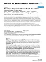
Figure 2 Age-related induction of MHC II and inflammation-
response transcripts. Bioinformatic analysis of transcriptomic
expression data identified significant upregulation the MHC II
antigen presentation pathway and associated inflammatory
signaling factors in hippocampal synaptosomes derived from both
cognitively intact and cognitively impaired aged rats compared to
adults (ANOVA with SNK post hoc testing, p < 0.05). Notably, no
differences were observed between aged intact and aged impaired
rats, which clustered separately from adults but did not cluster by
cognitive status. Log-scaled gene expression is presented relative to
the adult group mean per transcript (green: decreased; black:
unchanged; red: increased).
VanGuilder et al. Journal of Neuroinflammation 2011, 8:138
/>Page 9 of 21
Table 4 qPCR confirmation of age-related induction of MHC II pathway component genes
Gene All Aged vs. Adult (ratio, p-value t-
test)
Pearson Correlation to Morris
water maze
Aged Intact vs. Adult (ratio, p-value
SNK)
Aged Impaired vs. Adult(ratio, p-
value SNK)
Aged Impaired vs.
Aged Intact(ratio, p-value
SNK)
Syn CA1 CA3 DG Syn CA1 CA3 DG Syn CA1 CA3 DG Syn CA1 CA3 DG Syn CA1 CA3 DG
MHC II
Components
Cd74 6.58
###
5.61
###
3.61
###
2.43
###
- - - - 6.79*** 5.66*** 3.88*** 2.43*** 6.35*** 5.58*** 3.40*** 2.49*** - - - -
Ctse 1.69
##
1.45
##
1.75
###
1.48
###
- - - - 1.81* - 1.73*** 1.48* 1.77*** 1.54** - - - -
Hla-dmb 1.64
###
1.37
###
1.37
###
1.26
##
- - - - 1.57** 1.33** 1.39*** - 1.72*** 1.40*** 1.35*** 1.33** - - - 1.16*
Hla-dra 6.30
###
4.04
###
3.02
###
- - - - - 6.76*** 4.18*** 3.25*** - 5.75** 3.95*** 2.85*** - -
Hla-drb1 10.50
###
7.13
###
4.39
###
2.91
#
- - - - 10.70*** 7.12*** 4.77*** - 10.26*** 7.13*** 4.12*** 3.42** - - - -
Antigen
Recognition
C1s 1.84
#
1.21
#
1.56
###
- - 1.59** - - - 1.53*** - - - - -
C3 3.47
###
2.00
###
1.96
###
1.69
###
- - - - 3.40** 2.07*** 2.22*** 1.52*** 3.55** 1.94*** 1.78*** 1.81*** - - - 1.19*
C4a 4.18
###
1.77
###
1.51
###
- - - - - 3.83** 1.67*** 1.58*** - 4.59** 1.85*** 1.47*** - -
Serping1 2.71
###
2.03
###
2.27
###
1.54
###
- - - - 2.79*** 2.18*** 2.52*** 1.49** 2.63*** 1.92*** 2.09*** 1.58*** - - - -
Tlr2 1.64
##
- 1.54## - - - - - 1.55* - 1.66* - 1.73* - 1.46* - -
Tlr4 1.51
##
1.35
#
1.96
###
- - - - - 1.41* - 2.00*** - 1.60* - 1.93*** - -
Tlr7 1.69
##
1.34
##
1.73
###
- - - - - 1.65* 1.34* 1.74*** - 1.73* 1.35* 1.72*** - -
Immune Cell Activating
Ccr5 1.96
#
- - - - - - -
Erbb3 1.28
###
1.34
###
1.20
#
1.40
#
- - - - 1.26** 1.43*** - - 1.30** 1.28** - 1.47** - - - -
Fcgr2a 1.45
#
1.19
#
1.30
###
- - 1.30** 1.30** - -
Fcgr2b 1.96
##
- 1.61
###
- - - - - 2.08* - 1.63*** - 1.82* - 1.60*** - -
Inflammation Response
Cp 1.67
#
- 1.56
###
- - - - - 1.92* - 1.64*** 1.50*** - -
Hsbp1 2.87
###
1.68
###
2.09
###
- - - - - 3.13** 1.65** 2.25*** - 2.58*** 1.70** 1.97*** - -
Icam1 1.46
#
1.55
###
1.43
###
- - 1.68*** 1.64*** 1.46** 1.31** - -
Lgals3 3.42
##
2.64
###
1.98
###
1.84
##
- - - - 3.68*** 2.62*** 2.18*** 1.63* 3.03*** 2.66*** 1.84*** 2.00** - - - -
S100a6 1.52
#
2.03
###
2.21
###
- - 2.00*** 2.48*** - - 2.06*** 2.02*** - -
qPCR confirmation of transcriptomic findings was performed in hippocampal synaptosomes and expanded to include hippocampal subregion dissections. Age-based comparisons (aged intact and aged impaired
versus adult) were statistically analyzed by two-tailed t-test with BHMTC (
#
p < 0.05;
##
p < 0.01;
###
p < 0.001). Three-group data (adult, aged intact, aged impaired) were statistically analyzed by one-way ANOVA with
BHMTC, and pairwise comparisons were evaluated by SNK post hoc testing to identify potential regulation of gene expression with cognitive decline (*p < 0.05; **p < 0.01; ***p < 0.001). Correlation of gene
expression with Morris water maze performance was assessed by Pearson product moment analysis with BHMTC, which demonstrated no significant association of MHC II pathway gene expression and cognitive
impairment. Data are presented as ratios of group means. Individual group gene expression data is presented in Table 5.
VanGuilder et al. Journal of Neuroinflammation 2011, 8:138
/>Page 10 of 21
were evenly distributed throughout DG, CA1 and CA3
in adult rats. Few CD74
+
activated microglia were
observed in adult hippocampi. In both aged cognitively
intact and aged cognitively impaired rats, increased Iba1
immunoreactivity was apparent, as was a marked
increase in the appearance of activated (Iba1
+
/CD74
+
)
microglia. CD74-imunorea ctive cells were associated
with both synaptic and cell body (granule/pyramidal)
layers throughout all three hippocampal subregions.
Higher magnification of microglia revealed changes in
both protein expression and morphological characteris-
tics of activation (Figure 7A). Resting microglia were
immunoreactive to Iba1 but not CD74, and had numer-
ous thin, branched projections. Activated microgl ia
Table 5 Gene expression levels of age and cognitive status groups, presented as group mean (% of adult mean) ±
SEM
CA1 CA3 DG synaptosomes (whole-
hippocampus)
Aged Aged Aged Aged Aged Aged Aged Aged
Gene Adult Aged Intact Impaired Adult Aged Intact Impaired Adult Aged Intact Impaired Adult Aged Intact Impaired
C1s 100 ±
2.8
121 ±
6.1
125 ±
11.7
118 ±
6.6
100 ±
7.8
156 ±
7.6
159 ±
13.4
153 ±
9.3
100 ±
6.1
122 ±
4.8
117 ±
5.7
125 ±
6.9
100 ±
21.7
184 ±
21.8
182 ±
28.6
186 ±
35.3
C3 100 ±
6.6
200 ±
10.9
207 ±
16.4
194 ±
15.0
100 ±
7.9
196 ±
10.8
222 ±
17.8
178 ±
10.7
100 ±
8.1
169 ±
7.2
152 ±
10.2
181 ±
8.3
100 ±
23.2
347 ±
35.7
340 ±
27.6
355 ±
74.4
Serping1 100 ±
7.4
203 ±
13.4
218 ±
14.6
192 ±
20.2
100 ±
7.3
227 ±
11.5
252 ±
17.4
209 ±
13.1
100 ±
4.8
154 ±
6.3
149 ±
9.4
158 ±
8.7
100
±
6.9
271 ±
12.4
279 ±
19.9
263 ±
15.5
Cd74 100 ±
7.7
561 ±
33.3
566 ±
44.5
558 ±
47.9
100 ±
10.9
361 ±
20.5
388 ±
37.6
340 ±
21.2
100 ±
7.7
243 ±
17.2
233 ±
36.7
249 ±
18.2
100 ±
28.8
658 ±
48.9
679 ±
46.7
635 ±
95.8
C4a 100 ±
7.1
177 ±
7.0
167 ±
11.1
185 ±
8.6
100 ±
4.8
151 ±
4.6
158 ±
6.2
147 ±
6.2
100 ±
9.7
135 ±
10.3
117 ±
15.2
147 ±
13.0
100 ±
17.7
418 ±
44.6
383 ±
31.6
459 ±
88.3
Ccr5 100 ±
5.2
108 ±
5.1
106 ±
5.2
110 ±
7.8
100 ±
3.1
121 ±
6.3
117 ±
7.3
125 ±
10.3
100 ±
6.9
99 ±
7.0
88 ±
7.1
106 ±
10.4
100 ±
17.9
196
±
23.0
172 ±
18.8
220 ±
41.8
Cp 100 ±
5.4
93 ±
8.2
93 ±
6.9
93 ±
13.5
100 ±
3.9
156 ±
6.3
164 ±
9.2
150 ±
8.4
100 ±
9.2
110 ±
8.0
118 ±
13.7
103 ±
9.8
100 ±
10.8
167 ±
17.1
192 ±
28.6
146 ±
18.4
Ctse 100 ±
8.0
145 ±
10.2
141 ±
13.7
148 ±
14.4
100 ±
9.8
175 ±
7.4
173 ±
13.9
177 ±
8.7
100 ±
6.8
148 ±
6.8
140 ±
11.3
154 ±
8.4
100 ±
21.0
169 ±
12.0
181 ±
19.8
156 ±
11.4
Erbb3 100 ±
2.7
134 ±
5.1
143 ±
7.5
128 ±
6.5
100 ±
8.1
120 ±
5.1
128 ±
9.1
114 ±
5.6
100 ±
7.5
140 ±
7.8
129 ±
10.9
147 ±
10.3
100 ±
5.4
128 ±
4.0
126
±
6.5
130 ±
4.6
Fcgr2b 100 ±
3.2
108 ±
7.9
103 ±
6.4
112 ±
13.0
100 ±
9.0
161 ±
5.2
163 ±
12.0
160 ±
3.6
100 ±
4.2
124 ±
11.0
125 ±
19.8
123 ±
13.2
100 ±
20.8
196 ±
15.4
208 ±
22.6
182 ±
21.2
Fcgr2a 100 ±
4.4
119 ±
4.8
116 ±
3.8
120 ±
7.7
100 ±
5.2
130 ±
4.2
130 ±
6.4
130 ±
5.9
100 ±
5.1
115 ±
3.8
109 ±
5.0
120 ±
5.0
100 ±
15.2
145 ±
11.4
143 ±
10.7
147 ±
22.8
Hla-
dmb
100 ±
4.6
137 ±
4.6
133 ±
5.2
140 ±
7.3
100 ±
3.9
137 ±
3.7
139 ±
5.9
135 ±
4.9
100 ±
7.4
126 ±
4.4
115 ±
6.6
133 ±
4.7
100 ±
3.3
164 ±
8.3
157 ±
13.7
172
±
8.4
Hla-dra 100 ±
7.9
404 ±
19.0
418 ±
28.1
395 ±
26.5
100 ±
14.7
302 ±
23.4
325 ±
47.0
285 ±
23.4
100 ±
9.7
181 ±
22.4
190 ±
43.4
175 ±
26.4
100 ±
24.0
630 ±
51.8
676 ±
67.9
575 ±
79.8
Hsbp1 100 ±
12.8
168 ±
9.6
165 ±
14.0
170 ±
13.6
100 ±
8.7
209 ±
11.0
225 ±
18.7
197 ±
13.0
100 ±
9.5
109 ±
11.5
80 ±
14.2
130 ±
13.8
100 ±
17.7
287 ±
25.1
313 ±
30.1
258 ±
40.9
Icam1 100 ±
5.5
155 ±
7.6
168 ±
11.9
146 ±
9.4
100 ±
5.6
143 ±
4.2
164 ±
13.3
131 ±
5.7
100 ±
12.2
102 ±
6.5
100 ±
13.5
102 ±
6.5
100 ±
11.1
146 ±
11.8
145 ±
16.7
147 ±
18.3
Lgals3 100
±
4.9
264 ±
16.6
262 ±
21.4
266 ±
24.9
100 ±
7.3
198 ±
12.6
218 ±
16.3
184 ±
17.4
100 ±
10.5
184 ±
12.9
163 ±
10.0
200 ±
20.2
100 ±
21.4
342 ±
34.1
368 ±
54.6
303 ±
39.2
Hla-
drb1
100 ±
11.0
713 ±
57.0
712 ±
96.9
713 ±
73.8
100 ±
9.7
439 ±
34.8
477 ±
73.9
412 ±
30.1
100 ±
14.1
291 ±
40.2
225 ±
44.4
342 ±
59.0
100 ±
7.6
1050
± 67.3
1070
± 97.4
1026 ±
100.2
S100a6 100 ±
2.8
203 ±
11.5
200 ±
16.2
206 ±
16.9
100 ±
7.5
221 ±
13.4
248 ±
20.9
202 ±
15.5
100 ±
16.3
110 ±
8.3
88 ±
15.5
123 ±
7.4
100 ±
9.0
152 ±
13.1
161 ±
24.3
145 ±
15.5
Tlr2 100
±
6.3
106 ±
5.7
109 ±
9.8
104 ±
7.2
100 ±
11.2
154 ±
9.6
166 ±
18.9
146 ±
9.7
100 ±
16.6
172 ±
18.8
166 ±
36.7
176 ±
22.7
100 ±
13.0
164 ±
11.3
155 ±
19.2
173 ±
12.9
Tlr4 100 ±
2.7
135 ±
8.1
118 ±
12.8
143 ±
9.7
100 ±
8.4
196 ±
9.5
200 ±
19.7
193 ±
9.6
100 ±
8.7
119 ±
4.0
118 ±
5.9
120 ±
5.7
100 ±
8.3
151 ±
10.1
141 ±
4.9
160 ±
19.7
Tlr7 100 ±
3.5
134 ±
6.5
134 ±
17.2
135 ±
3.9
100 ±
8.1
173 ±
5.0
174 ±
9.6
172 ±
5.7
100 ±
11.6
94 ±
7.6
95 ±
11.6
93 ±
10.7
100 ±
13.3
169 ±
11.3
165 ±
16.6
173 ±
16.5
VanGuilder et al. Journal
of Neuroinflammation 2011, 8:138
/>Page 11 of 21
expressing both Iba1 and CD74 exhibited characteristics
of both mild activation (increased Iba 1 immunoreactiv-
ity, weak CD74 immunoreacti vity, enlarged somat a) and
moderate activation (increased Iba1 immunoreactivity,
intense CD74 immunoreactivity, enlarged somata, thick-
ened proximal processes, retracted distal processes). The
percentage of mildly- and moderately-activated micro-
glia relative to total microglia was calculated in each
subregion of adult, aged intact, and aged impaired rats
(Figure 7B). The percentage of activated microglia in
adult rats varied by subregion, with microglia in states
of mild or moderate activation reflecting 1-2% of total
microglia in CA1, 6% in CA3, and 2-3% in DG. Signifi-
cant age-related increa ses in both mildly-activated and
moderately activated microgl ia were evident in all three
subregions of both cognitively intac t and cognitively
impaired aged rats compar ed to adults (p < 0.001,
ANOVA,SNK),withnodifferencesineithermildor
moderate activation between aged intact and aged
impaired groups. In CA1 of both aged intact and aged
impaired rats, the percentage of both mildly- and mod-
erately-activated microglia increased by approximately
8-fold,to7-8%oftotalmicroglia.InCA3ofagedrats,
13-16% of microglia reflected either mild or moderate
activation regardless of cognitive status, representing a
2.5-fold increase in activation compared to adults. In
DG of both aged intact and aged impaired rats, mild
and moderate microglial activation increased by 6-fold
(12-13% of total microglia) compared to adults. No cor-
relation between the percentage of activated microglia
(mild or moderate) and MWM performance was
observed. No differences in the proportion of mildly-
activated to moder ately-activated microglia were
observed between groups in CA1, CA3 or DG. The den-
sity of microglia was the same (Iba1
+
cells/100 μm
2
)
across groups (Figure 7B) indicating that the increase in
activated microglial does not reflect proliferation or
infiltration, but rather that an increased percentage of
the existing microglial population is activated in the
aged hippocampus.
Examination of the negative control slides incubated
with secondary antibodies with primary antibodies
omitteddemonstratedthatimmunoreactivity to GFAP,
Iba1, and CD74 was antigen-specific, as no non-spec ific
background signal was detected on these controls
(Figure 8).
Discussion
Neuroinflammation (i.e.,expressionofinflammatory
response factors a nd glial activation), is a hall mark of
brain aging [46,47] and is implicated in the cognitive defi-
cits that accompany neurodegenerative conditions an d
profound brain insults (e.g., TBI and sepsis) [48-50]. Mar-
kers of neuroinf lammation (e.g., CD74, GFAP) have been
reported to increase in the hippocampus with age but the
manner in which these changes may contribute to cogni-
tive decline with normative aging remain to be deter-
mined. Here, we demonstrate that a coordinated induction
of MHC II immune response-associated genes and conco-
mitant astrocyte/microglial activation in the hippocampus
occurs with advanced aging in both cognitively intact and
impaired animals but does not correlate with age-related
deficits of cognitive function.
Figure 3 Age-related upregulation of hippocampal astrocyte
and microglial activation protein markers. (A) Immunoblotting for
GFAP, an intermediate filament protein expressed by astrocytes and
upregulated with inflammation, is significantly increased in CA1, CA3
and DG of both aged cognitively intact and aged cognitively
impaired rats compared to adults. (B) Iba1, a microglia-specific
calcium binding and actin-bundling protein, is significantly induced
in CA1 and CA3, but not DG of aged animals, regardless of cognitive
status, compared to adults. Insets depict representative immunoblot
images. *p < 0.05, **p < 0.01, ***p < 0.001, one-way ANOVA with
Student Newman Keuls post hoc testing, n = 7-10/group.
VanGuilder et al. Journal of Neuroinflammation 2011, 8:138
/>Page 12 of 21
In this work, bioinformatic analysis of the hippocampal
synaptosomal transcriptomes of adult and aged rats
behaviorally assessed for spatial learning and memory
identified 21 genes as components of an age-induced
MHC II-associated antigen presentation and response
pathway. Upregulation of these g enes with advanced
aging was confirmed by q PCR in whole-hippocampus
synaptosome fractions and in hippoca mpal subregion
(CA1, CA3 and DG) dissections. In agreement with our
findings, examination of primary microarray datasets
from previous transcriptomic studies of age-related
changes in hippocampal gene expression from humans
Figure 4 Age-induced activation of hippocampal astrocytes. Immunohistochemical visualization of GFAP revealed l ow-to-moderate
expression in astrocytes distributed throughout DG, CA1 and CA3 of adults. GFAP immunoreactivity was markedly increased, and astrocyte
morphology indicated activation, throughout the hippocampal formation of aged rats. No qualitative differences between aged intact and aged
impaired rats were evident, and no subregion-specific alterations in astrocyte activation were observed. Increased GFAP immunoreactivity was
associated with astrocyte hypertrophy, but not astrocyte proliferation, as total populations of GFAP
+
glia were not changed between groups in
CA1, CA3 or DG. Blue: Hoechst; Red: GFAP.
VanGuilder et al. Journal of Neuroinflammation 2011, 8:138
/>Page 13 of 21
[51], non-human primates [52], and rodents [53,54]
reveals upregulation of a number of immune response
factors, including inductio n of MHC II-associated genes,
that have not previously been systematically pursued in
follow-up studies. F or example, transcriptomic analyses
of Fischer 344 rat hippocampal gene expression reveals
age-related upregulation of MHC II alpha chain, Cd74,
Fcgr3, and C3 [55], as well as induction of MHC II beta
and invariant chains and multiple complement compo-
nents [56]. Importantly, no statistical relationships
between age-related increases in these specific inflamma-
tion-response genes and cognitive performance w ere
identified in these s tudies. Increased Cd74, C4a and C3
mRNA expression, which we observed in aged rats
Figure 5 Morphological characteristics of activated astrocytes in aged hippocampus. Higher magnification imaging revealed a stellate
morphology and low level of GFAP immunoreactivity in hippocampal astrocytes of adult rats. In hippocampus of both cognitively intact and
cognitively impaired aged rats, astrocytes exhibited intense GFAP immunoreactivity and adopted a highly ramified morphology with
hypertrophic processes, while maintaining spatial compartmentalization.
VanGuilder et al. Journal of Neuroinflammation 2011, 8:138
/>Page 14 of 21
compared to 12-month old adults, has also been observed
in the hippocampal transcriptome between young (4-6
months) and aged (24 months) Fischer 344 rats [57].
Our findings also share commonalities with previous
targeted gene expression studies of hippocampal aging.
Increased hippocampal expression of Hla-dra has been
reported in aged (24 months) versus young (3 months)
Fischer 3 44 × B rown Norway rats [58]. We have
expanded on this work by demonstrating that increased
expression of multiple MHC I I components [i.e., MHC
Figure 6 Activation of hippocampal microglia with aging. Immunohistochemical co-localization of the microglia-specific marker Iba1 and the
activation-specific marker CD74 (MHC II invariant chain) identified an increase in activated microglia in the aged hippocampus regardless of
cognitive status. In adults, Iba1
+
microglia were distributed throughout hippocampal subregions, with a greater population evident in CA3 than
in CA1 or DG. A small fraction of microglia exhibited mild activation indicated by low levels of CD74 immunoreactivity. In both aged cognitively
intact and aged cognitively impaired rats, a marked increase in the number of Iba1
+
/CD74
+
microglia was observed in all three subregions, in
the form of both mildly-activated and moderately-activated microglia. Activated microglia were associated with both synaptic and cell body-
containing hippocampal strata. Blue: Hoechst; Red: Iba1; Green: CD74; Orange: Iba1/weak CD74 co-expression (mild activation); Yellow: Iba1/high
CD74 co-expression (moderate activation).
VanGuilder et al. Journal of Neuroinflammation 2011, 8:138
/>Page 15 of 21
Figure 7 Age-related increase in activated microglial populations. (A) High-magnification visualization of resting/surveilling, or non-
activated, and activated microglia reveals differences in both immunoreactivity and morphology with aging. Non-activated microglia expressed
Iba1 but not CD74, and had long, thin, branched processes. Microglia undergoing mild activation displayed increased Iba1 immunoreactivity,
weak CD74 immunoreactivity, and enlarged somata, while moderately activated microglia demonstrated increased Iba1 immunoreactivity,
intense CD74 immunoreactivity, enlarged somata, thickened proximal processes, and retracted distal processes. Notably, activated microglia
maintained a ramified rather than ameboid morphology, indicating that microglial activation had not progressed to a reactive, phagocytic
phenotype. (B) Quantitation of activated microglia (calculated as the ratio of Iba1+/CD74
+
to total Iba1
+
cells) revealed significant increases in all
three hippocampal subregions studied. In adult rats, a small fraction of total microglia were mildly or moderately activated in CA3, with far fewer
activated microglia evident in CA1 and DG. The percentage of microglia undergoing mild or moderate activation was significantly increased, by
several fold, throughout the hippocampus of both aged cognitively intact and aged cognitively impaired rats compared to adults. No differences
between the degree of microglial activation (i.e., mild versus moderate) were observed between aged intact and aged impaired rats in CA1, CA3
or DG. Increased numbers of activated microglia in aged rats were not due to proliferation/infiltration, as total populations of microglia (Iba1
+
cells) were not different between groups. ***p < 0.001, one-way ANOVA with SNK post hoc testing.
VanGuilder et al. Journal of Neuroinflammation 2011, 8:138
/>Page 16 of 21
II alpha (Hla-dra), beta (Hla-drb1) and invariant (Cd74)
chains, and the MHC II antigen-loading cofactor (Hla-
dmb)], and multiple MHC II pathway-associated genes
occurs between mature adult (12 months) and aged (26-
28 months) rats. Similarly, the present study builds
upon a previous finding of increased toll-like receptor
(TLR) expression with aging in mouse whole-brain pre-
parations [59], by demonstrating that increased expres-
sion of TLRs 3, 4, and 7 occurs primari ly in hippocampal
synapses and in the hippocampal CA3 subregion, and
that TLR expression does not correlate with cognitive
impairment.
Our findings extend these previous reports by charac-
terizing a large set of MHC II components and asso-
ciated immune/inflammationresponsefactorsandby
demonstrating that increased expression of these genes
occurs in both cognitively intact and impaired animals
but that the extent of inducti on does not correlate with
cognitive deficits. Inter estingly, the magnitude of induc-
tion of these MHC II-me diated immu ne response genes
wasgreaterinCA1andCA3thaninDGinnearlyall
cases, suggesting that pyramidal cell-containing subre-
gions may undergo age-related neuroinflammation to a
greater extent tha n granule cell-containing regions.
Further, age-related increases in immune/inflammation
response gene expression were, in many cases, larger in
hippocampal synaptosome fractions than in dissected
hippocampal subregions. MHC II pathway gene expres-
sion in hippocampal synaptosomes likely stems largely
from astrocytic and microglial processes closely asso-
ciated with synaptic compartments, while downstream
inflammatory signaling factors may derive from the
synaptic terminals themselves. This suggests that heigh-
tened antigen processing/immune response within the
synaptic milieu may contribute to age-related dysfunc-
tion of hippocampal synapse s and potentially plays a
role in synapse loss [60-62].
Immunohistochemical studies of both human and non-
human primate aging have demonstrated upregulation of
MHC II alpha/beta chains in hippocampal microglia
[18,21,63,64]. We have observed, in both aged cognitively
intact and aged cognitively impaired rats, widespread mild
and moderate microglial activation indicated by increased
numbers of microglia expressing CD74 (i.e., the MHC II
invariant chain), and altered morphological and immunor-
eactive phenoty pes. While the percentage of activated
microglia (reflecting the ratio of CD74
+
/Iba1
+
to total
Iba1
+
microglia) was consistently increased by several fold
in all aged rats compared to adults, these glia maintained a
ramified morphology, rather than the ameboid,
150Pm
Hoechst Cy5
OverlayCy2
Figure 8 Assessment of n egativ e control signals in i mmunohistochemi cal experiments. To ensure that visualization of a strocytes and
microglia was not confounded by nonspecific secondary antibody binding, a negative control for the Cy2 (donkey-anti-mouse DyLight 488) and
Cy5 (donkey-anti-rabbit DyLight 649) channels was included with primary antibodies omitted. Individual channels are presented as inverted gray
scale. Weak, diffuse background signal was observed in the Cy2 channel, and no signal was apparent in the Cy5 channel. No nonspecific cell
staining in the absence of primary antibodies occurred. Blue: Hoechst; Green: Cy2; Red: Cy5.
VanGuilder et al. Journal of Neuroinflammation 2011, 8:138
/>Page 17 of 21
morphology typical of a phagocytic phenotype. This
suggests that the neuroinflammatory state in the aged hip-
pocampus, w hile elevated com pared to adults, is not severe
enough to induce a transition from a “surveilling” micro-
glial phenotype [65,66] to the reactive phenotype associated
with dramatic CNS insults and neurodegeneration [66-68].
In agreement with previous work, the total number of
microglia (Iba1
+
cells) did not increase with aging [69],
indicating that increases in Iba1 protein expression and
numbers of CD74+ microglia reflect age-related activation
rather than microglial proliferation or infiltration. Iba1/
CD74 co-labeling extends previous findings of increased
hippoc ampal microglial activation (CD74+ only) in com-
parisons of young (3-6 months) and aged (24+ months)
[70,71] by demonstrating that an increased percentage of
tot al hippocampal microglia are activated with advanced
aging compared to mature adults. Additionally, previous
studies [70-72] have generally compared young (3-6
months) and old (24+ months) rats, while our results
demonstrate that microglial ac tivation occurs between
mature adulthood (12 months) and advanced age (24-26
months). Furthermore, we extend a previous finding of
qualitative differences in populations of activated microglia
in the aged Wistar rat hippocampus [70] by demonstrating
that increased activation is evident across CA1, CA3, and
DG,andthat,withineachsubregion,thepercentagesof
qualitatively distinct activated microglia (mild and moder-
ate) are equivalent. Similar to the work by Gavilan and col-
leagues [70], fewer activated microglia were observed in
close proximity to pyramidal and granule cell layers than
associated with synapse-containing hippocampal layers.
While adding to the evidence of hippocampal microglial
activation with advanced aging, our findings also clearly
demonstrate that the examined marker of microglial acti-
vation (CD74) is not directly associated with deficits of spa-
tial learning and maze performance , in agreeme nt with
previous studies in the Long Evans rat [72].
We also observed increased astrocyte activation, as indi-
cated by increased GFAP expression and morphological
alterations, which occurred in the absence of astrocyte
proliferation. Studies of humans, nonhuman primates, and
rodent models of human aging have demonstrated
increased glial fibrillary acidic protein (GFAP) gene and
protein expression indicativeofastrocyteactivationand
age-related astrocyte hypertrophy, in agreement with our
findings [73-79]. Our observations agree with one of the
first investigations of age-related hippocampal astrocyte
activation, which demonstrated that these dystrophic
astrocy te processes are often oriented in the same direc-
tion [77]. Our work also revealed that while astrocyte acti-
vation increases throughout the hippocampus with
advanced aging, no cognitive decline-associated differences
in GFAP protein content or numbers of GFAP
+
astrocytes
are apparent between aged intact and aged impaired rats.
Our finding that MHC II pathway induction a nd glial
activation occur in aged rats regardless of cognitive status
raises the important question of whether neuroinflamma-
tion plays a role in the pathogenesis of cognitive decline
[47]. Pathological gliosis and inflammation are associated
with severe cognitive dysfunction in neurodegenerative/
advanced disease states, traumatic brain injury, and direct
inflammatory stimulation [17-24]. Hippocampal neuroin-
flammation has a lso been suggested to play a functional
role in the etiology of age-related cognitive impairment
[80,81]. A lesser degree of neuroinflammation occurs
with normative aging than with disease or injury, and
ranges from increased oxidative stress to induction of
inflammatory signal transducing factors including cyto-
kines, chemokines, complement, and stress response pro-
teins which we and others have previously described in
studies of hippocampal aging [12,25,27,55,82-84]. Age-
related alterations in these factors s uggest an allostatic
shift toward a heig htened basal neuroinflammatory state
in the aging hippocampus. Establishment and mainte-
nance of such a state has been termed “para-inflamma-
tion” and is thought to represent an adaptive response to
persistent sub-threshold stimuli such as cellular/tissue
stress [45].
The present work demonstrates that, while neuroin-
flammation at the level of MHC II pathway expression
and glial activation may represent an aspect of age-
related hippocampal dysregulation necessary for devel-
opment of cogn itive deficits, these measures do not cor-
relate to cognitiv e performance deficits. It is likely that
multiple age-related processes, including decreased neu-
rotransmission, glial dysfunction, and increased neuroin-
flammation, combine with cognitive decline-related
dysregulation of plasticity- and myelin-associated pro-
teinsandgenes[27,55],toformanadditivearrayof
insults to impede healthy neuronal function and learn-
ing and memory [3]. Thus far, the triggering event(s)
that cause the transition from an aged, cognitively intact
state to an aged, cognitively impaired state have
remained elusive. Additionally, the potential remains
that other measures of neuroinflammation, including
cytokines in both the CNS and systemically could
directly correlate to impaired spatial learning and mem-
ory. The importance of the interplay betw een systemic
inflammation, neuroinflammation, and brain function
with aging was recently underscored by a report demon-
strating inhibition of hippocampal neurogenesis by t he
systemic cytokine CCL11 [85]. Ultimately, our under-
standing of the functional roles of neuroinflammat ion in
both protective and harmful CNS processes must
advance in order to identify whether there are appropri-
ate points for interventions seeking modulate age-related
neuroinflammation with the goal of preventing or rever-
sing age-related hippocampal dysfunction.
VanGuilder et al. Journal of Neuroinflammation 2011, 8:138
/>Page 18 of 21
Conclusions
These data demonstrate the age-related induction of mild
to moderate neuroinflammation with aging in multiple
regions of the hippocampus. Analysis of neuroinflamma-
tory measures (MHC II pathway-associated gene expres-
sion, and astrocyte and microglial activation) in rats
assessed for spatial learning and memory by Morris water
maze testing revealed a lack of correlation between inflam-
mation and behavioral performance, suggesting that these
aspects of hippocampal neuroinflammation alone are not
sufficient to induce age-related impairment of spatial
learning and memory. Future work building on this
demonstration of age-related induction of MHC II path-
way and inflammatory response factor expression and glial
activation in both cognitively intact and cognitively
impaired aged animals will need to examine if and how
age-related hippocampal changes such as those described
here combine with cognition-specific alterations to ulti-
mately impair spatial learning and memory.
Abbreviations
CA1: cornu ammonis 1; CA3: cornu ammonis 3; DG: dentate gyrus; MHC II:
major histocompatibility complex class II; CLIP: class II-associated invariant
chain peptide; GFAP: glial fibrillary acidic protein; Iba1: ionized calcium
binding adaptor molecule 1: SNK: Student Newman Keuls; BHMTC:
Benjamini-Hochberg multiple testing correction
Acknowledgements
This work was support by funding from the National Institute on Aging,
National Institutes of Health (5R01AG026607) and Donald W. Reynolds
Foundation. The authors wish to thank Winston Wolfe for technical
assistance.
Author details
1
Department of Pharmacology, Pennsylvania State Univ ersity College of
Medicine, 500 University Drive, Hershey, Pennsylvania, 17057, USA.
2
Donald
W. Reynolds Department of Geriatric Medicine, Reynolds Oklahoma Center
on Aging, University of Oklahoma Health Sciences Center, 975 NE 10
th
Street, BRC-1303, Oklahoma City, Oklahoma, 73104, USA.
Authors’ contributions
HDV, WMF, and WES conceived and designed the studies. JAF conducted
behavioral testing, and HY and JPW performed hippocampus dissections.
HDV, GVB, RMB executed and analyzed the experiments. HDV and WMF
wrote, and all authors edited, read, and approved the final manuscript.
Competing interests
The authors declare that they have no competing interests.
Received: 27 July 2011 Accepted: 11 October 2011
Published: 11 October 2011
References
1. Schaie KW: Intellectual Development in Adulthood: The Seattle Longitudinal
Study Cambridge: Cambridge University Press; 1996.
2. Hedden T, Gabrieli JD: Insights into the ageing mind: a view from
cognitive neuroscience. Nat Rev Neurosci 2004, 5:87-96.
3. Vanguilder HD, Freeman WM: The hippocampal neuroproteome with
aging and cognitive decline: past progress and future directions. Front
Aging Neurosci 2011, 3:8.
4. Shing YL, Rodrigue KM, Kennedy KM, Fandakova Y, Bodammer N, Werkle-
Bergner M, Lindenberger U, Raz N: Hippocampal subfield volumes: age,
vascular risk, and correlation with associative memory. Front Aging
Neurosci 2011, 3:2.
5. Poe BH, Linville C, Riddle DR, Sonntag WE, Brunso-Bechtold JK: Effects of
age and insulin-like growth factor-1 on neuron and synapse numbers in
area CA3 of hippocampus. Neuroscience 2001, 107:231-238.
6. Kantarci K, Senjem ML, Lowe VJ, Wiste HJ, Weigand SD, Kemp BJ, Frank AR,
Shiung MM, Boeve BF, Knopman DS, Petersen RC, Jack CR Jr: Effects of age
on the glucose metabolic changes in mild cognitive impairment. AJNR
Am J Neuroradiol 2010, 31:1247-1253.
7. O’Callaghan RM, Griffin EW, Kelly AM: Long-term treadmill exposure
protects against age-related neurodegenerative change in the rat
hippocampus. Hippocampus 2009, 19:1019-1029.
8. Squier TC: Oxidative stress and protein aggregation during biological
aging. Exp Gerontol 2001, 36:1539-1550.
9. Sonntag WE, Lynch CD, Cooney PT, Hutchins PM: Decreases in cerebral
microvasculature with age are associated with the decline in growth
hormone and insulin-like growth factor 1. Endocrinology 1997,
138:3515-3520.
10. Gavilan MP, Vela J, Castano A, Ramos B, del Rio JC, Vitorica J, Ruano D:
Cellular environment facilitates protein accumulation in aged rat
hippocampus. Neurobiol Aging 2006, 27:973-982.
11. Shi L, Linville MC, Tucker EW, Sonntag WE, Brunso-Bechtold JK: Differential
effects of aging and insulin-like growth factor-1 on synapses in CA1 of
rat hippocampus. Cereb Cortex 2005, 15:571-577.
12. Vanguilder HD, Yan H, Farley JA, Sonntag WE, Freeman WM: Aging alters
the expression of neurotransmission-regulating proteins in the
hippocampal synaptoproteome. J Neurochem 2010, 113:1577-1588.
13. Ben Achour S, Pascual O: Glia: the many ways to modulate synaptic
plasticity. Neurochem Int 2010, 57:440-445.
14. Bessis A, Bechade C, Bernard D, Roumier A: Microglial control of neuronal
death and synaptic properties. Glia 2007, 55
:233-238.
15.
Neumann H, Kotter MR, Franklin RJ: Debris clearance by microglia: an
essential link between degeneration and regeneration. Brain 2009,
132:288-295.
16. Streit WJ: Microglial-neuronal interactions. J Chem Neuroanat 1993,
6:261-266.
17. Buchanan JB, Sparkman NL, Chen J, Johnson RW: Cognitive and
neuroinflammatory consequences of mild repeated stress are
exacerbated in aged mice. Psychoneuroendocrinology 2008, 33:755-765.
18. Godbout JP, Chen J, Abraham J, Richwine AF, Berg BM, Kelley KW,
Johnson RW: Exaggerated neuroinflammation and sickness behavior in
aged mice following activation of the peripheral innate immune system.
FASEB J 2005, 19:1329-1331.
19. Hein AM, Stasko MR, Matousek SB, Scott-McKean JJ, Maier SF,
Olschowka JA, Costa AC, O’Banion MK: Sustained hippocampal IL-1beta
overexpression impairs contextual and spatial memory in transgenic
mice. Brain Behav Immun 2010, 24:243-253.
20. Huang Y, Henry CJ, Dantzer R, Johnson RW, Godbout JP: Exaggerated
sickness behavior and brain proinflammatory cytokine expression in
aged mice in response to intracerebroventricular lipopolysaccharide.
Neurobiol Aging 2008, 29:1744-1753.
21. Sloane JA, Hollander W, Moss MB, Rosene DL, Abraham CR: Increased
microglial activation and protein nitration in white matter of the aging
monkey. Neurobiol Aging 1999, 20:395-405.
22. Fuller S, Steele M, Munch G: Activated astroglia during chronic
inflammation in Alzheimer’s disease–do they neglect their
neurosupportive roles? Mutat Res 2010, 690:40-49.
23. Peters O, Schipke CG, Philipps A, Haas B, Pannasch U, Wang LP, Benedetti B,
Kingston AE, Kettenmann H: Astrocyte function is modified by
Alzheimer’s disease-like pathology in aged mice. J Alzheimers Dis 2009,
18:177-189.
24. Wilson CJ, Finch CE, Cohen HJ: Cytokines and cognition–the case for a
head-to-toe inflammatory paradigm. J Am Geriatr Soc 2002, 50:2041-2056.
25. Freeman WM, Vanguilder HD, Bennett C, Sonntag WE: Cognitive
performance and age-related changes in the hippocampal proteome.
Neuroscience 2009, 159:183-195.
26. Gallagher M, Rapp PR: The use of animal models to study the effects of
aging on cognition. Annu Rev Psychol 1997, 48:339-370.
27. Vanguilder HD, Farley JA, Yan H, Van Kirk CA, Mitschelen M, Sonntag WE,
Freeman WM: Hippocampal dysregulation of synaptic plasticity-
VanGuilder et al. Journal of Neuroinflammation 2011, 8:138
/>Page 19 of 21
associated proteins with age-related cognitive decline. Neurobiol Dis
2011, 43:201-212.
28. Gorelick PB: Role of inflammation in cognitive impairment: results of
observational epidemiological studies and clinical trials. Ann N Y Acad Sci
2010, 1207:155-162.
29. Comim CM, Constantino LC, Barichello T, Streck EL, Quevedo J, Dal-Pizzol F:
Cognitive impairment in the septic brain. Curr Neurovasc Res 2009,
6:194-203.
30. Bianchi ME: DAMPs, PAMPs and alarmins: all we need to know about
danger. J Leukoc Biol 2007, 81:1-5.
31. Rocha N, Neefjes J: MHC class II molecules on the move for successful
antigen presentation. EMBO J 2008, 27:1-5.
32. Aloisi F, Ria F, Penna G, Adorini L: Microglia are more efficient than
astrocytes in antigen processing and in Th1 but not Th2 cell activation.
J Immunol 1998, 160:4671-4680.
33. Hamo L, Stohlman SA, Otto-Duessel M, Bergmann CC: Distinct regulation
of MHC molecule expression on astrocytes and microglia during viral
encephalomyelitis. Glia 2007, 55:1169-1177.
34. Conde JR, Streit WJ: Microglia in the aging brain. J Neuropathol Exp Neurol
2006, 65:199-203.
35. Tremblay ME, Majewska AK: A role for microglia in synaptic plasticity?
Commun Integr Biol 2011, 4:220-222.
36. Bruce-Keller AJ: Microglial-neuronal interactions in synaptic damage and
recovery. J Neurosci Res 1999, 58:191-201.
37. Lee SC, Moore GR, Golenwsky G, Raine CS: Multiple sclerosis: a role for
astroglia in active demyelination suggested by class II MHC expression
and ultrastructural study. J Neuropathol Exp Neurol 1990, 49:122-136.
38. Mitschelen M, Garteiser P, Carnes BA, Farley JA, Doblas S, Demoe JH,
Warrington JP, Yan H, Nicolle MM, Towner R, Sonntag WE: Basal and
hypercapnia-altered cerebrovascular perfusion predict mild cognitive
impairment in aging rodents. Neuroscience 2009, 164:918-928.
39. Maei HR, Zaslavsky K, Teixeira CM, Frankland PW: What is the Most
Sensitive Measure of Water Maze Probe Test Performance? Front Integr
Neurosci 2009, 3:4.
40. Terry AV Jr: Spatial Navigation (Water Maze) Tasks. In Methods of Behavior
Analysis in Neuroscience second edition. Edited by: Buccafusco JJ. Boca
Raton: CRC Press; 2009:.
41. Newton IG, Forbes ME, Legault C, Johnson JE, Brunso-Bechtold JK,
Riddle DR: Caloric restriction does not reverse aging-related changes in
hippocampal BDNF. Neurobiol Aging
2005, 26:683-688.
42.
Brucklacher RM, Patel KM, Vanguilder HD, Bixler GV, Barber AJ, Antonetti DA,
Lin CM, LaNoue KF, Gardner TW, Bronson SK, Freeman WM: Whole genome
assessment of the retinal response to diabetes reveals a progressive
neurovascular inflammatory response. BMC Med Genomics 2008, 1:26.
43. Vanguilder HD, Bixler GV, Kutzler L, Brucklacher RM, Bronson SK, Kimball SR,
Freeman WM: Multi-modal proteomic analysis of retinal protein
expression alterations in a rat model of diabetic retinopathy. PLoS One
2011, 6:e16271.
44. Bixler GV, Vanguilder HD, Brucklacher RM, Kimball SR, Bronson SK,
Freeman WM: Chronic insulin treatment of diabetes does not fully
normalize alterations in the retinal transcriptome. BMC Med Genomics
2011, 4:40.
45. Medzhitov R: Origin and physiological roles of inflammation. Nature 2008,
454:428-435.
46. de Magalhaes JP, Curado J, Church GM: Meta-analysis of age-related gene
expression profiles identifies common signatures of aging. Bioinformatics
2009, 25:875-881.
47. Lucin KM, Wyss-Coray T: Immune activation in brain aging and
neurodegeneration: too much or too little? Neuron 2009, 64:110-122.
48. Khandaker GM, Jones PB: Cognitive and functional impairment after
severe sepsis. JAMA 2011, 305:673-674.
49. Brooks WM, Stidley CA, Petropoulos H, Jung RE, Weers DC, Friedman SD,
Barlow MA, Sibbitt WL, Yeo RA: Metabolic and cognitive response to
human traumatic brain injury: a quantitative proton magnetic resonance
study. J Neurotrauma 2000, 17:629-640.
50. Dash PK, Orsi SA, Zhang M, Grill RJ, Pati S, Zhao J, Moore AN: Valproate
administered after traumatic brain injury provides neuroprotection and
improves cognitive function in rats. PLoS One 2010, 5:e11383.
51. Berchtold NC, Cribbs DH, Coleman PD, Rogers J, Head E, Kim R, Beach T,
Miller C, Troncoso J, Trojanowski JQ, Zielke HR, Cotman CW: Gene
expression changes in the course of normal brain aging are sexually
dimorphic. Proc Natl Acad Sci USA 2008, 105:15605-15610.
52. Blalock EM, Grondin R, Chen KC, Thibault O, Thibault V, Pandya JD,
Dowling A, Zhang Z, Sullivan P, Porter NM, Landfield PW: Aging-related
gene expression in hippocampus proper compared with dentate gyrus
is selectively associated with metabolic syndrome variables in rhesus
monkeys. J Neurosci 2010, 30:6058-6071.
53. Terao A, Apte-Deshpande A, Dousman L, Morairty S, Eynon BP, Kilduff TS,
Freund YR: Immune response gene expression increases in the aging
murine hippocampus. J Neuroimmunol 2002, 132:99-112.
54. Lee CK, Weindruch R, Prolla TA: Gene-expression profile of the ageing
brain in mice.
Nat Genet 2000, 25:294-297.
55.
Blalock EM, Chen KC, Sharrow K, Herman JP, Porter NM, Foster TC,
Landfield PW: Gene microarrays in hippocampal aging: statistical
profiling identifies novel processes correlated with cognitive
impairment. J Neurosci 2003, 23:3807-3819.
56. Kadish I, Thibault O, Blalock EM, Chen KC, Gant JC, Porter NM, Landfield PW:
Hippocampal and cognitive aging across the lifespan: a bioenergetic
shift precedes and increased cholesterol trafficking parallels memory
impairment. J Neurosci 2009, 29:1805-1816.
57. Rowe WB, Blalock EM, Chen KC, Kadish I, Wang D, Barrett JE, Thibault O,
Porter NM, Rose GM, Landfield PW: Hippocampal expression analyses
reveal selective association of immediate-early, neuroenergetic, and
myelinogenic pathways with cognitive impairment in aged rats. J
Neurosci 2007, 27:3098-3110.
58. Frank MG, Barrientos RM, Biedenkapp JC, Rudy JW, Watkins LR, Maier SF:
mRNA up-regulation of MHC II and pivotal pro-inflammatory genes in
normal brain aging. Neurobiol Aging 2006, 27:717-722.
59. Letiembre M, Hao W, Liu Y, Walter S, Mihaljevic I, Rivest S, Hartmann T,
Fassbender K: Innate immune receptor expression in normal brain aging.
Neuroscience 2007, 146:248-254.
60. Kumar A, Foster TC: Neurophysiology of Old Neurons and Synapses. In
Brain Aging: Models, Methods, and Mechanisms. Edited by: Riddle DR. Boca
Raton: CRC Press; 2007:.
61. Kumar A, Thinschmidt JS, Foster TC, King MA: Aging effects on the limits
and stability of long-term synaptic potentiation and depression in rat
hippocampal area CA1. J Neurophysiol 2007, 98:594-601.
62. Barnes CA, Suster MS, Shen J, McNaughton BL: Multistability of cognitive
maps in the hippocampus of old rats. Nature 1997, 388:272-275.
63. Sheffield LG, Berman NE: Microglial expression of MHC class II increases
in normal aging of nonhuman primates. Neurobiol Aging 1998, 19:47-55.
64. Simpson JE, Ince PG, Higham CE, Gelsthorpe CH, Fernando MS, Matthews F,
Forster G, O’Brien JT, Barber R, Kalaria RN, Brayne C, Shaw PJ, Stoeber K,
Williams GH, Lewis CE, Wharton SB: Microglial activation in white matter
lesions and nonlesional white matter of ageing brains. Neuropathol Appl
Neurobiol 2007, 33:670-683.
65. Hanisch UK, Kettenmann H: Microglia: active sensor and versatile effector
cells in the normal and pathologic brain. Nat Neurosci 2007, 10:1387-1394.
66. Nimmerjahn A, Kirchhoff F, Helmchen F: Resting microglial cells are highly
dynamic surveillants of brain parenchyma in vivo. Science 2005,
308:1314-1318.
67. Neniskyte U, Neher JJ, Brown GC: Neuronal death induced by nanomolar
amyloid beta is mediated by primary phagocytosis of neurons by
microglia. J Biol Chem 2011, E-Published.
68. Witting A, Muller P, Herrmann A, Kettenmann H, Nolte C: Phagocytic
clearance of apoptotic neurons by Microglia/Brain macrophages in vitro:
involvement
of lectin-, integrin-, and phosphatidylserine-mediated
recognition. J Neurochem 2000, 75:1060-1070.
69. Long JM, Kalehua AN, Muth NJ, Calhoun ME, Jucker M, Hengemihle JM,
Ingram DK, Mouton PR: Stereological analysis of astrocyte and microglia
in aging mouse hippocampus. Neurobiol Aging 1998, 19:497-503.
70. Gavilan MP, Revilla E, Pintado C, Castano A, Vizuete ML, Moreno-Gonzalez I,
Baglietto-Vargas D, Sanchez-Varo R, Vitorica J, Gutierrez A, Ruano D:
Molecular and cellular characterization of the age-related
neuroinflammatory processes occurring in normal rat hippocampus:
potential relation with the loss of somatostatin GABAergic neurons. J
Neurochem 2007, 103:984-996.
71. Ogura K, Ogawa M, Yoshida M: Effects of ageing on microglia in the
normal rat brain: immunohistochemical observations. Neuroreport 1994,
5:1224-1226.
VanGuilder et al. Journal of Neuroinflammation 2011, 8:138
/>Page 20 of 21
72. Nicolle MM, Gonzalez J, Sugaya K, Baskerville KA, Bryan D, Lund K,
Gallagher M, McKinney M: Signatures of hippocampal oxidative stress in
aged spatial learning-impaired rodents. Neuroscience 2001, 107:415-431.
73. Bernal GM, Peterson DA: Phenotypic and gene expression modification
with normal brain aging in GFAP-positive astrocytes and neural stem
cells. Aging Cell 2011, 10:466-482.
74. Bjorklund H, Eriksdotter-Nilsson M, Dahl D, Rose G, Hoffer B, Olson L: Image
analysis of GFA-positive astrocytes from adolescence to senescence. Exp
Brain Res 1985, 58:163-170.
75. David JP, Ghozali F, Fallet-Bianco C, Wattez A, Delaine S, Boniface B, Di MC,
Delacourte A: Glial reaction in the hippocampal formation is highly
correlated with aging in human brain. Neurosci Lett 1997, 235:53-56.
76. Geinisman Y, Bondareff W, Dodge JT: Hypertrophy of astroglial processes
in the dentate gyrus of the senescent rat. Am J Anat 1978, 153:537-543.
77. Landfield PW, Rose G, Sandles L, Wohlstadter TC, Lynch G: Patterns of
astroglial hypertrophy and neuronal degeneration in the hippocampus
of ages, memory-deficient rats. J Gerontol 1977, 32:3-12.
78. O’Callaghan JP, Miller DB: The concentration of glial fibrillary acidic
protein increases with age in the mouse and rat brain. Neurobiol Aging
1991, 12:171-174.
79. Sloane JA, Hollander W, Rosene DL, Moss MB, Kemper T, Abraham CR:
Astrocytic hypertrophy and altered GFAP degradation with age in
subcortical white matter of the rhesus monkey. Brain Res 2000, 862:1-10.
80. Ownby RL: Neuroinflammation and cognitive aging. Curr Psychiatry Rep
2010, 12:39-45.
81. Yirmiya R, Goshen I: Immune modulation of learning, memory, neural
plasticity and neurogenesis. Brain Behav Immun 2011, 25:181-213.
82. Poon HF, Shepherd HM, Reed TT, Calabrese V, Stella AM, Pennisi G, Cai J,
Pierce WM, Klein JB, Butterfield DA: Proteomics analysis provides insight
into caloric restriction mediated oxidation and expression of brain
proteins associated with age-related impaired cellular processes:
Mitochondrial dysfunction, glutamate dysregulation and impaired
protein synthesis. Neurobiol Aging 2006, 27:1020-1034.
83. Van Kirk CA, Vanguilder HD, Young M, Farley JA, Sonntag WE, Freeman WM:
Age-related alterations in retinal neurovascular and inflammatory
transcripts. Mol Vis 2011, 17:1261-1274.
84. Calabrese V, Scapagnini G, Ravagna A, Colombrita C, Spadaro F,
Butterfield DA, Giuffrida Stella AM: Increased expression of heat shock
proteins in rat brain during aging: relationship with mitochondrial
function and glutathione redox state. Mech Ageing Dev 2004, 125:325-335.
85. Villeda SA, Luo J, Mosher KI, Zou B, Britschgi M, Bieri G, Stan TM,
Fainberg N, Ding Z, Eggel A, Lucin KM, Czirr E, Park JS, Couillard-Despres S,
Aigner L, Li G, Peskind ER, Kaye JA, Quinn JF, Galasko DR, Xie XS, Rando TA,
Wyss-Coray T:
The ageing systemic milieu negatively regulates
neurogenesis and cognitive function. Nature 2011, 477:90-94.
doi:10.1186/1742-2094-8-138
Cite this article as: VanGuilder et al.: Concurrent hippocampal induction
of MHC II pathway components and glial activation with advanced
aging is not correlated with cognitive impairment. Journal of
Neuroinflammation 2011 8:138.
Submit your next manuscript to BioMed Central
and take full advantage of:
• Convenient online submission
• Thorough peer review
• No space constraints or color figure charges
• Immediate publication on acceptance
• Inclusion in PubMed, CAS, Scopus and Google Scholar
• Research which is freely available for redistribution
Submit your manuscript at
www.biomedcentral.com/submit
VanGuilder et al. Journal of Neuroinflammation 2011, 8:138
/>Page 21 of 21