báo cáo hóa học: " Brain microvascular pericytes are immunoactive in culture: cytokine, chemokine, nitric oxide, and LRP-1 expression in response to lipopolysaccharide" ppt
Bạn đang xem bản rút gọn của tài liệu. Xem và tải ngay bản đầy đủ của tài liệu tại đây (2.67 MB, 9 trang )
RESEARC H Open Access
Brain microvascular pericytes are immunoactive
in culture: cytokine, chemokine, nitric oxide, and
LRP-1 expression in response to
lipopolysaccharide
Andrej Kovac
2,4
, Michelle A Erickson
1,3
and William A Banks
1,2,3*
Abstract
Background: Brain microvascular pericytes are important constituents of the neurovascular unit. These cells are
physically the closest cells to the microvascular endothelial cells in brain capillaries. They significantly contribute to
the induction and maintenance of the barrier functions of the blood-brain barrier. However, very little is known
about their immu ne activities or their roles in neuroinflammation. Here, we focused on the immunological profile
of brain pericytes in culture in the quiescent and immune-challenged state by studying their production of
immune mediators such as nitric oxide (NO), cytokines, and chemokines. We also examined the effects of immune
challenge on pericyte expression of low density lipoprotein receptor-related protein-1 (LRP-1), a protein involved in
the processing of amyloid precursor protein and the brain-to-blood efflux of amyloid-b peptide.
Methods: Supernatants were collected from primary cultures of mouse brain pericytes. Release of nitric oxide (NO)
was measured by the Griess reaction and the level of S-nitrosylation of pericyte proteins measured with a modified
“biotin-switch” method. Specific mitogen-activated protein kinase (MAPK) pathway inhibitors were used to
determine involvement of these pathways on NO production. Cytokines and chemokines were analyzed by
multianalyte technology. The expression of both subunits of LRP-1 was analyzed by western blot.
Results: Lipopolysaccharide (LPS) induced release of NO by pericytes in a dose-dependent manner that was
mediated through MAPK pathways. Nitrative stress resulted in S-nitrosylation of cellular proteins. Eighteen of
twenty-three cytokines measured were released constitutively by pericytes or with stimulation by LPS, including
interleukin (IL)-12, IL-13, IL-9, IL-10, granulocyte-colony stimulating factor, granulocyte macrophage-colony
stimulating factor, eotaxin, chemokine (C-C motif) ligand (CCL)-3, and CCL-4. Pericyte expressions of both subunits
of LRP-1 were upregulated by LPS.
Conclusions: Our results show that cultured mouse brain microvascular pericytes secrete cytokines, chemokines,
and nitric oxide and respond to the innate immune system stimulator LPS. These immune pro perties of pericytes
are likely important in their communication within the neurovascular unit and provide a mechanism by which they
participate in neuroinflammatory processes in brain infections and neurodegenerative diseases.
Keywords: mouse brain pericytes, LPS, neurovascular unit, cytokines, chemo kines, LRP-1, Alzheimers disease, nitric
oxide
* Correspondence:
1
Geriatrics Research Education and Clinical Center, Veterans Affairs Puget
Sound Health Care System, Seattle, Washington, USA
Full list of author information is available at the end of the article
Kovac et al. Journal of Neuroinflammation 2011, 8:139
/>JOURNAL OF
NEUROINFLAMMATION
© 2011 Kovac et al; licensee BioMed Central Ltd. This is an Open Access article distributed under the terms of the Creative Commons
Attribution License ( .0), which permits unrestricted use, distribution, and reproduction in
any medium, provided the original work is properly cited.
Background
The blood-brain barrier (BBB) is a selective barrier that is
created by the endothelial cells in cerebral microvessels.
Endothelial cells and supporting cells such as astrocytes,
pericytes, neurons, and perivascular microglia are orga-
nized together to form the “neurovascular unit” which is
ess ential for induction, function, and support of the BBB
[1]. In contrast to the considerable knowledge chara cter-
izing the crosstalk among brain endothelial cells, astro-
cytes, and microglia within the neurovascular unit during
inflammation, very little is known about the role played
by the brain microvascular pericyte.
Among the cells of the neurovascular unit, brain
microvascular pericytes are physically the cells closest to
brain endothelial cells, wrapping around them, joined to
them by gap junctions, and interfacing with them by
peg-and-socket structures [2,3]. These cells are also
essential for the induction of the barrier properties of
the BBB and attrition of pericytes during the neovascu-
larization process [4] or aging [5] can lead to increased
vascular permeability. Furthermore, it has been
described that pericytes regul ates BBB-specific gene
expression in endothelial cells and induces polarization
of astrocyte end-feets [6].
The exact contribution of pericytes to regulation of brain
blood capillary flow is still not adequately examined. Early
ultrastructural studies showed that cerebellar pericytes con-
tains microfilaments similar to actin- and myosin-contain-
ing muscle fibers [7,8]. Furthermore, it has been described
that at least some subpopulations of brain pericytes express
contractile proteins such as a-smooth muscle actin and
non-muscle myosin [9,10]. More recently, using the acute
brain tissue preparation, Peppiatt et al., showed dilatation
of cerebe llar pericytes as an respo nse to glutamate stimula-
tion [11]. Studies on cultured pericytes support contractile
role of these cells however the expression of contractile
proteins such as a-smooth muscle actin seems to be chan-
ged after cultivation [12].
Several in-vitro studies exist that demonstrated that
pericytes are multipotent cells. Pericytes isolated from
adult brains can differentiate into cells of neural lineage
[13]. Cultured brain pericytes express macrophage mar-
kers ED-2 and CD11b and to exhibit pha gocytic activity,
thus expressing immune cell properties [14].
During pathological conditions such as sepsis, peri-
cytes detach from the basal lamina which leads to
increased cerebrovascular permeability. Activation o f
pericytes through TLR-4 has been suggested to be
responsible for this process [15].
Here, we focused on the immunological profile of cul-
tured mouse brain pericytes in the quiescent and
immune-challenged state. We studied production of
immune mediators such a s nitric oxide (NO), cytokines,
and chemokines. We also examined the effects of immune
activation on p ericyte expression of low density lipopro-
tein receptor-related protein-1 (LRP-1), an immune-
modulated processor of amyloid precursor protein and a
brain-to-blood efflux pump for amyloid beta peptide.
Methods
Mouse brain pericytes culture
Primary mouse brain microvascular pericytes were pre-
pared according to Nakagawa et al [16]. Briefly, cultures of
mouse cerebrovascular pericytes were obtained by a pro-
longed, 2-week culture of isolated brain microvessel frag-
ments, containing pericytes and endothelial cells. Pericyte
survival and proliferation was favored by selective culture
conditions using uncoated dishes and DMEM F12 supple-
mented with 20% fetal calf serum (Sigma, USA), L-gluta-
mine (2 mM, G IBCO, USA) and gentamicin (Sigma,
USA). Culture medium was changed twice a week.
Cell stimulation
Mouse brain microvascular pericyte cultures (p2-p8)
were stimulated with lipopolysaccharide from Salmo-
nella typhimurium (L6511; Sigma, USA) for 4, 8, and 24
hours. For MAPK pathways study, SB203580 (p38
MAPK inhibitor, Tocris, USA), PD98059 (MAPKK/MEK
inhibitor, Tocris, USA), UO126 (MEK-1/MEK-2 inhibi-
tor, Tocris, USA), SP600126 (c-Jun N-Terminal kinase
inhibitor, Sigma, USA) and PTDC (NF-Binhibitor,
Sigma, USA) were added to the pericytes cultivated in
96 well plates 1 h before cell stimulation with LPS.
Nitrite assay and detection of S-nitrosylated proteins
Nitri te, a downstre am product of nitric oxide (NO), was
measured by the Griess reaction in culture supernatants
as an indicator of NO production. Brief ly, 50 ul of cell
culture medium was incubated with 100 ul of Griess
reagent A (1% sulfanilamide, 5% phosphoric acid; Sigma,
USA) for 5 min, followed by addition of 100 ul of Griess
reagent B (0.1% N-(1-naphtyl) ethylenediamine; Sigma,
USA) for 5 min. The absorbance was determined at 540
nm using a microplate reader.
Assessment o f S-nitrosylation was done by a modification
of the “bio tin-switch” method. Cells were wa shed in PBS
and lysed in lysis buffer contain NEM (N-ethylmaleimide)
to block free thiol groups. S-nitrosothiols were then
reduced, biotinylated and visualized after SDS-PAGE/wes-
tern blot using a streptavidin-based detection system (Cay-
man Chemical Company, USA). Membranes were
digitalized with a LAS4000 CCD imaging system (GE
Healthcare, USA) and analyzed by ImageQuant TL
software.
Measurement of cytokines and chemokines
Concentrations of cytokines and chemokines secreted
intotheculturemediaweremeasuredbyacommercial
Kovac et al. Journal of Neuroinflammation 2011, 8:139
/>Page 2 of 9
magnetic bead based Multiplex ELISA kit (Bioplex,
Biorad, USA) according to the manufacturer’s protocol.
Immunocytochemistry
Pericytes grown on glass cover slips (12 mm diameter)
were washed in PBS and fixed with 4% PFA for 10 min
at 4°C. Cells were permeabilized with 0.2% TRITON-
X100, blocked with 5% BSA, and then incubated with
anti-a smooth muscle actin antibody (Abcam, USA),
anti-CD13 antibody (Abcam, USA), Griffonia simplicifo-
lia lectin-FITC (Sigma, USA), anti-factor VIII antibody
(Sigma, USA) and anti-GFAP antibody (Abcam, USA)
followed by incubation wi th corresponding ALEXA
Fluor-488 or Alexa Fluor-546 conjugated secondary
antibody (Invitrogen-Molecular Probes, USA). Finally,
slides were mounted in fluorescence mounting media
and photo graphed with a Nikon ECLIPSE E800 fluores-
cence microscope.
Western blotting
For LRP-1, pericyte extracts were run on a 3-8% Tris-
acetate gel (non-reducing conditions), transferred onto
nitrocellulose membranes (Invitrogen, USA), and probed
first with a LRP-1 primary antibody that recognizes the
large subunit (Sigma, USA) and then with a LRP-1 pri-
mary antibody that recognizes the small subunit (Epi-
tomics, 2703-1). SYPRO Ruby (Invitrogen, USA)
staining of membranes was u sed to verify uniformity of
protein loading [17]. Incubation with primary antibodies
was foll owed by horseradish peroxida se-conjugated sec-
ondary antibody (Santa Cruz, USA). As positive and
negative controls, respectively, MEF-1 (SV40 trans-
formed mouse embryo fibroblasts, ATCC, USA) and
PEA-13 (mouse embryo fibroblasts, ATCC, USA) cell
lysates were loaded onto the gel. The enhanced chemilu-
minescence western blot was digitalized with a LAS4000
CCD imaging s ystem (GE Healthcare, USA) and ana-
lyzed by ImageQuant TL software.
Data analysis
Values are presented as means ± SEM. More than two
means were compared by one- way ANOVA followed by
Tukey’s multiple comparison test (Prism 5.0 software,
GraphPad, inc, San Diego, CA). Differences at P < 0.05
were accepted as statistically significant.
Results
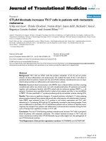
Characterization of purity of primary mouse brain
pericyte cultures
Purity of isolated primary mouse brain pericytes was
analyzed by immunocytochemical staining of cultures.
We evaluated the presence of contaminating astrocytes,
microglia and endothelial cells. More than 95% of cells
in cultures was positive for the pericyte markers a-
smooth muscle actin [14,18] (Figure 1A) a nd CD13
(aminopeptidase N) [19-22] (Figure 1B). Results demon-
strated that there was no contamination of our primary
pericyte cultures either with astrocytes (Figure 1C),
microglia (Figure 1D) or endothelial cells (Figure 1E).
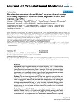
LPS induces nitric oxide production via MAPK pathways
in mouse brain pericytes
Activation of immune cells is accompanied by produc-
tion of different immune mediators. Thus, we studied
the effect of LPS on production of nitric oxide (NO)
and various cytokines and chemokines by cultured pri-
mary brain pericytes. Pericytes were treated for 4, 8 and
24 h with different concentrations of the LPS and nitrite
(a downstream product of NO) concentration in cell
culture media was measured. LPS at concentrations of
0.1 and 1 μg/ml after 8 and 24 h significantly induced
NO release (for example, 24 h results: controls: 0.5 ±
0.15 uM at 24 h; 0.1 ug/ml LPS: 4.3 ± 0.77 uM; 1 ug/ml
LPS: 6.4 ± 0.98 uM; n = 8/group). There was no change
in NO production at 4 h. (Figure 2A) Production of
reactive nitrogen species led to increased S-nitrosylation
of pericyte proteins (2.4× in 0.1 ug/ml LPS vs CTRL, n
= 3) (Figure 2B).
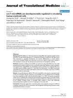
To identify the signal transduction pathway responsible
for production of reactive nitrogen species, we tested sev-
eral MAPK inhibitors and the NF-B inhibitor PDTC for
their ability to reduce NO production by pericytes. Pre-
incubation of cells with SB203580 (at 20 uM; p38 MAPK
inhibitor), PD98059 (at 5 and 50 uM; MAPKK/MEK inhi-
bitor), UO126 (at 5 and 20 uM; MEK-1/MEK-2 inhibi-
tor), SP600126 (at 50 uM; c-Jun N-Terminal kinase
inhibitor) and PTDC (at 5 uM) significantly inhibited
production of NO by cultured brain pericytes (Figure 3).
These results indicated involvement of the MAPK signal-
ing pathway in LPS-induced NO production.
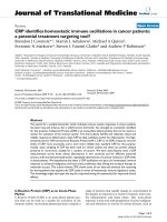
LPS stimulates cytokine and chemokine release by
primary mouse brain pericytes
Pericytes spontaneously released several interleukins
(IL), including IL-9, IL-10, IL-12(p70), IL-13, and IL-17.
Levels of IL-1 alpha, IL-3 , and IL-12(p40) were not
detectable. Other cytokines and chemokines that were
detected were tumor necrosis factor-alpha, interferon-
gamma, granulocyte-colony stimulating factor, granulo-
cyte macrophage-colony stimulating factor, eotaxin,
CCL-3 and CCL-4. To further characterize pericyte
immune capacity, we determined the effect of LPS on
the release of cytokines and chemokines. The results
(Figure 4) showed that stimulation of primary mouse
brain pericyte cultures with 0.1 and 1 ug/ml LPS
resulted in significant release of pro-inflam matory cyto-
kines such as IL-1a, TNF-a, IL-3, IL-9 and IL-13 (4 h, 8
h and 24 h) and anti-inflammatory cytokines such as IL-
Kovac et al. Journal of Neuroinflammation 2011, 8:139
/>Page 3 of 9
10 (4 h, 8 h, 24 h). Additionally, LPS-stimulated peri-
cytes significantly increased their secretion of I L12 het-
erodimer (p70) and of its p40 subunit. Moreover,
activated pericytes produced more chemokines such as
G-CSF, eotaxin, CCL-3, CCL-4 (4 h, 8 h and 24 h) and
MCP-1,KC,CCL-5(4h,8h,24h;datanotshown)in
comparison to unstimulated control cells. Of the
detected cytokines, only the increase in IL-17 was not
significant. There was no detectable constitutive or LPS-
induced production of IL-1b, IL-2, IL-4 and IL-5 by
brain pericytes.
LPS induces up-regulation of LRP-1 expression in brain
pericytes
Neuroinflammation plays an important role in neuro-
degeneration. Here, we analyzed the effect of LPS on
Figure 1 Determination of the purity of the pericyte culture. A primary culture of pericytes isolated from mouse brain microvessels was
labeled with anti-a smooth muscle actin antibody (pericyte marker; red) (Panel A), anti-CD13 antibody (pericyte marker; green) (Panel B), anti-
GFAP antibody (astrocytes marker; green) (Panel C), Griffonia simplicifolia lectin (microglial marker; green) (Panel D) or anti-factor VIII antibody
(endothelial cell marker; green) (Panel E) and counterstained with nuclear stain DAPI (blue). Visual observation of immunostained cells in pericyte
cultures demonstrates that they primarily consist of a a-smooth muscle actin/CD13 positive pericytes. No contamination with microglia,
astrocytes or endothelial cells was detected. Scale bar: 40 μm.
Figure 2 Release of nitric oxide and nitrosative stress in primary brain pericytes after LPS stimulation. Brain pericytes were stimulated for
4, 8, and 24 h with LPS (0.1 and 1 ug/ml), media collected, and analyzed for NO production by the Griess reaction. LPS (0.1 ug/ml and 1 μg/ml)
induced a significant NO release from cells after 8 and 24 hours (A). Nitrative stress was accompanied by massive S-nitrosylation of cellular
proteins (B). Values of nitrite accumulation from treated cells represent the mean ± SEM of two independent experiments conducted in
tetraplicates. *P < 0.05, **P < 0.01, ***P < 0.001 vs. untreated cells.
Kovac et al. Journal of Neuroinflammation 2011, 8:139
/>Page 4 of 9
expression of LRP-1 in peric ytes. Stimulation of cells
with LPS (1 ug/ml) fo r 24 hours significantly increased
expression of both subunits of LRP-1 protein (Figure
5A representative WB and quantification Figure 5B).
The MEF1 (LRP-1 wild type) and PEA13 (LRP-1
knockout) cells were used as positive and negative
controls respectively for LRP-1 antibodies.
Discussion
In this work, we focused on the characterization of the
immunological properties of mouse brain pericytes
under inflammatory conditions induced by LPS. We
have used primary mouse brain pericytes as a model cell
culture for our studies. These cells were isolated by
modifications of the method for isolat ion of microcapil-
laries from mouse brains. However, such isolation pro-
cedures potentially can lead to cultures that are
contaminated with adjacent cell types such as astrocytes,
endothelial cells, and juxtavascular microglia; further-
more, the presence of these contaminating cells can lead
to erroneous results [23,24]. Staining with markers for
microglia, astrocytes and endothelial cells that are not
expressed by pericytes [18], showed that our cultures
were free of these cell types.
Nitric oxide (NO) is a signaling molecule and immune
mediator that is released from glial and endothelial cells
with activation. Microglia and astrocytes are common
sources of N O in the brain during CNS inflammatory
processes [25]. Production of large amounts of NO by
iNOS-2 can lead to generalized nitrosative stress in cells
and to posttra nslational modification of protein residues
by S-nitrosy lation. S-nitrosylation mediates many of the
biological effects of NO. This posttranslational modifica-
tion causes specific physiological or pathophysiolog ical
activities by modifying protein thiols [ 26]. S-nitrosylated
of peptides or proteins are involved in many human dis-
eases such as type II diabetes, Alzheimer’sdisease,and
Parkinson’s disease [27]. Our results demonstrated that
LPS strongly induces production of nitric oxide and
nitrosative stress in brain pericytes. Furthermore, we
found increased S-nitrosylation of pericyte proteins. It
will be important to further analyze and study those
pericyte proteins w hich are affected by increased S-
nitrosylation of their thiol residues.
Mitogen-activated protein kinase (MAPK) signal trans-
duction pathways be long to the most prevalent mechan-
isms of eukaryotic ce lls that respond to extracellular
sti muli [28]. We used several MAPK pathway inhibitors
to analyze the involvement of these pathways in the
release of nitric oxide by brain pericytes in response to
LPS. Our results clearly showed that production of NO
was blocked by pre-incubation of pericytes with these
drugs. These results agree with those obtained from
lung microvascular pericytes [29] and indicate that simi-
lar mechanisms are involved in activation of brain
microvascular pericytes by LPS.
Another interesting finding of our study is related to
the production of important signaling molecules, cyto-
kines and chemokines by pericytes. Of 23 cytokines
and chemokines that we studied, 18 w ere secreted by
brain pericytes constitutively or in response to LPS sti-
mulation.LPSisderivedfromthebacterialcoatof
gram negative bacteria and is a strong stimulant of the
innate immune system. Among the several cytokines
and chemokines whose production was increased by
LPS, IL-12, IL-13, and IL-9 are of particular interest
with regard to pericyte communication within the neu-
rovascular unit. IL-12 plays a critical role in the early
inflammatory response to infection. An increased pro-
duction of IL-12 is involved in the pathogenesis of a
number of autoimmune inflam matory diseases (multi-
ple sclerosis, arthritis, insulin dependent diabetes)
[30-32]. IL-12 consists of two subunits (p40 and p35)
which are linked together by a disulfide bond to give
heterodimeric p70 molecule [33]. We showed that
brain pericytes release substantial amounts of both the
heterodimeric p70 molecule and p40 subunits after
LPS stimulation. Release of the p40 subunit was higher
than release of the hete rodimeric p70 molecule itself.
Interestingly, the p40 subunit of IL12 can link together
Figure 3 Involvement of MAPK pathways in nitric oxide
production by pericytes after LPS stimulation. Brain pericytes
were stimulated for 4, 8, and 24 h with LPS (0.1 and 1 ug/ml). MAPK
pathway inhibitors were added to the culture medium 1 h before
LPS treatment. Media was collected and analyzed for NO production
by Griess reaction. Addition of MAPK pathways inhibitors significantly
reduced NO production by LPS treated pericytes. Values represent
the mean ± SEM of two independent experiments conducted in
tetraplicates. *P < 0.05, ***P < 0.001 vs. untreated cells.
Kovac et al. Journal of Neuroinflammation 2011, 8:139
/>Page 5 of 9
and this homodimeric form has been shown to
increase expression of leukocyte chemoattractant factor
(IL-16) in microglia [34].
IL-9 is another pleiotropic cytokine whose production
was markedly increased after LPS stimulation of brain
pericytes. IL-9 is mainly produced by T lymphocytes
and mediates allergic inflammation in tissues such as
the lung and intestine [35]. In the CNS, the IL-9 recep-
tor complex is present on astrocytes and IL-9 stimulated
astrocytes express CCL-20 chemokine which promotes
infiltration of Th17 cells into the CNS [36].
IL-13 is known as an anti-inflammatory cytokine that
is produced by microglia but not astrocytes or neurons
after direct injection of LPS into the cortex. Neurons
are required for IL- 13 production by microglia and pro-
duction of IL-13 by microglia leads to the death of acti-
vated microglia and enhancement of neuronal survival
[37]. In our study, IL-13 p roductio n by bra in pericytes
Figure 4 Release of cytokines and chemokines from primary brain pericytes constitutively and after LPS stimulation. Brain pericytes
were stimulated for 4, 8, and 24 h with LPS (0.1 and 1 ug/ml). Media was collected and cytokine and chemokine concentrations were
determined via commercial magnetic bead immunoassay. Addition of LPS at 0.1 ug/ml concentration induced significant changes in production
of several pro-inflammatory cytokines and chemokines from brain pericytes. Values of cytokine production represent the mean ± SEM of two
independent experiments conducted in triplicates *P < 0.05, **P < 0.01, ***P < 0.001 vs. untreated cells.
Kovac et al. Journal of Neuroinflammation 2011, 8:139
/>Page 6 of 9
was elevated after LPS treatment; this shows that peri-
cytes are a source of IL-13 as well.
Additionally, compared to published results f rom LPS
treated mouse microglia [38], production of IL1-a and
TNF-alpha, a two typical proinflammatory cytokines, by
brain pericytes was low. This shows that although peri-
cytes and microglia both respond to LPS, the profile of
cytokines released is different.
Recently an interesting study comparing the gene pro-
file expression of different cell components of neurovas-
cular unit in adult or during the development was
published. The study revealed several important genes
that are involved in pericyte-endothelial signaling such
as transforming growth factor beta superfamily members
bmp5 and nodal [39]. It would be interesting to perform
such study with immune-challenged neurovascular unit
as well.
Neurodegenerative processes are closely associated
with neuroinflammation [40]. In Alzheimer’ sdisease,
increased production and impaired transport lead to
accumulation of toxic amyloid beta peptide deposits
along the vascular system in patients affected by this
dis ease. LRP-1 at the brain endothelial cell is an impor-
tant transporter for the brain-to-blood efflux of amyloid
beta peptide [41] and in neurons is important in the
processing of amyloid precursor protein [42,43]. It has
been shown previously that human brain pericytes
express LRP-1 and that the expression is increased after
incubation of cells with amyloid beta peptide [44]. It is
likely that pericyte LRP-1 contributes to the up take and
processing of amyloid beta peptide and amyloid precur-
sor protein. Interestingl y, accumulation of amyloid beta
peptide within the pericyte bodies have been previously
described for early onset familial [45,46] and for spora-
dic Alzheimer’s disease [47]. In line with these observa-
tions, we analyzed the expression of LRP-1 in brain
pericytes during brain infl ammation. We demonstrated
that the expression of both subunits of LRP-1 is
increased in brain pericytes under inflammatory
conditions.
Conclusions
In conclusion, our results as presented here show that
cultured mouse brain pericytes secreting NO, cytokines,
and chemokines and responding to LPS stimulation. We
also showed that pericytes in-vitro express LRP-1, an
important regulator of the levels of amyloid beta peptide
in the brain, and that expression is influenced by LPS.
These immunoactive properties of cultured pericytes
suggest mechanisms by which they can act as an integral
part of the neurovascular unit during brain inflamma-
tory processe s such as brai n infections and neurodegen-
erative processes.
List of abbreviations
BBB: blood-brain barrier; NO: nitric oxide; LRP-1: lipoprotein receptor-related
protein-1; CD11B: cluster of differentiation molecule 11B; LPS:
lipopolysaccharide; GFAP: glial fibrillary acidic protein; iNOS-2: inducible NO
synthase-2; MAPK: mitogen-activated protein kinase.
Acknowledgements and funding
Supported by VA Merit Review, RO1 AG029839, and R01 DK083485.
Author details
1
Geriatrics Research Education and Clinical Center, Veterans Affairs Puget
Sound Health Care System, Seattle, Washington, USA.
2
Division of
Gerontology and Geriatric Medicine, Department of Internal Medicine,
University of Washington, Seattle, Washington, USA.
3
Department of
Pharmacological and Physiological Sciences, Saint Louis University School of
Figure 5 LPS induce up-regulation of LRP-1 expre ssion in brain pericytes. Primary brain pericytes were stimulated for 24 h with LPS (0.1
and 1 ug/ml). After 24 h, expression of both LRP-1 subunits was analyzed by western blot as described in the Material and methods. LPS at 1
ug/ml concentration induced significant increases in expression of the large (515 kDa) and small (85 kDa) subunits of LRP-1. A representative
western blot (A) and density quantification (B) based on ratios between the antibody signal (LRP-1 85 or 515 kDa) and total protein loading per
lane (SYPRO) is shown. Lane designation: 1-PEA13 (LRP-1 knockout as negative control), 2-MEF1 (LRP-1 wild type as positive control), 3-CTRL, 4-
LPS 0.1 ug/ml, 5-LPS 1 ug/ml. Values represent the mean ± SEM of two independent experiments * P < 0.05 vs. untreated cells, n = 5.
Kovac et al. Journal of Neuroinflammation 2011, 8:139
/>Page 7 of 9
Medicine, St. Louis, MO USA.
4
Institute of Neuroimmunology, Slovak
Academy of Sciences, Bratislava, Slovakia.
Authors’ contributions
AK designed the study, performed the bulk of the experiments and analyzed
all data. AK and WB wrote the manuscript. ME performed the western blot
analysis. All authors have read and approved the final version of this
manuscript.
Competing interests
The authors declare that they have no competing interests.
Received: 17 August 2011 Accepted: 13 October 2011
Published: 13 October 2011
References
1. Neuwelt E, Abbott NJ, Abrey L, Banks WA, Blakley B, Davis T, Engelhardt B,
Grammas P, Nedergaard M, Nutt J, et al: Strategies to advance
translational research into brain barriers. Lancet Neurol 2008, 7:84-96.
2. Bonkowski D, Katyshev V, Balabanov RD, Borisov A, Dore-Duffy P: The CNS
microvascular pericyte: pericyte-astrocyte crosstalk in the regulation of
tissue survival. Fluids Barriers CNS 2011, 8:8.
3. Mae M, Armulik A, Betsholt C: Getting to Know the Cast-Cellular
Interactions and Signaling at the Neurovascular Unit. Curr Pharm Des
2011, 9:9.
4. Daneman R, Zhou L, Kebede AA, Barres BA: Pericytes are required for
blood-brain barrier integrity during embryogenesis. Nature 2010,
468:562-566.
5. Bell RD, Winkler EA, Sagare AP, Singh I, LaRue B, Deane R, Zlokovic BV:
Pericytes control key neurovascular functions and neuronal phenotype
in the adult brain and during brain aging. Neuron 2010, 68:409-427.
6. Armulik A, Genove G, Mae M, Nisancioglu MH, Wallgard E, Niaudet C, He L,
Norlin J, Lindblom P, Strittmatter K, et al: Pericytes regulate the blood-
brain barrier. Nature 2010, 468:557-561.
7. Ho KL: Ultrastructure of cerebellar capillary hemangioblastoma. IV.
Pericytes and their relationship to endothelial cells. Acta Neuropathol
1985, 67:254-264.
8. Le Beux YJ, Willemot J: Actin- and myosin-like filaments in rat brain
pericytes. Anat Rec 1978, 190:811-826.
9. Bandopadhyay R, Orte C, Lawrenson JG, Reid AR, De Silva S, Allt G:
Contractile proteins in pericytes at the blood-brain and blood-retinal
barriers. J Neurocytol 2001, 30:35-44.
10. Dalkara T, Gursoy-Ozdemir Y, Yemisci M: Brain microvascular pericytes in
health and disease. Acta Neuropathol 2011, 122:1-9.
11. Peppiatt CM, Howarth C, Mobbs P, Attwell D: Bidirectional control of CNS
capillary diameter by pericytes. Nature 2006, 443:700-704.
12. Hamilton NB, Attwell D, Hall CN: Pericyte-mediated regulation of capillary
diameter: a component of neurovascular coupling in health and disease.
Front Neuroenergetics 2010, 2:5.
13. Dore-Duffy P, Katychev A, Wang X, Van Buren E: CNS microvascular
pericytes exhibit multipotential stem cell activity. J Cereb Blood Flow
Metab 2006, 26:613-624.
14. Balabanov R, Washington R, Wagnerova J, Dore-Duffy P: CNS microvascular
pericytes express macrophage-like function, cell surface integrin alpha
M, and macrophage marker ED-2. Microvasc
Res 1996, 52:127-142.
15. Nishioku T, Dohgu S, Takata F, Eto T, Ishikawa N, Kodama KB, Nakagawa S,
Yamauchi A, Kataoka Y: Detachment of brain pericytes from the basal
lamina is involved in disruption of the blood-brain barrier caused by
lipopolysaccharide-induced sepsis in mice. Cell Mol Neurobiol 2009,
29:309-316.
16. Nakagawa S, Deli MA, Kawaguchi H, Shimizudani T, Shimono T, Kittel A,
Tanaka K, Niwa M: A new blood-brain barrier model using primary rat
brain endothelial cells, pericytes and astrocytes. Neurochem Int 2009,
54:253-263.
17. Hagiwara M, Kobayashi K, Tadokoro T, Yamamoto Y: Application of SYPRO
Ruby- and Flamingo-stained polyacrylamide gels to Western blot
analysis. Anal Biochem 2010, 397:262-264.
18. Guillemin GJ, Brew BJ: Microglia, macrophages, perivascular
macrophages, and pericytes: a review of function and identification. J
Leukoc Biol 2004, 75:388-397.
19. Alliot F, Rutin J, Leenen PJ, Pessac B: Pericytes and periendothelial cells of
brain parenchyma vessels co-express aminopeptidase N,
aminopeptidase A, and nestin. J Neurosci Res 1999, 58:367-378.
20. Cai J, Kehoe O, Smith GM, Hykin P, Boulton ME: The angiopoietin/Tie-2
system regulates pericyte survival and recruitment in diabetic
retinopathy. Invest Ophthalmol Vis Sci 2008, 49:2163-2171.
21. Ramsauer M, Krause D, Dermietzel R: Angiogenesis of the blood-brain
barrier in vitro and the function of cerebral pericytes. Faseb J 2002,
16:1274-1276.
22. Armulik A, Genove G, Betsholtz C: Pericytes: developmental, physiological,
and pathological perspectives, problems, and promises. Dev Cell 2011,
21:193-215.
23. Krueger M, Bechmann I: CNS pericytes: concepts, misconceptions, and a
way out. Glia 2010, 58:1-10.
24. Saura J: Microglial cells in astroglial cultures: a cautionary note. J
Neuroinflammation 2007, 4:26.
25. Murphy S: Production of nitric oxide by glial cells: regulation and
potential roles in the CNS. Glia 2000, 29:1-13.
26. Foster MW: Methodologies for the characterization, identification and
quantification of S-nitrosylated proteins. Biochim Biophys Acta 2011, 4:4.
27. Foster MW, Hess DT, Stamler JS: Protein S-nitrosylation in health and
disease: a current perspective. Trends Mol Med 2009, 15:391-404.
28. Koistinaho M, Koistinaho J: Role
of p38 and p44/42 mitogen-activated
protein kinases in microglia. Glia 2002, 40:175-183.
29. Kim CO, Huh AJ, Kim MS, Chin BS, Han SH, Choi SH, Jeong SJ, Choi HK,
Choi JY, Song YG, Kim JM: LPS-induced vascular endothelial growth
factor expression in rat lung pericytes. Shock 2008, 30:92-97.
30. Constantinescu CS, Goodman DB, Hilliard B, Wysocka M, Cohen JA: Murine
macrophages stimulated with central and peripheral nervous system
myelin or purified myelin proteins release inflammatory products.
Neurosci Lett 2000, 287:171-174.
31. Swardfager W, Lanctot K, Rothenburg L, Wong A, Cappell J, Herrmann N: A
meta-analysis of cytokines in Alzheimer’s disease. Biol Psychiatry 2010,
68:930-941.
32. Zipris D, Greiner DL, Malkani S, Whalen B, Mordes JP, Rossini AA: Cytokine
gene expression in islets and thyroids of BB rats. IFN-gamma and IL-
12p40 mRNA increase with age in both diabetic and insulin-treated
nondiabetic BB rats. J Immunol 1996, 156:1315-1321.
33. Jana M, Dasgupta S, Pal U, Pahan K: IL-12 p40 homodimer, the so-called
biologically inactive molecule, induces nitric oxide synthase in microglia
via IL-12R beta 1. Glia 2009, 57:1553-1565.
34. Jana M, Pahan K: IL-12 p40 homodimer, but not IL-12 p70, induces the
expression of IL-16 in microglia and macrophages. Mol Immunol 2009,
46:773-783.
35. Goswami R, Kaplan MH: A brief history of IL-9. J Immunol 2011,
186:3283-3288.
36. Zhou Y, Sonobe Y, Akahori T, Jin S, Kawanokuchi J, Noda M, Iwakura Y,
Mizuno T, Suzumura A: IL-9 promotes Th17 cell migration into the central
nervous system via CC chemokine ligand-20 produced by astrocytes. J
Immunol 2011, 186:4415-4421.
37. Shin WH, Lee DY, Park KW, Kim SU, Yang MS, Joe EH, Jin BK: Microglia
expressing interleukin-13 undergo cell death and contribute to neuronal
survival in vivo. Glia 2004, 46:142-152.
38. Lu X, Ma L, Ruan L, Kong Y, Mou H, Zhang Z, Wang Z, Wang JM, Le Y:
Resveratrol differentially modulates inflammatory responses of microglia
and astrocytes. J Neuroinflammation 2010, 7:46.
39. Daneman R, Zhou L, Agalliu D, Cahoy JD, Kaushal A, Barres BA: The mouse
blood-brain barrier transcriptome: a new resource for understanding the
development and function of brain endothelial cells. PLoS One 2010, 5:
e13741.
40. Eikelenboom P, Bate C, Van Gool WA, Hoozemans JJ, Rozemuller JM,
Veerhuis R, Williams A: Neuroinflammation in Alzheimer’s disease and
prion disease. Glia 2002, 40:232-239.
41. Zlokovic BV: Clearing amyloid through the blood-brain barrier.
J
Neurochem 2004, 89:807-811.
42.
Bu G, Maksymovitch EA, Nerbonne JM, Schwartz AL: Expression and
function of the low density lipoprotein receptor-related protein (LRP) in
mammalian central neurons. J Biol Chem 1994, 269:18521-18528.
43. Lillis AP, Van Duyn LB, Murphy-Ullrich JE, Strickland DK: LDL receptor-
related protein 1: Unique tissue-specific functions revealed by selective
gene knockout studies. Physiological Reviews 2008, 88:887-918.
Kovac et al. Journal of Neuroinflammation 2011, 8:139
/>Page 8 of 9
44. Wilhelmus MM, Otte-Holler I, van Triel JJ, Veerhuis R, Maat-Schieman ML,
Bu G, de Waal RM, Verbeek MM: Lipoprotein receptor-related protein-1
mediates amyloid-beta-mediated cell death of cerebrovascular cells. Am
J Pathol 2007, 171:1989-1999.
45. Wegiel J, Wisniewski HM: Tubuloreticular structures in microglial cells,
pericytes and endothelial cells in Alzheimer’s disease. Acta Neuropathol
1992, 83:653-658.
46. Wisniewski HM, Wegiel J, Wang KC, Lach B: Ultrastructural studies of the
cells forming amyloid in the cortical vessel wall in Alzheimer’s disease.
Acta Neuropathol 1992, 84:117-127.
47. Szpak GM, Lewandowska E, Wierzba-Bobrowicz T, Bertrand E, Pasennik E,
Mendel T, Stepien T, Leszczynska A, Rafalowska J: Small cerebral vessel
disease in familial amyloid and non-amyloid angiopathies: FAD-PS-1
(P117L) mutation and CADASIL. Immunohistochemical and
ultrastructural studies. Folia Neuropathol 2007, 45:192-204.
doi:10.1186/1742-2094-8-139
Cite this article as: Kovac et al.: Brain microvascular pericytes are
immunoactive in culture: cytokine, chemokine, nitric oxide, and LRP-1
expression in response to lipopolysaccharide. Journal of
Neuroinflammation 2011 8:139.
Submit your next manuscript to BioMed Central
and take full advantage of:
• Convenient online submission
• Thorough peer review
• No space constraints or color figure charges
• Immediate publication on acceptance
• Inclusion in PubMed, CAS, Scopus and Google Scholar
• Research which is freely available for redistribution
Submit your manuscript at
www.biomedcentral.com/submit
Kovac et al. Journal of Neuroinflammation 2011, 8:139
/>Page 9 of 9