báo cáo hóa học:" Differential expression of type X collagen in a mechanically active 3-D chondrocyte culture system: a quantitative study" docx
Bạn đang xem bản rút gọn của tài liệu. Xem và tải ngay bản đầy đủ của tài liệu tại đây (380.94 KB, 10 trang )
BioMed Central
Page 1 of 10
(page number not for citation purposes)
Journal of Orthopaedic Surgery and
Research
Open Access
Research article
Differential expression of type X collagen in a mechanically active
3-D chondrocyte culture system: a quantitative study
Xu Yang
†
, Peter S Vezeridis
†
, Brian Nicholas, Joseph J Crisco,
Douglas C Moore and Qian Chen*
Address: Orthopaedic Research Laboratories, Department of Orthopaedics, Brown Medical School/Rhode Island Hospital, Providence, RI 02903,
USA
Email: Xu Yang - ; Peter S Vezeridis - ; Brian Nicholas - ;
Joseph J Crisco - ; Douglas C Moore - ; Qian Chen* -
* Corresponding author †Equal contributors
Abstract
Objective: Mechanical loading of cartilage influences chondrocyte metabolism and gene
expression. The gene encoding type X collagen is expressed specifically by hypertrophic
chondrocytes and up regulated during osteoarthritis. In this study we tested the hypothesis that
the mechanical microenvironment resulting from higher levels of local strain in a three dimensional
cell culture construct would lead to an increase in the expression of type X collagen mRNA by
chondrocytes in those areas.
Methods: Hypertrophic chondrocytes were isolated from embryonic chick sterna and seeded
onto rectangular Gelfoam sponges. Seeded sponges were subjected to various levels of cyclic
uniaxial tensile strains at 1 Hz with the computer-controlled Bio-Stretch system. Strain distribution
across the sponge was quantified by digital image analysis. After mechanical loading, sponges were
cut and the end and center regions were separated according to construct strain distribution. Total
RNA was extracted from the cells harvested from these regions, and real-time quantitative RT-
PCR was performed to quantify mRNA levels for type X collagen and a housing-keeping gene 18S
RNA.
Results: Chondrocytes distributed in high (9%) local strain areas produced more than two times
type X collagen mRNA compared to the those under no load conditions, while chondrocytes
located in low (2.5%) local strain areas had no appreciable difference in type X collagen mRNA
production in comparison to non-loaded samples. Increasing local strains above 2.5%, either in the
center or end regions of the sponge, resulted in increased expression of Col X mRNA by
chondrocytes in that region.
Conclusion: These findings suggest that the threshold of chondrocyte sensitivity to inducing type
X collagen mRNA production is more than 2.5% local strain, and that increased local strains above
the threshold results in an increase of Col X mRNA expression. Such quantitative analysis has
important implications for our understanding of mechanosensitivity of cartilage and mechanical
regulation of chondrocyte gene expression.
Published: 06 December 2006
Journal of Orthopaedic Surgery and Research 2006, 1:15 doi:10.1186/1749-799X-1-15
Received: 07 March 2006
Accepted: 06 December 2006
This article is available from: />© 2006 Yang et al; licensee BioMed Central Ltd.
This is an Open Access article distributed under the terms of the Creative Commons Attribution License ( />),
which permits unrestricted use, distribution, and reproduction in any medium, provided the original work is properly cited.
Journal of Orthopaedic Surgery and Research 2006, 1:15 />Page 2 of 10
(page number not for citation purposes)
Background
Cartilage in human joints is subjected to various loads
that regulate chondrocyte metabolism and cartilage extra-
cellular matrix protein composition. The mechanical
stress placed on cartilage in vivo plays an important role in
the regulation of chondrocyte proliferation, differentia-
tion, and hypertrophy. One of the ways in which this reg-
ulation occurs is through complex control of chondrocyte
gene expression. Mechanical loading of cartilage is sensed
by chondrocytes embedded within extracellular matrix.
Mechanical signals then activate mechanotransduction
pathways to alter gene expression [1-3]. These chondro-
cyte mechanoregulatory pathways are hypothesized to
involve several levels of signaling, including transduction
through ion channels [2], activation of transcription fac-
tors [4], and alteration of microtubules in the cytoskele-
ton [5].
Previous study using the Bio-Stretch culture system has
demonstrated that chondrocytes subjected to tensile
strain maintain their chondrocyte phenotype [2]. These
cells are stimulated first to proliferate and then to mature
and hypertrophy by the cyclic uniaxial tensile strain
induced by the device [2]. We identified the type X colla-
gen gene as one of the mechanosensitive genes in cartilage
[2]. Type X collagen is a marker for hypertrophic cartilage
since its mRNA is greatly up regulated in hypertrophic
chondrocytes. Interestingly, type X collagen mRNA is
induced in articular chondrocytes during osteoarthritic
pathogenesis [6-9]. It is not clear how type X collagen
mRNA expression is stimulated only in a specific part of
cartilage, e.g., the hypertrophic region and/or the osteoar-
thritic lesion. Elucidation of the differential expression of
type X collagen regulated by mechanical loading will pro-
vide a clearer understanding of the mechanoregulatory
pathways involved in normal and pathogenic cartilage
processes.
Our previous study has shown that type X collagen mRNA
is significantly up regulated in response to 5% overall
matrix deformation at 1 Hz in a 3-D chondrocyte culture
system after 48 hours cyclic loading [2]. The specific load-
ing strain and frequency were chosen because they stimu-
late the proliferation and differentiation of growth plate
chondrocytes [2]. In the present study, we test the hypoth-
esis that various local strains in different regions of the 3D
scaffold result in different levels of type X collagen mRNA
expression by chondrocytes in those areas.
Methods
Chondrocyte isolation
Primary cultures of early hypertrophic chondrocytes were
established from 17-day-old embryonic chick sterna as
described previously [10,11]. Chondrocytes from the
cephalic part of chick sterna were used in the examination
of type X collagen mRNA levels. Briefly, sternal cartilage
pieces were enzymatically dissociated using 0.1% trypsin
(Sigma, St. Louis, MO, USA), 0.3% collagenase (Wor-
thington, Freehold, NJ), and 0.1% type I testicular
hyaluronidase (Sigma). After an incubation of 30 min at
37°C and 5% CO
2
, the media was replaced and the incu-
bation was continued at 37°C for an additional 1 h.
Chondrocytes were centrifuged and suspended at 5 × 10
6
cells/ml in Ham's F-12 medium (Life Technologies, Grand
Island, NY, USA) containing 10% fetal bovine serum
(HyClone, Logan, UT, USA). One hundred μl of cell sus-
pension was added into each sponge.
3D chondrocyte culture
Gelfoam sponges (Dupont, Delaware) were cut into rec-
tangular pieces (2 cm × 2 cm), assembled in cell culture
chambers, and seeded with chondrocytes as described pre-
viously [2]. The Bio-Stretch device (ICCT Technologies,
Markham, ON, Canada) stretched the chondrocyte-
seeded sponges at different overall strains (the extent of
the deformation of the entire sponge) at 1 Hz with a duty
cycle of 25%. Control chondrocyte-seeded sponges were
maintained under identical test conditions with the
exception that the sponges were not mechanically loaded.
After 48 h of culture, sponges were washed once in HBSS,
and 2 mm lengths from the fixed and free ends of each
sponge (high strain) were cut and separated from the
center area (low strain) (see Fig. 1 and 3). 2 mm lengths
were examined since mechanical characterization of the
Gelfoam sponge demonstrated that local strain decreased
to a constant level of one-half overall strain 2 mm from
each edge of the sponge. Chondrocytes were harvested by
digestion of collagen sponge samples with 0.03% colla-
genase in HBSS for 20 min at 37°C. Cells were collected
by centrifugation at 1000 rpm for 7 min and then resus-
pended in HBSS and counted with a hemacytometer
(American Optical Corporation, Buffalo, NY, USA). Each
of the four groups (non-stretch/stretch, center/ends) con-
tained n = 5 samples.
Analysis of type X collagen mRNA levels
Total RNA was extracted from cells with RNeasy mini kits
(Qiagen, Valencia, CA, USA). Quantification of the type X
collagen mRNA was performed by real-time quantitative
reverse transcriptase PCR (RT-PCR). 1 μg total RNA was
used for each reverse transcriptase reaction in a reaction
buffer containing 1 μl oligo(dT) and 1 μl 10 mM dNTP
Mix (Invitrogen, Carlsbad, CA, USA). Real-time quantita-
tive PCR amplification was performed using SYBR Green
I (Finnzymes, Keilaranta, Finland) with DNA Engine
Opticon 2 Continuous Fluorescence Detection System
(MJ Research, Waltham, MA, USA). Primers used in
amplification of type X collagen mRNA are shown in
Table 1. Type X collagen mRNA levels were normalized to
housekeeping gene 18S RNA levels. Since the level of 18S
Journal of Orthopaedic Surgery and Research 2006, 1:15 />Page 3 of 10
(page number not for citation purposes)
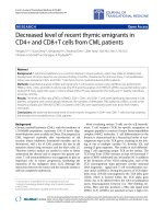
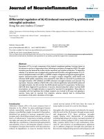
Photograph and line drawing of the Gelfoam sponge loaded in a square petri dish with a 6 by 7 grid of dots marked on surfaceFigure 1
Photograph and line drawing of the Gelfoam sponge loaded in a square petri dish with a 6 by 7 grid of dots marked on surface.
The stationary clamp edge is on left, and mobile plastic clip-metal bar assembly is on right.
Journal of Orthopaedic Surgery and Research 2006, 1:15 />Page 4 of 10
(page number not for citation purposes)
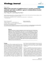
A. Chondrocytes from the ends of the sponge that experienced higher local strain had a statistically significant increase in type X collagen mRNA production in comparison to the corresponding region under no load conditionsFigure 2
A. Chondrocytes from the ends of the sponge that experienced higher local strain had a statistically significant increase in type
X collagen mRNA production in comparison to the corresponding region under no load conditions. (*: p < 0.05) n = 5. Type X
collagen mRNA production was not significantly affected by loading in the center region of the sponge. B. Chondrocytes from
both the clip end and the clamp end of the sponge had a statistically significant increase in type X collagen mRNA production in
comparison to their corresponding regions under no load conditions. (*: p < 0.05) n = 5. Type X collagen mRNA expression
levels in hypertrophic chondrocytes cultured in a sponge were subjected to 5% overall strain. ColX mRNA was quantified
using real-time quantitative RT-PCR. The mRNA levels were normalized to 18S RNA levels, which served as the internal con-
trol.
0
0.5
1
1.5
2
2.5
3
3.5
Nonload Load
Relative Type X Collagen mRNA
(normalized to 18S)
Center
Ends
*
0
0.5
1
1.5
2
2.5
3
3.5
4
4.5
Nonload Load
Relative Type X Collagen mRNA
(normalized to 18S)
Clamp End
Center
Clip End
*
*
A
B
Journal of Orthopaedic Surgery and Research 2006, 1:15 />Page 5 of 10
(page number not for citation purposes)
RNA is constant in all the cells, the normalized value
reflected the relative level of type X collagen mRNA in
each cell regardless of the cell number. Calculation of the
type X collagen mRNA values was performed as previously
described [2]. The 18S RNA was amplified at the same
time and used as an internal control. The cycle threshold
(Ct) values for 18S RNA and that of samples were meas-
ured and calculated by computer software (PE ABI). Rela-
tive transcript levels were calculated as x = 2
-ΔΔCt
, in which
ΔΔCt = ΔE – ΔC, and ΔE = Ct
exp
-Ct
18s
; ΔC = Ct
ctl
-Ct
18s
.
Western blot analysis
Western blot analysis was performed with collected cell
lysates from cell culture. Cell lysates were extracted using
4 M urea, 50 mM Tris at pH 7.5. For non-reducing condi-
tion, collected samples were mixed with standard 2× SDS
gel-loading buffer. For reducing conditions, the loading
buffer contains 5% b-mercaptoethanol and 0.05 M DTT.
Samples were boiled for 10 minutes before loaded onto
10% SDS-PAGE gels. After electrophoresis, proteins were
transferred onto Immobilon-PVDF membrane (Millipore
Corp., Bedford, MA, USA) in 25 mM Tris, 192 mM gly-
cine, and 15 % methanol. The membranes were blocked
in 2% bovine serum albumin fraction V (Sigma Co., St.
Louis, MO, USA) in PBS for 30 minutes and then probed
with antibodies. The primary antibodies used were a pol-
yclonal antibody against Col X [10], and a monoclonal
antibody against β-actin. Horseradish peroxidase conju-
gated goat anti-mouse or goat anti-rabbit IgG (H+L) (Bio-
Rad Laboratories, Melville, NY, USA), diluted 1:3,000,
was used as a secondary antibody. Visualization of immu-
noreactive proteins was achieved using the ECL Western
blotting detection reagents (Amersham Corp., Heights, IL,
USA) and exposing the membrane to Kodak X-Omat AR
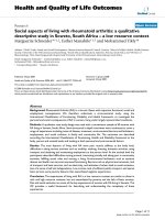
Distribution of surface strains in a typical sponge (4.3% overall strain in this example)Figure 3
Distribution of surface strains in a typical sponge (4.3% overall strain in this example). The local strains in the central region
were found to be dramatically lower than the strain in either end region. Strain values are reported as mean ± one standard
deviation.
Initial Marker Position (mm)
024681012
Strain (%)
0
2
4
6
8
10
12
end region end regioncentral region
Journal of Orthopaedic Surgery and Research 2006, 1:15 />Page 6 of 10
(page number not for citation purposes)
film. Molecular weights of the immunoreactive proteins
were determined against two different sets of protein
marker ladders.
Quantification of strain distribution across the sponge
Strain distribution was determined for collagen Gelfoam
sponges (n = 4) loaded in the culture dish of the Bio-
Stretch electromagnetic system (ICCT Technologies,
Markham, ON, Canada). Gelfoam sponge (Upjohn,
Kalamazoo, MI, USA) was cut into rectangular pieces (20
mm × 20 mm × 6 mm). A-plastic clip assembly with an
imbedded metal bar was attached to one end of the
sponge and the other end of the sponge was fixed to the
culture dish with a plastic clamp leaving approximately a
12 mm length of exposed sponge. Using a fine tipped per-
manent marker, a 6 by 7 grid of dots was placed on each
sponge to provide marker points for measurement of
sponge strain distribution (Fig. 1). The sponge was then
pre-soaked with Hanks' Balanced Salt Solution (HBSS,
GIBCO, Grand Island, NY) overnight at 37°C and 5%
CO
2
.
Sponges were deformed using power settings on the Bio-
Stretch system of 20%, 30%, 40%, 50%, 60%, and 70%.
Digital images of each sponge were captured in the
unstretched and maximally stretched state at each power
setting in 16-bit gray-scale at 16× magnification using a
Polaroid DMC2 digital microscope camera (Polaroid,
Wayland, MA, USA) connected to a Leica M26 stereomi-
croscope (Leica, Bannockburn, IL, USA). Scion Image soft-
ware (Scion, Frederick, MD, USA) was used to analyze the
sponge images. Using this software, each image was
thresholded to assign x- and y-coordinate values to the
centroid of each marker point. The x- and y-coordinate
values of points along the clamp edge and clip edge were
also recorded. The x-direction was defined in the direction
of the principal tensile load and the y-direction was in the
perpendicular direction. The local strain was calculated as
a change in length between unstretched and stretched
positions as a percent of the unstretched state. Strain val-
ues were calculated for all combinations of adjacent
marker points. The strain in the transverse direction (y
direction) was zero at both ends because the sponge was
clamped at each end and ranged from undetectable values
at the lower power to very small values at maximum
power. Thus all strain values reported here in are those in
the x-direction. Strain values are reported with respect to
their initial unstretched position on the sponge and are
the averages of the strain values for that specific column
(y-direction) of marker points.
Statistical analysis
Two-tailed t-tests were used to compare type X collagen
mRNA levels from mechanically loaded chondrocytes in
the Gelfoam sponge to those in the corresponding region
under non-load conditions. Col X mRNA levels from
chondrocytes in the center or end regions of the sponge in
response to different strains were analyzed by one-way
ANOVA with Dunnett Multiple Comparison post-hoc
test. For these calculations, p < 0.05 was considered to be
statistically significant.
Results
Type X collagen mRNA expression in response to 5%
overall strain
We have shown previously that hypertrophic chondro-
cytes significantly increased their Col X mRNA production
in response to 5% overall strain following 48 h cyclic
uniaxial mechanical loading [2]. However, we found that
type X collagen mRNA levels were not up regulated by
chondrocytes in the center region of sponges, defined as
the central region 2 mm from each end, in response to
cyclic mechanical loading (Figure 2). In contrast, hyper-
trophic chondrocytes from the 2 mm areas at the ends of
the sponge (end region) produced more than 2 times of
type X collagen mRNA compared to those in the end
region of non-loaded sponge (Figure 2A). Chondrocytes
from both ends of the sponge produced significantly
higher levels of Col X mRNA under loading conditions
than the corresponding regions under non-load condi-
tions (Figure 2B). Therefore, the increase of Col X mRNA
level in response to 5% overall strain was attributed to the
chondrocytes residing in the end regions, but not those in
the central region of the sponge.
Strain distribution across the collagen sponge
Quantification of the surface strains of a Gelfoam sponge
indicated that mechanical property was different in the
end region vs. the central region of collagen scaffold. Ten-
sile loading of the sponge by the Bio-Stretch system
resulted in a highly non-uniform strain distribution – the
strain in the end region was much higher than the strain
Table 1: Oligonucleotide primer sequences used for real-time quantification RT-PCR detection of type X collagen mRNA
Gene Primer Sequence
Type X collagen Forward 5'-AGTGCTGTCATTGATCTCATGGA-3'
Reverse 5'-TCAGAGGAATAGAGACCATTGGATT-3'
18S RNA Forward 5'-CGGCTACCACATCCAAGGAA-3'
Reverse 5'-GCTGGAATTACCGCGGCT-3'
Journal of Orthopaedic Surgery and Research 2006, 1:15 />Page 7 of 10
(page number not for citation purposes)
in the central region (Figure 3). As a result, 5% overall
strain caused 2.5% local strain in the central region and
9% local strain in the end region of a sponge. However,
the strain in the central region of the sponge was nearly
constant. This constant strain in the central region was
consistently 1/2 of the overall strain values across a wide
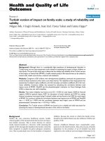
range of overall strain values tested. Specifically, for the six
groups of overall strain values tested, the ratio of central
strain to overall strain was 0.497 ± 0.067 (Figure 4).
Type X collagen expression in response to different overall
strains
To determine whether type X collagen mRNA production
was affected by the overall strain of a sponge, we quanti-
fied Col X mRNA levels from both central and end regions
of the sponges subjected to different overall strains includ-
ing 0% (non-load), 2.5%, 5%, and 7.5% (Figure 5A). For
the central region, only the Col X mRNA value from the
7.5% overall strain group was significantly (p = 0.02)
higher than that from the central region of non-loaded
sponge (0% strain group). This indicated a local strain at
3.75% (half of the overall strain) is required for up regu-
lation of Col X mRNA. For the end regions, samples from
5% and 7.5% overall strain groups, but not that from
2.5% overall strain group, had significantly (p < 0.01)
higher Col X mRNA levels than that from the end region
of non-loaded sample. Therefore, Col X mRNA produc-
tion was increased with increasing local strains regardless
of the region of sponge. We also quantified Col X protein
production by chondrocytes in the center and end regions
of the sponge subjected to different overall strains (Figure
5B). Western blot analysis indicated that Col X protein
levels were up regulated in the samples from higher strain
regions (5% End, 7.5% Center, and 7.5% End). Thus
increasing overall strains results in an increase of Col X
protein production.
Relationship between strains in the central region versus overall strainsFigure 4
Relationship between strains in the central region versus overall strains. The strain values in the central region were approxi-
mately 1/2 (0.5 ± 0.07; n = 4) of the overall strain across a wide range of overall strain values generated by various power set-
tings on the Bio-Stretch System. Each point in the graph represents a different power level tested.
Overall Strain (%)
024681012
Central Strain (%)
0
1
2
3
4
5
6
7
Journal of Orthopaedic Surgery and Research 2006, 1:15 />Page 8 of 10
(page number not for citation purposes)
A. Type X collagen mRNA expression levels in hypertrophic chondrocytes cultured in different sponges subjected to different overall strainsFigure 5
A. Type X collagen mRNA expression levels in hypertrophic chondrocytes cultured in different sponges subjected to different
overall strains. Quantifying ColX mRNA was performed using real-time quantitative RT-PCR. The mRNA levels were normal-
ized to 18S RNA, which served as the internal control. Chondrocytes from the central region of sponges subjected to 7.5%
overall strain (3.75% local strain) had a significant increase in type X collagen mRNA production compared to the central
region of non-loaded (0% strain group) sponges (n = 3/group; #: p = 0.02). Chondrocytes from the end region of the sponges
subjected to 5% or 7.5% overall strains had a significant increase in type X collagen mRNA production in comparison to the
end region of non-loaded (0% strain group) sponge (n = 3/group; *: p < 0.01). B. Western blot analysis of type X collagen from
hypertrophic chondrocytes cultured in different sponges subjected to different overall strains. β-actin was used as an internal
control of a housekeeping protein. Note the increasing strains result in an increase of type X collagen protein level while the
level of β-actin remains constant. C: the center region of sponge; and E: the end region of sponge. Data shown are representa-
tive of those from three independent experiments.
0
0.5
1
1.5
2
2.5
3
02.557.5
Overall Strain (%)
Relative Type X Collagen mRNA
(normalized to 18S)
Center
Ends
*
*
#
A
B
Journal of Orthopaedic Surgery and Research 2006, 1:15 />Page 9 of 10
(page number not for citation purposes)
Discussion
This study tested the hypothesis that mechanical microen-
vironment resulting from higher magnitudes of local
strain within a three-dimensional chondrocyte culture
system leads to increased type X collagen mRNA expres-
sion by chondrocytes in those areas. This hypothesis was
tested in two ways: 1) in a single sponge in response to dif-
ferent local strains, and 2) in different sponges in response
to different overall strains. Data from both tests supported
the conclusion that induction of Col X mRNA was
resulted from an increasing local strain above a certain
threshold.
First, taking advantage of the non-uniform strain distribu-
tion property of the sponge, we demonstrated that type X
collagen mRNA expression in hypertrophic chondrocytes
subjected to cyclic matrix deformation is dependent on
differential local strains within the same sponge. Under
identical culture conditions, chondrocytes in the region
experiencing high local strain produced higher levels of
type X collagen mRNA than those under non-loaded con-
ditions, while there was no significant difference of Col X
production between the region experienced low local
strain and that under no strains. Interestingly, non-uni-
form strain distribution as described for the collagen
sponge exists in articular cartilage, with the highest strain
observed in the end zones of cartilage [12,13]. The system
utilized in the present study exerts differential local strains
within the collagen scaffold of implanted chondrocytes.
This property is significant in that it allows for differential
strains within a single cell culture chamber, thereby limit-
ing variation in the cell culture environment of the
chondrocytes. However, one precaution is the local strain
values measured in the present study represent surface
strains, because the strains on the interior of the sponge in
the end region could not be determined. Furthermore,
there is not necessarily a distinct transition from an area
of high strain to an area of low strain within the sponge
scaffold.
To overcome this shortcoming, we tested sponges sub-
jected to different overall strain magnitudes. Type X colla-
gen mRNA was quantified and compared from the central
regions of the sponges that experienced relatively constant
local strains (1/2 of the overall strain). We show that only
the center region sample subjected to 7.5% overall strain
(3.75% local strain) had a significant increase of type X
collagen mRNA level compared to non-loaded control.
This result is consistent with the data from the single
sponge experiment showing that only local strain more
than 2.5% resulted in a significant increase of type X col-
lagen synthesis. This suggests that the threshold of cyclic
mechanical induction of type X collagen mRNA produc-
tion is greater than 2.5% local strain. This in vitro observa-
tion may have implications for the in vivo situations in
cartilage. Since type X collagen is a marker of hypertrophic
cartilage and osteoarthritic cartilage, our data suggest that
mechanical strain above certain threshold (2.5%) may
contribute to activation of hypertrophic phenotype dur-
ing endochondral ossification.
Osteoarthritis has been described as a loss of regulation of
chondrocyte maturation, in which chondrocytes are not
prevented from progressing from mature chondrocytes to
hypertrophic chondrocytes and then through endochon-
dral ossification [12]. Thus, osteoarthritic chondrocytes
may share some common properties with embryonic
chondrocytes used in this study. Our data suggest that
increased local strain beyond a certain threshold in the
osteoarthritic lesion may also contribute to the local acti-
vation of type X collagen synthesis, similar to its activation
in the hypertrophic region. Future studies need to deter-
mine whether the threshold of mechanical activation of
Col X gene expression is the same between growth plate
chondrocytes and the osteoarthritic chondrocytes.
Applied to in vivo cartilage function, these results may
indicate that certain mechanosensitive gene expression
pathways have a threshold for mechanical induction. Dif-
ferential stress experienced within joint cartilage could be
responsible for differential activation of genes involved in
matrix remodeling. In support of this hypothesis, applica-
tion of mechanical stress to normal chondrocytes has
revealed that high magnitude cyclic tensile load causes an
imbalance between matrix metalloproteinases (MMPs)
and tissue inhibitors of matrix metalloproteinases
(TIMPs), and an increases of the expression of proinflam-
matory cytokines IL-1β and TNF-α [14-16]. Thus, differen-
tial gene expression activated by local high stress may
contribute to osteoarthritic degeneration of some areas of
cartilage while other areas remain viable. This may
account for heterogeneity of osteoarthritic lesion distribu-
tion within a single piece of cartilage or even heterogene-
ity within osteoarthritic lesions.
Commonly used systems for application of mechanical
load to chondrocytes include systems that exert tensile
strain, shear stress, hydrostatic pressure, and compressive
force [17]. These various forms of mechanical loading dif-
ferentially up or down regulate cartilage extracellular
matrix proteins. For example, studies using cyclic tensile
strain have demonstrated an upregulation of several
markers of hypertrophic chondrocytes, including type X
collagen [2]. Type X collagen up regulation is also found
in articular chondrocytes subjected to hydrostatic pressure
[18]. Comparison of cyclic tensile strain and hydrostatic
pressure found that while both mechanical forces signifi-
cantly up regulate type X collagen expression, cyclic ten-
sion exerts a more pronounced effect on type X collagen
up regulation [18]. In addition, examination of the in vivo
Journal of Orthopaedic Surgery and Research 2006, 1:15 />Page 10 of 10
(page number not for citation purposes)
forces exerted on articular cartilage reveals that cyclic ten-
sile strain is analogous to the force created tangential to
the joint surface where it articulates and at the cartilage-
bone interface where type X collagen is expressed [17].
Thus, cyclic tensile strain is a suitable mechanical loading
model for investigation of type X collagen.
Tensile strains applied on a 3D construct in one dimen-
sion may lead to compression in the other dimensions.
Cyclic compression has also been shown to regulate
chondrocyte gene expression [15]. Furthermore, mechan-
ical loading-induced matrix deformation, as measured by
the strain of the sponge, leads to a change of the chondro-
cyte microenvironment within matrix, which includes
fluid flow shear stress, streaming potential, hydrostatic
pressure, and nutrient transport. All of these factors may
contribute to mechanical signaling of chondrocytes [17].
Since our 3D culture system contains these biophysical
factors, alteration of the local matrix strain may lead to
changes of the microenvironment comprising these fac-
tors. It is particularly interesting to link our finding to pre-
vious observations [19-21], which suggest that high
interstitial fluid flow may be responsible for increased
gene expression in local areas. Thus, our data lend support
to the idea that altered mechanical microenvironment in
cartilage may lead to local activation of gene expression in
those areas. Furthermore, the non-uniform strain distri-
butions in Gelfoam sponges, as described in this study,
have implications for biomechanical and tissue engineer-
ing studies that employ such scaffoldings [2,3,22-26].
Acknowledgements
This work was supported by grants from NIH (AG17021, AG 14399),
Arthritis Foundation, and the RIH Orthopaedic Foundation, Inc.
References
1. Sah RL, Kim YJ, Doong JY, Grodzinsky AJ, Plaas AH, Sandy JD: Bio-
synthetic response of cartilage explants to dynamic com-
pression. Journal of Orthopaedic Research 1989, 7:619-636.
2. Wu Q, Chen Q: Mechanoregulation of chondrocyte prolifera-
tion, maturation and hypertrophy: ion-channel dependent
transduction of matrix deformation signals. Experimental Cell
Research 2000, 256:383-391.
3. Wu QZYCQ: Indian hedgehog is an essential component of
mechanotransduction complex to stimulate chondrocyte
proliferation. The Journal of Biological Chemistry 2001,
276:35290-35296.
4. Sironen RK, Karjalainen HM, Elo MA, Kaarniranta K, Torronen K,
Takigawa M, Helminen HJ, Lammi MJ: cDNA array reveals mech-
anosensitive genes in chondrocytic cells under hydrostatic
pressure. Biochimica et Biophysica Acta 2002, 1591:45-54.
5. Jortikka MO, Parkkinen JJ, Inkinen RI, Karner J, Jarvelainen HT, Neli-
markka LO, Tammi MI, Lammi MJ: The role of microtubules in
the regulation of proteoglycan synthesis in chondrocytes
under hydrostatic pressure. Archives of Biochemistry & Biophysics
2000, 374:172-180.
6. Girkontaite I, Frischholz S, Lammi P, Wagner K, Swoboda B, Aigner
T, Vondermark K: Immunolocalization Of Type X Collagen In
Normal Fetal and Adult Osteoarthritic Cartilage With Mon-
oclonal Antibodies. Matrix Biology 1996, 15:231-238.
7. Hoyland JA, Thomas JT, Donn R, Marriott A, Ayad S, Boot-Handford
RP, Grant ME, Freemont AJ: Distribution of type X collagen
mRNA in normal and osteoarthritic human cartilage. Bone &
Mineral 1991, 15:151-163.
8. von der Mark K, Kirsch T, Nerlich A, Kuss A, Weseloh G, Gluckert
K, Stoss H: Type X collagen synthesis in human osteoarthritic
cartilage. Indication of chondrocyte hypertrophy. Arthritis &
Rheumatism 1992, 35:806-811.
9. Walker GD, Fischer M, Gannon J, Thompson RCJ, Oegema TRJ:
Expression of type-X collagen in osteoarthritis. Journal of
Orthopaedic Research 1995, 13:4-12.
10. Chen Q, Johnson DM, Haudenschild DR, Goetinck PF: Progression
and recapitulation of the chondrocyte differentiation pro-
gram: cartilage matrix protein is a marker for cartilage mat-
uration. Developmental Biology 1995, 172:293-306.
11. Leboy PS, Sullivan TA, Menko AS, Enomoto M: Ascorbic acid
induction of chondrocyte maturation. Bone & Mineral 1992,
17:242-246.
12. Wong M, Carter DR: Articular cartilage functional histomor-
phology and mechanobiology: a research perspective. Bone
2003, 33:1-13.
13. Chen SS, Falcovitz YH, Schneiderman R, Maroudas A, Sah RL: Depth-
dependent compressive properties of normal aged human
femoral head articular cartilage: relationship to fixed charge
density. Osteoarthritis & Cartilage 2001, 9:561-569.
14. Jin G, Sah RL, Li YS, Lotz M, Shyy JY, Chien S: Biomechanical reg-
ulation of matrix metalloproteinase-9 in cultured chondro-
cytes. Journal of Orthopaedic Research 2000, 18:899-908.
15. Sah RL, Grodzinsky AJ, Plaas AHK, Sandy JD: Effects of static and
dynamic compression on matrix metabolism in cartilage
explants. In Articular Cartilage and Osteoarthritis Edited by: Kuettner
KE, Schleyerbach R, Peyron JG and Hascall VC. New York, Raven
Press; 1992:373-392.
16. Honda K, Ohno S, Tanimoto K, Ijuin C, Tanaka N, Doi T, Kato Y,
Tanne K: The effects of high magnitude cyclic tensile load on
cartilage matrix metabolism in cultured chondrocytes. Euro-
pean Journal of Cell Biology 2000, 79:601-609.
17. Carter DR, Wong M: Modelling cartilage mechanobiology. Phil-
osophical Transactions of the Royal Society of London - Series B: Biological
Sciences 2003, 358:1461-1471.
18. Wong MSMGK: Cyclic tensile strain and cyclic hydrostatic
pressure differentially regulate expression of hypertrophic
markers in primary chondrocytes. Bone 2003, 33:685-693.
19. Buschmann MD, Kim YJ, Wong M, Frank E, Hunziker EB, Grodzinsky
AJ: Stimulation of Aggrecan Synthesis in Cartilage Explants
by Cyclic Loading Is Localized to Regions of High Interstitial
Fluid Flow1,. Archives of Biochemistry and Biophysics 1999, 366:
1-7.
20. Buschmann MD, Gluzband YA, Grodzinsky AJ, Hunziker EB:
Mechanical compression modulates matrix biosynthesis in
chondrocyte/agarose culture. Journal of Cell Science 1995,
108:1497-1508.
21. Quinn TM, Grodzinsky AJ, Buschmann MD, Kim YJ, Hunziker EB:
Mechanical compression alters proteoglycan deposition and
matrix deformation around individual cells in cartilage
explants. J Cell Sci 1998, 111:573-583.
22. Liu M, Xu J, Souza P, Tanswell B, Tanswell AK, Post M: The effect of
mechanical strain on fetal rat lung cell proliferation: compar-
ison of two- and three-dimensional culture systems. In Vitro
Cellular & Developmental Biology Animal 1995, 31:858-866.
23. Liu M, Montazeri S, Jedlovsky T, Van Wert R, Zhang J, Li RK, Yan J:
Bio-stretch, a computerized cell strain apparatus for three-
dimensional organotypic cultures. In Vitro Cellular & Developmen-
tal Biology Animal 1999, 35:87-93.
24. Geiger M, Li RH, Friess W: Collagen sponges for bone regener-
ation with rhBMP-2. Advanced Drug Delivery Reviews 2003,
55:1613-1629.
25. Still J, Glat P, Silverstein P, Griswold J, Mozingo D: The use of a col-
lagen sponge/living cell composite material to treat donor
sites in burn patients. Burns 2003, 29:837-841.
26. Ito T, Nakamura T, Suzuki K, Takagi T, Toba T, Hagiwara A, Kihara
K, Miki T, Yamagishi H, Shimizu Y: Regeneration of hypogastric
nerve using a polyglycolic acid (PGA)-collagen nerve conduit
filled with collagen sponge proved electrophysiologically in a
canine model. International Journal of Artificial Organs 2003,
26:245-251.