Báo cáo hóa học: " Decreased level of recent thymic emigrants in CD4+ and CD8+T cells from CML patients" pdf
Bạn đang xem bản rút gọn của tài liệu. Xem và tải ngay bản đầy đủ của tài liệu tại đây (754.45 KB, 8 trang )
Li et al. Journal of Translational Medicine 2010, 8:47
/>Open Access
RESEARCH
BioMed Central
© 2010 Li et al; licensee BioMed Central Ltd. This is an Open Access article distributed under the terms of the Creative Commons Attri-
bution License ( which permits unrestricted use, distribution, and reproduction in any
medium, provided the original work is properly cited.
Research
Decreased level of recent thymic emigrants in
CD4+ and CD8+T cells from CML patients
Yangqiu Li*
1,2
, Suxia Geng
1,3
, Qingsong Yin
1
, Shaohua Chen
1
, Lijian Yang
1
, Xiuli Wu
1
, Bo Li
1
, Xin Du
3
,
Christian A Schmidt
4
and Grzegorz K Przybylski*
4,5
Abstract
Background: T-cell immunodeficiency is a common feature in cancer patients, which may relate to initiation and
development of tumor. Based on our previous finding, to further characterize the immune status, T cell proliferative
history was analyzed in CD4+ and CD8+ T cells from chronic myeloid leukemia (CML) patients.
Methods: Quantitative analysis of δRec-ψJα signal joint T cell receptor excision circles (sjTRECs) was performed in
PBMCs, CD3+, CD4+ and CD8+T cells by real-time PCR, and the analysis of 23 TRBV-D1 sjTRECs was performed by semi-
nested PCR. Forty eight CML cases in chronic phase (CML-CP) were selected for this study and 17 healthy individuals
served as controls.
Results: The levels of δRec-ψJα sjTRECs in PBMCs, CD3+, CD4+, and CD8+ T cells were significantly decreased in CML
patients, compared with control groups. Moreover, the numbers of detectable TRBV subfamily sjTRECs, as well as the
frequency of particular TRBV-BD1 sjTRECs in patients with CML were significantly lower than those from healthy
individuals.
Conclusions: We observed decreased levels of recent thymic emigrants in CD4+ and CD8+ T cells that may underlay
the persistent immunodeficiency in CML patients.
Background
Chronic myeloid leukemia (CML), with the incidence of
1.5/100,000 population, represents 15% of newly diag-
nosed leukemia cases in adults in China. The prognosis in
CML improved markedly after introduction of abl
tyrosine kinase inhibitors (Immatinib mesylate and its
derivatives), still a lot of CML patients die due to abl
mutation related drug resistance and the blast crisis [1].
Therefore further studies are needed in order to better
understand the disease and to improve the patient out-
come. T cell immunodeficiency was suggested to play an
important role in tumor progression, facilitating the
expansion of the malignant clone [2,3], although the
interaction between the tumor and the immune system is
still not completely understood.
Most circulating mature T-cells use the α/β heterodi-
meric T cell receptor (TCR) for specific recognition of
antigenic peptides in context of major histocompatibility
complex (MHC) molecules. T cell differentiation in the
thymus is characterized by a hierarchical order of rear-
rangement steps in the TCR genes, resulting in the join-
ing of one of multiple variable (V), diversity (D), and
joining (J) gene segments. This results in each differenti-
ating T cell expressing unique TCR on the surface. The
TCR beta locus (TRB) contains at least 64 functional V
genes (TRBV) subdivided into 24 families [4]. In addition
to the formation of the V(D)J coding joint, each of the
TCR rearrangement steps generates circular episomal
DNA fragments - signal joint T cell recombination exci-
sion circles (sjTRECs). During the process of TCR alpha-
delta locus (TRAD) rearrangement, the TCR delta gene
(TRD), which is located within the TCR alpha gene
(TRA), has to be deleted before the TRA recombination
starts. Rearrangement between two TRD deleting ele-
ments, δRec and ψJα, produces a δRec-ψJα signal joint
TRECs [5-9]. sjTRECs are assumed to have a high over-
* Correspondence: ,
1
Institute of Hematology, Medical College, Jinan University, Guangzhou,
510632, China
Full list of author information is available at the end of the article
Li et al. Journal of Translational Medicine 2010, 8:47
/>Page 2 of 8
time stability, but they can not multiply and consequently
are diluted during T cell proliferation. A maximum of two
sjTRECs can be present within one αβ T cell if the corre-
sponding rearrangement event occurs in both alleles and
if the cell did not divide upon the rearrangement.
sjTRECs are exported from thymus to the periphery
within recent thymic emigrants (RTEs), therefore, the fre-
quency of sjTRECs is considered to be the most accurate
marker of T-cell neogenesis. Quantitative detection of
sjTRECs can be applied for direct measurement of thymic
output and proliferative history of T cells [6]. Over the
last decade the technique was used to evaluate T-cell
immune reconstitution in different immunodeficiency
diseases [6,10-13]. To assess the proliferative history in
different TRBV subfamilies of T cells, quantitative analy-
sis of TRBV-BD sjTRECs has been developed [12,14,15].
The first sjTREC analysis in hematopoietic malignancy
was reported by Petridou et al [16], who compared the
sjTREC values in childhood B-ALL and T-ALL. Signifi-
cant reduction of sjTREC values was observed in T-ALL,
whereas children with B-ALL had slightly but insignifi-
cantly lower sjTRECs values compared with healthy con-
trols. In another study, consistent with the reduction of
naïve T cells, thymopoiesis (measured by sjTRECs levels)
was significantly lower in 73 children with ALL than in
normal controls [17]. However, little data exist regarding
the proliferative history of T cells in myeloid leukemia
patients. Recently, we published the first analysis of the
sjTRECs-content in patients with acute myeloid leukemia
(AML) [18]. Our previous study showed decreased δRec-
ψJα sjTRECs level and skewed TRBV repertoire in
peripheral blood mononuclear cells (PBMCs) from 20
CML cases [19]. Since the high number of CML cells in
the blood might have influenced the results, in the pres-
ent study, in order to more precisely characterize the
immune status in chronic myeloid leukemia (CML), we
analyzed both δRec-ψJα sjTRECs and TRBV-BD sjTRECs
in sorted CD4+ and CD8+ T cells from CML patients.
Materials and methods
Samples
Forty eight newly diagnosed chronic phase CML patients,
33 males and 15 females (13-81 years old; median age: 30
years) were included in this study. BCR-ABL fusion gene
was detected in all samples by RT-PCR. Seventeen
healthy individuals: 6 males and 11 females (25-51 years
old, median age: 28 years) served as controls. The sam-
ples were collected at Dept. of Hematology, Guangdong
Province People's Hospital, all the procedures were con-
ducted according to the guidelines of the Medical Ethics
committees of the health bureau of Guangdong Province
of China. sjTRECs were measured in PBMCs from all 48
cases, and CD4+ and CD8+ T cells from 19 cases. TRBV
sjTRECs were determined in PBMCs, CD4+ and CD8+ T
cells from 10 patients. The clinical data of the patients are
listed in Table 1.
Mononuclear cells isolation
Peripheral blood mononuclear cells (PBMCs) were iso-
lated from CML patients and healthy individuals by
Ficoll-Hypaque gradient centrifugation.
CD3+ cells determination
CD3+ T cells percentage from PBMCs was determined
by indirect immune fluorescent analysis. The PLP-fixed
cytospin preparations were incubated with 200 μg/ml of
murine anti-CD3 mAb (Boster Biological Technology
Ltd, Wuhan, China), washed and incubated with 1:50
dilution of fluorescein labeled goat anti-mouse Ig (Boster
Biological Technology Ltd, Wuhan, China). The slides
were counterstained with Mayer's hematoxylin for 30
min. All slides were blindly evaluated using the fluores-
cent microscope (Nikon WFX-II, Nikon Ltd, Japan); 200
cells were counted.
T cells sorting
The CD4+ and CD8+ T cells from 19 CML cases and 17
healthy individuals were sorted using CD4 and CD8
monoclonal antibody and MACS
®
Magnetic Cell sorting
technique (Miltenyi Biotec, Bergisch Gladbach, Ger-
many). After CD4+ and CD8+ T cells sorting, the purity
was determined by indirect immune fluorescent analysis.
The positive cells were around 95% to 97%.
DNA extraction
Total DNA from distinct cell populations was extracted
using QIAamp
®
DNA Blood Mini Kit (QIAGEN, Ger-
many), the quality of RNA was analyzed in 0.8% agarose
gel stained with ethidium bromide and the concentration
was determined by spectrophotometric analysis at 260
and 280 nm (Lambda 45 UV/VIS Spectrometer, Perkin
Elmer USA).
Real-time quantitative PCR (RQ-PCR)
Quantitative detection of δRec-ψJα sjTRECs in DNA
from PBMCs and sorted CD4+ or CD8+ T cells was pre-
formed by real-time PCR using the ABI PRISM 7700
Sequence Detector TaqMan (PE Biosystems, Foster City,
CA). PCR was performed as described by previous stud-
ies [15,20]. To precisely determine the percentage of cells
carrying sjTREC we constructed a duplex vector includ-
ing a fragment of the δRec-ψJα (sjTREC) and a fragment
of the RAG2 gene used as a reference. The RAG2 was
cloned first in the T-A acceptor site and subsequently the
sjTREC was cloned in to the EcoRV restriction site of the
TOPO TA Vector (Invitrogen, Groning, The Nether-
lands). Based on the DNA concentration, measured by
spectrophotometry and confirmed by a quantitative gel
eletrophoresis, standard dilutions of the vector from 10
7
Li et al. Journal of Translational Medicine 2010, 8:47
/>Page 3 of 8
Table 1: Clinical data of CML patients
No. sex age WBC
(×10
9
/L)
Blast+pro
myelocyte
cells (%)
Platelets
(×10
9
/L)
CD3+% CD4+/
CD8+
cells sorted
C1 F 49 213.59 9 147 28.91 Yes
C2 M 16 351.16 0 345 4.71 Yes
C3 M 58 59.93 6 144 11.01 Yes
C4 M 20 124 0 605 18.2 Yes
C5 M 25 256.82 6 109 13.8 Yes
C6 M 15 333.95 8.5 208 10.8 Yes
C7 F 31 294.91 3 252 10.46 Yes
C8 M 30 118.55 5 440 9.6 Yes
C9 M 24 244.05 9 750 12.04 Yes
C10 M 61 279 10 993 11.8 Yes
C11 F 30 99.8 6.5 378 2.1 Yes
C12 M 38 103.66 1 181 10.4 Yes
C13 F 20 450.45 1 396 12.1 Yes
C14 M 42 81.6 6 85 14.18 Yes
C15 M 73 196 8 1531 9.1 Yes
C16 M 31 129 3.5 285 28.4 Yes
C17 M 22 76.6 5.5 171 19.64 Yes
C18 M 20 112.7 5 596 42.5 Yes
C19 F 19 7 4 125 28.0 Yes
C20 M 61 44.9 12 77 13.6 No
C21 F 13 314.78 3 640 32 No
C22 M 59 18.54 2 695 56.89 No
C23 M 50 31.5 0 163 36.51 No
C24 M 35 5.1 5 283 38.6 No
C25 F 66 62.87 2 657 7.8 No
C26 F 30 160 16 842 12.4 No
C27 F 26 114.17 2 222 19.1 No
C28 M 26 5.3 0 118 44.7 No
C29 M 15 185.9 3 291 10.5 No
C30 M 27 101.5 3 326 42.5 No
C31 F 21 29.7 2 296 26.67 No
C32 M 22 111.92 0 115 9.17 No
C33 F 75 267 7 258 11.2 No
C34 M 29 0.08 2 34 18.75 No
C35 M 26 61.67 9 661 31.5 No
C36 M 43 170 0 671 40.66 No
C37 M 36 43.87 6 69 32.07 No
C38 M 38 58.55 0 3363 18.95 No
C39 M 29 132.4 10.5 1221 26.9 No
C40 F 55 130.21 4 204 14.2 No
C41 M 44 485.1 1 514 27.2 No
C42 F 16 1.39 0 46 38.2 No
Li et al. Journal of Translational Medicine 2010, 8:47
/>Page 4 of 8
to 10
1
copies were prepared [15,20]. In brief, PCR of 25 μl
total volume was performed with approximately 100 ng
of genomic DNA, 25 pmol of each primers (TREC-1 and
TREC-2 for sjTRECs, RAG2-for and RAG2-back for
RGA2 amplification), 10 nmol each dNTP, 1.5 U Ampli-
Taq Gold (Applied Biosystems, Branchburg, New Jersey,
USA), 5 pmol of 6FAM-TAMRA probe and PCR Buffer
including 4.5 mM MgCl
2
. After the initial denaturation at
95°C for 5 min, 45 cycles consisting of 95°C for 30 sec and
67°C for 1 min were performed. If no TRECs were
detected in a sample, PCR was repeated with more DNA.
TRBV-BD1 sjTRECs detection by semi-nest PCR
Twenty three TRBV-BD1 sjTRECs were amplified by
semi-nest PCR from different amounts of genomic DNA
(1.3 μg, 325 ng or 65 ng, corresponding to 2 × 10
5
, 5 × 10
4
or 1 × 10
4
cells respectively) isolated from PBMCs, CD4+
and CD8+ T cells. Two nested 5' TRBD1 primers, located
upstream of the segment, and twenty three 3' TRBV
primers (BV1-19 and BV21-24) were used [15,20]. Since
the TRBV20-BD1 rearrangement occurs by inversion, it
does not generate a sjTREC. In the first round PCR, 2 μl
of genomic DNA were amplified in a 10 μl reaction mix-
ture containing: 0.375 μM external sense and antisense
primers, 0.1 mM dNTP, 1.5 mM MgCl
2
, 1× PCR buffer
and 1 U Taq polymerase (GoTaq
®
Flexi DNA polymerase,
Promega, Madison, WI, USA) using the DNA thermal
cycler. After 3 min denaturation at 94°C, 30 PCR cycles
were performed, each cycle consisting of 94°C for 1.5
min, 65°C for 1 min and 72°C for 1 min, and a final 6 min
elongation at 72°C. Then, the products were stored at
4°C. In the second round PCR, 25 cycles of amplification
were carried out with 2 μl of the first PCR products, the
same BV primer and the internal sense BD1 primer.
Statistical analysis
Univariate analyses were done using the Mann-Whitney
test to compare the numbers of δRec-ψJα sjTRECs and
detectable TRBV-BD1 sjTRECs in CML and healthy con-
trol groups. The chi square test was used to compare the
frequency of TRBV-BD1 sjTRECs in PBMCs in CML and
healthy control groups. Pearson correlation and linear
regression analysis were used to estimate the correlation
between age and sjTRECs numbers.
Results
Decreased level of δRec-ψJα sjTRECs in PBMCs, CD4+ and
CD8+ cells from CML patients
The absolute numbers of sjTRECs and RAG2 were mea-
sured in two independent assays and sjTREC content per
1000 PBMCs was calculated using a formula n = 2 × 1000
× [sjTREC(1)+sjTREC(2)]/[RAG2(1)+RAG2(2)] [15]. The
absolute numbers of sjTRECs in T cells were determined
by the percentage of CD3-positive cells (n = sjTRECs/
1000 PBMCs÷CD3
+
%). The CD3+ percentage in PBMCs
from healthy individuals was 62.32 ± 4.72%, and 22.89 ±
13.76% in PBMCs from CML patients. The sjTRECs lev-
els in PBMCs, CD3+, CD4+ and CD8+ T cells from
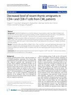
patients with CML are shown in Figure 1. In comparison
with the sjTRECs in healthy individuals (3.76 ± 3.42 cop-
ies/1000 PBMCs, 5.87 ± 4.96 copies/1000 CD3+ cells,
5.62 ± 6.45 copies/1000 CD4+ T cells, 6.79 ± 7.1 copies/
1000 CD8+T cells), a dramatic reduction of sjTRECs val-
ues was found in patients with CML (0.23 ± 0.38 copies/
C43 F 35 102.85 1 335 41.7 No
C44 F 25 33.44 6 470 35.0 No
C45 M 81 30.3 0 747 9.02 No
C46 M 38 154 1 485 13.75 No
C47 M 30 2.32 0 46 41.9 No
C48 M 25 7.63 11 139 49.3 No
Table 1: Clinical data of CML patients (Continued)
Figure 1 Comparison of the sjTRECs levels in patients with CML
and healthy individuals (HI). A: The sjTRECs levels in PBMCs; B: The
sjTRECs levels in CD4+ and CD8+ T cells respectively.
Li et al. Journal of Translational Medicine 2010, 8:47
/>Page 5 of 8
1000 PBMCs, 1.34 ± 1.63 copies/1000 CD3+ cells, 1.49 ±
1.88/1000 CD4+ T cells, 2.52 ± 2.43 copies/1000 CD8+ T
cells) (p < 0.0001, p < 0.0001, p = 0.0115 and p = 0.0129,
respectively).
The numbers of sjTRECs in PBMCs and sorted T cells
from CML were higher in females than in males. The val-
ues were: the PBMCs group: 0.19 ± 0.25 copies/1000cells
in male (n = 33) versus 0.43 ± 0.56 copies/1000cells in
female (n = 15) (p = 0.0467), in the CD3+T cells group:
1.05 ± 1.21 copies/1000cells in male (n = 33) versus 1.97 ±
2.25 copies/1000cells in female (n = 15) (p = 0.0712), in
the CD4+T cells group: 1.4 ± 2.08 copies/1000cells in
male (n = 14) versus 1.74 ± 1.31 copies/1000cells in
female (n = 5) (p = 0.739), and in the CD8+T cells group:
1.66 ± 1.63 copies/1000cells in male (n = 14) versus 4.95 ±
2.82 copies/1000cells in female (n = 5) (p = 0.0053). Simi-
lar results were found in healthy individual group (data
not shown). Although the differences between genders
were quite obvious, they were not statistically significant,
except for PBMCs and CD8+ cells in CML patients.
Lower frequencies of 23 TRBV-BD1 sjTRECs in PBMCs, CD4+
and CD8+ cells from CML patients
The TRBV-BD1 sjTRECs from TRBV1-19 and TRBV21-
24 were analyzed by semi-nested PCR, using different
amounts of DNA (corresponding to 2 × 10
5
, 5 × 10
4
or 1 ×
10
4
cells respectively). Samples were amplified to estimate
the frequency of TCR TRBV-BD1 sjTRECs and the
sequences of the junction regions of each TRBV-BD1
sjTRECs were confirmed by PCR products direct
sequencing (data not shown).
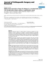
The number of detectable TRBV subfamily sjTRECs
differed significantly between CML and healthy control
in 2 × 10
5
, 5 × 10
4
and 1 × 10
4
PBMCs or in 1 × 10
4
of
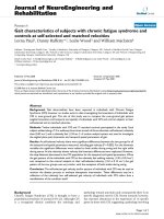
CD4+ and CD8+ T cells (Figure 2). Comparison of the
frequencies of 23 TRBV-BD1 sjTRECs in PBMCs
between CML patients and normal controls at different
amounts of DNA level showed that the frequencies of the
most TRBV subfamily sjTRECs were significantly lower
than those from healthy individuals, especially at the
higher cellular concentration (2 × 10
5
PBMCs) (Figure 3).
But the significant difference was found only in few sub-
families (BV2, BV10, BV12 and BV14 in CD8+T cells)
when comparing the frequency of TRBV subfamily
Figure 3 Comparison the frequencies of 23 TRBV-BD1 sjTRECs in PBMCs between CML patients and healthy controls (HI) at different
amounts of DNA level (n = 10). Note: *: compare to normal control p < 0.05, **: compare to normal control p < 0.01.
Figure 2 The number of detectable subfamilies of TRBV-BD1
sjTRECs in from CML patients and healthy controls. A: The subfam-
ily numbers of TRBV-BD1 sjTRECs in PBMCs; B: The subfamily numbers
of TRBV-BD1 sjTRECs in CD4+ and CD8+ T cells (1 × 10
4
cells) respective-
ly.
Li et al. Journal of Translational Medicine 2010, 8:47
/>Page 6 of 8
sjTRECs in CD4+ and CD8+ T cells at 1 × 10
4
concentra-
tion between both group (Figure 4).
Discussion
In patients with CML, cellular immune deficiency is a
common feature which may be due to decreased output
of recent thymic emigrants, the abnormal expression of T
cell receptor repertoire and, may in part, due to altered
expression of TCR-CD3 complex. Our previous study
showed decreased δRec-ψJα sjTRECs level and skewed
TRBV repertoire in peripheral blood mononuclear cells
(PBMCs) from CML patients [19,21]. And TCR ζ chain
expression was decreased in T cells from patients with
CML [22,23].
In order to further evaluate the T-cell immune func-
tion, the T cell proliferative history in CML patients was
analyzed. The sjTRECs-content in PBMCs and CD3+ T
cells from 48 CML cases was determined. The results
confirmed our previous smaller study [19]. We showed a
dramatic reduction of sjTRECs values in CML patients.
In some cases no sjTRECs could be detected in 40 000 T
cells. This suggests poor thymic output in CML patients,
which may be even more pronounced than in ALL
patients [16]. To date there are only a few papers describ-
ing TRECs level in hematopoietic malignancies [16,17].
The exact value of sjTRECs level in PBMCs from CML
patients are influenced by contaminating normal non-T
cells and leukemia blast cells; therefore the sjTRECs
numbers were normalized with the percentage of
CD3+cells in the analyzed samples. Furthermore, we ana-
lyzed sjTRECs in sorted CD4+ and CD8+ T cells. This is
the most sensitive and accurate method for quantitation
of naïve T-cells. It allows also the comparison of sjTRECs
levels in CD4+ and CD8+ subsets. The levels of sjTRECs-
expressing CD4+ and CD8+ T cells were significantly
decreased in CML patients, as compared with age and sex
Figure 4 Comparison the frequencies of 23 TRBV-BD1 sjTRECs in CD4+ (A) and CD8+T cells (B) between CML patients and healthy controls
(HI) (n = 10). Note: *: compare to normal control p < 0.05, **: compare to normal control p < 0.01.
Li et al. Journal of Translational Medicine 2010, 8:47
/>Page 7 of 8
matched healthy individuals. The decrease of sjTRECs
levels was similar in both T cell subsets. These findings
suggest that an impaired thymic output function and, as a
consequence, an altered ability to maintain T cell homeo-
stasis, which may play an important role in the immuno-
deficiency in CML patients. However, whether this is due
to the clonal expansion of T-cells to antigens, for example
leukemia associated antigens, or reflects the impairment
of immune function associated with the malignancy,
remains an open question [7,24-27].
Pido-Lopez et al showed that the decline in number of
recent thymic emigrants in the blood with increasing age
is gender-linked [28]. Peripheral blood from female con-
tained significantly higher levels of sjTRECs per CD3+ T
cell than blood from males. Also in children, the number
of sjTRECs was higher in healthy girls than in healthy
boys, and a similar pattern was evident in T-cell malig-
nancies [16]. In the present study, we observed slightly,
but in-significantly higher sjTRECs levels in healthy
females, however, the number of sjTRECs was statisti-
cally higher in PBMCs and CD8+ T cells from female
CML patients.
The majority of studies published previously focused
only on the total thymic output, as measured by quantita-
tive analysis of δRec-ψJα sjTRECs [6]. This approach
doesn't allow the evaluation of the complexity of thymic
output in different TRBV subfamily naïve T cells, which is
an important factor in immune competence. In this study,
we analyzed the total 23 subfamilies of TRBV-DB1
sjTRECs in PBMCs, CD4+ and CD8+ T-cells from CML
patients by a semi-nested PCR. The results indicate that
the percentage of cases positive for TRBV-DB1 sjTRECs
varies in different BV subfamilies in healthy controls; the
highest for TRBV1, 3, 4, 10, 12-14, 17 and V21, which
could be detected in all 10 samples (at 2 × 10
5
PBMCs).
The most important observation in this study was the sig-
nificantly lower frequency of 23 TRBV-BD1 sjTRECs in
PBMCs, as well as in CD4+ and CD8+ T cells from CML
patients as compared with healthy individuals, indicating
poor thymic output in CML patients. The results further
support and explain the significant reduction of recent
thymic emigrant numbers in peripheral blood of CML
patients, as measured by quantitative detection of δRec-
ψJα sjTRECs.
In conclusion, this is, to our knowledge, the first char-
acterization of thymic output function in CD4+ and
CD8+ T cells from CML patients based on analyses of
both δRec-ψJα sjTRECs and TRBV-DB1 subfamily spe-
cific sjTRECs. We showed a prominent decrease of
sjTRECs levels in CML, indicating the reduction of recent
thymic emigrants affects the majority of TRBV subfami-
lies.
Competing interests
The authors declare that they have no competing interests.
Authors' contributions
YQL, CAS and GKP were responsible for study design and data management.
SXG and SHC performed the real-time PCR, QSY and LJY performed the semi-
nested PCR, XLW and BL performed the statistical analysis, XD collected sam-
ples. All authors read and approved the final manuscript.
Acknowledgements
The authors thank Prof. Dr. John Yeuk-Hon Chan for critical reading of this man-
uscript. The study was sponsored by grants from National Natural Science
Foundation of China (No. 30270579) and Natural Science Foundation of
Guangdong Province (No.23001, 9251063201000001).
Author Details
1
Institute of Hematology, Medical College, Jinan University, Guangzhou,
510632, China,
2
Key Laboratory for Regenerative Medicine of Ministry of
Education, Jinan University, Guangzhou, 510632, China,
3
Department of
Hematology, Guangdong Province People's Hospital, Guangzhou 510080,
China,
4
Department of Hematology and Oncology, Ernst-Moritz-Arndt
University Greifswald, Greifswald 17487, Germany and
5
Institute of Human
Genetics, Polish Academy of Sciences, Poznan, Poland
References
1. Jabbour E, Cortes J, Kantarjian H: Treatment selection after imatinib
resistance in chronic myeloid leukemia. Target Oncol 2009, 4:3-10.
2. Hadden JW: Immunodeficiency and cancer: prospects for correction.
Int Immunopharmacol 2003, 3:1061-71.
3. Costello RT, Rey J, Fauriat C, Gastaut JA, Olive D: New approaches in the
immunotherapy of haematological malignancies. Eur J Haematol 2003,
70:333-45.
4. Pannetier C, Even J, Kourilsky P: T-cell repertoire diversity and clonal
expansions in normal and clinical samples. Immunol Today 1995,
16:176-81.
5. De Villartay JP, Hockett RD, Coran D, Korsmeyer SJ, Cohen DI: Deletion of
the human T-cell receptor δ-gene by a site specific recombination.
Nature 1988, 335:170-4.
6. Douek DC, McFarland RD, Keiser PH, Gage EA, Massey JM, Haynes BF, Polis
MA, Haase AT, Feinberg MB, Sullivan JL, Jamieson BD, Zack JA, Picker LJ,
Koup RA: Changes in thymic function with age and during the
treatment of HIV infection. Nature 1998, 396:690-5.
7. Hazenberg MD, Verschuren MC, Hamann D, Miedema F, van Dongen JJ: T
cell receptor excision circles as markers for recent thymic emigrants:
basic aspects, technical approach, and guidelines for interpretation. J
Mol Med 2001, 79:631-40.
8. Geenen V, Poulin JF, Dion ML, Martens H, Castermans E, Hansenne I,
Moutschen M, Sekaly RP, Cheynier R: Quantification of T cell receptor
rearrangement excision circles to estimate thymic function: an
important new tool for endocrine-immune physiology. J Endocrinol
2003, 176:305-11.
9. Al-Harthi L, Marchett G, Steffens CM, Poulin J, Sekaly R, Landay A:
Detection of T cell receptor circles (TRECs) as biomarkers for de novo T
cell synthesis using a quantitative polymerase chain reaction-enzyme
linked immunosorbent assay (PCR-ELISA). J Immunol Methods 2000,
237:187-97.
10. Douek D, Vescio RA, Betts MR, Brenchley JM, Hill BJ, Zhang L, Berenson JR,
Collins RH, Koup RA: Assessment of thymic output in adults after
haematopoietic stem-cell transplantation and prediction of T-cell
reconstitution. Lancet 2000, 355:1875-81.
11. Svaldi M, Lanthaler AJ, Dugas M, Lohse P, Pescosta N, Straka C, Mitterer M:
T-cell receptor excision circles: a novel prognostic parameter for the
outcome of transplantation in multiple myeloma patients. Br J
Haematol 2003, 122:795-801.
12. Poulin JF, Sylvestre M, Champagne P, Dion ML, Kettaf N, Dumont A,
Lainesse M, Fontaine P, Roy DC, Perreault C, Sékaly RP, Cheynier R:
Evidence for adequate thymic function but impaired naive T-cell
survival following allogeneic hematopoietic stem cell transplantation
Received: 28 November 2009 Accepted: 14 May 2010
Published: 14 May 2010
This article is available from: 2010 Li et al; licensee BioMed Central Ltd. This is an Open Access article distributed under the terms of the Creative Commons Attribution License ( which permits unrestricted use, distribution, and reproduction in any medium, provided the original work is properly cited.Journal of Tr anslational Medi cine 2010, 8:47
Li et al. Journal of Translational Medicine 2010, 8:47
/>Page 8 of 8
in the absence of chronic graft-versus-host disease. Blood 2003,
102:4600-7.
13. Przybylski GK, Kreuzer KA, Siegert W, Schmidt CA: No recovery of T-cell
receptor excision circles (TRECs) after non-myeloablative allogeneic
hematopoietic stem cell transplantation is correlated with the onset of
GvHD. J Appl Genet 2007, 48:397-404.
14. Poulin JF, Viswanathan MN, Harris JM, Komanduri KV, Wieder E, Ringuette
N, Jenkins M, McCune JM, Sékaly RP: Direct evidence for thymic function
in adult humans. J Exp Med 1999, 190:479-86.
15. Li Y, Chen S, Yang L, Yin Q, Geng S, Wu X, Schmidt CA, Przybylski GK: TRAV
and TRBV repertoire, clonality and the proliferative history of umbilical
cord blood T cells. Transplant Immunol 2007, 18:151-8.
16. Petridou E, Klimentopoulou AE, Moustaki M, Kostrikis LG, Hatzakis A,
Trichopoulos D: Recent thymic emigrants and prognosis in T- and B-cell
childhood hematopoietic malignancies. Int J Cancer 2002, 101:74-7.
17. Haining WN, Evans J, Seth N, Callaway G, Wucherpfennig K, Nadler L,
Guinan E: Rapid T Cell Response to Vaccination Can Occur without
Antibody Response in Children Post HSCT. Blood 2004, 104(11):614a.
18. Li Y, Yin Q, Yang L, Chen S, Geng S, Wu X, Zhong L, Schmidt CA, Przybylski
GK: Reduced levels of recent thymic emigrants in acute myeloid
leukemia patients. Cancer Immunol Immunother 2009, 58:1047-55.
19. Geng SX, Li YQ, Chen SH, Yang LJ, Yin QS, Wu XL, Zhang XL: Peripheral
blood naive T cell level and its T cell receptor Vbeta repertoire usage
profile in patients with chronic myelogenous leukemia. Zhonghua Xue
Ye Xue Za Zhi 2005, 26:413-6. (Chinese)
20. Kimmig S, Przybylski GK, Schmidt CA, Laurisch K, Mowes B, Radbruch A,
Thiel A: Two subsets of naive T helper cells with distinct T cell receptor
excision circle content in human adult peripheral blood. J Exp Med
2002, 195:789-94.
21. Wang L, Zhu K, Zha X, Chen S, Yang L, Chen S, Li Y: Evolution of T-cell
clonality in a patient with Ph-negative acute lymphocytic leukemia
occurring after interferon and imatinib therapy for Ph-positive chronic
myeloid leukemia. J Hematol Oncol 2010, 3:14.
22. Rossi E, Matutes E, Morilla R, et al.: Zeta chain and CD28 are poorly
expressed on T lymphocytes from chronic lymphocytic leukaemia.
Leukemia 1996, 10:494-7.
23. Chen S, Yang L, Chen S, Li Y: TCR ζ chain expression in T cells from
patients with CML. Hematology 2009, 14:95-100.
24. Farace F, Orlanducci F, Dietrich PY, Gaudin C, Angevin E, Courtier MH,
Bayle C, Hercend T, Triebel F: T cell repertoire in patients with B chronic
lymphocytic leukemia. Evidence for multiple in vivo T cell clonal
expansions. J Immunol 1994, 153:4281-90.
25. Serrano D, Monteiro J, Allen SL, Kolitz J, Schulman P, Lichtman SM,
Buchbinder A, Vinciguerra VP, Chiorazzi N, Gregersen PK: Clonal
expansion within the CD4+CD57+ and CD8+CD57+ T cell subsets in
chronic lymphocytic leukemia. J Immuno 1997, 158:1482-9.
26. Alatrakchi N, Farace F, Frau E, Carde P, Munck JN, Triebel F: T-cell clonal
expansion in patients with B-cell lymphoproliferative disorders. J
Immunother 1998, 21:363-70.
27. Li Y: Recent thymic output function in patients with hematological
malignancy. Hematology 2005, 10:297-305.
28. Pido-Lopez J, Imami N, Aspinall R: Both age and gender affect thymic
output: more recent thymic migrants in females than males as they
age. Clin Exp Immunol 2001, 125:409-13.
doi: 10.1186/1479-5876-8-47
Cite this article as: Li et al., Decreased level of recent thymic emigrants in
CD4+ and CD8+T cells from CML patients Journal of Translational Medicine
2010, 8:47