báo cáo hóa học:" Tadpole system as new lumbar spinal instrumentation" pot
Bạn đang xem bản rút gọn của tài liệu. Xem và tải ngay bản đầy đủ của tài liệu tại đây (1.01 MB, 6 trang )
BioMed Central
Page 1 of 6
(page number not for citation purposes)
Journal of Orthopaedic Surgery and
Research
Open Access
Technical Note
Tadpole system as new lumbar spinal instrumentation
Yuichi Kasai*
1
, Tadashi Inaba
2
, Koji Akeda
1
and Atsumasa Uchida
1
Address:
1
Department of Orthopaedic Surgery, Mie University Graduate School of Medicine, 2-174 Edobashi, Tsu city, Mie prefecture, 514-8507,
Japan and
2
Department of Mechanical Engineering, Mie University, Tsu city, Mie prefecture, Japan
Email: Yuichi Kasai* - ; Tadashi Inaba - ; Koji Akeda - ;
Atsumasa Uchida -
* Corresponding author
Abstract
Background: There have been reports of serious complications associated with pedicle screw
fixation, including nerve root injuries caused by accidental screw insertion. We have developed a
new system of lumbar spinal instrumentation that we call Tadpole system
®
. The purposes of this
report were to show the results of a biomechanical study and the short-term outcome of a clinical
study, as well as to determine the usefulness of this system.
Methods: The Tadpole system
®
lumbar spinal fusion is a hook-and-rod system according to which
the spine is stabilized using 2 sets of 2 spinous processes each that are held in place by 4 hooks
tandemly connected to a rod. The biomechanical study was done using 5 human lumbar cadaveric
spines, and the range of motion (ROM) was examined in a non-treatment model, an injured model,
a pedicle screw fixation model and a Tadpole system
®
model. For the short-term clinical study the
Tadpole system
®
was used in 31 patients, and the factors analyzed were operation time, time
required for spinal instrumentation, amount of intraoperative bleeding, postoperative
improvement rate of the Japanese Orthopaedic Association (JOA) score for lumbar spinal
disorders, instrumentation failure, spinous process fracture, spinal fluid leakage, nerve root injury,
postoperative infection, and bone fusion 2 years after the operation.
Results: The ROM in the Tadpole system
®
model was slightly bigger than that in the pedicle screw
fixation model, but smaller than that in the normal control model. These biomechanical data
indicated that the Tadpole system
®
provided fairly good stability. The mean operation time was 79
min, the mean time required for spinal instrumentation was 8 min, and the mean amount of
intraoperative bleeding was 340 mL. The mean postoperative improvement rate of JOA score was
70.9 ± 24.8%. Instrumentation failure (dislocation of a hook) occurred in one patient, and none of
the patients developed spinous process fracture, spinal fluid leakage, nerve root injury, or
postoperative infection. Two years after the operation, bone union was confirmed in 29 of the 31
patients (93.5%).
Conclusion: We conclude that this system is a useful, easy-to-use and safe spinal instrumentation
technique for lumbar fusion surgery.
Published: 12 September 2008
Journal of Orthopaedic Surgery and Research 2008, 3:41 doi:10.1186/1749-799X-3-41
Received: 19 June 2008
Accepted: 12 September 2008
This article is available from: />© 2008 Kasai et al; licensee BioMed Central Ltd.
This is an Open Access article distributed under the terms of the Creative Commons Attribution License ( />),
which permits unrestricted use, distribution, and reproduction in any medium, provided the original work is properly cited.
Journal of Orthopaedic Surgery and Research 2008, 3:41 />Page 2 of 6
(page number not for citation purposes)
Background
Lumbar spinal instrumentation has been widely used for
pedicle screw fixation (PSF), with generally favorable clin-
ical outcomes. However, there have been reports of seri-
ous complications associated with this method, including
nerve root injuries caused by accidental screw insertion
[1,2]. We have developed a new system of lumbar spinal
instrumentation, which we call Tadpole system
®
(Kisco
DIR Co., Ltd., Osaka, Japan), that uses the spinous proc-
esses as anchors.
In the present report, we describe the Tadpole system
®
, the
results of a biomechanical study and the short-term out-
come of a clinical study.
Tadpole system
Overview
The Tadpole system
®
is used in spinal fusion to treat lum-
bar spinal canal stenosis and lumbar degenerative spond-
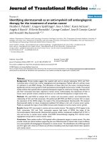
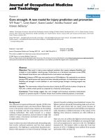
ylolisthesis. It is a hook-and-rod system of spinal
instrumentation in which the spine is stabilized using 2
sets of 2 spinous processes each that are held in place by 4
hooks tandemly connected to a rod (Figure 1). The hooks
are 7 to 15 mm in length, and the rods are 4 to 12 cm in
length. The hook is connected to the rod by tightening a
nut that is attached to the hook. Because the hook resem-
bles a tadpole, we named this system "Tadpole".
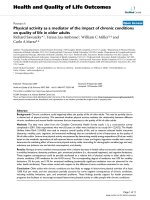
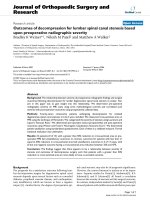
With the Tadpole system
®
, implants can be positioned
using a unilateral approach (Figure 2), and a sufficient
base for posterolateral fusion can be retained due to its
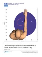
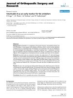
placement toward the median line. This system is com-
monly used for two-level fusion (Figure 3) between L3
and L5 or between L2 and L4, or for single-level fusion
(Figure 4) between L3 and L4 or L4 and L5. However, this
system is not applicable for fusions that include the sacral
spine, because the spinous process of S1 is too small to be
fixed with the hook. It is neither applicable in patients
with spondylolysis or severe spinal instability, nor for cor-
rection of spinal alignment.
Operative techniques when using the Tadpole system
®
Surgery is performed with the patient in the prone posi-
tion and under general anesthesia. The paravertebral mus-
cles are disclosed after a posterior median incision of the
lumbar spinal region. Laminectomy or spinal decompres-
sion by fenestration is then performed. If necessary, trans-
foraminal lumbar interbody fusion (TLIF) is performed
with interbody cages before using the Tadpole system
®
. It
Tadpole system
®
; hook-and-rod system of spinal instrumen-tationFigure 1
Tadpole system
®
; hook-and-rod system of spinal instrumen-
tation.
Tadpole system
®
positioned using a unilateral approachFigure 2
Tadpole system
®
positioned using a unilateral approach.
X-rays of a case of two-level fusionFigure 3
X-rays of a case of two-level fusion
Journal of Orthopaedic Surgery and Research 2008, 3:41 />Page 3 of 6
(page number not for citation purposes)
is important to minimize resection of the spinous proc-
esses, because excessive resection may result in their frac-
ture or increased instability of the hook. In particular, an
excessive cut of the spinous process of L5 should be
avoided because this is frequently smaller than that of L3
and L4.
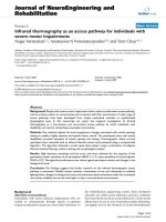
After spinal decompression, guide holes for hooks are
made on the interspinous ligament using a starter awl
(Figure 5A; in this case holes were made between L3 and
L4, and between L5 and S1). The size of the hook is then
determined using a frontal-view preoperative plain lum-
bar radiograph. The cranial and caudal hooks are held
with a hook holder and inserted into the interspinous lig-
ament (Figure 5B), and then the stability of the hooks in
the interspinous ligament should be confirmed.
Next, 2 hooks are connected in the center to a rod, and the
rod is connected to the previously inserted cranial and
caudal hooks while holding the 2 hooks with a hook
holder (Figure 5C). The hooks are tightened temporarily
while compression is maintained between the cranial and
caudal hooks (Figure 5D). It is important not to apply
excessive compression to avoid a spinous process fracture.
Thereafter, the 2 hooks for the stabilization of each
spinous process are tightened temporarily, pinching each
spinous process (Figure 5E). Finally, the screws of all
hooks are tightened using a screwdriver (Figure 5F).
In patients who do not require TLIF treatment, the supe-
rior articular process of the portion to be fixed, the lateral
portion of the intervertebral joint, and the base of the
transverse process should be completely decorticated, and
posterolateral fusion should be implemented using a
removed piece of the ilium or a local bone.
Biomechanical study using the Tadpole system
®
Method
The specimens used were 5 human lumbar cadaveric
spines consisting of L2 to L5 removed from donor bodies,
which were autopsied in our university hospital; the spec-
imens were used with the consent of the donors' family
members. Both ends of each lumbar spine were mounted
with dental resin and set on a moment-loading instru-
ment. Four experimental models were made step by step
from each lumbar spine. The experimental models
included the non-treatment model (normal control
model), an injured model (with resection of the lateral
facet joint between L3 and L4), a PSF model generated
from the injured model with pedicle screw fixation
(Absorbing Shock Device
®
, KiscoMedica, Saint Priest,
France), and a Tadpole system
®
(TS) model created from
the injured model. A marker was placed on the vertebral
bodies of L3 and L4, and a moment load of 5 N.m was
applied to each experimental model for forward flexion,
backward flexion, left side bending, right side bending,
left rotation, and right rotation. The movement of the
markers on the vertebral bodies of L3 and L4 after appli-
cation of a moment load and with no load was photo-
graphed with a digital camera. The ROM for backward and
forward flexion, right and left side bending, and right and
left rotation between L3 and L4 was determined using a
Microsoft Office Visio
®
image processing software.
Results
The mean ROM of the five cadaveric spines is shown in
Table 1. The ROM for all backward and forward flexions
and right and left side bending was smaller in the TS
model and PSF model than in the injured model. The
ROM in the TS model was slightly bigger than that in the
PSF model, but smaller than that in the normal control
model. These data indicated that the Tadpole system
®
pro-
vided fairly good stability.
Short-term clinical study using the Tadpole system
®
Subjects and methods
The subjects were 31 patients (19 men and 12 women)
who underwent spinal fusion using the Tadpole system
®
and were followed up ≥ 2 years in our clinic. Twenty-seven
patients had lumbar spinal canal stenosis and 4 had
spondylolisthesis. Intervertebral fusion of L2–L4, L3–L5,
L3–L4 and L4–L5 was performed in 4, 22, 1 and 4
patients, respectively; 8 of the 31 patients underwent sur-
gery via a unilateral approach, and all patients underwent
posterolateral fusion. The mean age at the time of the
operation was 73.3 years (range: 59 to 84 years). The
mean follow-up period was 2 years 3 months (range: 2
years to 2 years 9 months).
The factors analyzed in this study were operation time,
time required for spinal instrumentation, amount of
X-rays of a case of single-level fusion Figure 4
X-rays of a case of single-level fusion
Journal of Orthopaedic Surgery and Research 2008, 3:41 />Page 4 of 6
(page number not for citation purposes)
intraoperative bleeding, Japanese Orthopaedic Associa-
tion (JOA) score for lumbar spinal disorders on a 29-point
scale before and 2 years after the operation, postoperative
improvement rate of JOA score (%, Hirabayashi method),
instrumentation failure, spinous process fracture, spinal
fluid leakage, nerve root injury, postoperative infection,
and bone fusion 2 years after the operation. Evaluation of
lumbar spinal fusion is partially subjective, therefore, the
X-ray images were assessed by two independent observers
and it was determined that fusion had been achieved
when both observers confirmed there was no pseudoar-
throsis. Pseudoarthrosis was diagnosed when any gap in
the fusion mass on antero-posterior or oblique radio-
graphs was seen, or if there were more than two degrees of
Operative techniques for Tadpole system® Figure 5
Operative techniques for Tadpole system® a; Guide holes for hooks are made on the interspinous ligament using a
starter awl
b; The hook is held with a hook holder, and it is inserted into the interspinous ligament
c; The 2 hooks are connected in the center to a rod, and the rod is connected to the cranial and caudal hooks.
d; The hooks are tightened temporarily while compression is maintained between the cranial and caudal hooks.
e; The set of 2 hooks for the stabilization of a spinous process is tightened temporarily, pinching each spinous process
f; The screws of all hooks are tightened using a screwdriver
Table 1: Mean range of motion of each model on the biomechanical study
(mean± SD)
ROM of forward – backward flextion ROM of right – left side bending ROM of right – left side rotaton
Normal control model 7.2° ± 4.8° 7.8° ± 3.8° 13.8° ± 7.1°
Injured model 18.6° ± 6.2° 22.1° ± 8.8° 24.8° ± 10.4°
PSF model 4.3° ± 2.9° 2.5° ± 1.3° 3.4° ± 2.6°
TS model 3.4° ± 2.2° 5.1° ± 2.5° 7.9° ± 5.2°
Normal control model; non-treatment model
Injured model; model resected the facet joint between L3 and L4
PSF model; model generated from the injured model with pedicle screw fixation
TS model; model created from the injured model with the Tadpole system
®
Journal of Orthopaedic Surgery and Research 2008, 3:41 />Page 5 of 6
(page number not for citation purposes)
motion on flexion-extension films. The rate of consistency
of fusion status grading achieved by the two observers was
100%, and interjudge reliability was very good.
Results
The mean operation time was 79 ± 41 min (± S.D.), the
mean time required for spinal instrumentation was 8 ± 3
min, and the mean amount of intraoperative bleeding was
340 ± 278 mL. The mean JOA score was 14.2 ± 7.1 points
before the operation, and 24.7 ± 3.8 points after the oper-
ation; the mean postoperative improvement rate was 70.9
± 24.8%. Dislocation of a hook occurred in one patient,
however, this patient showed solid spinal fusion one year
after the surgery. None of the patients developed spinous
process fracture, spinal fluid leakage, nerve root injury, or
postoperative infection. Two years after the operation,
bone union was confirmed in 29 of the 31 patients
(93.5%).
Discussion
Although clinical studies have generally shown favorable
outcomes for lumbar fusion surgery using pedicle screw
fixation, there have been reports of complications includ-
ing spinal fluid leakage (4%), nerve injury (2%), deep
infection (4–5%), and instrumentation failure (3 – 12%)
[1,2]. Another disadvantage of this technique is radiation
exposure of operators and patients, because X-ray images
are taken during the surgery.
Among several methods of posterior spinal instrumenta-
tion, the use of the spinous processes as anchors provides
less biomechanical strength than pedicle screw fixation
[3], but produces no complications such as spinal fluid
leakage or nerve injury, and requires no excessive excision
of the paraspinal muscles [4]. In addition, the relatively
low invasiveness of spinal instrumentation using the
spinous processes is a key advantage, because such
method is associated with shortened operation time,
reduced bleeding, and reduced length of subsequent hos-
pital stay. Thus, the Tadpole system
®
we have developed,
which is an easy-to-perform fixation technique that is less
invasive than other techniques, may become widely
accepted and can be expected to result in good cost-effec-
tiveness.
Spinal instrumentation using the spinous processes as
anchors has been performed for approximately 50 years
[5,6]. In case studies, the Daab plate and the Wilson plate
have been used, and large-scale studies have not been con-
ducted as yet. The pull-out strength of the spinous process
wiring technique developed by Drummond et al. [7] was
reported to be 30% to 45%, compared with sublaminar
wiring. This suggests that sublaminar wiring is preferable
to spinous process wiring [3]. Coe et al. [8] reported that
the fracture load of the spinous process is one-fifth to one-
half of that of the vertebral arch. Because the biomechan-
ical strength of the spinous process is not high, there is
limited flexibility in spinal instrumentation using the
spinous process as an anchor, but recent studies of lumbar
fusion surgery using the Lumbar Alligator Spinal system
®
[9] and CD Horizon Spire spinous process plate
®
[4,10]
have shown relatively favorable clinical outcomes. In a
biomechanical study carried out by Shepherd et al. [11],
holding the spinous process with a hook provided suffi-
cient holding ability. Thus, the available evidence indi-
cates that the Tadpole system
®
has advantages over other
systems.
The mean intervertebral fusion rate in the present study
was as high as the 96% attained with lumbar fusion sur-
gery using pedicle screw fixation [12-14]. The Lumbar
Alligator Spinal system
®
with spinal instrumentation
using the spinous process as an anchor was associated
with an intervertebral fusion rate of 92.7% (104 of 107
patients) [9], and the intervertebral fusion rate of our Tad-
pole system
®
was 93.5%, which is quite good.
In future studies, we plan to: 1) use data from long-term
follow-up clinical studies; 2) increase the number of study
patients; 3) use clinical data for a unilateral approach; 4)
examine postoperative changes in vertebral bodies adja-
cent to fixed vertebrae; and 5) attempt to establish the cri-
teria for deciding between pedicle screw fixation, the
Tadpole system
®
, and no spinal instrumentation.
Conclusion
The clinical outcomes of the Tadpole system
®
were gener-
ally favorable. Therefore, we conclude that this system is a
useful, easy-to-use and safe spinal instrumentation tech-
nique for lumbar fusion surgery.
Competing interests
Yuichi Kasai, the inventor of Tadpole system, receives roy-
alities from Kisco DIR Co., Ltd. resulting from its sale.
Authors' contributions
YK and TI have made substantial contributions to concep-
tion and design, or acquisition of data, or analysis and
interpretation of data. YK and KA have been involved in
drafting the manuscript or revising it critically for impor-
tant intellectual content. AU has given final approval of
the version to be published.
References
1. Esses SI, Sachs BL, Dreyzin V: Complication associated with the
technique of pedicle screw fixation. A selected survey of ABS
members. Spine 1993, 18:2231-2238.
2. Jutte PC, Castelein RM: Complications of pedicle screws in lum-
bar and lumbosacral fusion in 105 consecutive primary oper-
ations. Eur Spine J 2002, 11:594-598.
3. Heller KD, Prescher A, Schneider T, Block FR, Frost R: Stability of
different wiring techniques in segmental spinal instrumenta-
Publish with BioMed Central and every
scientist can read your work free of charge
"BioMed Central will be the most significant development for
disseminating the results of biomedical research in our lifetime."
Sir Paul Nurse, Cancer Research UK
Your research papers will be:
available free of charge to the entire biomedical community
peer reviewed and published immediately upon acceptance
cited in PubMed and archived on PubMed Central
yours — you keep the copyright
Submit your manuscript here:
/>BioMedcentral
Journal of Orthopaedic Surgery and Research 2008, 3:41 />Page 6 of 6
(page number not for citation purposes)
tion; An experimental study. Arch Orthop Trauma Surg 1998,
117:96-99.
4. Wang JC, Haid RW, Miller JS, Robinson JC: Comparison of CD
HORIZON SPIRE spinous process plate stabilization and
pedicle screw fixation after anterior lumbar interbody
fusion. J Neurosurg Spine 2006, 4:132-136.
5. Bostman O, Myllynen P, Riska EB: Posterior spinal fussion using
internal fixation with the Daab plate. Acta OrthopScand 1984,
55:310-314.
6. Wilson PD, Straub LR: Lumbosacral fusion with metallic plate
fixation. Instr Course Lect 1952, 9:52-57.
7. Drummond DS, Keene JS: Spinous process segmental spinal
instrumentation. Orthopedics 1988, 11:1403-1410.
8. Coe JD, Warden KE, Herzig MA, McAfee PC: Influence of bone
mineral density on the fixation of thoracolumbar implants: A
comparative study of transpedicular screws, laminar hooks,
and spinous process wires. Spine 1990, 15:902-907.
9. Fuji T, Hosono N, Kato Y: The Lumbar Alligator Spinal System
– A simple and less invasive device for posterior lumbar fix-
ation. In Spinal Reconstruction. Clinical examples of applied basic science,
biomechanics and engineering Edited by: Kai-Uwe Lewandrowski. New
York London; Informa; 2007:81-90.
10. Wang JC, Spencer D, Robinson JC: SPIRE spinous process plate:
biomechanical evaluation of a novel technology. J Neurosurg
Spine 2006, 4:160-164.
11. Shepend DET, Leahy JC, Mathias KJ, Wilkinson SJ, Hukins DW:
Spinous process strength. Spine 2000, 25:319-323.
12. Fischgrund JS, Markay M, Herkowitz HN, Brower R, Montgomery
DM, Kurz LT: Degenerative lumbar spondylolisthesis with spi-
nal stenosis; a prospective, randomized study compareing
decompressive laminectomy and arthrodesis with and with-
out spinal instrumentation. Spine 1997, 22:2807-2812.
13. Thomson K, Christensen FBm, Eiskjaer SP, Hansen ES, Fruensgaard S,
Bunger CE: The effect of pedicle screw instrumentation on
functional outcome and fusion rates in posterolateral fusion;
a prospective randomized clinical study.
Spine 1997,
22:2813-2822.
14. Zdeblick TA: A prospective, randomized study of lumbar
fusion; Preliminary results. Spine 1993, 18:983-991.