Báo cáo hóa học: " Does Japanese encephalitis virus share the same cellular receptor with other mosquito-borne flaviviruses on the C6/36 mosquito cells?" pot
Bạn đang xem bản rút gọn của tài liệu. Xem và tải ngay bản đầy đủ của tài liệu tại đây (335.59 KB, 7 trang )
BioMed Central
Page 1 of 7
(page number not for citation purposes)
Virology Journal
Open Access
Hypothesis
Does Japanese encephalitis virus share the same cellular receptor
with other mosquito-borne flaviviruses on the C6/36 mosquito cells?
Junping Ren
†
, Tianbing Ding
†
, Wei Zhang, Jianhua Song and Wenyu Ma*
Address: Department of Microbiology, Fourth Military Medical University, 17 Changle West Road, Xi'an, 710032, People's Republic of China
Email: Junping Ren - ; Tianbing Ding - ; Wei Zhang - ;
Jianhua Song - ; Wenyu Ma* -
* Corresponding author †Equal contributors
Abstract
Japanese encephalitis virus (JEV) is a member of mosquito-borne Flaviviridae. To date, the
mechanisms of the early events of JEV infection remain poorly understood, and the cellular
receptors are unidentified. There are evidences that the structure of the virus attachment proteins
(VAP), envelope glycoprotein of mosquito-borne flaviviruses is very similar, and the vector-virus
interaction of mosquito-borne flaviviruses is also very similar. Based on the studies previously
demonstrated that the similar molecules present on the mosquito cells involved in the uptake
process of JEV, West Nile virus (WNV) and Dengue virus (DV), it is proposed that the same
receptor molecules for mosquito-borne flaviviruses (JEV, WNV and DV) may present on the
surface of C6/36 mosquito cells. By co-immunoprecipitation assay, we investigated a 74-KDa
protein on the C6/36 cells binds JEV, and the mass spectrometry results indicated it may be heat
shock cognate protein 70(HSC70) from Aedes aegypti. Based upon some other viruses use of heat
shock protein 70 (HSP70) family proteins as cell receptors, its possible HSC70's involvement in the
fusion of the JEV E protein with the C6/36 cells membrane, and known form of cation channels in
the interaction of HSC70 with the lipid bilayer, it will further be proposed that HSC70 as a
penetration receptor mediates JEV entry into C6/36 cells.
1 Background
Japanese Encephalitis Virus (JEV) is an enveloped positive
single stranded RNA virus belonging to genus Flavivirus in
the family Flaviviridae. It is the most common agent of
viral encephalitis, causing an estimated 50,000 cases
annually, of which 15,000 will die and up to 50% of sur-
vivors are left with severe residual neurological complica-
tions [1,2]. Most cases occur in southern and eastern Asia,
but the geographical area affected by JEV is expanding.
Outbreaks have been reported in Saipan islands, Torres
Straits islands and on Australia mainland in recent years
[3-5]. Cases have also occurred among travelers and US
servicemen to Asia [6,7].
The first step in virus infection requires interaction
between the virus attachment proteins (VAP) and cellular
receptors. The interaction of VAP and its cellular receptors
is known to contribute to host range, tissue tropism and
viral pathogenesis. The characteristic and function of virus
receptor, once ascertained, may ultimately lead to the pro-
duction of effective antiviral agents. But what are the cel-
lular receptors for JEV? How the cellular receptors for JEV
mediate JEV entry into the host cells?
To look for the answers to these questions, the authors
made a detailed analysis on the JEV receptors, based upon
previous flaviviruses receptor research results on Dengue
Published: 6 September 2007
Virology Journal 2007, 4:83 doi:10.1186/1743-422X-4-83
Received: 11 June 2007
Accepted: 6 September 2007
This article is available from: />© 2007 Ren et al; licensee BioMed Central Ltd.
This is an Open Access article distributed under the terms of the Creative Commons Attribution License ( />),
which permits unrestricted use, distribution, and reproduction in any medium, provided the original work is properly cited.
Virology Journal 2007, 4:83 />Page 2 of 7
(page number not for citation purposes)
virus (DV) and West Nile virus (WNV). By analyzing and
summarizing the known characteristics of flaviviruses
receptors on mosquito cells, we raised a hypothesis stating
that a 74-KDa heat shock cognate protein 70(HSC70)
may act as a penetration receptor for JEV on mosquito
cells.
2 Discussion
2.1 The same receptor molecule(s) for mosquito-borne
flaviviruses (JEV, WNV and DV) might present on the
surface of mosquito cells
The classic notion of a virus binding to a single receptor to
enter cells is being overtaken by the more complex con-
ception. One of the complex concepts is that a virus parti-
cle can use multiple (individual) receptors during cell
entry. Several viruses use at least two different receptors to
interact with their host cells: (i) the binding receptors,
which in general allow the virus particle to rapidly attach
to the cell surface, and (ii) receptors that are used by the
virus after binding to the cell, which are referred to using
different terminology, such as post-binding, post-attach-
ment, entry, fusion, internalization, secondary or co-
receptors, depending on the function that they are known
or proposed to play during the process of virus infection.
In some instances, such as in the case of human immun-
odeficiency virus-1 (HIV-1), herpes simplex virus 1 and 2,
adenovirus and measles virus [8-12], the multiple interac-
tions that take place between the virus and cell surface
molecules have been proposed to occur in a sequential
manner. A prominent example of a dual receptor require-
ment occurs with HIV-1 binding. HIV-1 uses CD4 to bind
the cell surface and chemokine so-called co-receptors,
such as CXCR4 and CCR5, to facilitate the conformational
alterations in envelope glycoproteins that culminate in
fusion of the viral envelope and cell membrane.
Another complex concept is that very different viruses
may use identical receptors. Although in most cases indi-
vidual viruses have their own distinct receptors, in some
cases the same receptor can be used by quite different
viruses. Perhaps the best studied example of this is the
coxsackie-adenovirus receptor or CAR [13]. CAR is a
member of the immunoglobulin superfamily and medi-
ates both attachment and entry of these two viruses.
Another example is the integrins, that have been identi-
fied as attachment and entry receptors for several
viruses[14], including reovirus (β1 integrins), echovirus
(α2β1), foot-and-mouth disease virus (αvβ1, αvβ3, and
αvβ6), hantaviruses NY-1 and Sin Nombre virus (β3
integrins), Kaposi sarcoma herpesvirus (α3β1), rotavirus
(α2β1, αvβ3, αxβ2, and α4β3) and cytomegalovirus
(α2β1, α6β1, and αvβ3). Integrins are a family of cell sur-
face receptors that consist of α and β subunits. Integrins
mediate cellular adhesion to the extracellular matrix
(ECM), regulate cellular trafficking, and transducer both
outside-in and inside-out signaling events.
Flaviviruses package their positive-strand RNA genome
into particles consisting of a rigid outer protein shell and
an underlying lipid membrane. The major envelope glyc-
oprotein, E, and a small membrane protein, M, form the
outer shell. As the principal envelope component, E is
responsible for receptor binding and membrane fusion.
Flavivirus E proteins belong to the structurally conserved
"class II" fusion proteins, which are also found in alphavi-
ruses. Crystal structures of four class II fusion proteins –
Tick-borne encephalitis virus (TBEV) E [15,16], Dengue
virus (DV) E [17-19], Semliki Forest virus (SFV) E1
[20,21], and West Nile virus (WNV) E [22,23] – before
and after their fusogenic conformational rearrangements
provide us with a detailed molecular picture of the fusion
mechanism of these viruses. Based on the work of the
above, it has been demonstrated that the E protein of the
mature flaviviruses forms homodimers in an anti-parallel
manner (head-to-tail orientation). Each monomer is
folded into three distinct domains (Fig 1A and 1B),
namely domain I (DI) – the central N-terminal domain;
domain II (DII) – the dimerization domain; and domain
III (DIII) – the immunoglobulin (Ig)-like domain. DIII of
E protein consists of 100 amino acids (residues 303–395)
of the C-terminal. This domain has been suggested to be
the receptor recognition and binding domain due to a
number of reasons. The Ig-like fold present in the DIII
protein is commonly associated with structures that have
an adhesion function. This domain extends perpendicu-
larly to the surface of the virus with a tip projecting further
from the virion surface than any part of the E protein. In
addition, studies have demonstrated that both recom-
binant DIII proteins and antibodies generated against DIII
of E protein of flaviviruses can inhibit entry of the flavivi-
ruses into target cells [24-26]. In addition, flaviviruses
with mutations in DIII of the E protein show either atten-
uated virulence or the ability to escape immune neutrali-
zation [27-30].
Neuroinvasiveness is a common feature of mosquito-
borne flaviviruses infections where Culex mosquitoes are
the predominant mosquito vector. In natural infection,
mosquito-borne flaviviruses are first deposited in the
mosquito vector and then in a human host bitten by the
vector during a blood meal. Therefore, it is necessary to
study receptors in mosquito cells to determine which
binding proteins serve as true virus receptors. Several stud-
ies on the mosquito-borne flaviviruses, DV [31-36], WNV
[37], have been made significant progress in identifying a
number of putative cellular receptors [glycolsaminogly-
cans, DC-SIGN, laminin receptor, glucose regulated pro-
tein 78 (GRP78), heat shock protein 90 (HSP90) and heat
shock protein 70 (HSP70), αVβ3 integrin] in different
Virology Journal 2007, 4:83 />Page 3 of 7
(page number not for citation purposes)
mammalian cell types. However, the search for cellular
receptor in mosquito cells has been less successful. A
series of protein bands of different molecular masses on
the mosquito cells (C6/36 cells) or mosquito tissues were
observed to bind to DV [38-42], WNV [43] and JEV [44].
Two glycoproteins of apparent molecular weights of 40-
and 45-kDa expressed on the surface of C6/36 cells and
mosquito tissues have been previously identified by del
Angel and colleagues [38-40] as potential Dengue virus
serotype 4 (DV-4) receptor proteins. The 45-kDa protein
has been shown to be widely expressed in mosquito tis-
sues and antibodies against this protein specifically
inhibit DV-4 infection. They demonstrated that periodate
treatment of C6/36 cells protein extract modified the
molecular weight of the 40- and 45-kDa glycoproteins to
a 38-kDa protein, which was also able to bind DV-4.
Therefore, the 40- and 45-kDa molecules are probably the
glycosylated forms of the 38-kDa molecule. Sakoonwa-
tanyoo and colleagues [41] identified a laminin-binding
protein of 50-kDa on the surface of C6/36 cells as a candi-
date DV 2, 3, and 4 receptor protein. The authors have
noticed that the marker used in their study gives higher
calculated molecular weights than other markers. As such,
they thought that the protein of 50-kDa identified by
them and the 45-kDa protein identified by del Angel et al
are probably the same protein. Another two proteins with
molecular masses of 80- and 67-kDa on the midgut cells
of Aedes aegypti and C6/36 cells have been recently identi-
fied as receptors for the four serotypes of DV [42]. Inter-
estingly, Chu et al [43] identified proteins (55-kDa, 70-
kDa, 95-kDa and 140-kDa) implicated in the binding and
internalization of WNV, of which two (70- and 95-kDa)
were proposed to be part of the receptor complex for mos-
quito-borne flaviviruses (WNV, JEV and DV) on C6/36
cells, as antibodies against these proteins blocked WNV,
JEV and DV-2 entry into C6/36 cells. More recently, Boon-
sanay et al [44] identified several proteins on the surface
of C6/36 cells binding to JEV, and predominant band was
noted at 53-kDa, while other bands in the range of 150 to
200-kDa and minor bands of approximately 35-, 51- and
80-kDa were also observed. Soluble laminin produced a
marginal, but dose-dependent inhibition of JEV infection,
suggesting laminin role in virus-receptor interaction.
Collectively, on the surface of C6/36 cells or mosquito tis-
sues the molecular masses approximately 38-, 40-, 45-,
50-kDa proteins have been identified as a candidate DV
receptor protein, and are probably the same protein, lam-
inin-binding protein. Another two proteins, molecular
masses approximately 67 to 70-kDa and 80 to 95-kDa are
probably a part of the receptor complex for mosquito-
borne flaviviruses (JEV, WNV and DV). The evidences are
listed as following: (i) the structure of VAP, envelope glyc-
oprotein of mosquito-borne flaviviruses is very similar
described as above; (ii) the vector-virus interaction of
mosquito-borne flaviviruses is very similar. Based on the
studies previously demonstrated that the similar mole-
cules present on the mosquito cells involved in the uptake
process of JEV, WNV and DV, it is proposed that the same
receptor molecules for mosquito-borne flaviviruses (JEV,
WNV and DV) may present on the surface of mosquito
cells.
2.2 Heat shock cognate protein 70 (HSC70) may be a
penetration receptor mediate JEV entry into C6/36 cells
To date, there has been little progress in identifying the
nature of the molecules involved in the initial JEV entry,
except that one report previously identified a 74-kDa pro-
tein expressed by Vero cells that may be involved in the
uptake process [45], and another report identified several
proteins on the surface of C6/36 cells binding to JEV [44].
However, the detailed characteristics and functioning of
JEV receptor still remain a mystery.
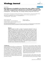
Summary of the Structural Organization and Different Conforma-tions of the Flavivirus Envelope Protein E (obtained the kind per-mission from the copyright holder to reproduce figures that have previously been published on [51])Figure 1
Summary of the Structural Organization and Different Conforma-
tions of the Flavivirus Envelope Protein E (obtained the kind per-
mission from the copyright holder to reproduce figures that have
previously been published on [51]). (A) Schematic top view of the
organization of the sE protein dimer as present at the surface of
mature virions, color-coded according to the three domains (DI,
DII, and DIII). The fusion peptide (FP) is indicated in orange. (B)
Crystal structure (top view) of the TBEV E ectodomain (termed
"sE") dimer. (C) Schematic side view of the DV E dimer at the sur-
face of mature virions, with the "stem" and TM C-terminal
polypeptide segments (missing in the truncated sE form) indicated
in green. The viral lipid bilayer is illustrated with lipids belonging to
the outer and inner leaflets colored blue and pink, respectively.
Cryo-electron microscopy 3D reconstructions have shown that
the stem forms two α-helices (H1 and H2) lying on the viral mem-
brane, followed by the two transmembrane (TM) segments. (D)
Schematic representation illustrating the proposed organization of
full-length DV E in its postfusion conformation. In this model, the
α-helices of the stem interact with the body of the trimer, in the
grooves between adjacent, parallel DIIs. The lipid bilayer as well as
the stem and TM segments is drawn as in (C).
Virology Journal 2007, 4:83 />Page 4 of 7
(page number not for citation purposes)
We tried to investigate those proteins binding to JEV on
the surface of C6/36 cells. Several positive proteins were
observed in co-immunoprecipitation assay, and corre-
sponding bands were then subject to mass spectrometry
(data not published so far). The results indicated only a
74-kDa band may be HSC70 from Aedes aegypti. Amaz-
ingly, after transferred to nitrocellulose membrane, the
74-kDa protein from C6/36 cells was shown to bind only
with HSC70 antibody but not with HSP70 antibody.
Thus, we preferred that the 74-kDa HSC70 may be a cellu-
lar receptor for JEV. Further experiments directed to con-
firm the 74-KDa protein's activity as a putative cellular
receptor for JEV are being performed in our laboratory.
Such judgment we made is reasonably deducible due to
the following reasons.
First, some other viruses, including human T lympho-
tropic virus type 1 (HTLV-1) [46], coxsackievirus A9 [47],
rotavirus [48] and Dengue virus [35,36] have been
reported to use HSP70 family proteins as cell receptors.
Specifically, GRP78 and HSP70 have been described as a
member of a receptor complex on the mammalian cells
for mosquito-borne flavivirus, Dengue virus. The HSP70
family is composed of four highly conserved proteins:
HSP70, HSC70, GRP75 and GRP78. These proteins serve
a variety of roles, such as acting as molecular chaperones
facilitating the assembly of multi-protein complexes, par-
ticipating in the translocation of polypeptides across cell
membranes and to the nucleus, and aiding in the proper
folding of nascent polypeptide chains. Virus proliferation
depends on the successful recruitment of host cellular
components for their own replication, protein synthesis,
and virion assembly. HSP70 chaperones, as central com-
ponent of the cellular chaperone network, are frequently
recruited by viruses. Although HSP70 and HSC70 do not
contain export signal peptide sequences, and more impor-
tantly depend in their chaperone function on repetitive
cycles of ATP hydrolysis, they are found on the cell surface
of a number of different cell types including tumor cells,
virus infected cells, spermatogenic cells, epidermal cells,
arterial smooth muscle cells, monocytes and B cells [49].
Second, HSC70 as chaperones might participate in the
conformational changes of JEV envelope glycoprotein
membrane fusion. Flaviviruses enter cells by receptor-
mediate endocytosis, and the acidic pH in the endosome
triggers the fusion of the viral envelope with the mem-
brane of the target cells [50]. The crystal structures of the
E ectodomain (termed "sE") were determined for four fla-
viviruses in both their prefusion and postfusion confor-
mation [15-23]. Flaviruses use only a single envelope
glycoprotein (E) to mediate the membrane fusion during
virus entry. Stiasny et al [51] recently reported a model to
explain the conformational changes in E protein mem-
brane fusion. In the prefusion form, as shown in Figure 1A
and 1B, the three domains of sE are aligned along a rod-
like molecule, with the C terminus and the fusion peptide
(FP) lying at the two distal ends of the molecule. In full-
length E, the sE segment connects to the C-terminal trans-
membrane (TM) segments via an element of about 50
amino acids (called "stem") that contains two α-helices,
H1 and H2, which are peripherally attached to the viral
envelope (Figure 1C). In the postfusion form, as shown in
Figure 1D and 1E is converted into a more stable trimeric
conformation. The structure of sE in the trimer shows that
DIII is displaced from its original location and thus
becomes positioned at the side of DI with its C terminus
pointing toward the FP. This domain is the one that
undergoes the most significant displacement in the dimer-
to-trimer transition. In this scenario, it is tempting to spec-
ulate that a protein with chaperone activity, like HSC70,
could have a pivotal role to help in these processes. By
binding JEV E protein through DIII, it appears that HSC70
not only serves as an anchor on the cell membrane, but
also modifies the conformational changes of dimer-to-
trimer. This idea is consistent with the known functions of
the HSC70 protein. For this reason, it is rational to
hypothesize that HSC70 as a chaperone might participate
in this proposed transition of JEV E protein.
Finally, HSC70 as a penetration receptor may mediate JEV
entry into the C6/36 cells. To infect, a virus must first
attach itself to the surface of a susceptible cell. The mole-
cules to which viruses bind constitute a diverse collection
of cellular proteins, carbohydrates, or lipids. Some of
them merely serve as attachment factors that concentrate
viruses on the cell's surface. Others are true receptors in
that they not only bind viruses but are also responsible for
guiding the bound viruses into endocytic pathways and
for transmitting signals to the cytoplasm. These receptors
can also serve as cues that induce conformational changes
leading to membrane fusion and viral penetration [52].
All members of the HSP70 family contain three structural
and functional domains [49]. The domain at the N termi-
nus of the molecule (44-kDa) binds and hydrolyzes ATP.
The subsequent region (18-kDa) participates in the inter-
action with target proteins (peptide binding domain). The
C terminus of the molecule (10-kDa) seems to be
involved in the association with co-chaperone molecules
such as DnaJ. The interaction of HSP70s with peptides is
modulated by the presence and hydrolysis of ATP. Thus,
ATP is necessary for the recognition of the peptide,
whereas hydrolysis of ATP to ADP increases the affinity for
the peptide. The interaction of HSP70s with membranes
may be necessary for the translocation of polypeptides
across these lipid barriers. HSC70 is also known to inter-
act with lipids, and it has been shown that this protein is
able to form cation channels in acidic phospholipid
membranes [53]. The HSC70 channel activity is ATP-
Virology Journal 2007, 4:83 />Page 5 of 7
(page number not for citation purposes)
dependent and is reversibly blocked by ADP. This channel
has cationic selectivity. Perhaps the interaction of HSP70
with lipids is important in the processes of translocation
and folding of membrane proteins. In addition, hydro-
phobic patches of HSC70, which are thought to be likely
regions for interaction with membrane lipids, have been
observed in the C-terminal of the ATP binding domain
[54] and in the N-terminal of the peptide binding site
[55], which may be involved in the interaction with the
lipid bilayer.
Therefore, based upon HSC70's possible involvement in
the fusion of the JEV E protein with the C6/36 cell mem-
brane as described above, and its known form of cation
channels in the interaction of HSC70 with the lipid
bilayer, it is reasonably proposed that HSC70 as a penetra-
tion receptor mediate JEV entry into C6/36 cells.
3 Conclusion
Based upon several lines of evidence, it is reasonable to
infer that mosquito-borne flaviviruses may share the same
receptor molecule(s) on mosquito cells, because these
viruses must replicate in mosquito cells first before inject-
ing into host animals, including human. Compared with
other mosquito-borne flaviviruses, Dengue is not a typical
neuron-invasive virus, while JEV and WNV are character-
istic with similar genome, neuron-invasive, and biological
properties. Yet, it is also notable that more and more case
reports recently indicated a increasing of Dengue infection
manifested as encephalitis with unknown mechanism
[56,57]. However, the discrepancy of pathogenesis and
clinical manifestations of these viruses strongly suggests
that these viruses must have different receptor(s) and
pathogenesis on human. Even so, identification and char-
acterization of receptor on mosquito cells is the prologue
to final elucidation of flavivirus pathogenesis on human.
To confirm that HSC70 is a receptor for JEV on C6/36
mosquito cells, we are trying to do several experiments in
our laboratory to: (i) make sure that JEV E protein inter-
acts with HSC70 from C6/36 cell membranes by pull-
down assay; (ii) test if antibodies against HSC70 block
JEV infection, or inhibition of HSC70 expression by small
RNA interference technique decrease JEV infection; and
(iii) further define the specific interaction site(s) of
HSC70 and JEV E protein.
Additionally, future research should define the role of
HSC70 in JEV entry, identify any other co-receptors of JEV
if existing, determine the route of JEV entry, and reveal the
specific mechanism of JEV internalization. A combination
of standard biochemical and molecular tools, together
with the use of other technologies, such as RNA interfer-
ence, as well as high-resolution structural cryo-electron
microscopy and X-ray crystallography, will be required to
gain insight into the elaborate mechanism employed by
JEV to enter cells. The unveiled domain of JEV E protein in
the conformational change interactions with cellular
receptors could be a target of neutralizing antibodies or
antiviral drugs. Such complexity pertaining to virus entry
may make discovering treatments targeting this stage of
infectious cycle more challenging, but the specificity
involved in the processes, once ascertained, may ulti-
mately lead to the production of effective antiviral agents
or developments of new viral vaccines.
Competing interests
The author(s) declare that they have no competing inter-
ests.
Authors' contributions
JR and TD produced the ideas and drafted the manuscript.
WZ and JS helped to comment on the manuscript. WM
finalized the manuscript. All authors read and approved
the final manuscript.
Acknowledgements
The authors give a special thanks to Dr. Guangyu Li for encouragement and
critical review of the manuscript. This work was supported by Natural Sci-
ence Foundation of China (No.30600526, No.30400378, No. 30470091).
References
1. Tsai TF: New initiatives for the control of Japanese encephali-
tis by vaccination: minutes of a WHO/CVI meeting, Bang-
kok, Thailand, 13–15 October 1998. Vaccine 2000, 18(Suppl
2):1-25.
2. Solomon T: Control of Japanese encephalitis – within our
grasp? NEJM 2006, 355(9):869-871.
3. Paul WS, Moore PS, Karabatsos N, Flood SP, Yamada S, Jackson T,
Tsai TF: Outbreak of Japanese encephalitis on the island of
Saipan. J Infect Dis 1993, 167(5):1053-1058.
4. Hanna JN, Ritchie SA, Phillips DA, Shield J, Bailey MC, Mackenzie JS,
Poidinger M, McCall BJ, Mills PJ: An outbreak of Japanese
encephalitis in the Torres Strait Australia, 1995. Med J Austra
1996, 165(5):256-260.
5. Hanna JN, Ritchie SA, Phillips DA, Lee JM, Hills SL, van den Hurk AF,
Pyke AT, Johansen CA, Mackenzie JS: Japanese encephalitis in
north Queensland, Australia, 1998. Med J Aust 1999,
170(11):533-536.
6. Burdon JT, Stanley PJ, Lloyd G, Jones NC: A case of Japanese
encephalitis. J Infect 1994, 28(2):175-179.
7. Saito M, Sunagawa T, Makino Y, Tadano M, Hasegawa H, Kanemura
K, Zamami Y, Killenbeck BJ, Fukunaga T: Three Japanese encepha-
litis cases in Okinawa, Japan, 1991. Southeast Asian. J Tropical
Medicine Public Health 1999, 30(2):277-279.
8. Freed EO: HIV-1 and the host cell: an intimate association.
Trends Microbiol 2004, 12(4):170-177.
9. Spear PG, Longnecker R: Herpesvirus entry: an update. J Virol
2003, 77(19):10179-10185.
10. Bender FC, Whitbeck JC, de Leon MP, Lou H, Eisenberg RJ, Cohen
GH: Specific association of glycoprotein B with lipid rafts dur-
ing herpes simplex virus entry. J Virol 2003, 77(17):9542-9552.
11. Wu E, Nemerow GR: Virus yoga: the role of flexibility in virus
host cell recognition. Trends Microbiol 2004, 12(4):162-169.
12. Oldstone MB, Homann D, Lewicki H, Stevenson D: One, two, or
three step: measles virus receptor dance. Virology 2002,
299(2):162-163.
13. Bergelson JM, Cunningham JA, Droguett G, Kurt-Jones EA, Krithivas
A, Hong JS, Horwitz MS, Crowell RL, Finberg RW: Isolation of a
common receptor for coxsackie B viruses and adenoviruses
2 and 5. Science 1997, 275(5304):1320-1323.
Virology Journal 2007, 4:83 />Page 6 of 7
(page number not for citation purposes)
14. Maginnis MS, Forrest JC, Kopecky-Bromberg SA, Nemerow GR,
Bergelson JM, Dermody TS: Beta1 integrin mediates internaliza-
tion of mammalian reovirus. J Virol 2006, 80(6):2760-2770.
15. Rey FA, Heinz FX, Mandl C, Kunz C, Harrison SC: The envelope
glycoprotein from tick-borne encephalitis virus at 2 Å reso-
lution. Nature 1995, 375(6529):291-298.
16. Heinz FX, Rey FA: Structure of a flavivirus envelope glycopro-
tein in its low-pH-induced membrane fusion conformation.
EMBO J 2004, 23(4):728-738.
17. Modis Y, Ogata S, Clements D, Harrison SC: A ligand-binding
pocket in the dengue virus envelope glycoprotein. Proc Natl
Acad Sci USA 2003, 100(12):6986-6991.
18. Zhang W, Chipman PR, Corver J, Johnson PR, Zhang Y, Mukhopad-
hyay S, Baker TS, Strauss JH, Rossmann MG, Kuhn RJ: Visualization
of membrane protein domains by cryo-electron microscopy
of dengue virus. Nat Struct Biol 2003, 10(11):907-912.
19. Modis Y, Ogata S, Clements D, Harrison SC: Structure of the den-
gue virus envelope protein after membrane fusion. Nature
2004, 427(6972):313-319.
20. Gibbons DL, Vaney MC, Roussel A, Vigouroux A, Reilly B, Lepault J,
Kielian M, Rey FA: Conformational change and protein-protein
interactions of the fusion protein of Semliki Forest virus.
Nature 2004, 427(6972):320-325.
21. Lescar J, Roussel A, Wien MW, Navaza J, Fuller SD, Wengler G, Wen-
gler G, Rey FA: The fusion glycoprotein shell of Semliki Forest
virus: an icosahedral assembly primed for fusogenic activa-
tion at endosomal pH. Cell 2001, 105(1):137-148.
22. Kanai R, Kar K, Anthony K, Gould LH, Ledizet M, Fikrig E, Marasco
WA, Koski RA, Modis Y: Crystal structure of West Nile virus
envelope glycoprotein reveals viral surface epitopes. J Virol
2006, 80(22):11000-11008.
23. Nybakken GE, Nelson CA, Chen BR, Diamond MS, Fremont DH:
Crystal structure of the West Nile virus envelope glycopro-
tein. J Virol 2006, 80(23):11467-11474.
24. Chu JJ, Rajamanonmani R, Li J, Bhuvanakantham R, Lescar J, Ng ML:
Inhibition of West Nile virus entry by using a recombinant
domain III from the envelope glycoprotein. J Gen Virol 2005,
86(Pt2):405-412.
25. Hung JJ, Hsieh MT, Young MJ, Kao CL, King CC, Chang W: An exter-
nal loop region of domain III of dengue virus type 2 envelope
protein is involved in serotype-specific binding to mosquito
but not mammalian cells. J Virol 2004, 78(1):378-388.
26. Chin JF, Chu JJ, Ng ML: The envelope glycoprotein domain III of
dengue virus serotypes 1 and 2 inhibit virus entry. Microbes
and Infection 2007, 9(1):1-6.
27. Lin CW, Wu SC: A functional epitope determinant on domain
III of the Japanese encephalitis virus envelope protein inter-
acted with neutralizing-antibody combining sites. J Virol 2003,
77(4):2600-2606.
28. Mandl CW, Allison SL, Holzmann H, Meixner T, Heinz FX: Attenu-
ation of tick-borne encephalitis virus by structure-based site-
specific mutagenesis of a putative flavivirus receptor binding
site. J Virol 2000, 74(20):9601-9609.
29. Ni H, Ryman KD, Wang H, Saeed MF, Hull R, Wood D, Minor PD,
Watowich SJ, Barrett AD: Interaction of yellow fever virus
French neurotropic vaccine strain with monkey brain: char-
acterization of monkey brain membrane receptor escape
variants. J Virol 2000, 74(6):2903-2906.
30. Sanchez IJ, Ruiz BH: A single nucleotide change in the E protein
gene of dengue virus 2 Mexican strain affects neurovirulence
in mice. J Gen Virol 1996, 77(Pt10):2541-2545.
31. Chen Y, Maguire T, Hileman RE, Fromm JR, Esko JD, Linhardt RJ,
Marks RM: Dengue virus infectivity depends on envelope pro-
tein binding to target cell heparan sulfate. Nat Med 1997,
3(8):866-871.
32. Navarro-Sanchez E, Altmeyer R, Amara A, Schwartz O, Fieschi F,
Virelizier JL, Arenzana-Seisdedos F, Desprès P: Dendriticcell-spe-
cific ICAM3-grabbing non-integrin is essential for the pro-
ductive infection of human dendritic cells bymosquito-cell-
derived dengue viruses. EMBO Rep 2003, 4(7):723-728.
33. Tassaneetrithep B, Burgess TH, Granelli-Piperno A, Trumpfheller C,
Finke J, Sun W, Eller MA, Pattanapanyasat K, Sarasombath S, Birx DL,
Steinman RM, Schlesinger S, Marovich MA: DC-SIGN (CD209)
mediates dengue virus infection of human dendritic cells. J
Exp Med 2003, 197(7):823-829.
34. Thepparit C, Smith DR: Serotype-specific entry of dengue virus
into liver cells: identification of the 37-kilodalton/67-kilodal-
ton high affinity laminin receptor as a dengue virus serotype
1 receptor. J Virol 2004, 78(22):12647-12656.
35. Jindadamrongwech S, Thepparit C, Smith DR: Identification of
GRP 78 (BiP) as a liver cell expressed receptor element for
dengue virus serotype 2. Arch Virol 2004, 149(5):915-927.
36. Reyes-Del Valle J, Chavez-Salinas S, Medina F, Del Angel RM: Heat
shock protein 90 and heat shock protein 70 are components
of dengue virus receptor complex in human cells. J Virol 2005,
79(8):4557-4567.
37. Chu JJ, Ng ML: Characterization of a 105-kDa plasma mem-
brane associated glycoprotein that is involved in West Nile
virus binding and infection. Virology 2003, 312(2):458-469.
38. Salas-Benito JS, Del Angel RM: Identification of two surface pro-
teins from C6/36 cells that bind dengue type 4 virus. J Virol
1997, 71(10):7246-7252.
39. Yazi Mendoza M, Salas-Benito JS, Lanz-Mendoza H, Hernandez-Mar-
tinez S, Del Angel RM: A putative receptor for dengue virus in
mosquito tissues: localization of a 45-kDa glycoprotein. Am J
Trop Med Hyg 2002, 67(1):76-84.
40. Reyes-del Valle J, Del Angel RM: Isolation of putative dengue
virus receptor molecules by affinity chromatography using a
recombinant E protein ligand. J Virol Methods 2004,
116(1):95-102.
41. Sakoonwatanyoo P, Boonsanay V, Smith DR: Growth and produc-
tion of the dengue virus in C6/36 cells and identification of a
laminin-binding protein as a candidate serotype 3 and 4
receptor protein. Intervirology 2006, 49(3):161-172.
42. Mercado-Curiel RF, Esquinca-Aviles HA, Tovar R, Díaz-Badillo A,
Camacho-Nuez M, Muñoz Mde L: The four serotypes of dengue
recognize the same putative receptors in Aedes aegypti mid-
gut and Ae. albopictus cells. BMC Microbiol 2006, 6:85-94.
43. Chu JJ, Leong PW, Ng ML: Characterization of plasma mem-
brane-associated proteins from Aedes albopictus mosquito
(C6/36) cells that mediate West Nile virus binding and infec-
tion. Virology 2005, 339(2):249-260.
44. Boonsanay V, Smith DR: Entry into and production of the Japa-
nese encephalitis virus from C6/36 cells. Intervirology 2007,
50(2):85-92.
45. Kimura T, Kimura-Kuroda J, Nagashima K, Yasui K: Analysis of
virus-cell binding characteristics on the determination of
Japanese encephalitis virus susceptibility. Arch Virol 1994,
139(3–4):239-251.
46. Sagara Y, Ishida C, Inoue Y, Shiraki H, Maeda Y: 71-kilodalton heat
shock cognate protein acts as a cellular receptor for syncy-
tium formation induced by human T-cell lymphotropic virus
type 1. J Virol 1998, 72(1):535-541.
47. Triantafilou K, Fradelizi D, Wilson K, Triantafilou M: GRP78, a core-
ceptor for coxsackievirus A9, interacts with major histocom-
patibility complex class I molecules which mediate virus
internalization. J Virol 2002, 76(2):633-643.
48. Guerrero CA, Bouyssounade D, Zarate S, Isa P, López T, Espinosa R,
Romero P, Méndez E, López S, Arias CF: Heat shock cognate pro-
tein 70 is involved in rotavirus cell entry. J Virol 2002,
76(8):4096-4102.
49. Mayer MP: Recruitment of Hsp70 chaperones: a crucial part of
viral survival strategies. Rev Physiol Biochem Pharmacol 2005,
153:1-46.
50. Heinz FX, Allison SL: The machinery for flavivirus fusion with
host cell membrane. Curr Opin Microbiol 2001, 4(4):450-455.
51. Stiasny K, Kössl C, Lepault J, Rey FA, Heinz FX: Characterization
of a structural intermediate of flavivirus membrane fusion.
PLoS Pathog 2007, 3(2):191-199.
52. Smith AE, Helenius A: How viruses enter animal cells. Science
2004, 304(4):137-242.
53. Arispe N, De Maio A: ATP and ADP modulate a cation channel
formed by Hsc70 in acid phospholipid membranes. J Biol Chem
2000, 275(40):30839-30843.
54. Flaherty KM, DeLuca-Flaherty C, McKay DB: Three-dimensional
structure of the ATPase fragment of a 70 K heat-shock cog-
nate protein. Nature 1990, 346(6285):623-628.
55. Morshauser RC, Hu W, Wang H, Pang Y, Flynn GC, Zuiderweg ER:
High-resolution solution structure of the 18 kDa substrate-
binding domain of the mammalian chaperone protein
HSC70. J Mol Biol 1999, 289(5):1387-403.
Publish with BioMed Central and every
scientist can read your work free of charge
"BioMed Central will be the most significant development for
disseminating the results of biomedical research in our lifetime."
Sir Paul Nurse, Cancer Research UK
Your research papers will be:
available free of charge to the entire biomedical community
peer reviewed and published immediately upon acceptance
cited in PubMed and archived on PubMed Central
yours — you keep the copyright
Submit your manuscript here:
/>BioMedcentral
Virology Journal 2007, 4:83 />Page 7 of 7
(page number not for citation purposes)
56. Solomon T, Dung NM, Vaughn DW, Kneen R, Thao LT, Raengsakul-
rach B, Loan HT, Day NP, Farrar J, Myint KS, Warrell MJ, James WS,
Nisalak A, White NJ: Neurological manifestations of dengue
infection. Lancet 2000, 355(3):1053-1059.
57. Misra UK, Kalita J, Syam UK, Dhole TN: Neurological manifesta-
tions of dengue virus infection. J Neurol Sci 2006, 244(1–
2):117-22.