Báo cáo hóa học: " Differential expression of papillomavirus L1 proteins encoded by authentic and codon modified L1 genes in methylcellulose-treated mouse keratinocytes" pptx
Bạn đang xem bản rút gọn của tài liệu. Xem và tải ngay bản đầy đủ của tài liệu tại đây (567.21 KB, 6 trang )
BioMed Central
Page 1 of 6
(page number not for citation purposes)
Virology Journal
Open Access
Short report
Differential expression of papillomavirus L1 proteins encoded by
authentic and codon modified L1 genes in methylcellulose-treated
mouse keratinocytes
Xiao Wang, Bo Li and Kong-Nan Zhao*
Address: Diamantina Institute for Cancer, Immunology & Metabolic Medicine, University of Queensland, Research Extension, Building 1, Princess
Alexandra Hospital, Woolloongabba, Queensland 4102, Australia
Email: Xiao Wang - ; Bo Li - ; Kong-Nan Zhao* -
* Corresponding author
Abstract
Papillomaviruses (PVs) are double-stranded DNA viruses that infect keratinocytes in differentiating
epithelia and induce hyperproliferative lesions. Here, we used methylcellulose to induce cell
differentiation of primary mouse keratinocytes (KCs) in in vitro culture and assessed the expression
of authentic and codon-modified version of L1 capsid genes from two PV types (HPV6b and BPV1).
Based on the quantitative RT-PCR analysis, methylcellulose treatment did not influence the
transcriptional expression of both authentic and codon-modified L1 genes in KCs. Western blot
showed that methylcellulose significantly increased the levels of the L1 proteins expressed from
two authentic L1 genes. Conversely, methylcellulose dramatically decreased L1 protein expression
in KCs transfected with two codon-modified L1 expression constructs. These data suggest that L1
protein expression is associated with KC differentiation induced by methylcellulose treatment and
regulated at the post-transcriptional level.
Findings
Papillomaviruses (PVs) are double-stranded DNA viruses
that infect keratinocytes in differentiating epithelia and
induce hyperproliferative lesions [1]. Amplification of PV
DNA and transcription of PV late genes is activated in
suprabasal cells of differentiated epithelium, indicating
that the PV life cycle is closely linked to host cell differen-
tiation [2]. This link has posed a substantial barrier to the
study of PV in the laboratory because PVs cannot be prop-
agated in conventional cell lines. Different raft culture sys-
tems that mimick keratinocyte differentiation in vitro have
been developed to study viral gene transcription [3] and
to achieve differentiation-specific viral amplification and
virion morphogenic stages [4] and to produce virions
from infected cells for sexually transmitted HPV types
[5,6]. However, the yield of infectious virus is very low in
those systems. Because raft culture is a time-consuming
technique, it cannot be used for rapid analysis of multiple
constructs [7].
Recently, we established mouse primary KCs culture sys-
tem to express PV L1 proteins by transient transfection of
authentic or codon modified L1 gene expression con-
structs [8]. Using the KC culture system, we proved that
KC differentiation differentially regulates expression of PV
authentic and codon modified L1 genes [8,9]. Methylcel-
lulose is a cell differentiation enhancer widely used in the
study of KC differentiation [10-12]. Human KCs grown in
methylcellulose semisolid medium for 48 h were induced
to differentiate and express involucrin a terminal KC dif-
Published: 25 November 2007
Virology Journal 2007, 4:127 doi:10.1186/1743-422X-4-127
Received: 5 October 2007
Accepted: 25 November 2007
This article is available from: />© 2007 Wang et al; licensee BioMed Central Ltd.
This is an Open Access article distributed under the terms of the Creative Commons Attribution License ( />),
which permits unrestricted use, distribution, and reproduction in any medium, provided the original work is properly cited.
Virology Journal 2007, 4:127 />Page 2 of 6
(page number not for citation purposes)
ferentiation marker [13,14]. In HPVs, Flores and Lambert
reported that HPV 16 DNA replication was promoted and
virus-like particles were detected when HPV-16-positive
cervical epithelial cells were grown in medium containing
1.68% methylcellulose for 2 to 10 days [15]. Methylcellu-
lose also induced HPV31-positive epithelial cells to
express two KC terminal differentiation markers involu-
crin and transglutaminase [16]. However, no HPV 31 L1
protein expression was detected in HPV-infected KCs
treated by methylcellulose although L1 mRNA was well
transcribed [16]. In this work, we investigated effects of
methylcellulose treatment on expression of PV L1 genes in
our established mouse primary KC culture system. Four
PV L1 gene expression constructs including two authentic
(Nat) L1 gene plasmids (pcDNA3HPV6b Nat L1 and
pcDNA3BPV1 Nat L1) and two codon modified (Mod) L1
gene plasmids (pcDNA3HPV6b Mod L1, and
pcDNA3BPV1 Mod L1) were used in the experiments as
previously described [8].
We prepared primary KCs from new-born mouse skin as
previously described [8]. The primary mouse KCs were
directly grown in semisolid KC-SF complete medium
(Gibco, Australia) containing 0, 0.8% and 1.6% methyl-
cellulose for two days. Resulting morphological changes
of the cultured KCs after methylcellulose treatment were
clearly observed, which included the changes of cell sizes
and shapes (Fig 1A). The morphological changes of the
cultured KCs were tightly associated with methylcellulose
concentrations. The KCs changed their morphologies
more dramatically in medium containing 1.6% methyl-
cellulose than in medium containing 0.8% methylcellu-
lose (Fig 1A). Immunofluorescence microscopy revealed
further that the KCs grown in methylcellulose-free
medium for two days showed weak involucrin signals, but
expression of involucrin was significantly up-regulated in
the KCs grown in medium containing methylcellulose
(Fig. 1B). Methylcellulose treatment also resulted the KCs
to change expression patterns of the other KC differentia-
tion markers by reducing expression of basal keratins K14
and increasing expression of keratins K1 and K10 (data
not shown). These data indicate that methylcellulose
could induce mouse primary KCs to rapidly differentiate,
consistent with previous observations for human KCs
[13,14].
We first examined whether and how post-transfection
treatment of methylcellulose affected expression of the
four PV L1 expression constructs in the transiently L1-
transfected KCs. Separated batches of 1 × 10
6
cells of the
freshly isolated mouse primary KCs were respectively
transfected with 2 μg of each of the four PV L1 plasmid
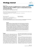
Cell morphology and expression of involucrin in the primary mouse KCs grown in KC-SF medium containing different concen-tration of methylcellulose for 2 daysFigure 1
Cell morphology and expression of involucrin in the primary mouse KCs grown in KC-SF medium containing
different concentration of methylcellulose for 2 days. (A). Gross cell morphology. Images ×200.(B). Immunofluores-
cence micrograph showing involucrin (green), β-tubulin (red) and nuclei (blue). Images ×400.
$
0HWK\OFHOOXORVH
%
0HWK\OFHOOXORVH
Virology Journal 2007, 4:127 />Page 3 of 6
(page number not for citation purposes)
DNAs using lipofectamine (Invitrogen, Australia). The L1-
transfected KCs were incubated in basal KC medium for
18 h, and then grown in semisolid KC-SF complete
medium containing different concentrations of methyl-
cellulose (0%, 0.8% and 1.6%) for 48 h. The L1-trans-
fected KCs treated with methylcellulose were harvested for
analysing L1 gene expression at both transcriptional and
translational levels (Fig 2). Total mRNAs were extracted
from the L1-transfected KCs and reverse-transcribed into
cDNA using a reverse transcription kit (Promega, Aus-
tralia). The cDNAs were analyzed by quantitative RT-PCR
for the L1 and tubulin transcripts using SYBR
®
Greet PCR
kit (Qiagen, Australia). The specificity of RT-PCR was
determined by the Rotor Gene Software and agarose gel
electrophoresis (Fig 2A). As shown in Fig 2A, L1 tran-
scripts were present at a similar level in L1-transfected KCs
Effects of post-transfection treatment of methylcellulose on expression of PV L1 genes in mouse primary KCsFigure 2
Effects of post-transfection treatment of methylcellulose on expression of PV L1 genes in mouse primary KCs.
Newly isolated mouse primary KCs, respectively transfected with four PV L1 expression constructs, were grown in basal KC
medium for 18 h and in 3:1 medium for 24 h. The L1-tranfected KCs were suspended in semisolid KC medium containing dif-
ferent concentration of methylcellulose for 48 h and harvested for analysis of L1 gene expression. (A). L1 transcripts were
assessed by quantitative RT-PCR. β-tubulin transcript was analysed as an internal control. Up panel: Representative electro-
phoresis of the L1 and tubulin mRNA qRT-PCR products. Lower panel: Results are shown with the means (± S.E.M) of duplicate
transfection assays from two separate experiments. (B). Expression of the L1 proteins analysed by Western blot is represent-
ative of duplicate transfection from two separate experiments. β-tubulin (Tub) was used as comparable loading control.
%39+39E
$
1DW/0RG/
/
7XE
0HWK\OFHOOXORVH
5HODWLYH/P51$
ǻǻ&
7
/
7XE
5HODWLYH/P51$
ǻǻ&
7
%
/
,
QYR
7XE
/
,
QYR
7XE
1D
W
/0RG/
0HWK\OFHOOXORVH
Virology Journal 2007, 4:127 />Page 4 of 6
(page number not for citation purposes)
treated with 0%, 0.8% and 1.6% methylcellulose. These
data indicate that L1 transcription was unrelated to meth-
ylcellulose treatment. Western blot analysis was used to
measure L1 protein expression. Protein (40 μg) extracted
from the L1-transfected KCs with or without methylcellu-
lose treatment was separated by SDS-PAGE and blotted
onto PVDF membrane. The blots were probed with either
anti-HPV L1 monoclonal antibody (BD PharMingen, Aus-
tralia) or anti-involucrin polyclonal antibody (Covance,
USA) at 4°C over night. Blots were then incubated with
horseradish-peroxidase(HRP)-conjugated goat anti-
mouse IgG or HRP- conjugated goat anti-rabbit IgG
(Sigma, Australia) followed by a chemiluminescence
analysis (ECL, Amersham, Australia). Western blots
showed that significantly up-regulated expression of
involucrin was associated with the methylcellulose treat-
ment (Fig 2B), indicating that methylcellulose enhanced
L1-transfected KC differentiation. In the absence of meth-
ylcellulose treatment, only weak signals of the L1 proteins
were detected in KCs transfected with the two PV Nat L1
expression constructs, but the levels of L1 proteins were
significantly higher in KCs transfected with the two PV
Mod L1 expression constructs (Fig 2B). Post-transfection
treatment of methylcellulose dramatically decreased L1
protein expression in KCs transfected with the Mod PV L1
constructs (Fig 2B). Conversely, methylcellulose signifi-
cantly increased the levels of the L1 proteins encoded by
the two PV Nat L1 genes (Fig 2B). The down- and up-reg-
ulated responses of the L1 protein expression from the PV
Mod and Nat L1 genes to post-transfection treatment of
methylcellulose were clearly dose-dependent and associ-
ated with the differentiation status of the L1-tranfected
KCs. The results support our previous study that gene
codon composition in part determined differentiation-
dependent expression of the PV L1 proteins in KCs [8].
We next examined how pre-transfection treatment of
methylcellulose affected transcriptional and translational
expressions of the four PV L1 expression constructs in
transiently L1-transfected KCs. Freshly isolated primary
mouse KCs were grown in semisolid KC-SF complete
medium containing 0, 0.8% and 1.6% methylcellulose for
two days. The KCs were recovered from semisolid KC-SF
complete medium by multiple dilutions with serum-free
F medium and PBS followed by centrifugation. The meth-
ylcellulose-treated KCs after recovered were respectively
transfected with each of the four PV L1 plasmid DNAs
using lipofectamine as mentioned above. At 48 h post-
transfection, the L1-transfected KCs were harvested for
analysing L1 transcripts and proteins (Fig. 3). Again,
quantitative RT-PCR was used to examine transcripts of
both Nat and Mod PV L1 genes, with no remarkable differ-
ences observed (Fig. 3A). Meantime, significantly up-reg-
ulated expression of involucrin was detected by Western
blot analysis (data not shown). Western blot showed fur-
ther that methylcellulose-induced KC differentiation
resulted in dramatically down-regulated expression of the
L1 proteins from the two PV Mod L1 genes (Fig. 3B), but
significantly up-regulated expression of the L1 proteins
from the two PV Nat L1 genes (Fig. 3B). These data indi-
cate that pre-transfection treatment of methylcellulose
also differentially regulated expression of the L1 proteins
from PV Nat or Mod L1 genes, confirming further that
expression of the L1 proteins from PV Nat and Mod L1
genes is differentially associated with the differentiation
status of the KCs.
The close association of the HPV life cycle with the differ-
entiation state of its host cell is demonstrated by the
restriction of late gene transcription and amplification of
viral DNA to suprabasal epithelial cells. The study of HPVs
in cell culture has been hindered because of the difficulty
in recreating the three-dimensional structure of the epi-
thelium on which the virus depends to complete its life
cycle. Although raft culture system can provide a spatial
separation of cells for the study of HPV life cycle [3], it is
technically challenging and requires extended periods of
time for KC growth and differentiation. Meantime, it is
hard to isolate separate layers in raft culture system. We
developed the simple mouse primary KCs culture system
to successfully express PV L1 proteins by transient trans-
fection of the L1 expression constructs [8]. We reported
that primary KCs in culture undergo cell differentiation to
regulate expression of the PV L1 genes. Here, we demon-
strated that suspension of mouse primary KCs in methyl-
cellulose resulted in the rapid cell differentiation. As a
model inducer of KC differentiation, use of methylcellu-
lose has allowed us to characterize expression of targeted
genes including L1 and involucrin in only 2–3 days
instead of the 2 weeks required for raft culture and to
study the mechanisms which regulate differentiation-
dependent expression of the PV late genes. Methylcellu-
lose did not influence L1 mRNA transcription in L1-trans-
fected KCs, thus, the results confirmed that the expression
of the L1 protein was post-transcriptionally regulated,
consistent with previous studies [4,17]. Our results dem-
onstrated further that the L1 gene codon composition cor-
related with the differentiation-dependent expression of
the L1 protein in L1-transfected KCs grown in KC-SF com-
plete medium with or without methylcellulose. This cor-
relation can be well explained by our previous
observations that composition of aminoacyl-tRNA pool
changes during cell differentiation, which differentially
favors translation of PV authentic and codon-modified L1
genes [18].
In conclusion, we established a methylcellulose culture
system in the mouse primary KCs and demonstrated that
methylcellulose enhanced KC differentiation. Methylcel-
lulose did not influence L1 transcription but differentially
Virology Journal 2007, 4:127 />Page 5 of 6
(page number not for citation purposes)
Effects of pre-transfection treatment of methylcellulose on expression of the PV L1 genes in mouse primary KCsFigure 3
Effects of pre-transfection treatment of methylcellulose on expression of the PV L1 genes in mouse primary
KCs. Newly isolated mouse KCs were suspended in semisolid KC medium containing different concentration of methylcellu-
lose for 48 h. They were then transfected with the four PV L1 gene expression constructs and grown in KC-SF complete
medium for 48 h before harvested for analysis of L1 gene expression. (A). L1 transcripts were assessed by quantitative RT-
PCR. β-tubulin transcript was analysed as an internal control. Up panel: Representative electrophoresis of the L1 and tubulin
mRNA qRT-PCR products. Lower panel: Results are shown with the means (± S.E.M) of duplicate transfection assays from two
separate experiments. (B). Expression of the L1 proteins analysed by Western blot is representative of duplicate transfection
from two separate experiments. β-tubulin (Tub) was used as comparable loading control.
$
/
7XE
5HODWLYH/P51$
ǻǻ&
7
1D
W
/0RG/
5H
0HWK\OFHOOXORVH
ODWLYH/P51$
ǻǻ&
7
/
7XE
%39 +39E
/
7XE
/
7XE
0HWK\OFHOOXORVH
1
D
W
/0RG/
%
Publish with Bio Med Central and every
scientist can read your work free of charge
"BioMed Central will be the most significant development for
disseminating the results of biomedical research in our lifetime."
Sir Paul Nurse, Cancer Research UK
Your research papers will be:
available free of charge to the entire biomedical community
peer reviewed and published immediately upon acceptance
cited in PubMed and archived on PubMed Central
yours — you keep the copyright
Submit your manuscript here:
/>BioMedcentral
Virology Journal 2007, 4:127 />Page 6 of 6
(page number not for citation purposes)
regulated translation of the authentic and codon-modi-
fied L1 genes. These data support our previous study that
L1 expression in response to differentiation is regulated at
the post-transcriptional level.
Abbreviations
KCs : keratinocytes;
PVs : Papillomaviruses.
Competing interests
The author(s) declare that they have no competing inter-
ests.
Authors' contributions
XW and BL conducted experiments together. XW wrote
the manuscript. KNZ designed and coordinated the
research efforts and edited the manuscript. All co-authors
read and approved the final manuscript.
Acknowledgements
This work was funded in part by a National Health and Medical Research
Council of Australia Industry Research Fellowship (301256 to KNZ) and
the Queensland Cancer Fund (401623 to KNZ).
References
1. Spink KM, Laimins LA: Induction of the human papillomavirus
type 31 late promoter requires differentiation but not DNA
amplification. J Virol 2005, 79:4918-4926.
2. Doorbar J: The papillomavirus life cycle. J Clin Virol 2005, 32
Suppl 1:S7-15.
3. McCance DJ, Kopan R, Fuchs E, Laimins LA: Human papillomavi-
rus type 16 alters human epithelial cell differentiation in
vitro. Proc Natl Acad Sci U S A 1988, 85:7169-7173.
4. Frattini MG, Lim HB, Laimins LA: In vitro synthesis of oncogenic
human papillomaviruses requires episomal genomes for dif-
ferentiation-dependent late expression. Proc Natl Acad Sci U S
A 1996, 93:3062-3067.
5. Dollard SC, Wilson JL, Demeter LM, Bonnez W, Reichman RC, Bro-
ker TR, Chow LT: Production of human papillomavirus and
modulation of the infectious program in epithelial raft cul-
tures. OFF. Genes Dev 1992, 6:1131-1142.
6. Meyers C, Frattini MG, Hudson JB, Laimins LA: Biosynthesis of
human papillomavirus from a continuous cell line upon epi-
thelial differentiation. Science 1992, 257:971-973.
7. McLaughlin-Drubin ME, Christensen ND, Meyers C: Propagation,
infection, and neutralization of authentic HPV16 virus. Virol-
ogy 2004, 322:213-219.
8. Zhao KN, Gu W, Fang NX, Saunders NA, Frazer IH: Gene codon
composition determines differentiation-dependent expres-
sion of a viral capsid gene in keratinocytes in vitro and in
vivo. Mol Cell Biol 2005, 25:8643-8655.
9. Fang NX, Gu W, Ding J, Saunders NA, Frazer IH, Zhao KN: Calcium
enhances mouse keratinocyte differentiation in vitro to dif-
ferentially regulate expression of papillomavirus authentic
and codon modified L1 genes. Virology 2007, 365:187-197.
10. Watt FM, Jones PH: Expression and function of the keratinoc-
yte integrins.
Dev Suppl 1993:185-192.
11. Kubler MD, Jordan PW, O'Neill CH, Watt FM: Changes in the
abundance and distribution of actin and associated proteins
during terminal differentiation of human epidermal kerati-
nocytes. J Cell Sci 1991, 100 ( Pt 1):153-165.
12. Zhu S, Oh HS, Shim M, Sterneck E, Johnson PF, Smart RC: C/EBP-
beta modulates the early events of keratinocyte differentia-
tion involving growth arrest and keratin 1 and keratin 10
expression. Mol Cell Biol 1999, 19:7181-7190.
13. Green H: Terminal differentiation of cultured human epider-
mal cells. Cell 1977, 11:405-416.
14. Adams JC, Watt FM: Fibronectin inhibits the terminal differen-
tiation of human keratinocytes. Nature 1989, 340:307-309.
15. Flores ER, Lambert PF: Evidence for a switch in the mode of
human papillomavirus type 16 DNA replication during the
viral life cycle. J Virol 1997, 71:7167-7179.
16. Ruesch MN, Stubenrauch F, Laimins LA: Activation of papilloma-
virus late gene transcription and genome amplification upon
differentiation in semisolid medium is coincident with
expression of involucrin and transglutaminase but not kera-
tin-10. J Virol 1998, 72:5016-5024.
17. Cumming SA, Repellin CE, McPhillips M, Radford JC, Clements JB,
Graham SV: The human papillomavirus type 31 late 3'
untranslated region contains a complex bipartite negative
regulatory element. J Virol 2002, 76:5993-6003.
18. Gu W, Ding J, Wang X, de Kluyver RL, Saunders NA, Frazer IH, Zhao
KN: Generalized substitution of isoencoding codons shortens
the duration of papillomavirus L1 protein expression in tran-
siently gene-transfected keratinocytes due to cell differenti-
ation. Nucleic Acids Res 2007, 35:4820-4832.