Báo cáo hóa học: " Bioinformatic analysis suggests that the Orbivirus VP6 cistron encodes an overlapping gene" potx
Bạn đang xem bản rút gọn của tài liệu. Xem và tải ngay bản đầy đủ của tài liệu tại đây (376.51 KB, 10 trang )
BioMed Central
Page 1 of 10
(page number not for citation purposes)
Virology Journal
Open Access
Research
Bioinformatic analysis suggests that the Orbivirus VP6 cistron
encodes an overlapping gene
Andrew E Firth
Address: Department of Biochemistry, BioSciences Institute, University College Cork, Cork, Ireland
Email: Andrew E Firth -
Abstract
Background: The genus Orbivirus includes several species that infect livestock – including
Bluetongue virus (BTV) and African horse sickness virus (AHSV). These viruses have linear dsRNA
genomes divided into ten segments, all of which have previously been assumed to be
monocistronic.
Results: Bioinformatic evidence is presented for a short overlapping coding sequence (CDS) in the
Orbivirus genome segment 9, overlapping the VP6 cistron in the +1 reading frame. In BTV, a 77–79
codon AUG-initiated open reading frame (hereafter ORFX) is present in all 48 segment 9
sequences analysed. The pattern of base variations across the 48-sequence alignment indicates that
ORFX is subject to functional constraints at the amino acid level (even when the constraints due
to coding in the overlapping VP6 reading frame are taken into account; MLOGD software). In fact
the translated ORFX shows greater amino acid conservation than the overlapping region of VP6.
The ORFX AUG codon has a strong Kozak context in all 48 sequences. Each has only one or two
upstream AUG codons, always in the VP6 reading frame, and (with a single exception) always with
weak or medium Kozak context. Thus, in BTV, ORFX may be translated via leaky scanning. A long
(83–169 codon) ORF is present in a corresponding location and reading frame in all other Orbivirus
species analysed except Saint Croix River virus (SCRV; the most divergent). Again, the pattern of
base variations across sequence alignments indicates multiple coding in the VP6 and ORFX reading
frames.
Conclusion: At ~9.5 kDa, the putative ORFX product in BTV is too small to appear on most
published protein gels. Nonetheless, a review of past literature reveals a number of possible
detections. We hope that presentation of this bioinformatic analysis will stimulate an attempt to
experimentally verify the expression and functional role of ORFX, and hence lead to a greater
understanding of the molecular biology of these important pathogens.
Background
The Orbivirus genus is one of ≥ 12 genera within the family
Reoviridae. The Reoviridae have segmented linear dsRNA
genomes. There are 9–12 segments [1] and these are usu-
ally, but not always, monocistronic. Subgenomic RNAs
are unknown. Orbivirus genomes have 10 segments. Many
species infect ruminants while some infect humans.
Transmission is via arthropods – including midges, ticks
and mosquitoes. The type species is Bluetongue virus
(BTV) which causes severe and sometimes fatal disease,
particularly in sheep. BTV is endemic in many tropical
countries, but there have also been recent outbreaks in
Published: 14 April 2008
Virology Journal 2008, 5:48 doi:10.1186/1743-422X-5-48
Received: 25 March 2008
Accepted: 14 April 2008
This article is available from: />© 2008 Firth; licensee BioMed Central Ltd.
This is an Open Access article distributed under the terms of the Creative Commons Attribution License ( />),
which permits unrestricted use, distribution, and reproduction in any medium, provided the original work is properly cited.
Virology Journal 2008, 5:48 />Page 2 of 10
(page number not for citation purposes)
Europe [2,3]. Another species is African horse sickness
virus (AHSV) which is a fatal disease of horses. AHSV is
endemic in many parts of sub-Saharan Africa, but has
made incursions into Europe [4]. Recent outbreaks of BTV
in Europe may be a consequence of climate change –
allowing the midge vectors to expand their range [5].
The Orbivirus proteins, structure, assembly and replication
have been reviewed in [6-8]. The BTV core is composed of
two major proteins (VP3 and VP7). Transcription com-
plexes – composed of three minor proteins (VP1 –
polymerase, VP4 – capping enzyme, and VP6 – helicase)
are located inside the core. Transcription occurs within
the intact core and full-length capped mRNAs from each
of the genome segments are fed out into the cytoplasm for
translation. An outer capsid (VP2 and VP5) surrounds the
core, but is removed during cell entry. There are four non-
structural proteins – NS1, NS2 and NS3/3A. VP6 is a
hydrophilic, basic protein that binds dsRNA and other
nucleic acids and functions as the viral helicase [9-13]. In
some, but not all, BTV serotypes, VP6 migrates as a
closely-spaced doublet [14]. This is apparently due to the
fact that in these serotypes the first VP6 AUG codon has
weak Kozak context while a second in-frame AUG codon
has medium context.
The genomes of RNA viruses are under strong selective
pressure to compress maximal coding and regulatory
information into minimal sequence space. Thus overlap-
ping CDSs are particularly common in such viruses. Such
CDSs can be difficult to detect using conventional gene-
finding software [15], especially when short. The software
package MLOGD, however, was designed specifically for
locating short overlapping CDSs in sequence alignments
and overcomes many of the difficulties with alternative
methods [15,16]. MLOGD includes explicit models for
sequence evolution in double-coding regions as well as
models for single-coding and non-coding regions. It can
be used to predict whether query ORFs are likely to be
coding, via a likelihood ratio test, where the null model
comprises any known CDSs and the alternative model
comprises the known CDSs plus the query ORF. MLOGD
has been tested extensively using thousands of known
virus CDSs as a test set, and it has been shown that, for
overlapping CDSs, a total of just 20 independent base var-
iations are sufficient to detect a new CDS with ~90% con-
fidence.
Using MLOGD, we recently identified – and subsequently
experimentally verified – a new short CDS in the Potyviri-
dae that overlaps the polyprotein cistron but is translated
in the +2 reading frame [17]. When we applied MLOGD
to the Orbivirus genome we also found evidence for a short
CDS overlapping the VP6 cistron. Here we describe the
bioinformatic analysis.
Results
Identification in BTV using MLOGD
The putative new CDS, ORFX, was first identified in a BTV
sequence alignment, using MLOGD. In the RefSeq [Gen-
Bank: NC_006008
] (1049 nt), ORFX has coords 182 415
(77 codons) and therefore is completely contained within
the VP6 cistron (16 1005), overlapping it in the +1 read-
ing frame (Figure 1). When applied to an alignment of 48
BTV sequences (see Methods; pairwise divergences ≤0.21
base variations per nucleotide and total alignment diver-
gence ~0.77 independent base variations per column in
the ORFX region), MLOGD detected a strong coding sig-
nature for ORFX (Figures 2, 3). There are ~180 independ-
ent base variations across the alignment in the ORFX
region, thus providing MLOGD with a robust signal. For-
mally, and within the MLOGD model, p < 10
-40
. Indeed
Figure 2 shows four non-overlapping – and hence com-
pletely independent – positively scoring windows in the
ORFX region. Moreover, the MLOGD results showed that,
within the ORFX region, ORFX is more conserved at the
amino acid level than VP6 (Figure 2). Finally, inspection
of the MLOGD output showed that the ORF is present in
all of the 48 sequences (i.e. no premature termination
codons; Figure 2).
Nucleotide sequence analysis in BTV
In the 48-sequence BTV alignment (not shown), one can
observe the following:
• The ORFX AUG initiation codon is present in all 48
sequences and is at the same location in the alignment. All
have 'G' at +4; 46/48 have 'A' at -3 and 2/48 have 'G' at -
3, giving the ORFX AUG codon a strong Kozak context
[18].
• As far as amino acid constraints in the VP6 reading frame
are concerned, there is no reason for the ORFX AUG
codon to be conserved. In every sequence, the overlapping
VP6-frame codons are gAU_Ggu. GAU codes for Asp, but
Asp could also be encoded by GAC.
• Many sequences contain ORFX-frame termination
codons just two codons 5' of the AUG codon. Thus initia-
tion of ORFX at an upstream non-AUG codon, or via other
non-canonical mechanisms, appears unlikely.
• ORFX is always in the +1 frame relative to the VP6 read-
ing frame.
• The length of ORFX is 77 aa in 44/48 sequences (UAG
termination codon) and 79 aa in 4/48 sequences (UAA
termination codon). The alignment is gap-free within
ORFX.
Virology Journal 2008, 5:48 />Page 3 of 10
(page number not for citation purposes)
• All AUG codons upstream of the ORFX AUG codon are
in the VP6 reading frame. There are a maximum of two
upstream AUG codons in any given sequence, and the
Kozak contexts of the upstream AUG codons are nearly
always weak or medium (Table 1).
• There is only a single AUG codon (in a single sequence)
in the purine-rich ~70 nt region (Figure 4) directly
upstream of the ORFX AUG codon.
Nucleotide sequence analysis in other Orbivirus RefSeqs
The five non-BTV Orbivirus GenBank RefSeqs (see Meth-
ods) were inspected for a long ORF in the same location
and reading frame as ORFX relative to the annotated VP6
CDS. Such an ORF was found in all RefSeqs except SCRV
(Figure 5). The ORFX lengths are 143, 111, 113 and 83
codons in, respectively, AHSV, PHSV, YUOV and PALV.
We propose (see Discussion) that ORFX is not present in
SCRV. The following AUG codons are (potentially) used
in the various RefSeqs (Kozak contexts – in parantheses –
are assumed to be 'strong' if there is 'G' at +4 and an 'A' or
'G' at -3, 'medium' if one of these is present, and 'weak' if
neither are present):
BTV: AUG1 (weak) and AUG2 (medium) in VP6 frame.
AUG3 (strong) in ORFX frame. AUG[4-10] also in ORFX
frame.
AHSV: AUG1 (weak) in VP6 frame. AUG2 (strong) in
ORFX frame. AUG[3-10] also in ORFX frame.
PALV: AUG1 (weak) in VP6 frame. AUG2 (strong) in
ORFX frame. AUG[3-7] also in ORFX frame.
PHSV: AUG1 (weak) in VP6 frame. AUG2 (medium) in
ORFX frame (1 codon ORF). AUG3 (medium) in +2
frame (10 codon ORF). AUG4 (weak) and AUG5 (strong)
in ORFX frame. AUG[6-7] also in ORFX frame.
YUOV: AUG1 (weak) in VP6 frame. AUG2 (medium) in
ORFX frame (1 codon ORF). AUG3 (medium) in +2
frame (21 codon ORF; overlaps AUG4 [strong; +2 frame]
and AUG5 [medium; VP6 frame]). AUG6 (medium),
AUG7 (strong), AUG8 (strong) and AUG9 (medium) in
ORFX frame.
SCRV: AUG1 (medium) and AUG2 (strong) in VP6 frame.
AUG3 (medium) in ORFX frame (1 codon ORF). AUG4
(medium), AUG5 (strong) and AUG6 (strong) in VP6
frame. AUG7 (weak) and AUG8 (strong) in ORFX frame
(ORFXa; Figure 5). AUG9 (weak) and AUG10 (weak) in
ORFX frame (ORFXb; Figure 5).
MLOGD analysis of ORFX coding potential
MLOGD can not be used effiectively on an alignment of
the six RefSeqs because the pairwise divergences are too
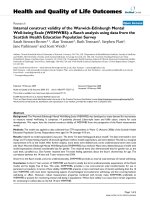
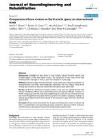
Genome map for BTVFigure 1
Genome map for BTV. The putative new coding sequence – ORFX – is located on segment 9 (RNA9), in the +1 reading
frame relative to the overlapping VP6 cistron. Molecular masses are based on the unmodified amino acid sequences.
NS3/3A
VP6 (36 kDa)
NS2 (41 kDa)
VP7 (39 kDa)
VP5 (59 kDa)
NS1 (64 kDa)
VP4 (75 kDa)
VP3 (103 kDa)
VP2 (111 kDa)
VP1 (150 kDa)
(26 kDa)
RNA10
RNA9
RNA8
RNA7
RNA5
RNA6
RNA4
RNA3
RNA2
RNA1
5′
5′
5′
5′
5′
5′
5′
5′
5′
5′
3′
3′
3′
3′
3′
3′
3′
3′
3′
3′
ORFX (+1 frame; 9.5 kDa)
Untranslated regions
Annotated CDSs
Putative CDS
200 nt
Virology Journal 2008, 5:48 />Page 4 of 10
(page number not for citation purposes)
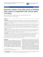
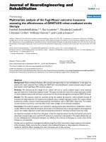
MLOGD statistics for the alignment of 48 BTV sequencesFigure 2
MLOGD statistics for the alignment of 48 BTV sequences. The input alignment comprised a CLUSTALW [39] align-
ment of the VP6 amino acid sequences only, back-translated to nucleotide sequences. (1) The positions of alignment gaps in
each of the 48 sequences. In fact most of the alignment is ungapped, though a few sequences are incomplete. (2)–(4) The posi-
tions of stop codons in each of the 48 sequences in each of the three forward reading frames. Note the conserved absence of
stop codons in the +0 frame (i.e. the VP6 CDS) and in the +1 frame in the ORFX region. (5)–(8) MLOGD sliding-window
plots. Window size = 20 codons. Step size = 10 codons. Each window is represented by a small circle (showing the likelihood
ratio score for that window), and grey bars showing the width (ends) of the window. See [16] for further details of the
MLOGD software. In (5)–(6) the null model, in each window, is that the sequence is non-coding, while the alternative model
is that the sequence is coding in the window frame. Positive scores favour the alternative model. There is a strong coding sig-
nature in the +0 frame (5) throughout the VP6 CDS, except where the VP6 CDS overlaps ORFX. In this region there is a
strong coding signature in the +1 frame (6) indicating that ORFX is subject to stronger functional constraints than the overlap-
ping section of VP6. In (7)–(8) the null model, in each window, is that only the VP6 frame is coding, while the alternative model
is that both the VP6 frame and the window frame are coding. Only the +1 (7) and +2 (8) frames are shown because the +0
frame is the VP6 frame which is included in the null model. Scores are generally negative with occasional random scatter into
low positive scores, except for the ORFX region which has consecutive high-positively scoring windows (7). Note that there
are four non-overlapping – and hence completely independent – positively scoring windows in the ORFX region (7). Formally,
and within the MLOGD model, p < 10
-40
. (9) Genome map for the reference sequence [GenBank: NC_006008]. (10) Phyloge-
netically summed sequence divergence (mean number of base variations per nucleotide) for the sequences that contribute to
the statistics at each position in the alignment. In any particular column, some sequences may be omitted from the statistical
calculations due to alignment gaps. Statistics in regions with lower summed divergence (i.e. partially gapped regions) have a
lower signal-to-noise ratio.
positions of
alignment gaps
(1)
positions of
stop codons
(triangles)
(2)
Frame = +0
(3)
Frame = +1
(4)
Frame = +2
MLOGD log likelihood ratio per 20 codon window
positive values
=> coding
negative values
=> non−coding
−40
−20
0
20
40
(5)
Frame = +
0
null model =
non−coding
−40
−20
0
20
40
(6)
Frame = +
1
null model =
non−coding
−40
−20
0
20
40
(7)
Frame = +
1
null model =
VP6 CDS
−40
−20
0
20
40
(8)
Frame = +
2
null model =
VP6 CDS
VP6 CDS
ORFX
(9)
summed
divergence of
contributing
sequences
0 200 400 600 800 1000
0.0
0.5
1.0
(10)
mean number
of mutations
per column
alignment coordinate (nt)
Virology Journal 2008, 5:48 />Page 5 of 10
(page number not for citation purposes)
great. However it can be used on other within-species
alignments. Alignments were constructed for (a) the 48
BTV sequences, (b) the 3 AHSV sequences, (c) the 11
PALV sequences (183 nt, partial), and (d) the PHSV and
YUOV RefSeqs (see Methods). PHSV and YUOV are the
two most-closely related of the six RefSeqs and are not too
divergent for MLOGD. MLOGD results for ORFX are given
in Table 2 and Figure 3. ORFX is present in all the aligned
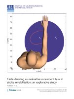
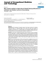
MLOGD statistics for BTV, AHSV, PALV and PHSV/YUOV alignmentsFigure 3
MLOGD statistics for BTV, AHSV, PALV and PHSV/YUOV alignments. Output plots from MLOGD used in the
'Test Query CDS' mode, applied to the ORFX region in BTV, AHSV, PALV and PHSV/YUOV sequence alignments. See [16] for
full details of the MLOGD software. The null model comprises the VP6 CDS and the query CDS is ORFX. In each plot, the top
panel displays the raw log(LR) statistics at each alignment position. There is a separate track for each reference – non-refer-
ence sequence pair (labelled at the right, together with the pairwise divergences; albeit not legible for the BTV alignment since
it contains so many – i.e. 48 – sequences). Stop codons (of which there are none except 3' terminal ones) in each of the VP6
and ORFX reading frames, and alignment gaps for each sequence, are marked on the appropriate tracks. The second panel dis-
plays the Σ
tree
log(LR) statistic at each alignment position, where 'tree' represents a phylogenetic tree – see [16]. The third and
fourth panels display sliding window means of the statistics in the first and second panels, respectively. The fifth panel shows
the locations of the null and alternative model CDSs (i.e. VP6 and ORFX, respectively). The sixth panel shows the summed
mean sequence divergence (base variations per alignment nt column) for the sequence pairs that contribute to the Σ
tree
log(LR)
statistic at each alignment position. This is a measure of the information available at each alignment position (e.g. partially
gapped regions have lower summed mean sequence divergence). The predominantly positive values in the fourth panel indicate
that ORFX is subject to functional constraints, at the amino acid level, over the majority of its length.
BTV segment 9; mean base variations per column over tree = 0.77
stops in null model stops in alternate model
gaps
sequence
v. reference
pairs
divergence
(mean #
muts per nt
)
log likelihood ratio
per nucleotide
positive values favour
the alternate model
negative values favour
the null model
DQ289042
DQ289041
DQ289043
A22393
DQ289044
U55779
U55781
U55784
U55788
U55794
U55797
U55800
U55801
DQ289045
U55778
U55782
U55787
AF403418
DQ289050
L08672
U55780
U55793
U55796
L08668
L08670
U55790
U55792
U55795
AY493691
U55785
AF403421
DQ289047
DQ289048
D10905
L08671
U55786
DQ825671
DQ825669
L08669
DQ825668
AF403419
AF403420
AY124373
U55799
AF403423
DQ832170
DQ289046
0.17
0.17
0.17
0.12
0.21
0.03
0
0.02
0.02
0.02
0
0
0
0.18
0.01
0.03
0.02
0.02
0.16
0.02
0.01
0.02
0.02
0.07
0.02
0.03
0.02
0.02
0.15
0.01
0.06
0.16
0.16
0.18
0.02
0.03
0.06
0.05
0.07
0.06
0.05
0.06
0.06
0.06
0.04
0.04
0.16
NC_006008 (reference)
0
1
2
3
4
sum over
phylogenetic
tree
running mean
(window size 21 nt)
scale−bar
s
raw score
s
8
0
running
means
0
8
0.0
0.2
0.4
0.6
0.8
sum over
phylogenetic
tree
Alternate model CDSs
Null model CDSs
summed
divergence o
f
contributing
sequence pairs
0 200 400 600 800 1000
0.0
0.2
0.4
0.6
0.8
1.0
1.2
mean number
of mutations
per column
alignment coordinate (nt)
AHSV segment 9; mean base variations per column over tree = 0.06
stops in null model stops in alternate model
gaps
sequence
v. reference
pairs
divergence
(mean #
muts per nt
)
log likelihood ratio
per nucleotide
positive values favour
the alternate model
negative values favour
the null model
AM883170
U19881
0.05
0.03
NC_006019 (reference)
0.0
0.2
0.4
0.6
0.8
sum over
phylogenetic
tree
running mean
(window size 21 nt)
scale−bar
s
raw score
s
0.25
0
running
means
0
0.25
0.00
0.05
0.10
0.15
sum over
phylogenetic
tree
Alternate model CDSs
Null model CDSs
summed
divergence o
f
contributing
sequence pairs
0 200 400 600 800 1000 1200
0.00
0.02
0.04
0.06
0.08
mean number
of mutations
per column
alignment coordinate (nt)
PALV partial segment 9; mean base variations per column over tree = 0.23
stops in null model stops in alternate model
gaps
sequence
v. reference
pairs
divergence
(mean #
muts per nt
)
log likelihood ratio
per nucleotide
positive values favour
the alternate model
negative values favour
the null model
AB034675
AB034676
AB034678
AB034682
AB034677
AB034681
AB034679
AB034680
AB034683
AB034684
0.01
0.05
0.03
0.11
0.04
0.03
0.03
0.03
0.12
0.11
NC_005992 (reference)
0.0
0.5
1.0
1.5
2.0
sum over
phylogenetic
tree
running mean
(window size 21 nt)
scale−bar
s
raw score
s
2
0
running
means
0
2
0.0
0.1
0.2
0.3
0.4
sum over
phylogenetic
tree
Alternate model CDSs
Null model CDSs
summed
divergence o
f
contributing
sequence pairs
0 50 100 150
0.00
0.05
0.10
0.15
0.20
0.25
0.30
0.35
mean number
of mutations
per column
alignment coordinate (nt)
PHSV+YUOV segment 9; mean base variations per column over tree = 0.56
stops in null model stops in alternate model
gaps
sequence
v. reference
pairs
divergence
(mean #
muts per nt
)
log likelihood ratio
per nucleotide
positive values favour
the alternate model
negative values favour
the null model
NC_007664
0.56
NC_007753 (reference)
0.0
0.5
1.0
1.5
sum over
phylogenetic
tree
running mean
(window size 21 nt)
scale−bar
s
raw score
s
0.5
0
running
means
0
0.5
0.0
0.1
0.2
0.3
sum over
phylogenetic
tree
Alternate model CDSs
Null model CDSs
summed
divergence o
f
contributing
sequence pairs
0 200 400 600 800 1000
0.0
0.2
0.4
0.6
0.8
mean number
of mutations
per column
alignment coordinate (nt)
Virology Journal 2008, 5:48 />Page 6 of 10
(page number not for citation purposes)
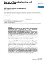
Nucleotide frequencies for segment 9Figure 4
Nucleotide frequencies for segment 9. Nucleotide frequencies in 60 nt running windows along each Orbivirus segment 9
RefSeq. 'A' – red, 'C' – green, 'G' – blue, 'U' – purple. Horizontal black bars represent the locations of the VP6 CDS and ORFX
(the grey bar represents ORFXb in SCRV). Except for SCRV, the sequences are A- or AG-rich, but they also have an A-rich
peak just upstream of ORFX.
nucleotide usage
0.0 0.1 0.2 0.3 0.4 0.5 0.6
BTV
AHSV
PALV
0 200 600 1000
0.0 0.1 0.2 0.3 0.4 0.5 0.6
PHSV
0 200 600 1000
YUOV
0 200 600 1000
SCRV
nucleotide index
Table 1: Kozak contexts of VP6 AUG codons in BTV. Kozak contexts of AUG codons upstream of ORFX in BTV for the 34 segment 9
sequences which appear to contain the complete 5'UTR. Kozak contexts are assumed to be 'strong' if there is 'G' at +4 and an 'A' or
'G' at -3, 'medium' if one of these is present, and 'weak' if neither are present.
One upstream AUG codon Two upstream AUG codons
First Strength Number First Second Strength Number
-3 +4 -3 +4 -3 +4
G C medium 5 C U U G weak-medium 15
A A medium 1 C U G C weak-medium 9
C U weak 1 C U A A weak-medium 1
CU A G weak-strong 1
CUC C weak-weak 1
Virology Journal 2008, 5:48 />Page 7 of 10
(page number not for citation purposes)
sequences (no premature termination codons) and, in
each alignment, MLOGD detects a strong coding signature
for ORFX. ORFX is longest in the three AHSV sequences –
the maximal lengths being 143 codons in [Gen-
bank:NC_006019
], 154 codons in [Gen-
bank:AM883170
], and 169 codons in
[Genbank:U19881
].
Analysis of the ORFX peptide sequence
Application of blastp [19] to the ORFX peptide sequences
for the six RefSeqs revealed no similar amino acid
sequences in GenBank (14 Mar 2008), while tblastn iden-
tified only the ORFX region in other Orbivirus sequences
(as expected). Application of InterProScan [20] to the six
sequences returned no hits (protein motifs, domains etc).
The ORFX amino acid sequence appears to have greater
amino acid conservation than the overlapping region of
the VP6 CDS (e.g. Figure 2). In a comparison between
[Genbank:NC_006008
] and three divergent BTV
sequences – [Genbank:DQ289044
], [Genbank:D10905]
and [Genbank:DQ825671
], all three showed greater
amino acid conservation (relative to NC_006008
) in the
ORFX frame than in the VP6 frame in the ORFX region.
Specifically, there was respectively 87%, 78% and 100%
amino acid identity in the ORFX frame, but only 58%,
73% and 83% identity in the VP6 frame. Similarly, in a
comparison of [Genbank:NC_007753
] (PHSV) with
[Genbank:NC_007664
] (YUOV), there were 32 amino
acid identities in ORFX while, in the corresponding region
of VP6, there were only 22 amino acid identities.
Discussion
Due to the segmented nature of their genomes, the Reoviri-
dae may escape a fundamental problem that many other
eukaryotic viruses face – how to circumvent the host cell's
general rule of 'one functional protein per mRNA'. None-
theless, of the 352 Reoviridae RefSeqs in GenBank (10 Mar
2008; 33 species × 9–12 segments per species), ~5% are
multicistronic. Among these are a few examples of fully
overlapping genes apparently translated via leaky scan-
ning, for example in Phytoreovirus segment S12 or S9 [21]
and mammalian Orthoreovirus segment S1 [22,23].
For optimal leaky scanning [24], one would expect the
VP6 CDS to initiate at AUG1 with weak context and ORFX
to initiate at AUG2 with strong context. This indeed is the
situation in the AHSV and PALV RefSeqs. Although there
are two upstream VP6-frame AUG codons in many BTV
serotypes, leaky scanning still appears fairly straightfor-
ward in this virus as a translational mechanism for ORFX
(though potentially at a much lower abundance than
VP6). In the YUOV and PHSV RefSeqs, leaky scanning
may be possible, but requires scanning through or trans-
lation and reinitiation of two upstream short ORFs. It is
interesting, and possibly relevant, that in another Reoviri-
Segment 9 genome maps for six Orbivirus speciesFigure 5
Segment 9 genome maps for six Orbivirus species. Genome maps for segment 9 of the six Orbivirus RefSeqs in Gen-
Bank, showing the location of putative ORFX homologues. In SCRV, no long ORF was found in the right location and frame;
the two ORFs indicated here are separated by a stop codon. A phylogenetic tree for the six Orbivirus VP6 amino acid
sequences (columns with alignment gaps excluded; neighbour-joining tree; numbers indicate bootstrap support [out of 1000];
scale bar represents the number of substitutions per site; tree produced with CLUSTALX [39]) is given at left.
NC_006005: Saint Croix river virus (SCRV) − VP6
NC_007664: Yunnan orbivirus (YUOV) − VP6
NC_007753: Peruvian horse sickness virus (PHSV) − VP6
NC_005992: Palyam virus (PALV) − VP6
NC_006019: African horse sickness virus (AHSV) − VP6
NC_006008: Bluetongue virus (BTV) − VP6
ORFXa ORFXb
ORFX
ORFX
ORFX
ORFX
ORFX
5′
5′
5′
5′
5′
5′
3′
3′
3′
3′
3′
3′
100 nt
0.1
914
463
1000
4
18
17
20
18
16
702
1034
1021
838
1127
1005
101
175
165
144
148
182
379
516
500
395
579
415
217
Virology Journal 2008, 5:48 />Page 8 of 10
(page number not for citation purposes)
dae species – Avian reovirus – a novel, as yet not fully
understood, scanning-independent ribosome migration
mechanism is used to bypass two upstream CDSs in order
to translate the 3'-proximal CDS on the tricistronic S1
mRNA [25,26].
IRESs have not been reported in the Reoviridae and, at this
genomic location, use of an IRES would seem unlikely.
However, it has been shown that a variety of poly-purine
A-rich sequences – such as (GAAA)
16
– can serve as effi-
cient IRESs without the requirement for a complex RNA
secondary structure such as in the Picornaviridae IRESs
[27], so it is interesting to note that there is an A-rich poly-
purine tract just upstream of ORFX in all species except
SCRV (Figure 4). In the BTV RefSeq, for example, the 68 nt
immediately preceding ORFX comprise 32 A, 7 C, 25 G
and 4 U nucleotides. In fact the entire sequences (except
SCRV) are A- or AG-rich (Table 3). Nonetheless the region
just upstream of ORFX is a peak in A-richness (Figure 4).
Admittedly, this could be due to many other reasons (e.g.
just amino acid coding constraints in VP6) and there is no
strong reason to suspect an IRES here.
SCRV lacks a long ORF in the correct reading frame and
location for an ORFX homologue. The number (six) and
contexts (3 are strong) of upstream AUG codons make
conventional leaky scanning to 'ORFXa' (38 codons; Fig-
ure 5) extremely unlikely. It is quite possible, therefore,
that no ORFX homologue is present in SCRV. This is not
too surprising – SCRV segment 9 is the most divergent,
and the shortest, of the six RefSeqs (Figure 5) [28]. SCRV
is also the only species of the six which is tick-borne
instead of insect-borne (BTV, AHSV and PALV are trans-
mitted by midges; YUOV by mosquitoes).
At ~9.5 kDa, the putative ORFX product in BTV is too
small to appear on most published protein gels. Nonethe-
less there are unidentified low molecular mass bands in a
number of reported gels [29-32], often running near the
dye front, that may represent ORFX product. Furthermore,
ref. [33] (in vitro translation of the individual segments)
noted, with reference to excluded data, that segment 9
may encode a low molecular weight protein in addition to
VP6.
The ORFX product is largest in AHSV (~17 kDa in [Gen-
Bank:NC_006019
] and ~20 kDa in [GenBank:U19881]).
Ref. [34] (in vitro translation of the individual AHSV seg-
ments, and comparison with proteins extracted from
infected cell lysate) clearly identified an additional non-
structural protein translated from segment 9 – termed
Table 3: Nucleotide frequencies for segment 9. Mean nucleotide
frequencies for the six Orbivirus segment 9 RefSeqs in GenBank.
RefSeq Species A% C% G% U%
NC_006008 BTV 32 16 33 19
NC_006019 AHSV 32 16 32 20
NC_005992 PALV 36 16 26 23
NC_007753 PHSV 41 13 24 22
NC_007664 YUOV 36 18 25 20
NC_006005 SCRV 25 27 24 25
Table 2: ORFX MLOGD statistics. MLOGD statistics for ORFX in different Orbivirus alignments. These statistics were derived using
MLOGD in the 'Test Query CDS' mode (Figure 3) – specifically testing the coding potential of the whole ORFX – rather than the
'Sliding Window' mode used for Figure 2.
Species Reference
1
N
seqs
Length ln(LR)
2
var/nt
3
ln(LR)/nt
4
BTV NC_006008 48 234 nt 101.8 0.77 0.44 180 0.21
AHSV NC_006019 3 429 nt 15.8 0.06 0.04 26 0.05
PALV NC_005992 11 180
7
nt 29.7 0.23 0.16 41 0.12
PHSV/YUOV NC_007753 2 336 nt 33.0 0.56 0.10 189 0.56
1. GenBank reference sequence used for MLOGD.
2. Total MLOGD log likelihood score – positive values indicate that ORFX is likely to be coding. Formally, exp(ln(LR)) gives
, which may be equated to if equal Bayesian priors are assumed. These probabilities are,
however, subject to the assumptions of the MLOGD sequence evolution model [15]. Nonetheless, extensive tests with known single-coding and
double-coding sequences indicate that 'N
var
≥ 20' and 'ln(LR)/nt ≥ × var/nt' signals robust detection of an overlapping same-strand CDS [16] (and
unpublished data).
3. Alignment divergence per nucleotide – i.e. mean number of independent base variations per alignment column in the ORFX region.
4. Log likelihood score per alignment column.
5. Approximate total number of independent base variations in ORFX region.
6. Maximum pairwise divergence from the chosen reference sequence.
7. Alignment of PALV partial sequences – does not cover the entire ORFX region.
N
var
5
div
max
6
P alignment ORFX coding)
P alignment ORFX noncoding
(|
(| )
P ORFX coding)
P ORFX noncoding
(
()
1
6
Virology Journal 2008, 5:48 />Page 9 of 10
(page number not for citation purposes)
'NS3' – migrating ~1.5 kDa behind the 'NS4/4A' proteins
(equivalent to NS3/3A in our notation) translated from
segment 10. 'NS3' is a good candidate for ORFX product
migrating a little slower than expected, possibly as a result
of post-translational modification. The protein labelled
'VP6' in ref. [34] appears to be a truncated version of VP5
(translated from the same segment as VP5, and both were
shown to have similar partial protease digestion prod-
ucts). Interestingly the VP6 protein (our notation) is not
visible as a product of segment 9 translation in Fig. 6 of
ref. [34], but may be visible in Fig. 7 of ref. [34] (migrating
next to NS2), unless this is cross-contamination. An addi-
tional segment 9 product (~20 kDa), migrating ahead of
'NS4/4A', is also visible (albeit fainter) in Fig. 7 of ref.
[34]. If the 'NS3' band is post-translationally modified
ORFX product, then this band could be unmodified ORFX
product.
Ref. [35] also identified a number of low molecular mass
proteins in AHSV-infected cells – in particular P23, P20
and P21. Ref. [35] equated two of these (P20 and P21) to
the segment 10 products NS3/3A (~24/~22 kDa in
AHSV). The third protein may be ORFX product.
In addition to its small size, the fact that ORFX product
has not been widely reported suggests that it may be
present only in low abundance and/or only expressed at
certain stages (e.g. only in the insect vector) or cellular
locations.
Conclusion
We have identified a conserved ORF (ORFX) overlapping
the Orbivirus VP6 CDS in the +1 reading frame. ORFX
ranges from 77–169 codons in length, depending on spe-
cies, and is present in all Orbivirus segment 9 sequences
analysed except for the highly divergent species SCRV. The
software package MLOGD – designed specifically for
identifying and analysing overlapping CDSs – finds a
strong coding signature for ORFX when applied to BTV,
AHSV, PALV and PHSV/YUOV sequence alignments. The
location and Kozak context of the VP6 and ORFX initia-
tion codons is generally consistent with a leaky scanning
model for ORFX translation. ORFX product bears no
homology to known proteins.
We hope that presentation of this bioinformatic analysis
will stimulate an attempt to experimentally verify the
expression and functional role of ORFX product. Initial
verification could be by means of immunoblotting with
ORFX-specific antibodies or gel purification of ORFX
product from virus-infected cell protein extracts, followed
by mass spectrometry.
Methods
In GenBank, there are whole-genome RefSeqs for six Orbi-
virus species: Bluetongue virus (BTV), African horse sick-
ness virus (AHSV), Peruvian horse sickness virus (PHSV),
Yunnan orbivirus (YUOV), Palyam virus (PALV) and
Saint Croix river virus (SCRV). All six genomes comprise
10 segments. The segments homologous to BTV segment
9 (encoding VP6) were identified by finding the best
blastp-match, among the 10 BTV translated segments, for
the longest ORF in each of the 50 non-BTV segments. The
identifications were verified, where possible, by informa-
tion in the GenBank-file headers and in the literature
(AHSV [36]; YUOV [37]; PALV [38]; SCRV [28]).
As of 11 May 2007, there were 1273 Orbivirus sequences
in GenBank (i.e. including partial sequences), however
most of these are not segment 9. Incidently, none of these
sequences has more than one CDS annotated. Segment 9
sequences were extracted (a) using the GenBank-file DEF-
INITION headers, and (b) by finding the best blastp-
match for the longest ORF in each sequence among the 10
BTV translated segments. These were supplemented with
all GenBank (16 Mar 2008) tblastn matches to the ORFX
peptide sequences from the six RefSeqs (providing one
additional recent sequence). After removing duplicate
sequences, the following segment 9 sequences were
found: (1) the 6 RefSeqs for BTV, AHSV, PHSV, YUOV,
PALV and SCRV (all complete); (2) 47 other BTV
sequences (mostly complete VP6 CDS; all cover ORFX
completely; ~34 contain the full 5' UTR); (3) 2 other
AHSV sequences (full genome); and (4) 10 PALV partial
sequences (183 nt, completely contained in the ORFX
region).
The GenBank accession numbers are as follows: BTV –
NC_006008
, A22393, AF403418, AF403419, AF403420,
AF403421
, AF403423, AY124373, AY493691, D10905,
DQ289041
, DQ289042, DQ289043, DQ289044,
DQ289045
, DQ289046, DQ289047, DQ289048,
DQ289050
, DQ825668, DQ825669, DQ825671,
DQ832170
, L08668, L08669, L08670, L08671, L08672,
U55778
, U55779, U55780, U55781, U55782, U55784,
U55785
, U55786, U55787, U55788, U55790, U55792,
U55793
, U55794, U55795, U55796, U55797, U55799,
U55800
, U55801; AHSV – NC_006019, U19881,
AM883170
; PHSV – NC_007753; YUOV – NC_007664;
PALV – NC_005992
, AB034675, AB034676, AB034677,
AB034678
, AB034679, AB034680, AB034681,
AB034682
, AB034683, AB034684; SCRV – NC_006005.
Competing interests
The author(s) declare that they have no competing inter-
ests.
Virology Journal 2008, 5:48 />Page 10 of 10
(page number not for citation purposes)
Authors' contributions
AEF carried out the bioinformatics analyses and wrote the
manuscript.
Acknowledgements
We thank John F Atkins for providing encouragement and facilities. This
work was supported by an award from Science Foundation Ireland to John
F Atkins.
References
1. Attoui H, Mohd Jaafar F, Belhouchet M, Biagini P, Cantaloube JF, de
Micco P, de Lamballerie X: Expansion of family Reoviridae to
include nine-segmented dsRNA viruses: isolation and char-
acterization of a new virus designated Aedes pseudoscutella-
ris reovirus assigned to a proposed genus (Dinovernavirus).
Virology 2005, 343:212-223.
2. Enserink M: Emerging infectious diseases. During a hot sum-
mer, bluetongue virus invades northern Europe. Science 2006,
313:1218-1219.
3. Landeg F: Bluetongue outbreak in the UK. Vet Rec 2007,
161:534-535.
4. Mellor PS, Hamblin C: African horse sickness. Vet Res 2004,
35:445-466.
5. Purse BV, Mellor PS, Rogers DJ, Samuel AR, Mertens PP, Baylis M:
Climate change and the recent emergence of bluetongue in
Europe. Nat Rev Microbiol 2005, 3:171-181.
6. Roy P: Bluetongue virus proteins. J Gen Virol 1992, 73:3051-3064.
7. Roy P: Functional mapping of Bluetongue virus proteins and
their interactions with host proteins during virus replication.
Cell Biochem Biophys 2008, 50:143-157.
8. Mertens PP, Diprose J: The bluetongue virus core: a nano-scale
transcription machine. Virus Res 2004, 101:29-43.
9. Roy P, Adachi A, Urakawa T, Booth TF, Thomas CP: Identification
of bluetongue virus VP6 protein as a nucleic acid-binding
protein and the localization of VP6 in virus-infected verte-
brate cells. J Virol 1990, 64:1-8.
10. Hayama E, Li JK: Mapping and characterization of antigenic
epitopes and the nucleic acid-binding domains of the VP6
protein of bluetongue viruses. J Virol 1994, 68:3604-3611.
11. Stäuber N, Martinez-Costas J, Sutton G, Monastyrskaya K, Roy P:
Bluetongue virus VP6 protein binds ATP and exhibits an
RNA-dependent ATPase function and a helicase activity that
catalyze the unwinding of double-stranded RNA substrates.
J Virol 1997, 71:7220-7226.
12. Kar AK, Roy P: Defining the structure-function relationships of
bluetongue virus helicase protein VP6.
J Virol 2003,
77:11347-11356.
13. de Waal PJ, Huismans H: Characterization of the nucleic acid
binding activity of inner core protein VP6 of African horse
sickness virus. Arch Virol 2005, 150:2037-2050.
14. Wade-Evans AM, Mertens PP, Belsham GJ: Sequence of genome
segment 9 of bluetongue virus (serotype 1, South Africa) and
expression analysis demonstrating that different forms of
VP6 are derived from initiation of protein synthesis at two
distinct sites. J Gen Virol 1992, 73:3023-3026.
15. Firth AE, Brown CM: Detecting overlapping coding sequences
with pairwise alignments. Bioinformatics 2005, 21:282-292.
16. Firth AE, Brown CM: Detecting overlapping coding sequences
in virus genomes. BMC Bioinformatics 2006, 7:75.
17. Chung BYW, Miller WA, Atkins JF, Firth AE: An overlapping
essential gene in the Potyviridae. Proc Natl Acad Sci U S A 2008,
105:5897-5902.
18. Kozak M: An analysis of 5'-noncoding sequences from 699 ver-
tebrate messenger RNAs. Nucleic Acids Res 1987, 15:8125-8148.
19. Altschul SF, Gish W, Miller W, Myers EW, Lipman DJ: Basic local
alignment search tool. J Mol Biol 1990, 215:403-410.
20. Zdobnov EM, Apweiler R: InterProScan – an integration plat-
form for the signature-recognition methods in InterPro. Bio-
informatics 2001, 17:847-848.
21. Suzuki N, Sugawara M, Nuss DL, Matsuura Y: Polycistronic (tri- or
bicistronic) phytoreoviral segments translatable in both
plant and insect cells. J Virol 1996, 70:8155-8159.
22. Jacobs BL, Atwater JA, Munemitsu SM, Samuel CE: Biosynthesis of
reovirus-specified polypeptides. The s1 mRNA synthesized
in vivo is structurally and functionally indistinguishable from
in vitro-synthesized s1 mRNA and encodes two polypep-
tides, sigma 1a and sigma 1bNS. Virology 1985, 147:9-18.
23. Doohan JP, Samuel CE: Biosynthesis of reovirus-specified
polypeptides. Analysis of ribosome pausing during transla-
tion of reovirus S1 and S4 mRNAs in virus-infected and vec-
tor-transfected cells. J Biol Chem 1993, 268:18313-18320.
24. Kozak M: Pushing the limits of the scanning mechanism for
initiation of translation. Gene 2002, 299:1-34.
25. Shmulevitz M, Yameen Z, Dawe S, Shou J, O'Hara D, Holmes I, Dun-
can R: Sequential partially overlapping gene arrangement in
the tricistronic S1 genome segments of avian reovirus and
Nelson Bay reovirus: implications for translation initiation. J
Virol 2002, 76:609-618.
26. Racine T, Barry C, Roy K, Dawe SJ, Shmulevitz M, Duncan R: Leaky
scanning and scanning-independent ribosome migration on
the tricistronic S1 mRNA of avian reovirus. J Biol Chem 2007,
282:25613-25622.
27. Dorokhov YL, Skulachev MV, Ivanov PA, Zvereva SD, Tjulkina LG,
Merits A, Gleba YY, Hohn T, Atabekov JG: Polypurine (A)-rich
sequences promote cross-kingdom conservation of internal
ribosome entry. Proc Natl Acad Sci USA 2002, 99:5301-5306.
28. Attoui H, Stirling JM, Munderloh UG, Billoir F, Brookes SM, Bur-
roughs JN, de Micco P, Mertens PP, de Lamballerie X: Complete
sequence characterization of the genome of the St Croix
River virus, a new orbivirus isolated from cells of Ixodes
scapularis. J Gen Virol 2001, 82:795-804.
29. Gorman BM, Taylor J, Walker PJ, Davidson WL, Brown F: Compar-
ison of bluetongue type 20 with certain viruses of the blue-
tongue and Eubenangee serological groups of orbiviruses. J
Gen Virol 1981, 57:251-261.
30. Mertens PP, Brown F, Sangar DV: Assignment of the genome
segments of bluetongue virus type 1 to the proteins which
they encode. Virology 1984, 135:207-217.
31. Mecham JO, Dean VC, Jochim MM: Correlation of serotype spe-
cificity and protein structure of the five U.S. serotypes of
bluetongue virus. J Gen Virol 1986, 67:2617-2624.
32. French TJ, Inumaru S, Roy P:
Expression of two related nonstruc-
tural proteins of bluetongue virus (BTV) type 10 in insect
cells by a recombinant baculovirus: production of polyclonal
ascitic fluid and characterization of the gene product in BTV-
infected BHK cells. J Virol 1989, 63:3270-3278.
33. Grubman MJ, Appleton JA, Letchworth G Jr: Identification of blue-
tongue virus type 17 genome segments coding for polypep-
tides associated with virus neutralization and intergroup
reactivity. Virology 1983, 131:355-366.
34. Grubman MJ, Lewis SA: Identification and characterization of
the structural and nonstructural proteins of African horse-
sickness virus and determination of the genome coding
assignments. Virology 1992, 186:444-451.
35. Laviada MD, Arias M, Sánchez-Vizcaíno JM: Characterization of
African horsesickness virus serotype 4-induced polypeptides
in Vero cells and their reactivity in Western immunoblot-
ting. J Gen Virol 1993, 74:81-87.
36. Turnbull PJ, Cormack SB, Huismans H: Characterization of the
gene encoding core protein VP6 of two African horsesick-
ness virus serotypes. J Gen Virol 1996, 77:1421-1423.
37. Attoui H, Mohd Jaafar F, Belhouchet M, Aldrovandi N, Tao S, Chen B,
Liang G, Tesh RB, de Micco P, de Lamballerie X: Yunnan orbivirus,
a new orbivirus species isolated from Culex tritaeniorhyn-
chus mosquitoes in China. J Gen Virol 2005, 86:3409-3417.
38. Yamakawa M, Kubo M, Furuuchi S: Molecular analysis of the
genome of Chuzan virus, a member of the Palyam serogroup
viruses, and its phylogenetic relationships to other orbivi-
ruses. J Gen Virol 1999, 80:937-941.
39. Larkin MA, Blackshields G, Brown NP, Chenna R, McGettigan PA,
McWilliam H, Valentin F, Wallace IM, Wilm A, Lopez R, Thompson
JD, Gibson TJ, Higgins DG: Clustal W and Clustal X version 2.0.
Bioinformatics 2007, 23:2947-2948.