Báo cáo hóa học: " Kinematic analysis of the daily activity of drinking from a glass in a population with cervical spinal cord injury" potx
Bạn đang xem bản rút gọn của tài liệu. Xem và tải ngay bản đầy đủ của tài liệu tại đây (794.5 KB, 12 trang )
RESEARC H Open Access
Kinematic analysis of the daily activity of drinking
from a glass in a population with cervical spinal
cord injury
Ana de los Reyes-Guzmán
*
, Angel Gil-Agudo, Benito Peñasco-Martín, Marta Solís-Mozos,
Antonio del Ama-Espinosa, Enrique Pérez-Rizo
Abstract
Background: Three-dimensional kinematic analysis equipment is a valuable instrument for studying the execution
of movement during functional activities of the upper limbs. The aim of this study was to analyze the kinematic
differences in the execution of a daily activity such as drinking from a glass between two groups of patients with
tetraplegia and a control group.
Methods: A total of 24 people were separated into three groups for analysis: 8 subjects with metameric level C6
tetraplegia, 8 subjects with metameric level C7 tetraplegia and 8 control subjects (CG). A set of active mark ers that
emit infrared light were positioned on the upper limb. Two scanning units were used to record the sessions. The
activity of drinking from a glass was broken down into a series of clearly identifiable phases to facilitate analysis.
Movement times, velocities, and the joint angles of the shoulder, elbow and wrist in the three spatial planes were
the variables analyzed.
Results: The most relevant differences between the three groups were in the wrist. Wrist palmar flexion during the
back transport phase was greater in the patients with C6 and C7 tetraplegia than in the CG, whereas the highest
wrist dorsal flexion values were in forward transport in the subjects with C6 or C7 tetraplegia, who required
complete activation of the tenodesis effect to complete grasping.
Conclusions: A detailed description was made of the three-dimensional kinematic analysis of the task of drinking
from a glass in healthy subjects and in two groups of patients with tetraplegia. This was a useful application of
kinematic analysis of upper limb movement in a clinical setting. Better knowledge of the execution of this
movement in each of these groups allows therapeutic recommendations to be specifically adapted to the
functional deficit present. This information can be useful in designing wearable robots to compensate the
performance of AVD, such as drinking, in people with cervical SCI.
Background
Upper limb functionality is fundamental for the execu-
tion of basic activities of daily living (ADL) like drinking,
eating and personal hygiene. Impaired upper limb func-
tion is one of the most common sequelae in central ner-
vous system injury [1,2]. In the case of spinal cord injury
(SCI), the upper limb is affected in more than 50% of
cases [3]. Upper limb strength is impaired to some
extent in people who have suffered cervical SCI, making
it difficult for them to perform many ADLs essential for
their autonomy. They may require technical a ssistance.
Therefore, these patients experience sharp limitations in
their level of activity and participation in the social
setting, as people who have suffered another central
nervous system injury, such as stroke [4].
Until now, upper limb function has been evaluated
using a series of scales, such as the Fugl-Meyer Assess-
ment, Frenchay Arm Test, Motor Assessment Scale, Box
and Block Test, and the Nine-Hole Peg Test [5,6].
These tools are sensitive to gross functional changes,
but less sensitive in measuring small and more specific
* Correspondence:
Biomechanics and Technical Aids Unit, National Hospital for Spinal Cord
Injury. SESCAM, Finca la Peraleda s/n, Toledo, Spain
de los Reyes-Guzmán et al. Journal of NeuroEngineering and Rehabilitation 2010, 7:41
/>JNER
JOURNAL OF NEUROENGINEERING
AND REHABILITATION
© 2010 de los Reyes-Guzmán et al; licensee BioMe d C entral Ltd. This is an Open Access article distributed under the terms of the
Creative Commons Attribution License ( which permits unrestricted use, distribution, and
reproductio n in any medium , provided the original work is properly cited.
changes [7]. Moreover, the use of these scales is not
exempt from a degree of subjectivity.
In contrast with the lo wer limb, the upper limb has
extensive functionality due to the mobility of numerous
joints that can execute fine movements thanks to com-
plex neuromuscular control [7]. For that reason, objec-
tive measurement elements and exact systems of
movement analysis are necessary to be able to describe
upper limb activities more precisely and specifically. Bio-
mechanical analysis and, specifically, kinematic analysis
techniques are interesting tools for obtaining objective
data. At present, complex systems of kinematic analysis
allow the automated analysis of movement in three
dimensions. The biomechanical model of the lower limb
has been implemented for most equipment because gait
is one of the movements most analyzed by biomechanics
laboratories. Consequently, in order to analyze the
upper limb it is necessary to previously define and
develop the biomechanical model based on the activity
to be analyzed.
Kinematic studies have been made of the upper limb
in which reaching/grasping movements on a horizontal
plane as a free movement without arm support [8] and
with arm support [9-11] have been analyzed. However,
the analysis of purpose-oriented movements must b e
proposed because the musculoskeletal system has poten-
tially a larger number of ways to achieve the motor task,
permitting the organism to adapt to different environ-
men tal conditions. So, the musculoskeletal system takes
advantage of this feature of the motor apparatus by
select ing a desired trajectory and an interjoint coordina-
tion among many possible strategies to make goal-
oriented movements [12-14]. Studies have been
published on kinetic analysis of the shoulder and elbow
in healthy subjects performing a set of ADLs [15,16]
and on complete kinematic analysis of the upper limb
during the movement of drinking from a glass [7].
It has been confirmed that the characteristics of
movement can vary depending on the objective to be
completed. For example, the kinematics of the upper
limb is not the same in pointing to an object as when a
grasping function is added [11,17,18].
Several studies have been published recently on the
three-dimensional analysis of ADLs i n healthy subjects
[7,8,19,20]. Similar studies have been made in patients
with different neurological conditions [21-23].
Although there have been few reports in patients with
SCI, the results of the kinematics of grasping and the
movements of pointing toward an object in patients
with C6 tetraplegia have been described [24]. However,
we found no studies of the kinematic analysis of the
upper limb when performing a functional activity like
drinking from a glass in patients with different levels
of cervical SCI.
One working hypothesis has been that differences are
likely to exist between people with cervical SCI and peo-
ple without such an injury. On the other hand, the dif-
ferent levels of injury affect the upper limb musculatur e
differently and such differences should be manifested by
their respective movement patterns when executing the
drinking task. Identification of the different mobility pat-
terns could be useful in clinical practice to set therapeu-
tic goals appropriate to the severity of the injury.
Consequently, the objectives of the present study
were:
1. To compare the data obtained from kinematic ana-
lysis of the upper limb during the drinking task in peo-
ple with cervical SCI and a control group.
2. To compare the data obtained by kinematic analysis
of the upper limb during the drinking task between peo-
ple with two different levels of cervical SCI.
Methods
Population
Twenty-four subjects divided into three groups were
included in this study: a control group (CG), subjects
with metameric level C6 tetraplegia (C6 group) and sub-
jects with metameric level C7 t etraplegia (C7 group).
Each group contained 8 subjects. The demographic and
anthropometric characteristics of the CG were similar to
those of the two groups of patients with SCI (Table 1).
All subjects were right-handed. In the case of subjects
with C6 and C 7 tetraplegia, the etiology of injury was
trauma in every case. The patients screened had to fulfill
the following criteria to be included in the study: age 16
to 65 years, injury of at least 6 months’ duration and
level of injury C6 or C7 classified according to the
American Spinal Injury Association (ASIA) [25] scale
into grades A or B. Patients who presented any vertebral
deformity, joint restriction, surgery on any of the upper
limbs, balance disorders, dysmetria due to associated
neurologic disorders, visual acuity defects, cognitive defi-
cit, or head injury associated with the SCI were
excluded. The subjects were classified into C6 or C7 tet-
raplegia by a physical examin ation. The u pper limb
Motor Index was obtained [25], with the assessment of
thestrengthoffivemusclegroupsoftherightupper
limb by a physiotherapist. Each muscle group can be
evaluated between 0 (no function)-5 (normal function)
with a total of 25 points. All patients signed an informed
consent form before the study. The guidelines of the
declaration of Helsinki were followed in every case and
the study design was approved by the local ethics
committee.
Movement recording system and markers
Three-dimensional movement capture was recorded
with CodaMotion equipment (Charnwood Dynamics,
de los Reyes-Guzmán et al. Journal of NeuroEngineering and Rehabilitation 2010, 7:41
/>Page 2 of 12
Ltd, UK). This equipment has active markers that emit
infrared light, which was recorded by two scanning
units in this study. The marker images are displa yed on
a computer screen and projected as X, Y, and Z coordi-
nate values.
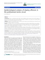
One of the cameras was placed in front of the table,
slightly to one side with respect to midline and contral-
ateral to the study side of the subject. The other camera
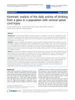
positioned laterally (Figure 1). The system was calibrated
by placing three active markers on the floor to serve as
the laboratory reference system. The coordinate system
was defined with the X-axis directed forward (ante-
riorly), the Y-axis upward (su periorly) and the Z-axis to
the side (laterally) [26]. The location o f the cameras and
markers was validated with a person sitting in the mea-
surement area to ensure that the markers were recorded
by least by one of the cameras throughout the drinking
activity.
Eighteen markers were used. Following the recom-
mendations of earlier studies, the body segments were
defined by placing 8 markers on the superficial bony
prominences of the right upper limb, which were easily
positioned in the different analyses [7,10,12,27,28].
These markers were placed on the head of the third
metacarpal, radial and ulnar styloid processes of the
wrist, lateral and medial epicondyles of the elbow,
right and left acromion and right iliac crest. The bio-
mechanical model of upper limb movement was com-
pleted with another 10 markers mounted on rigid
pieces that were placed on each body segment. These
pieces were used with the aim of minimizing any error
originated by possible marker displacement on the
skin. These pieces had to be light, comfortable for the
subject to w ear, and had to be fixed onto points where
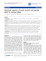
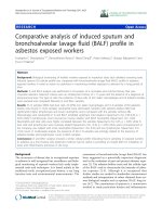
the least amount of movement was possible [22]. Four
markers were placed on the chest, three mounted on a
support and one directly on the skin; three markers
mounted on a support placed on the arm, and the last
three markers mounted on a support placed on the
forearm (Figure 2). The final position of the last 10
markers and the position of the cameras was the posi-
tion that yielded the best marker visibility to the scan-
ning cameras during the movement of drinking from a
glass and the best measurement results in the pro-
cessed recordings.
Experimental set-up and procedure
All subjects were right-handed and performed the
movement of the drinking task with the right arm.
Subjects with C6 or C7 tetraplegia sat in their own
wheelchairs and the control subjects sat in a conven-
tional wheelchair, Action3 Invacare (Invacare Corp,
Elyria OH, USA) with a configuration similar to that of
the wheelchair o f the subjects with tetraplegia. The
chair was placed before a table measuring 120 × 60 ×
72 cm. In every case, the subject-to-table distance wa s
18-20 cm and the angle between the seat and back was
90-100°. The starting po sition (position of calibration)
for all the subjects was defined as a position in which
Table 1 Demographic and functional characteristics of the sample analyzed (n = 24)
Control group (n = 8) C6 group (n = 8) C7 group (n = 8)
Sex (male)
†
345
Age (years)* 29.50 (4.00) 33.63 (13.03) 28.75 (9.82)
Height (cm)* 167.4 (3.24) 172.5 (8.91) 176.2 (8.89)
Weight (kg)* 62.00 (7.07) 70.25 (7.10) 68.37 (12.18)
Length of the right arm (cm)* 57.07 (2.34) 58.03 (3.21) 58.37 (3.90)
Injury etiology (Traumatic)
†
-88
Months since injury* - 8.50 (2.20) 7.50 (1.85)
ASIA (A)
†
-33
ASIA (B)
†
-55
Index Motor right arm (0-25)* 25.00 (0) 12.00 (2.07) 14.12 (2.03)
* Mean and standar desviation for continuous variables
† n for categorical variables
Figure 1 View from above of the set-up for the activity of
drinking from a glass. The XYZ coordinate system is visible. The
subject has the arm at the starting point.
de los Reyes-Guzmán et al. Journal of NeuroEngineering and Rehabilitation 2010, 7:41
/>Page 3 of 12
the subject’s trunk rested firmly against the back of the
chair. All subjects put their feet on the footrests with a
foot-leg angle of 90°. T he right upper arm was placed
against the trunk and the elbow was flexed 90° flexion
and in a ne utral pronation- supination, i. e., with t he
palm of the hand perpendicular to the table surface
and facing inward (medi ally). The ulnar side of the
wristrestedclosetothesurface of the table (Figure 1).
In every case, the sitting and table heights could be
adapted with the aim of obtaining the same starting
position for all the subject s. The subject rested the left
hand on the lap. A hard plastic glass measuring 6.5 cm
in diameter by 17.5 cm high was used. It was filled
with 1 dl of water and placed 18 cm from the edge of
the table where the subject was seated, in the area
marked on the table (Figure 1).
Each subject received an explanation about how to
perform the drinking task, which consisted of reaching
out for the glass from the starting position and grasping
it, raising the glass to the mouth, drinking, lowering the
glass to the pickup point, and returning the hand to the
starting position. All the subjects practiced the activity
twice to find a comfortable sitting position before the
movement exercise was recorded. This test confirmed
that the subjects could carry out the activity. Once this
phase was completed satisfactorily, a static calibration
recording was made. Using the static calibration record-
ing, we checked that each marker was visible to at least
one of the scanning cameras at all times. Movement
recordings were made as the subject executed the drink-
ing task at a comfortable, self-selected speed. Before a
recording was accepted as validated, we checked it to
ensure that the markers were visible at all times. Five
valid recordings of each subject were obtained for analy-
sis and processing.
The consistency and repeatability of the test protocol
was assessed by conducting a test-retest sequence with
four randomly selected control subjects. Test-retest
involved recording the action of the drinking task and
then removing the markers. The entire procedure was
repeated from the beginning fifteen minutes later.
Data processing
The reco rdings were processed with Visual 3D software
(C-Motion, Inc., USA), which involved using a signal
processing program to obtain signals of the movement
of different joints at a sampling frequency of 200 Hz,
the maximum allowed for the 18 markers used with the
two scanning units. Signals were filtered using a low-
pass Butterworth filter with a cutoff frequency of
1.5 Hz. The three best recordings were selected from
the five recordings made on the basis of best marker
visibility in each recording. The mean of these three
recordings yielded the final measurement value for
each subject. The human arm was modeled for t hree-
dimensional kinematic analysis in three segments, the
arm, forearm and hand, which were considered as rigid
solids [29]. A local coordinate system was defined for
each segment following the recommendations of the
International Society of Biomechanics [26]. In the arm,
the origin of the reference system was at the center of
the glenohumeral joint, 2 cm below the acromion. Also,
the Y-axis corresponded to the line that joined the mid-
point between the lateral and medial epicondyles and
the center of the glenohumeral joint in proximal direc-
tion and the Z-axis was the mediolateral axis pointing
to the right. In the forearm, the origin was at the mid-
point between both epicondyles of the elbow, the Y-axis
was formed by the line that joined the midpoint
between the radial and ulnar styloid processes with the
Figure 2 Actual marker positions on the subject. Figure show a) a frontal plane view (Y-Z) and b) a sagittal plane view (X-Y).
de los Reyes-Guzmán et al. Journal of NeuroEngineering and Rehabilitation 2010, 7:41
/>Page 4 of 12
midpoint between the lateral and medial epicondyles
proximally and the Z-axis was the line that joined both
epicondyles in the lateral direction. In the hand, the ori-
gin was at the midpoint between radial and ulnar styloid
ofthewrist,theY-axiswasthelinejoiningtheheadof
the third metacarpal with the midpoint between the
radial and ulnar styloid proce sses proximally and the Z-
axis joined both styloid processe s laterally. We obtained
trunk movement with respect to the laboratory coordi-
nate system, arm movement with respect to the trunk,
forearm movement with respect to the arm, and hand
movement with respect to the forearm using Euler angle
notation and a sequence of ZXY rotations of the trunk,
arm and hand, and ZYX rotations of the forearm.
In each recording, a complete cycle of the drinking
task was identified. The beginning of the cycle was the
onset o f displacement of the marker on the head of the
third metacarpal and the end of the cycle was the return
of the marker to the starting point after completing the
drinking task. As it happens with other cyclical move-
ments, such as walking, several phases were established
in the drinking task to facilitate task analysis. We used
phases and events delimiting the phases that have been
described previously: reaching, forward transport, drink-
ing, back transport and return [7].
Once the recordings were made and analyzed, the
results were described in terms of analysis of the follow-
ing variables:
• Movement times: the duration of each phase and
the complete cycle.
• Peak velocities: the velocities were obtained by cal-
culating the linear velocity with which the hand
moves in the phases of the cycle of reaching, forward
transport, back transport and return to start
position.
• Joint angles: flexion-extension and lateral inclina-
tion of the trunk; flexion-extension, abduction-
adduction and external-internal rotation of the
shoulder joint; flexion-extension and pronation-supi-
nation of the elbow joint; and dorsal-palmar flexion
of the wrist. For each joint angle, we calculated the
maximum, minimum, range of motion (ROM) and
moment in the complete drinking cycle in which
these values were reached.
• Coordination between the shoulder and elbow
joints, particularly between the shoulder flexion
angle and the elbow flexion angle, in the reaching
phase.
In order to compare the three groups analyzed, the
duration of the cycles was adjusted for time and
expressed as percentages. Consequently, data were
expressed in relation to the percentage of the drinking
task cycle that had lapsed (0-100% of the drinking task
cycle) when the movement was recorded.
Statistical analysis
A descriptive analysis was made of the clinical and func-
tional variables by calculating the median and interquar-
tile range of the quantitative variables and the
frequencies and percentages of the qualitative variables.
Given the limited number of participants, non-para-
metric methods were used. The Kruskal-Wallis test was
used to find possible differences in each variable
between the three groups analyzed; the Kruskal-Wallis
test began by testing the equivalence hypothesis between
groups. If the significance of the Kruskal-Wallis test is p
< 0.05, the equivalence of behavior between groups can
be rejected and a pairw ise comparison can be m ade
using the Mann-Whitney test. The Bonferroni correc-
tion was applied, which takes into account randomness
due to multiple comparisons.
The interrelation between the shoulder flexion angle
and the elbow flexion angle was analyzed in the reach-
ing phase using the Pearson correlation coefficient.
The repeatability of the protocol was evaluated with
the Student t test using a level of sign ificance of 0.05.
The mean difference between the test and retest values
was calculated for each of the four control subjects in
which test-retest consistency was analyzed with a 95%
confidence interval.
Data were analyzed statistically using the SPSS for
Windows (version 12.0) statistical package (SPSS Inc,
Chicago, IL, USA).
Results
Movement times
The duration of the complete cycle of the drinking
activity was more prolonged in the group of subjects
with C6 tetraplegia than in CG ( p < 0.05). In the phase
analysis the duration of the reaching phase was shorter
in CG than in subjects with C6 (p < 0.01) and C 7 tetra-
plegia (p < 0.05) (Table 2). In both groups of subjects
with tetraplegia, not only w as the reaching phase of
longer duration than in CG, it was also the longest
phase in the cycle. In the CG the longest phase in the
cycle was the back transport phase. The contribution of
each phase to the complete drinking task cycle in each
analyzed group is detailed in Tables 2.
Peak velocities
The peak velocities of the forward and back transport
phases were slower in subjects with C6 tetraplegia than
in subjects with C7 tetraplegia (p < 0.05 and p < 0.01,
respectively) and in CG (p < 0.05) (Table 2). In addition,
the peak velocity of the reaching phase was reached
later in controls than in the subjects with C6 tetraplegi a
de los Reyes-Guzmán et al. Journal of NeuroEngineering and Rehabilitation 2010, 7:41
/>Page 5 of 12
(p < 0.01) and C7 tetraplegi a (p < 0.05). In contrast, the
peak velocity of the forward transport phase was delayed
in the subjects with C6 tetraplegia (p < 0.01) and C7 tet-
raplegia (p < 0.01) compared to CG (Table 2).
Joint angles
In the sho ulder joint, only the mi nimum abduction
angle was greater in CG than in subjects with C7 tetra-
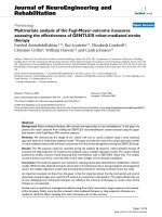
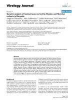
plegia (p < 0.01) (Table 3). In the elbow joint, the peak
minimum of flexion angle was smaller in the subjects
with C6 tetraplegia (p < 0.05) and C7 tetraplegia (p <
0.05) than in CG, but none of the differences in the
elbow flexion-extension ROM of the three groups was
statistically significant (Table 3 and Figure 3). None of
the joint angles analyzed in the trunk showed significant
differences (Table 3).
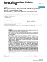
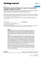
The wrist was the joint in which the most relevant dif-
ferences were found . The wrist palmar flexion angle was
greater in the two tetraplegia groups than in CG (p <
0.01), which made the ROM of wris t dorsal-palmar flex-
ion smaller in CG than in the subjects with C6 (p <
0.01) and C7 tetraplegia (p < 0.05). In CG, wrist palmar
flexion values were not even found in the cycle (Table
3). The maximum wrist palmar flexion in the thre e
groups was found in the back transport phase (at the
moment corresponding to 72.64% of the cycle of
subjects with C6 tetraplegi a, 73.45% of the cycle of sub-
jects with C7 tetraplegia and 71.38% of the cycle of CG)
(Figure 4). On the other hand, minimum wrist palmar
flexioninsubjectswithC6or C7 tetraplegia was in the
forward transport phase (at 36.54% and 22 .14% of the
cycle, respectively), and in CG it was in the back trans-
port phase (at 59.41% of the cycle) (Table 3).
Interjoint coordination between the shoulder and elbow
In the three study groups, there was a strong coordina-
tion between the shoulder and elbow joint angles. The
Pearson correlation index ranged from -0.95 (IR 0.08) to
-0.91 (IR 0.11) (Table 2). The negative value of the cor-
relation index meant that as shoulder flexion increased,
elbow extension also increased. The trajectory of these
correlations was c ontinuous, forming an almost linear
relation between the shoulder and elbow joint angles
(Figure 5).
Test-retest consistency
The statistical results of ten variables are shown in
Table 4. Mean retest values were within for the 95%
confidence interval of the first test. Based on this data,
we concluded that there were no differences between
the test and retest with a probability of 95%. However,
particularly for measures as maximum shoulder flexion,
Table 2 Duration of each phase of the drinking task cycle and peak velocities
Control group (n = 8) C6 group (n = 8) C7 group (n = 8)
Kinematic variables Median (IR) % mov time
Median (IR)
Median (IR) % mov time
Median (IR)
Median (IR) % mov time
Median (IR)
Movement times (s)
Reaching (+ grasping) 1.04 (0.33)
a,c
16.69 (3.86)
b,d
2.57 (0.98)
c
26.73(12.42)
b
1.66 (1.07)
a
21.83 (8.46)
d
Forward transport 0.91 (0.36) 31.55 (10.04)
a,b
1.20 (1.40) 41.76(10.97)
a
1.28 (0.81) 40.24 (8.46)
b
Drinking 1.37 (1.20) 54.97 (6.42) 1.96 (2.07) 59.75 (7.85) 1.41 (0.55) 54.48 (8.74)
Back transport 1.42 (0.39) 79.86 (4.92) 1.70 (0.96) 80.79 (5.58) 1.62 (1.18) 78.15 (8.24)
Returning 1.28 (0.25)
a
100 (0) 1.73 (0.68)
a
100 (0) 1.61 (0.62) 100 (0)
Total movement time 5.87 (1.80)
a
100 (0) 8.52 (5.16)
a
100 (0) 7.63 (4.25) 100 (0)
Peak velocity (PV) (m/s)
PV for reaching 0.66 (0.09) 5.68 (1.80)
a,c
0.56 (0.26) 2.57 (1.26)
c
0.67 (0.53) 3.42 (1.91)
a
PV for forward transport 0.69 (0.23)
a
22.07 (4.53)
c,d
0.44 (0.24)
a,b
32.69(10.75)
c
0.58 (0.60)
b
30.51 (13.11)
d
PV for back transport 0.72 (0.14)
a
63.39 (3.98) 0.54 (0.15)
a,c
67.60 (6.52) 0.79 (0.43)
c
60.73 (9.62)
PV for returning 0.63 (0.08) 87.82 (4.68)
c
0.52 (0.13) 92.46 (3.78)
c
0.52 (0.20) 89.40 (7.78)
Joints Coordination
Pearson index value -0.95(0.04) -0.91 (0.11) -0.95(0.08)
a, b (p < 0.05, with Bonferroni correction)
c, d (p < 0.01, with Bonferroni correction)
Joints Coordination: Coordination between shoulder and elbow flexion movements
Abbreviations: IR- Interquartile range
% mov ti me-% of total movement time
de los Reyes-Guzmán et al. Journal of NeuroEngineering and Rehabilitation 2010, 7:41
/>Page 6 of 12
maximum external rotation, maximum elbow flexion,
maximum pronation, even maximum wrist palmar flex-
ion, wide confidence intervals were obtained.
Discussion
The goal of this study was to analyze the three-dimen-
sional kinematic differences between two groups of peo-
ple with tetraplegia and a control group during the ADL
of drinking from a glass. The most relevant findings of
this study suggest that subjects with C6 tetraplegia per-
form the drinking task at a slower velocity and with
more prolonged phases. The greatest differences
between the two tetraplegia groups and controls were in
the wrist. A few studies have been made of the kine-
matic properties of the arms of patients with tetraplegia,
but none of them has analyzed an ADL [ 24,30,31]. The
slower velocity of subjects with C6 tetraplegia when
executing the drinking task coincides with the findings
Table 3 Angles of joints analyzed during the cycle of drinking task
Control group (n = 8) C6 group (n = 8) C7 group (n = 8)
Kinematic
variables
Median (IR) % mov time
Median (IR)
Median (IR) % mov time
Median (IR)
Median (IR) % mov time
Median (IR)
Shoulder
Max. Flexion 52.55 (21.31) 51.03 (13.60) 52.47 (11.29) 55.92 (10.92) 61.16 (22.77) 52.40 (8.66)
Min. Flexion -4.48 (10.70) 97.89 (3.34) 0.21 (28.41) 99.86 (2.02) -0.24 (11.01) 96.98 (8.45)
Range 59.53 (29.80) 45.49 (29.61) 63.75 (23.31)
Max. Abduction 29.77 (10.67) 83.67 (49.55) 23.00 (18.71) 60.35 (43.95) 29.37 (27.17) 69.15 (32.33)
Min. Abduction 13.55 (5.86)
c
13.35 (69.75) 8.40 (9.33) 28.08 (59.71) 0.13 (10.84)
c
13.51 (21.67)
Range 16.61 (14.98) 15.80 (14.79) 25.01 (16.19)
Max. Ext. Rot -4.74 (4.77) 95.68 (5.92) -9.90 (35.05) 91.13 (31.23) -16.98 (9.54) 97.15 (7.32)
Max. Int. Rot -45.59 (15.75) 68.62 (38.79) -38.71 (27.81) 48.42 (32.78) -48.89 (20.86) 63.81 (31.19)
Range 41.27 (8.29) 28.17 (17.06) 31.49 (36.22)
Elbow
Max. Flexion 128.20 (21.10) 46.25 (7.93)
a
113.40 (33.65) 53.84 (14.83)
a
114.05 (16.45) 45.30 (15.86)
Min. Flexion 64.42 (11.75)
a, b
77.27 (39.16) 42.54 (27.51)
a
79.15 (58.05) 42.11 (20.94)
b
75.58 (27.94)
Range 64.86 (27.45) 70.85 (20.64) 67.30 (26.56)
Max. Pronation 40.25 (22.37) 50.06 (4.79) 58.03 (33.33) 60.73 (30.52) 45.90 (23.73) 52.97 (16.02)
Min. Pronation 9.53 (22.75) 35.96 (48.62) 8.84 (22.20) 37.10 (58.00) -3.62 (22.60) 37.13 (49.71)
Range 33.07 (14.64) 55.17 (29.61) 49.59 (12.66)
Wrist
Max. Palmar Flexion -2.01 (10.84)
c, d
71.38 (55.94) 15.66 (24.92)
c
72.64 (12.24) 16.91 (16.79)
d
73.45 (11.97)
Min. Palmar Flexion -19.10 (5.41) 59.41 (53.51) -16.24 (15.04) 36.54 (14.83) -19.43 (13.27) 22.14 (31.51)
Range 14.98 (6.25)
a, b
33.46 (27.60)
a
37.58 (27.80)
b
Trunk
Max. Flexion -0.79 (7.03) 29.54 (28.50) 3.62 (18.66) 41.80 (58.28) 3.20 (15.66) 55.41 (33.10)
Min. Flexion -7.43 (11.84) 58.36 (22.15) -7.58 (14.10) 51.07 (16.99) -8.82 (9.87) 50.61 (13.38)
Range 6.38 (5.54) 7.86 (10.59) 11.37 (6.77)
Max. Lat. Incl. 5.01 (4.77) 57.34 (37.10) 5.21 (6.76) 65.45 (41.17) 10.28 (9.76) 48.34 (38.71)
Min. Lat. Incl. 2.82 (4.18) 63.99 (36.20) 2.50 (4.79) 35.42 (63.64) 6.17 (10.98) 24.26 (54.68)
Range 2.25 (1.93) 3.17 (5.26) 4.21 (3.52)
a, b (p < 0.05, with Bonferroni correction)
c, d (p < 0.01, with Bonferroni correction)
Abbreviations: IR - Interquartile Range
% mov ti me-% of movement time
de los Reyes-Guzmán et al. Journal of NeuroEngineering and Rehabilitation 2010, 7:41
/>Page 7 of 12
Figure 3 Elbow flexo-extension. Joint angles for elbow (extension-downward, flexion-upward). 0 degrees i s considered as full extension.
Figures 3a, 3b and 3c show the mean (continue thick line) and standard deviation (dashed line) of the CG, subjects with C6 tetraplegia and
subjects with C7 tetraplegia, respectively. The vertical lines delimit the duration of the phases for each group [1] reaching, [2] forward transport,
[3] drinking, [4] back transport, and [5] return to beginning.
Figure 4 Wrist dorsal-palmar flexion. Joint angles for wrist (dorsal flexion-downward, palmar flexion-upward). Figures 4a, 4b and 4c show the
mean (continue thick line) and standard deviation (dashed line) of the CG, subjects with C6 tetraplegia and subjects with C7 tetraplegia,
respectively. The vertical lines delimit the duration of the phases for each group [1] reaching, [2] forward transport, [3] drinking, [4] back
transport, and [5] return to beginning
de los Reyes-Guzmán et al. Journal of NeuroEngineering and Rehabilitation 2010, 7:41
/>Page 8 of 12
of previous studies that report that patients with C6 tet-
raplegia were slower than contro l subjects in performing
pointing movements on the horizontal plane [30,32]. On
the other hand, Wierzbicka et al. observed that the fast
elbow flexion movement, due to the lack of an antago-
nist, had an important effect on completion time of fast
goal-directed movements [31,33]. Finally, Laffont et al.
conc luded that in sp ite of some quantitative differ ences,
the kinematics of the hand during reaching and pointing
in quadriplegic patients are surprisingly simil ar to those
of control subjects [24].
However, more functional movements should be stu-
died. Previous studies of upper limb kinematics have
been made of control subjects performing ADLs such as
feeding, grooming and drinking [7,19,34,35]. These
movements are complex tasks in terms of kinematics
because they consist of several discrete movements.
Studies have analyzed upper limb kinematics in certain
specific groups, such as a normal pediatric population
[20] or groups of patients with conditions like cerebral
palsy or distal radius fracture performing certain func-
tional activities [22,36].
Figure 5 Shoulder-elbow joint coordination in the reaching phase for one randomly selected subject in the control group (red), C6
group (blue) and C7 group(black).
Table 4 Test-retest consistency for ten kinematic variables in 4 control subjects
Mean difference (95% CI) CI (95%) of mean difference T value p value
Max. Shoulder Flexion (°) -2.60 -19.37,13.95 -0.50 0.65
Max. Shoulder Abduction (°) -1.02 -7.54,5.50 -0.49 0.65
Max. External Rotation (°) -4.34 -12.87,18.20 -1.62 0.20
Max. Elbow Flexion (°) -2.48 -19.37,14.40 -0.46 0.67
Max. Pronation (°) 2.53 -12.33,17.40 0.54 0.62
Max. Wrist Palmar Flexion (°) -0.90 -10.30,8.49 -0.30 0.77
Max. Trunk Flexion (°) -1.67 -5.41,2.06 -1.42 0.25
Max. Trunk Lateral Inclination (°) -0.49 -3.11,2.12 -0.60 0.59
Movement time (s) -0.21 -0.65,0.22 -1.54 0.22
Peak velocity in reaching (m/s) -0.03 -0.09,0.03 -1.70 0.19
Abbreviations: CI - Confidence Interval
Value of the T statistic (Student t test).
de los Reyes-Guzmán et al. Journal of NeuroEngineering and Rehabilitation 2010, 7:41
/>Page 9 of 12
Much of the methodology developed in t he present
study followed the recommendations of a previous one
of healthy subjects in which five sequential phases of
drinking task were identified: reaching, forward trans-
port, drinking, back transport and returning [7]. How-
ever, the current experience has resolved previous
limitations and provides a full and detailed three-dimen-
sional kinematic analysis of the drinking task in contr ol
subjects and two groups of patients with tetraplegia,
analyzing the shoulder, elbow and wrist at all p ossible
joint angles except for lateral wrist inclination.
Using the upper limb model developed, we were able to
estimate the location of the center of the joints involved,
which made it possible to measure all the joint angles
described. Likewise, the use of mar kers mounted on rigid
pieces to position some of the markers helped t o reduce
tissue artifacts. These artifacts appear with limb displace-
ment when markers are placed on the skin surface.
It has been reported that trunk movement can act as
both a stabilizer and an integral component in position-
ing the hand close to the ta rget [37]. It ha s been shown
that hemiparetic subjects reaching within arm’slength
use a compensatory strategy that involves trunk displace-
ment [38,39]. In the present study, the glass was placed
within arm’s length and the subject could reach it with-
out separating the trunk from the back of the wheelchair.
Our findings confirmed those of earlier experience car-
ried out in contro l subjects, in which trunk displacement
was not relevant in the groups analyzed [36].
1. Movement times
The total duration of the drinking task was somewhat
shorter in our CG than in an earlier report, probably
because in the present study the palm of the hand was
closer to the drinking glass whereas in the earlier report
the wrist line was closer to the edge of the table [7].
However, both two studies had the same conclusion:
back transport is the most prolonged phase in controls
[7]. The duration of the drinking activity was longer in
subjects with C 6 tetraplegia compared to controls and
the duration of the reaching phase was longer in sub-
jects with C6 and C7 tetraple gia. As mentioned, the
reaching phase includes grasping. In order to grasp,
both groups of patients wit h tetraplegia developed a
compensatory strategy called “tenodes is,” in which these
patients extend the wrist to close the fingers p assively.
This pattern suggests that in subjects with tetraplegia
reaching and grasping are executed sequentially com-
pared to controls, who prepare for grasping during the
reaching phase [40].
2. Peak velocity
As the duration of the drinking task was shorter, the
velocity of each phase of the cycle in the controls was
somewhat faster than in a previous report [7]. T he
absence of triceps brachialis muscle activity in subjects
with C6 tetraplegia slows the velocity of the forward
transport and back transport phases, in which this mus-
cle controls the eccentric or concentric displacement of
the elbow in flexion-extens ion. As in an earlier study,
the peak velocity of the reaching phase was similar in
patients with tetraplegia and controls [24]. Another fac-
tor that could condition the v elocity of movements is
performing the movement with a load. The weakness of
the u pper limbs becomes more evident when raising an
object with a certain weight. In the absence of any addi-
tional load, peak velocity in the reaching phase is
reached earlier in groups of patients with tetraplegia.
However, in the forward transport phase in which the
glass of water is raised to the mouth, peak velocity is
notably faster in controls. It is difficult to compare the
velocities attained in other pathologies because they
have not been studied using the phases defined in our
study [23].
3. Joint angles
As in healthy subjects, but in contrast with subjects who
have experienced stroke and have a hemiparetic arm,
there was a strong coordination between shoulder and
elbow joint excursion in the reaching phase, indicating
good interjoint coordination in C6 and C7 tetraplegia
[10,12]. The wrist was th e joint with the most relevant
differences between the three groups. Wrist palmar flex-
ion angles were greater in both groups of subjects with
tetraplegia and the maximum wrist palmar flexion in
both cases was observed i n the back transport phase,
probably because no eccentric resistance is offered by
wrist e xtensor muscles as the glass is lowered from the
mouth to the table; passive wrist palmar flexion
occurred in both tetraplegia groups. The minimum wrist
palmar flexion angle was found in subjects with C6 or
C7 tetraplegia in the forward transport phase. This is
probably because at this time the subject required maxi-
mum wrist dorsal flexion to grasp a gl ass that has some
weight, which optimized the tenodesis effect and t he
ability to pick up an object. The elbow extension was
greater in both tetraplegia groups and occurred in the
back transport phase, perhaps also because elbow exten-
sion favored the tenodesis effect in the wrist.
4. Test-retest consistency
Mean retest va lues were within for the 95% confidence
interval of the first test. Based on this data, we con-
cluded that there were not differences between the test
and retest with a probability of 95%. However, for mea-
surements as maximum shoulder flex ion, maximum
external rotation, maximum elbow flexion, maximum
pronation, even maximum wrist palmar flexion, wide
de los Reyes-Guzmán et al. Journal of NeuroEngineering and Rehabilitation 2010, 7:41
/>Page 10 of 12
confi dence intervals were obtained. It could be probably
due to the natural large variation between the subjects
in those measurements. It is necessary to take into
account that people can perform a goal-oriented task
with many different combinations of individual joint
movements.
However, although the results obtained were sound,
for further research in this field, it would be good to
include another scanning unit to offer a view from
above with the aim of providing the best visibility of the
markers in the three space planes.
Robotic tools provide opportunities to study functional
adaptation after central nervous system injuries and can
provide objective measurements of the time-course of
changes in motor control of the affected limbs. Robot-
assisted therapy permits semi-autonomous practice of
therapeutic tasks [41]. In the last years, wearable tech-
nology has made an impact in the clinical setting and
recent studies have been focused on integrating this
technology with orthotic and prosthetic devices [42]. So,
wearable robots development can be one of the most
innovative therapeutic options for people with cervical
SCI, to improve upper limb functionality and so, facili-
tate the independence and quality of life during the per-
formance of ADL. In order to design these devices, it is
necessary to identify the movement patterns performed
by these patients during functional activities, such as the
drinking task. Then, these movement patterns can be
implemented into robotic devices to imitate or improve
these movements. In thi s research field, the present
study is of particular clinical relevance.
Conclusions
Kinematic analysis has shown great potential for use as
an outcome in clinical research to understand how func-
tional activities, such as drinking, are performed by
patients with upper limb impairment. The most relevant
differences were in the wrist, where the palmar flexion
values were greater in patients with C6 and C7 tetraple-
gia than in controls during the back transport phase,
whereas the highest wrist dorsal flexion value was in the
forward transport phase in subjects with C6 or C7 tetra-
plegia, in which complete activation of the tenodesis
effect is needed for grasping. This information can be
useful in designing wearable robots to compensate the
performance of AVD, such as drinking, in people with
cervical SCI.
Acknowledgements
This work was part of a project financed by FISCAM (Fundación para la
Investigación Sanitaria de Castilla-La Mancha, Spain) which does not have
any commercial interest in the results of this investigation. Ref no.: PI-2007-
09.
We thank Dr. Antonio Sánchez-Ramos (Head of Department of Physical
Medicine and Rehabilitation) for facilitating our work. We would like to
thank José Luis Rodríguez-Martín for his critical review of the manuscript
and methodology recommendations and Barbara Thomas and Elaine Van
Staalduinen for the revision of this manuscript in English.
Authors’ contributions
ARG contributed to the concept and design, planning of study, software
development, analysis and interpretation of the data, drafting and
completion of the manuscript. AGA contributed to design, analysis of the
data and completion of the manuscript. BPM contributed to the concept,
software development, design and acquisition of the data. MSM contributed
to the analysis and acquisition of the data. AAE contributed to the analysis
and acquisition of the data. EPR contributed to the software development.
All authors read and approved the manuscript to be published.
Competing interests
None of the authors of this paper has any conflict of interest in relation to
any sources of any kind pertinent to this study.
Received: 3 March 2010 Accepted: 20 August 2010
Published: 20 August 2010
References
1. Parker VM, Wade DT, Langton Hewer R: Loss of an arm function after
stroke: measurement, frequency and recovery. Int Rehabil Med 1986,
8(2):69-73.
2. Nakayama H, Jorgensen HS, Raaschou HO, Olsen TS: Compensation in
recovery of upper extremity function after stroke: the Copenhagen
Stroke Study. Arch Phys Med Rehabil 1994, 75(8):852-857.
3. Wyndaele M, Wyndaele JJ: Incidence, prevalence and epidemiology of
spinal cord injury: what learns a worldwide literature survey? Spinal Cord
2006, 44:523-529.
4. Broeks JG, Lankhorst GJ, Rumping K, Prevo AJ: The long-term outcome of
arm function after stroke: results of a follow-up study. Disabil Rehabil
1999, 21(8):357-364.
5. Wade DT: Measurement in neurological rehabilitation. Oxford medical
publications Oxford, Oxford Univ. Press 1992, 388.
6. Finch E: Physical rehabilitation outcome measures: a guide to enhanced
clinical decision making Hamilton, Ontario, Decker, 2 2002, ix:292.
7. Murphy MA, Sunnerhagen KS, Johnels B, Willen C: Three-dimensional
kinematic motion analysis of a daily activity drinking from a glass: a
pilot study. J Neuroeng Rehabil 2006, 3:18 [uroengrehab.
com/content/3/1/18].
8. Van Anden CJ, Wolterbeek N, Doorenbosch AM, Veeger HEJ, Harlaar J:
Complete 3D Kinematics of upper extremity functional tasks. Gait Posture
2008, 27:120-127.
9. McCrea PH, Eng JJ, Hodgson AJ: Biomechanics of reaching: clinical
implications for individuals with acquired brain injury. Disabil Rehabil
2002, 24(10):534-541.
10. Levin MF: Interjoint coordination during pointing movements is
disrupted in spastic hemiparesis. Brain 1996, 119(1):281-293.
11. Dwan LN, McIntosh AS: Kinematics of the upper limb: A reaching and
placing task with resistance in children. Gait & Posture 2006, 24(S):
S235-S238.
12. Cirstea MC, Levin MF: Compensatory strategies for reaching in stroke.
Brain 2000, 123(Pt 5):940-953.
13. Roby-Brami A, Feydy A, Combeaud M, Biryukova EV, Bussel B, Levin MF:
Motor compensation and recovery for reaching in stroke patients. Acta
Neurol Scand 2003, 107(5):369-381.
14. Roby-Brami A, Bennis N, Levin MF: Hand orientation for grasping and arm
joint rotation patterns in healthy subjects and hemiparetic stroke
patients. Brain Res 2003, 969(1-2):217-229.
15. Murray IA, Johnson GR: A study of the external forces and moments at
the shoulder and elbow while performing every day tasks. Clin Biomech
(Bristol Avon)
2004, 19(6):586-594.
16. Murgia A, Kyberd PJ, Chapell PH, Light CM: Marker placement to describe
the wrist movement during activities of daily living in cyclic tasks. Clin
Biomech (Bristol Avon) 2004, 19(4):248-254.
17. Safaee-Rad R, Shwedyk E, Quanbury AO, Cooper JE: Normal functional
range of motion of upper limb joints during performance of three
feeding activities. Arch Phys Med Rehabil 1990, 71(7):505-509.
de los Reyes-Guzmán et al. Journal of NeuroEngineering and Rehabilitation 2010, 7:41
/>Page 11 of 12
18. Trombly CA, Wu CY: Effect of rehabilitation tasks on organization of
movement after stroke. Am J Occup Ther 1999, 53(4):333-344.
19. Magermans DJ, Chadwick EKJ, Veeger HEJ, van der Helm FCT:
Requirements for upper extremity motions during activities of daily
living. Clin Biomech (Bristol, Avon) 2005, 20:591-599.
20. Petuskey K, Bagley A, Abdala E, James MA, Rab G: Upper extremity
kinematics during functional activities: Three-dimensional studies in a
normal pediatric population. Gait Posture 2007, 25:573-579.
21. Mosqueda T, James MA, Petuskey K, Bagley A, Abdala E, Rab G: Kinematic
assessment of the upper extremity in brachial plexus birth palsy. J
Pediatr Orthop 2004, 24(6):695-699.
22. Fitoussi F, Diop A, Maurel N, Laassel EM, Pennecot GF: Kinematic analysis
of the upper limb: a useful tool in children with cerebral palsy. J Pediatr
Orthop B 2006, 15(4):247-256.
23. Rönnqvist L, Rösblad B: Kinematic analysis of unimanual reaching and
grasping movements in children with hemiplegic cerebral palsy. Clin
Biomech (Bristol Avon) 2007, 22:165-17.
24. Laffont I, Briand E, Dizien O, Combeaud M, Bussel B, Revol M, Roby-Brami A:
Kinematics of prehension and pointing movements in C6 quadriplegic
patients. Spinal Cord 2000, 38(6):354-362.
25. Maynard FM, Bracken MB, Creasey G, Ditunno JF Jr, Donovan WH,
Ducker TB, Garber SL, Marino RJ, Stover SL, Tator CH, Waters RL,
Wilberger JE, Young W: International Standards for Neurological and
Functional Classification of Spinal Cord Injury. American Spinal Injury
Association. Spinal Cord 1997, 35:266-274.
26. Wu G, van der Helm F, Veeger HEJ, Makhsous M, Van Roy P, Anglin C,
Nagels J, Karduna A, McQuade K, Wang X, Werner F, Bucholz B: ISB
recommendation on definitions of joint coordinate systems of varius
joints for the reporting of human joint motion- Part II: shoulder, elbow,
wrist and hand. J Biomechanics 2005, 38:981-992.
27. Michaelsen SM, Luta A, Roby-Brami A, Levin MF: Effect of trunk restraint
on the recovery of reaching movements in hemiparetic patients. Stroke
2001, 32(8):1875-1883.
28. Turner-Stokes L, Reid K: Three-dimensional motion analysis of upper limb
movement in the bowing arm of string-playing musicians. Clin Biomech
(Bristol Avon) 1999, 14(6):426-433.
29. Biryukova EV, Roby-Brami A, Frolov AA, Mokhtari M: Kinematics of human
arm reconstructed from spatial tracking system recordings. J Biomech
2000, 33:985-995.
30. Popovic M, Popovic D: A new approach to reaching control for
quadriplegic patients. J Electromyogr Kinesiol 1994, 4:242-253.
31. Wierzbicka MM, Wiegner AW: Accuracy of motor responses in subjects
with and without control of antagonist muscle. J Neurophys 1996,
75(N86):2533-2541.
32. Popovic M, Tomovic R, Popovic D: Joint angle synergy in control arm
movements. Ser Aut Control 1993, 1:5-17.
33. Wierzbicka MM, Wiegner AW: Effects of weak antagonist on fast elbow
flexion movements in man. Exp Brain Res 1992, 91:509-519.
34. Packer TL, Peat M, Wyss U, Sorbie C: Examinating the elbow during
functional activities. Occup Ther J Res 1990, 10(6):323-333.
35. Cooper JE, Shweddyk E, Quandbury AO, Miller J, Hildebrand D: Elbow joint
restriction: effect on functional upper limb motion during performance
of three feeding activities. Arch Phys Med Rehabil 1993, 74:805-809.
36. Murgia A, Kyberd P, Barnhill T: The use of kinematic and parametric
information to highlight lack of movement and compensation in the
upper extremities during activities of daily living. Gait & Posture 2010,
31:300-306.
37. Kaminski TR, Bock C, Gentile AM: The coordination between trunk and
arm motion during pointing movements. Exp Brain Res 1995,
106(3):457-466.
38. Levin MF, Michaelsen SM, Cirstea CM, Roby-Brami A: Use of the trunk for
reaching targets placed within and beyond the reach in adult
hemiparesis. Exp Brain Res 2002, 143(2):171-180.
39. Michaelsen SM, Levin MF: Short-term effects of practice with trunk
restraint on reaching movements in patients with chronic stroke: a
controlled trial. Stroke 2004, 35(8):1914-1919.
40. Jaennerod M: The timing of natural prehension movements. J Mot Behav
1984, 16:235-254.
41. Johnson JMichelle: Recent trends in robot-assited therapy environments
to improve real-life functional performance after stroke. J Neuroeng
Rehabil 2006, 3:29 [ />42. Bonato P: Advances in wearable technology and applications in physical
medicine and rehabilitation. J Neuroeng Rehabil 2005, 2:2 [http://www.
jneuroengrehab.com/content/2/1/2].
doi:10.1186/1743-0003-7-41
Cite this article as: de los Reyes-Guzmán et al.: Kinematic analysis of the
daily activity of drinking from a glass in a population with cervical
spinal cord injury. Journal of NeuroEngineering and Rehabilitation 2010
7:41.
Submit your next manuscript to BioMed Central
and take full advantage of:
• Convenient online submission
• Thorough peer review
• No space constraints or color figure charges
• Immediate publication on acceptance
• Inclusion in PubMed, CAS, Scopus and Google Scholar
• Research which is freely available for redistribution
Submit your manuscript at
www.biomedcentral.com/submit
de los Reyes-Guzmán et al. Journal of NeuroEngineering and Rehabilitation 2010, 7:41
/>Page 12 of 12