Báo cáo hóa học: " Involvement of intracellular free Ca2+ in enhanced release of herpes simplex virus by hydrogen peroxide" ppt
Bạn đang xem bản rút gọn của tài liệu. Xem và tải ngay bản đầy đủ của tài liệu tại đây (464.07 KB, 9 trang )
BioMed Central
Page 1 of 9
(page number not for citation purposes)
Virology Journal
Open Access
Research
Involvement of intracellular free Ca
2+
in enhanced release of herpes
simplex virus by hydrogen peroxide
Emiko Arimoto
1
, Soichi Iwai
1
, Tetsuro Sumi
1
, Yuzo Ogawa
2
and
Yoshiaki Yura*
1
Address:
1
Department of Oral and Maxillofacial Surgery II, Osaka University Graduate School of Dentistry, Osaka, Japan and
2
Department of
Pathology, Osaka University Graduate School of Dentistry, Osaka, Japan
Email: Emiko Arimoto - ; Soichi Iwai - ; Tetsuro Sumi - ;
Yuzo Ogawa - ; Yoshiaki Yura* -
* Corresponding author
Abstract
Background: It was reported that elevation of the intracellular concentration of free Ca
2+
([Ca
2+
]i) by a calcium ionophore increased the release of herpes simplex virus type 1 (HSV-1).
Freely diffusible hydrogen peroxide (H
2
O
2
) is implied to alter Ca
2+
homeostasis, which further
enhances abnormal cellular activity, causing changes in signal transduction, and cellular dysfunction.
Whether H
2
O
2
could affect [Ca
2+
]i in HSV-1-infected cells had not been investigated.
Results: H
2
O
2
treatment increased the amount of cell-free virus and decreased the proportion of
viable cells. After the treatment, an elevation in [Ca
2+
]i was observed and the increase in [Ca
2+
]i
was suppressed when intracellular and cytosolic Ca
2+
were buffered by Ca
2+
chelators. In the
presence of Ca
2+
chelators, H
2
O
2
-mediated increases of cell-free virus and cell death were also
diminished. Electron microscopic analysis revealed enlarged cell junctions and a focal disintegration
of the plasma membrane in H
2
O
2
-treated cells.
Conclusion: These results indicate that H
2
O
2
can elevate [Ca
2+
]i and induces non-apoptotic cell
death with membrane lesions, which is responsible for the increased release of HSV-1 from
epithelial cells.
Background
Polymorphonuclear leukocytes (PMNs) have been
detected in the early cellular infiltrate at sites of herpes
simplex virus (HSV) infection [1]. It was also reported that
large numbers of PMNs infiltrated the mouse vaginal
mucosa within 24 h of the inoculation of HSV type 2 [2].
Activated inflammatory cells are a major source of oxida-
tive stress in inflammatory diseases and during secondary
inflammation after an initial toxic insult [3,4]. Exogenous
oxygen radicals can be also brought to the oral cavity, the
target of HSV type 1 (HSV-1) infection, for therapeutic
purpose [5-7]. These findings suggest that HSV-infected
epithelial cells can be exposed to oxygen radicals during
the infection cycle of HSV.
Freely diffusible hydrogen peroxide (H
2
O
2
) as an oxygen
radical can damage DNA directly by penetrating the cell
nucleus or indirectly by increasing the intracellular con-
centration of free Ca
2+
([Ca
2+
]i). The peroxidation of
membrane phospholipids leads to alterations in Ca
2+
Published: 31 August 2006
Virology Journal 2006, 3:62 doi:10.1186/1743-422X-3-62
Received: 05 June 2006
Accepted: 31 August 2006
This article is available from: />© 2006 Arimoto et al; licensee BioMed Central Ltd.
This is an Open Access article distributed under the terms of the Creative Commons Attribution License ( />),
which permits unrestricted use, distribution, and reproduction in any medium, provided the original work is properly cited.
Virology Journal 2006, 3:62 />Page 2 of 9
(page number not for citation purposes)
homeostasis, which further enhances abnormal cellular
activity, causing changes in signal transduction, and cellu-
lar dysfunction [8-12]. H
2
O
2
was cytotoxic to renal tubu-
lar epithelial cells and caused a sustained and
uncontrolled rise in [Ca
2+
]i that preceded substantial cell
injury or irreversible cell death [8].
With regard to viral infection and [Ca
2+
]i, many animal
viruses such as cytomegalovirus, poliovirus, coxsackie B3
virus, vaccinia virus, measles virus and rotavirus are
known to alter Ca
2+
homeostasis as a result of viral gene
expression [13-18]. [Ca
2+
]i is elevated after the binding of
HSV-1 to its cellular receptor [19]. In the previous study,
we found that a calcium ionophore, ionomycin, induced
Ca
2+
-dependent cell death and increased the virus release
from infected epithelial cells [20]. This suggests that Ca
2+
may be the stimulator of viral release. However, what
causes the elevation of [Ca
2+
]i in vivo has not been clari-
fied. In the present study, we examined the possibility that
H
2
O
2
could affect [Ca
2+
]i in HSV-1-infected epithelial
cells. The results suggest that H
2
O
2
is the candidate to pro-
mote the release of HSV-1 at the site of viral infection in a
[Ca
2+
]i-dependent manner.
Results
Effect of H
2
O
2
on the amounts of cell-free and cell-
associated virus
In the previous study, we treated HSV-1-infected cells with
a calcium ionophore, ionomycin, 18 h post infection
(p.i.) in order to detect its enhancing effect on the release
of HSV-1[20]. In this condition, most cells attached to the
plate and were releasing progeny viruses into culture
medium, although further incubation gradually increased
the number of detached cells. In the similar condition, we
examined the effect of H
2
O
2
on the release of HSV-1.
When FI cells were infected with HSV-1 at a multiplicity of
infection (MOI) of 2 plaque forming units (PFU)/cell, cul-
tured for 18 h and treated with H
2
O
2
at concentrations
ranging from 0.1 to 5 mM for 2 h, cell-free virus was
increased at 0.5, 1 and 5 mM; the increase at 1 and 5 mM
was significant as compared with the untreated control
(Fig. 1A). In contrast, the amount of cell-associated virus
was not significantly changed (Fig. 1B). In the absence of
H
2
O
2
, mean virus titers in cell-free and cell-associated
fractions were 4.6 × 10
6
and 1.1 × 10
8
PFU/ml. After treat-
ment with 1 mM H
2
O
2
for 2 h, mean virus titers in these
fractions were 2.6 × 10
7
and 1.1 × 10
8
PFU/ml, respec-
tively. A six-fold increase as compared with the untreated
control was observed in the cell-free fraction, but no
increase was observed in the cell-associated fraction. The
proportions of cell-free virus in the total amount of virus
in the presence or absence of H
2
O
2
were 22% and 4%,
respectively, indicating that H
2
O
2
markedly increased cell-
free virus in the cultures.
Effect of H
2
O
2
on [Ca
2+
] i in HSV-1-infected cells
It has been shown that H
2
O
2
caused a sustained and
uncontrolled rise in [Ca
2+
]i that preceded substantial cell
injury or irreversible cell death [8]. Whether H
2
O
2
could
affect the [Ca
2+
]i was examined at concentrations to
enhance the virus release. FI cells were infected with HSV-
1 at an MOI of 2 PFU/cell and cultured for 18 h. The mean
level of [Ca
2+
]i in HSV-1-infected cells was approximately
200 nM. When the infected cells were treated with 1 mM
H
2
O
2
, a significant rise in [Ca
2+
]i beginning approxi-
mately 30 sec after the exposure to H
2
O
2
was observed.
Subsequently, there was a secondary rise in [Ca
2+
]i, that
appeared within 40 sec; a maximal level (460 nM) was
attained in 6 min (Fig. 2A).
To determine the effect of calcium chelators, infected cells
were treated with an extracellular calcium chelating agent,
glycol-bis (beta-aminoethyl ether)-N',N',N',N'-tetraacetic
acid (EGTA), for 20 min until 18 h p.i., and then H
2
O
2
treatment was initiated. EGTA did not inhibit the imme-
diate rise in [Ca
2+
]i significantly, but suppressed the sec-
ondary rise at a low level (Fig. 2B). When HSV-1-infected
cells were exposed to an intercellular Ca
2+
chelator, 1,2-bis
(2-aminophenoxy)ethane- N',N',N',N'-tetraacetic acid
(BAPTA) or quin-2, for 20 min prior to the H
2
O
2
treat-
ment, both the initial and secondary rises in [Ca
2+
]i were
suppressed. Although the secondary rise was suppressed
by this treatment, the level of [Ca
2+
]i gradually increased
to 300–350 nM in 8 min (Fig. 2C and 2D).
Effect of H
2
O
2
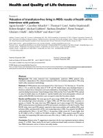
on the amount of cell-free virus and cell-asso-ciated virusFigure 1
Effect of H
2
O
2
on the amount of cell-free virus and
cell-associated virus. FI cells were infected with HSV-1 at
an MOI of 2 PFU/cells and cultured for 18 h. Thereafter, cells
were treated with H
2
O
2
at concentrations of 0.1, 0.5, 1 and 5
mM for 2 h, and the amounts of cell-free virus (A) and cell-
associated virus (B) in the cultures were determined by
plaque assay. Results were compared to those for the con-
trols and a percentage was calculated. Data are means ± SD
of three determinations. Differences of means were analyzed
with the unpaired t-test. * P < 0.05, ** P < 0.01 and ** P <
0.001 vs. samples exposed to H
2
O
2
only.
Virus titer (% of control)
0
200
400
600
800
1000
0.1 0.5 1 5
H
2
O
2
(mM)
A
0.1 0.5 1 5
H
2
O
2
(mM)
Virus titer (% of control)
0
200
400
600
800
1000
1200
1400
***
***
B
Virology Journal 2006, 3:62 />Page 3 of 9
(page number not for citation purposes)
Effect of buffering [Ca
2+
]i on H
2
O
2
-mediated
enhancement of viral release
The effect of Ca
2+
depletion on the release of HSV-1 was
examined. Eighteen hours after infection, cells were pre-
treated with 10 mM EGTA for 20 min to deplete extracel-
lular Ca
2+
. Thereafter, treatment with 1 mM H
2
O
2
for 2 h
was initiated. In this condition, the amount of cell-free
virus was 150% of that in the untreated control, whereas
it was increased to 450% of the control value by the treat-
ment with H
2
O
2
(Fig. 3A). The amounts of cell-free virus
in the presence of 50 μM BAPTA and 50 μM quin-2 were
250 % and 230 % of the control, respectively, indicating
that the H
2
O
2
-mediated increase was diminished by
BAPTA and quin-2. The amount of cell-associated virus in
the cultures was not significantly altered by H
2
O
2
in com-
bination with EGTA, BAPTA or quin-2 (Fig. 3B). When
HSV-1-infected cells were treated with EGTA, BAPTA or
quin-2 only, the amount of cell-free virus was unchanged
as compared with that in the untreated control (data not
shown).
Effect of H
2
O
2
on [Ca
2+
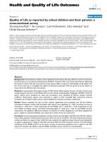
]i in HSV-1-infected cellsFigure 2
Effect of H
2
O
2
on [Ca
2+
]i in HSV-1-infected cells. HSV-1-infected FI cells were cultured for 18 h. Thereafter, the medium
was replaced with Hank's solution and the [Ca
2+
]i was monitored during treatment with 1 mM H
2
O
2
(A). Alternatively,
infected cells were treated with 10 mM EGTA (B), 50 μM BAPTA (C) or 50 μM quin-2 (D) for 20 min prior to treatment with
1 mM H
2
O
2
. Results are representative of 7 independent experiments.
0
100
200
300
400
500
30 180 360 540 (S)
1mM H
2
O
2
[Ca
2+
]i (nM)
0
100
200
300
400
500
30 180 360 540 (S)
1mM H
2
O
2
0
100
200
300
400
500
30 180 360 540 (S)
1mM H
2
O
2
[Ca
2+
]i (nM)
[Ca
2+
]i (nM)
[Ca
2+
]i (nM)
30 180 360 540 (S)
0
100
200
300
400
500
1mM H
2
O
2
A
B
C
D
Virology Journal 2006, 3:62 />Page 4 of 9
(page number not for citation purposes)
Effect of H
2
O
2
and buffering [Ca
2+
]i on cell viability
The effect of H
2
O
2
on cell viability was examined by
trypan blue exclusion. In mock-infected FI cells, the pro-
portion of trypan blue-positive dead cells was 8%. After
treatment with 1 mM H
2
O
2
for 2 h, 28% of cells were pos-
itive trypan blue (Fig. 4A). When cells were infected with
HSV-1 at an MOI of 2 PFU/cell and cultured for 20 h, 29%
of cells were stained. After the treatment with 1 mM H
2
O
2
from 18 to 20 h p.i., the proportion of dead cells was
increased to 56% (Fig. 4B). The only detectable morpho-
logical change of H
2
O
2
-treated cells was enlargement of
intercellular space due to cell rounding, irrespective of
HSV-1 infection.
To determine the effect of Ca
2+
chelators, HSV-1-infected
cells were pretreated with 10 mM EGTA, 50 μM BAPTA or
50 μM quin-2 for 20 min and then treated with 1 mM
H
2
O
2
for 2 h. In the presence of EGTA, BAPTA and quin-
2, the proportions of dead cells in H
2
O
2
-treated cultures
were 38%, 34% and 36%, respectively, indicating that
Ca
2+
chelators reversed the effect of H
2
O
2
(Fig. 5). When
HSV-1-infected cells were treated with EGTA, BAPTA or
quin-2 only, there were no changes in the proportion of
dead cells (data not shown).
Flow cytometric analysis of the H
2
O
2
-treated cells
A number of studies have shown that H
2
O
2
induced apop-
tosis with DNA fragmentation [8-11]. To clarify this issue,
DNA was labeled by propidium iodide (PI) and subjected
to flow cytometric analysis. In mock-infected cells treated
with 1 mM H
2
O
2
for 2 h, there were no apparent changes
in the pattern of the cell cycle as compared with the
untreated control (Fig. 6A and 6B). However, after treat-
ment for 24 h, a sub-G1 peak appeared (Fig. 6C), indicat-
ing the induction of DNA fragmentation. When FI cells
were infected with HSV-1 at an MOI of 2 PFU/cell and cul-
tured for 18 h, the profile of DNA content was different
from that of mock-infected cells. A broad peak was
observed at the position of G
0
/G
1
and the population of
Effect of Ca
2+
depletion on cell viabilityFigure 5
Effect of Ca
2+
depletion on cell viability. HSV-1-infected
FI cells were treated with 1 mM H
2
O
2
from 18 to 20 h p.i.
and then trypan blue-positive cells were determined. For the
depletion of extracellular Ca
2+
or [Ca
2+
]i, infected cells were
pretreated with 10 mM EGTA, 50 μM BAPTA or 50 μM
quin-2 for 20 min. Differences of means were analyzed with
the unpaired t-test. * P < 0.05 and ** P < 0.01 vs. samples
exposed to H
2
O
2
only.
ޓޓ
ޓޓޓޓ
ޓޓ
noneޓ H
2
O
2
H
2
O
2
H
2
O
2
H
2
O
2
ޓޓޓ
+ + +
EGTA ޓޓBAPTA quin-2
Cell death (%)
0
20
40
60
80
100
*
*
**
Effects of Ca
2+
depletion on viral releaseFigure 3
Effects of Ca
2+
depletion on viral release. HSV-1-
infected FI cells were treated with 1 mM H
2
O
2
from 18 to 20
h p.i. Alternatively, infected cells were pretreated with 10
mM EGTA, 50 μM BAPTA or 50 μM quin-2 for 20 min prior
to H
2
O
2
treatment for 2 h. After treatment with H
2
O
2
, the
amounts of cell-free virus (A) and cell- associated virus (B)
were determined. Results were compared to those for the
controls and a percentage was calculated. Data are means ±
SD of three determinations. Differences of means were ana-
lyzed with the unpaired t-test. * P < 0.05, ** P < 0.01 and ** P
< 0.001 vs. samples exposed to H
2
O
2
only.
㧴
㧞
㧻
㧞
㧴
㧞
㧻
㧞ޓ
㧴
㧞
㧻
㧞ޓޓ
㧴
㧞
㧻
㧞
㧗ޓޓ 㧗ޓ 㧗ޓޓޓ
EGTA BAPTA quin-2
A
0
100
200
300
400
500
Virus titer (% of control)
**
**
***
B
0
100
200
300
400
500
Virus titer (% of control)
㧴
㧞
㧻
㧞
㧴
㧞
㧻
㧞ޓ
㧴
㧞
㧻
㧞ޓޓ
㧴
㧞
㧻
㧞
㧗ޓޓ 㧗ޓ 㧗ޓޓޓ
EGTA BAPTA quin-2
Effect of H
2
O
2
on cell viabilityFigure 4
Effect of H
2
O
2
on cell viability. FI cells were treated with
1 mM H
2
O
2
and stained with trypan blue (A). HSV-1-infected
FI cells were treated with 1 mM H
2
O
2
from 18 to 20 h p.i.
and then trypan blue-positive cells were determined (B).
Data are means ± SD of three determinations. Differences of
means were analyzed with the unpaired t-test. * P < 0.05 and
** P < 0.01 vs. samples exposed to H
2
O
2
only.
H
2
O
2
(mM)
0 1 5
Cell death (%)
0
20
40
60
80
100
A
*
*
H
2
O
2
(mM)
Cell death (%㧕
B
**
**
0
20
40
60
80
100
0 1 5
Virology Journal 2006, 3:62 />Page 5 of 9
(page number not for citation purposes)
G
2
/M phase was decreased (Fig. 6D), indicating the distur-
bance of cell cycle due to HSV-1 infection. Even if infected
cells were treated with 1 mM H
2
O
2
for 2 h or 24 h, a spe-
cific sub-G1 peak was not demonstrated (Fig. 6E and 6F)
When HSV-1-infected cells were treated with 1 mM H
2
O
2
from 18 to 20 h after infection and subjected to Hoechst
staining and annexin V staining, increase of apoptotic
cells was not demonstrated (data not shown)
Electron microscopic observation
To gain further insight into the alterations caused by
H
2
O
2
, electron microscopy was used. The cultures were
fixed in situ and sections parallel to the dish surface were
prepared. HSV-1-infected cells had large vesicular nuclei
with dispersed chromatin. In the portion where cell-to-
cell interaction was tight, a large number of viral particles
were pooled in a narrow intercellular space (Fig. 7A and
7B). When HSV-1-infected cells were treated with 1 mM
H
2
O
2
from 18 to 20 h p.i., ruffling of the nuclear mem-
brane and clustering of condensed chromatin at the
nuclear periphery were observed, but the nuclear and
cytoplasmic density was apparently unaltered. Cell
shrinkage observed in apoptotic cells was not demon-
strated. Generally, cell-to-cell junctions were enlarged,
and as a consequence, viral particles pooled in the space
were lost (Fig. 7C). Although the integrity of most of the
plasma membrane was preserved, there were bubble-like
structures that arose from the cell membrane (Fig. 7E).
Occasionally, rapture of vacuoles containing organelles
was observed on the cell surface (Fig. 7D). A focal defect
of the plasma membrane was observed adjacent to trans-
port vesicles containing viral particles at cell periphery
(Fig. 7F and 7G).
Flow cytometric analysis of DNA fragmentationFigure 6
Flow cytometric analysis of DNA fragmentation. Untreated FI cells (A) and FI cells treated with 1 mM H
2
O
2
for 2 h (B)
or 24 h (C) were subjected to flow cytometric analysis. FI cells were infected with HSV-1 at an MOI of 2 PFU/cell and cultured
for 20 h (D). HSV-1-infected cells were treated with 1 mM H
2
O
2
from 18 to 20 h p.i. (E) or from 18 to 42 h p.i. (F). These
infected cells were also subjected to flow cytometric analysis.
A
B
C
D
E
F
Virology Journal 2006, 3:62 />Page 6 of 9
(page number not for citation purposes)
Discussion
We found that treatment with 1 mM H
2
O
2
for 2 h signifi-
cantly increased the amount of cell-free virus. If H
2
O
2
could affect the step of virus release only, the increase of
cell-free virus would be accompanied by the decrease of
cell-associated virus, but the amount of cell-associated
virus was not altered. This suggested that the total amount
of infectious virus in the cultures was rather increased.
Many factors such as cell proliferation and activity of pro-
tein and DNA synthesis will influence virus release and
infectivity. It is possible that oxidative stress promotes the
steps of transport and/or maturation of virus particles.
Alternatively, H
2
O
2
-induced increase of [Ca
2+
]i may have
an advantage of the infectivity of virions, because HSV-1
envelope was implicated to be sensitive to calcium deple-
tion [21]. In any case, it is apparent that the proportion of
cell-free virus in the cultures was markedly increased after
treatment with H
2
O
2
. H
2
O
2
must increase the release of
HSV-1 at the final step of viral replication.
H
2
O
2
exerts its effect through a second messenger, Ca
2+
,
which may play a critical role in cellular events [8-12] and,
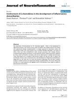
Electron microscopic observationFigure 7
Electron microscopic observation. FI cells were infected with HSV-1 at an MOI of 2 PFU/cell and cultured for 20 h (A, B).
The infected cells were also treated with 1 mM H
2
O
2
for 18 to 20 h p.i. (C to G). To examine cell-to-cell interaction, cultures
were fixed in situ and embedded in epoxy resin. Sections were cut parallel to the surface of the dishes. Bar, 1 μm
A
B
C D
E
F
G
Virology Journal 2006, 3:62 />Page 7 of 9
(page number not for citation purposes)
probably, the process of HSV-1 replication. In the present
study, there were two stages to the rise in [Ca
2+
]i ; an ini-
tial peak which appeared just after the addition of H
2
O
2
,
followed by a secondary increase which persisted for some
time. The removal of extracellular Ca
2+
by EGTA dimin-
ished the second rise in [Ca
2+
]i in response to H
2
O
2
, indi-
cating that the secondary increase was due to Ca
2+
influx.
The first peak was caused by the mobilization of Ca
2+
from
intracellular stores [12,20] and both rises in [Ca
2+
]i were
suppressed by the buffering agents BAPTA and quin-2
[22]. It is likely that H
2
O
2
increases [Ca
2+
]i through the
release of Ca
2+
from intracellular stores and Ca
2+
influx in
HSV-1-infected cells. Since the buffering of [Ca
2+
]i by Ca
2+
chelators diminished the effect of H
2
O
2
on the release of
HSV-1, we concluded that the enhanced viral release fol-
lowing H
2
O
2
treatment was ascribed to a Ca
2+
-mediated
mechanism.
Oxygen radicals act as an inducer of apoptosis by elevat-
ing [Ca
2+
]i [9,11]. We found that a short-term treatment
with H
2
O
2
increased the number of dead cells in HSV-1-
infected cultures and the effect was diminished in the
presence of calcium chelators. However, a specific sub-G
1
peak indicating apoptosis was not detected after
H
2
O
2
treatment for 2 h by a flow cytometric analysis.
Induction of apoptosis was not demonstrated by Hoechst
staining and annexin V staining. Thus, the H
2
O
2
-induced
cell death occurred in this situation was not apoptosis.
The apoptosis of HSV-1-infected cells by H
2
O
2
may be
prevented the function of anti-apoptotic genes such as
Us3, ICP27 and γ
1
34.5 of HSV-1 [23-25].
The plasma membrane is the primary target of cell injury
and the functional consequence of damage to this mem-
brane is a lethal influx of extracellular Ca
2+
into the cells
[26]. We also indicated that treatment of HSV-1-infected
epithelial cells with ionomycin induced the increase of
Ca
2+
influx, followed by cell death and the leakage of virus
particles [20]. In the present study, H
2
O
2
-induced cell
death was accompanied by the elevation of [Ca
2+
]i. Fur-
thermore, with the use of an electron microscope, mem-
brane protrusion, a bursting bubbles and a leakage of
virus particles in H
2
O
2
-treated cells were observed. Thus,
we concluded that the H
2
O
2
-induced cell death was char-
acterized by a focal disintegration of the plasma mem-
brane and partial loss of cytoplasmic contents, leading to
the enhanced release of virus particles to the extracellular
space. It should be also stated that the integrity of the
nucleus and cytoplasmic density were preserved to pro-
duce progeny virus and the release of virus particles dur-
ing the H
2
O
2
-induced cell death.
Another finding was that a number of cell-free viral parti-
cles were pooled at narrow cell junctions and were lost
after treatment with H
2
O
2
, because of the enlargement of
cell-to-cell junctions. As a function of a rise in [Ca
2+
]i, the
cytoskeletal architecture and rigid intercellular connec-
tions are altered [27,28], which will result in the libera-
tion of trapped viral particles from cell junctions. This
must contribute to the increase in the amount of cell-free
virus in HSV-1-infected cell cultures.
Oxygen radicals, such as H
2
O
2
, O
2
•-
and HO
•
, are highly
reactive molecules with unpaired electrons that are gener-
ated in normal physiological processes such as aerobic
metabolism or inflammation. PMNs generate both extra-
cellular and intracellular oxygen radicals and the released
oxygen radicals impair the host tissues [29,30]. The maxi-
mal H
2
O
2
concentration was reported to be 0.3 mM after
an activation of human PMNs [31]. Although 0.5 mM
H
2
O
2
increased cell-free virus (Fig. 1), we performed most
experiments at H
2
O
2
concentration of 1 mM. We specu-
late that a similar event would occur in vivo, because
other PMN-derived oxygen radicals such as O
2
•-
and HO
•
also exhibit cytotoxic effect [32]. In other systems to study
the neuronal cell death and renal tubular cell injury by
oxygen radicals, H
2
O
2
was used at 1 mM [8,10]. Histolog-
ical changes of skin vesicles due to HSV infection repre-
sent a combination of virally mediated cellular death and
associated inflammatory response [33]. Oxygen radicals
produced by inflammatory cells may promote the devel-
opment of herpetic vesicular lesions by increasing the
virus particles in the fluid. In mucosal lesions, more cell-
free virus particles would be released from the ulcerative
surface by the action of oxygen radicals and contribute to
the spread of viral infection. Oxygen radicals also act as
the mediators of anticancer agents [34,35]. This means
that HSV-1 infection, irrespective of primary and recurrent
infection, can be modified by antineoplasic agents, which
may lead to the development of oral mucositis during
antineoplastic chemotherapy [36]. From the aspect of
exogenous oxygen radicals, H
2
O
2
is used as a disinfectant,
hemostatic or bleaching agent for colored tooth at a con-
centration of approximate 1 M. It can be a stimulator of
viral release after a dilution to the level of mM in the oral
cavity.
Conclusion
Previously, we reported that a calcium ionophore, iono-
mycin, enhanced the release of HSV-1. Here, we indicated
that treatment with H
2
O
2
disrupted cell-to-cell interac-
tions, increased dead cells, and accelerated viral release
through a Ca
2+
-mediated mechanism. H
2
O
2
can be the
candidate that elevates [Ca
2+
]i and promotes the release of
HSV-1 in vivo.
Methods
Cell culture and virus
Oral squamous cell carcinoma FI cells [37] were used as
an epithelial cell line throughout the experiments. FI cells
Virology Journal 2006, 3:62 />Page 8 of 9
(page number not for citation purposes)
were grown in Dulbecco's modified Eagle's medium con-
taining 5% fetal bovine serum and supplemented with a
penicillin-streptomycin antibiotic mixture. The stock of
HSV-1 strain KOS was grown and infectivity was deter-
mined by plaque assay in Vero cells.
Preparation of cell-free viral and cell-associated viral
fractions
To measure the amounts of cell-free virus, FI cells were
infected with HSV-1 at an MOI of 2 PFU/cell. Thereafter,
the infected cells were cultured for 18 h and then treated
with H
2
O
2
. The culture plates were centrifuged at 400 × g
for 5 min and the supernatant was harvested as a cell-free
fraction and stored at -80°C until use. An equal volume of
medium was added to each culture plate. For the measure-
ment of cell-associated virus in a culture, the cells were
subjected to two cycles of freezing and thawing. They were
then centrifuged and the supernatant was harvested as a
cell-associated fraction and stored at -80°C. The viral titer
in each fraction was measured by assaying the formation
of plaques in Vero cell monolayers and means of three
determinations were obtained. Results were compared to
those for the untreated controls and a percentage value
was calculated. Differences of means were analyzed with
the unpaired t-test.
Measurement of [Ca
2+
]i
[Ca
2+
]i was measured using the fluorescent Ca
2+
indicator
fura-2, which was incorporated intracellularly as its ace-
toxymethyl ester (fura-2/AM; Calbiochem, Cambridge,
MA, USA). Cells were grown on glass-based plastic dishes
and incubated with 4 μM fura-2/AM in DMEM for 30 min
at 37°C. Cells were then washed in modified Hank's solu-
tion (Sigma) containing 137 mM NaCl, 3.5 mM KCl, 0.44
mM KH
2
PO
4
, 25 mM NaHCO
3
, 0.33 mM Na
2
HPO
4
and
0.5 mM CaCl
2
for a further 20 min at room temperature.
To deplete extracellular Ca
2+
, cells were treated with 10
mM EGTA (Calbiochem) for 10 min prior to the H
2
O
2
treatment. For buffering [Ca
2+
]i, cells were pretreated with
50 μM of the acetoxymethyl ester of BAPTA (BAPTA/AM;
Calbiochem) or 50 μM of the acetoxymethyl ester of quin-
2 (quin-2/AM; Calbiochem) for 10 min. After the addi-
tion of H
2
O
2
, [Ca
2+
]i was measured in individually iden-
tified fura-2-loaded cells using alternating excitation
wavelengths (340 and 380 nm) with an AQUACOSMOS
ratio imaging application software (HAMAMATSU Phot-
onics, Hamamatsu, Japan) and an inverted epifluores-
cence microscope (DIAPHOT 300, Nikon). In order to
evaluate its ability to quantify [Ca
2+
]i, the instrument was
tested on Ca
2+
buffer solutions (Molecular Probes) with
known values of [Ca
2+
]i, using fura-2/AM [38]; 7 cells
were monitored for each experiment.
Trypan blue staining
Cell viability was determined by trypan blue dye exclu-
sion analysis. Cells dissociated by the EDTA-trypsin solu-
tion were mixed with an equal volume of phosphate-
buffered saline containing 0.24% trypan blue and
observed with a microscope. We counted the numbers of
stained and unstained cells. Results were compared to
those for the untreated controls and a percentage value
was calculated. Differences of means were analyzed with
unpaired t-test.
Flow cytometric analysis
FI cells were dissociated in the EDTA-trypsin solution. Iso-
lated cells were added to ice-cold 70% ethanol and then
incubated at -20°C for 4 h. Thereafter, cells were centri-
fuged and incubated with phosphate-citrate buffer for 30
min at room temperature. They were again centrifuged,
incubated with 10 μg/ml PI and 10 μg/ml RNase A for 20
min at room temperature, and then analyzed with a Bec-
ton Dickinson FACSort (Becton Dickinson, San Jose, CA).
Electron microscopy
Cells grown on plastic dishes were fixed in 2% glutaralde-
hyde (TAAB, Berkshire, England) for 2 h, washed with
sodium cacodylate buffer and then postfixed in 1%
osmium tetroxide (TAAB) for 2 h. Thereafter, cells were
dehydrated in a graded series of ethanol and flat embed-
ded in epoxy resin. Sections were cut parallel to the surface
of the dishes. They were then stained with 4% uranyl ace-
tate and 0.1% lead citrate (TAAB) and examined with a
HITACHI H-7500 electron microscope.
Competing interests
The author(s) declare that they have no competing inter-
ests.
Authors' contributions
EA and YY conceived of the study, analyzed the results and
wrote the manuscript. SI measured [Ca
2+
]i; TS performed
flow cytometric analysis; YO carried out electron micro-
scopic study. All authors read and approved the final man-
uscript.
Acknowledgements
This work was supported in part by a Grant-in-aid (16390586) for Scientific
Research from the Ministry of Education, Science and Culture of Japan.
References
1. Watanabe D, Adachi A, Tomita Y, Yamamoto M, Kobayashi M, Nishi-
yama Y: The role of polymorphonuclear leukocyte infiltration
in herpes simplex virus infection of murine skin. Arch Dermatol
Res 1999, 291:28-36.
2. Milligan GN: Neutrophils aid in protection of the vaginal
mucosae of immune mice against challenge with herpes sim-
plex virus type 2. J Virol 1999, 73:6380-6386.
3. Repine JE, Cheronis JC, Rodell TC, Linas SL, Patt A: Pulmonary oxy-
gen toxicity and ischemia-reperfusion injury. A mechanism
in common involving xanthine oxidase and neutrophils. Am
Rev Respir Dis 1987, 136:483-485.
Publish with BioMed Central and every
scientist can read your work free of charge
"BioMed Central will be the most significant development for
disseminating the results of biomedical research in our lifetime."
Sir Paul Nurse, Cancer Research UK
Your research papers will be:
available free of charge to the entire biomedical community
peer reviewed and published immediately upon acceptance
cited in PubMed and archived on PubMed Central
yours — you keep the copyright
Submit your manuscript here:
/>BioMedcentral
Virology Journal 2006, 3:62 />Page 9 of 9
(page number not for citation purposes)
4. Werns SW, Lucchesi BR: Leukocytes, oxygen radicals, and myo-
cardial injury due to ischemia and reperfusion. Free Radic Biol
Med 1988, 4:31-37.
5. Persson LG, Mouhyi J, Berglundh T, Sennerby L, Lindhe J: Carbon
dioxide laser and hydrogen peroxide conditioning in the
treatment of periimplantitis: an experimental study in the
dog. Clin Implant Dent Relat Res 2004, 6:230-238.
6. Hannig C, Zech R, Henze E, Dreier S, Attin T: Peroxide release
into saliva from five different home bleaching systems in
vivo. Am J Dent 2005, 18:13-18.
7. Yalcin F, Gurgan S: Effect of two different bleaching regimens
on the gloss of tooth colored restorative materials. Dent
Mater 2005, 21:464-468.
8. Ueda N, Shah SV: Role of intracellular calcium in hydrogen per-
oxide-induced renal tubular cell injury. Am J Physiol 1992,
263:214-221.
9. Whittemore ER, Loo DT, Watt JA, Cotman CW: A detailed anal-
ysis of hydrogen peroxide-induced cell death in primary neu-
ronal culture. Neuroscience 1995, 67:921-932.
10. Oyama Y, Okazaki E, Chikahisa L, Nagano T, Sadakata C: Oxidative
stress-induced increase in intracellular Ca2+ and Ca(2+)-
induced increase in oxidative stress: an experimental model
using dissociated rat brain neurons. Jpn J Pharmacol 1996,
72:381-385.
11. Kim DK, Cho ES, Um HD: Caspase-dependent and -independ-
ent events in apoptosis induced by hydrogen peroxide. Exp
Cell Res 2000, 257:82-88.
12. Nam SH, Jung SY, Yoo CM, Ahn EH, Suh CK: H
2
O
2
enhances Ca
2+
release from osteoblast internal stores. Yonsei Med J 2002,
43:229-235.
13. Irurzun A, Arroyo J, Alvarez A, Carrasco L: Enhanced intracellular
calcium concentration during poliovirus infection. J Virol 1995,
69:5142-5146.
14. Keay S, Baldwin BR, Smith MW, Wasserman SS, Goldman WF:
Increases in [Ca
2+
]i mediated by the 92.5-kDa putative cell
membrane receptor for HCMV gp86. Am J Physiol 1995,
269:11-21.
15. Nokta M, Eaton D, Steinsland OS, Albrecht T: Ca
2+
responses in
cytomegalovirus-infected fibroblasts of human origin. Virol-
ogy 1987, 157:259-267.
16. Perez JF, Chemello ME, Liprandi F, Ruiz MC, Michelangeli F: Oncosis
in MA104 cells is induced by rotavirus infection through an
increase in intracellular Ca
2+
concentration. Virology 1998,
252:17-27.
17. Shainkin-Kestenbaum R, Winikoff Y, Chaimovitz C, Zimlichman S,
Sarov I: Inhibitory effect of the calcium antagonist, verapamil,
on measles and vaccinia replication in cell culture. Isr J Med
Sci 1993, 29:2-6.
18. van Kuppeveld FJ, Hoenderop JG, Smeets RL, Willems PH, Dijkman
HB, Galama JM, Melchers WJ: Coxsackievirus protein 2B modi-
fies endoplasmic reticulum membrane and plasma mem-
brane permeability and facilitates virus release. EMBO J 1997,
16:3519-3532.
19. Cheshenko N, Del Rosario B, Woda C, Marcellino D, Satlin LM,
Herold BC: Herpes simplex virus triggers activation of cal-
cium-signaling pathways. J Cell Biol 2003, 163:283-293.
20. Yura Y, Matsumoto R, Sumi T, Kusaka J: Effect of Ca
2+
-dependent
cell death on the release of gerpes simplex virus. Arch Virol
2003, 148:221-235.
21. Yanagi K, Harada S: Destabilization of herpes simplex virus
type 1 virions by local anesthetics, alkaline pH, and calcium
depletion. Arch Virol 1989, 108:151-159.
22. Cantoni O, Sestili P, Cattabeni F, Bellomo G, Pou S, Cohen M, Cerutti
P: Calcium chelator Quin 2 prevents hydrogen-peroxide-
induced DNA breakage and cytotoxicity. Eur J Biochem 1989,
182:209-212.
23. Galvan V, Roizman B: Herpes simplex virus 1 induces and blocks
apoptosis at multiple steps during infection and protects
cells from exogenous inducers in a cell-type-dependent man-
ner. Proc Natl Acad Sci USA 1998, 95:3931-3936.
24. Aubert M, O'Toole J, Blaho JA: Induction and prevention of
apoptosis in human HEp-2 cells by herpes simplex virus type
1. J Virol 1999, 73:10359-10370.
25. Munger J, Chee AV, Roizman B: The U(S)3 protein kinase blocks
apoptosis induced by the d120 mutant of herpes simplex
virus 1 at a premitochondrial stage. J Virol 2001, 75:5491-5497.
26. Kirkland JB: Lipid peroxidation, protein thiol oxidation and
DNA damage in hydrogen peroxide-induced injury to
endothelial cells: role of activation of poly(ADP-
ribose)polymerase. Biochim Biophys Acta 1991, 1092:319-325.
27. Weclewicz K, Kristensson K, Svensson L: Rotavirus causes selec-
tive vimentin reorganization in monkey kidney CV-1 cells. J
Gen Virol 1994, 75:3267-3271.
28. Brunet JP, Cotte-Laffitte J, Linxe C, Quero AM, Geniteau-Legendre
M, Servin A: Rotavirus infection induces an increase in intrac-
ellular calcium concentration in human intestinal epithelial
cells: role in microvillar actin alteration. J Virol 2000,
74:2323-2332.
29. Briheim G, Stendahl O, Dahlgren C: Intra- and extracellular
events in luminol-dependent chemiluminescence of poly-
morphonuclear leukocytes. Infect Immun
1984, 45:1-5.
30. Caldefie-Chezet F, Walrand S, Moinard C, Tridon A, Chassagne J,
Vasson MP: Is the neutrophil reactive oxygen species produc-
tion measured by luminol and lucigenin chemiluminescence
intra or extracellular? Comparison with DCFH-DA flow
cytometry and cytochrome c reduction. Clin Chim Acta 2002,
319:9-17.
31. Liu X, Zweier JL: A real-time electrochemical technique for
measurement of cellular hydrogen peroxide generation and
consumption: evaluation in human polymorphonuclear leu-
kocytes. Free Radic Biol Med 2001, 31:894-901.
32. Moseley R, Waddington RJ, Embery G: Degradation of gly-
cosaminoglycans by reactive oxygen species derived from
stimulated polymorphonuclear leukocytes. Biochim Biophys
Acta 1997, 1362:221-231.
33. Whitley RJ: Herpes simplex viruses. In Fields Virology Edited by:
Fields BN, Knipe DM, Howley PM. Philadelphia: Lippincott- Raven
Publishers; 1996:2297-2342.
34. Renschler MF: The emerging role of reactive oxygen species in
cancer therapy. Eur J Cancer 2004, 40:1934-1940.
35. Ramanathan B, Jan KY, Chen CH, Hour TC, Yu HJ, Pu YS: Resist-
ance to paclitaxel is proportional to cellular total antioxidant
capacity. Cancer Res 2005, 65:8455-8460.
36. Carrega G, Castagnola E, Canessa A, Argenta P, Haupt R, Dini G, Gar-
aventa A: Herpes simplex virus and oral mucositis in children
with cancer. Support Care Cancer 1994, 2:266-269.
37. Hasina R, Matsumoto K, Matsumoto-Taniura N, Kato I, Sakuda M,
Nakamura T: Autocrine and paracrine motility factors and
their involvement in invasiveness in a human oral carcinoma
cell line. Br J Cancer 1999, 80:1708-1717.
38. Helm PJ, Franksson O, Carlsson K: A confocal scanning laser
microscope for quantitative ratiometric 3D measurements
of [Ca
2+
] and Ca
2+
diffusions in living cells stained with Fura-
2. Pflugers Arch 1995, 429:672-681.