Báo cáo hóa học: " Genetic analysis of Thailand hantavirus in Bandicota indica trapped in Thailand" pptx
Bạn đang xem bản rút gọn của tài liệu. Xem và tải ngay bản đầy đủ của tài liệu tại đây (317.83 KB, 9 trang )
BioMed Central
Page 1 of 9
(page number not for citation purposes)
Virology Journal
Open Access
Research
Genetic analysis of Thailand hantavirus in Bandicota indica trapped
in Thailand
Jean-Pierre Hugot
1,2
, Angelina Plyusnina
3
, Vincent Herbreteau
2
,
Kirill Nemirov
4
, Juha Laakkonen
5
, Åke Lundkvist
4
, Yupin Supputamongkol
6
,
Heikki Henttonen
5
and Alexander Plyusnin*
3,4
Address:
1
OSEB, UMR 5202 du CNRS, Muséum National d'Histoire naturelle, Paris, France,
2
Institut de Recherche pour le Développement, Paris,
France,
3
Department of Virology, Haartman Institute, University of Helsinki, Finland,
4
Swedish Institute for Infectious Disease Control,
Stockholm, Sweden,
5
Finnish Forest Research Institute, Vantaa, Finland and
6
Siriraj Hospital, Bangkok, Thailand
Email: Jean-Pierre Hugot - ; Angelina Plyusnina - ; Vincent Herbreteau - ;
Kirill Nemirov - ; Juha Laakkonen - ; Åke Lundkvist - ;
Yupin Supputamongkol - ; Heikki Henttonen - ;
Alexander Plyusnin* -
* Corresponding author
Abstract
Sixty one tissue samples from several rodent species trapped in five provinces of Thailand were
examined for the presence of hantaviral markers by enzyme-immunoassay and immunoblotting.
Four samples, all from the great bandicoot rat Bandicota indica, were confirmed positive for the
hantaviral N-antigen. Two of them were trapped in Nakhon Pathom province, the other two in
Nakhon Ratchasima province, approximately 250 km from the other trapping site. When analysed
by RT-nested PCR, all four rodents were found positive for the hantaviral S- and M-segment
nucleotide sequences. Genetic analysis revealed that the four newly described wild-type strains
belong to Thailand hantavirus. On the phylogenetic trees they formed a well-supported cluster
within the group of Murinae-associated hantaviruses and shared a recent common ancestor with
Seoul virus.
Background
Hantaviruses (genus Hantavirus, family Bunyaviridae) are
robo (from ro
dent-borne) viruses that cause hemorrhagic
fever with renal syndrome (HFRS) in Eurasia and hantavi-
rus (cardio)pulmonary syndrome (HPS) in the Americas
[1-3]. In nature, hantaviruses are carried by rodents of
family Muridae, and each hantavirus species is predomi-
nantly associated with a unique rodent host species.
Transmission of the virus to humans occurs by inhalation
of virus-infected aerosols from excreta of persistently
infected animals. Currently three groups of hantavirus
species are recognized [3-5]. The first group is associated
with Murinae rodents (mice and rats of the Old World).
The hantaviruses that belonged to the second group are
carried by Sigmodontinae rodents (mice and rats of the
New World). The third group is associated with Arvicoli-
nae rodents (voles and lemmings of the north hemi-
sphere) and includes viruses from Europe, Asia and North
America. In addition to these three groups, the list of
hantaviral species includes Thottapalayam, so far the only
hantavirus found in association with a shrew, Suncus muri-
nus [6].
Published: 05 September 2006
Virology Journal 2006, 3:72 doi:10.1186/1743-422X-3-72
Received: 10 July 2006
Accepted: 05 September 2006
This article is available from: />© 2006 Hugot et al; licensee BioMed Central Ltd.
This is an Open Access article distributed under the terms of the Creative Commons Attribution License ( />),
which permits unrestricted use, distribution, and reproduction in any medium, provided the original work is properly cited.
Virology Journal 2006, 3:72 />Page 2 of 9
(page number not for citation purposes)
Since hantaviruses have been isolated from Murinae
rodents in North Asia and Europe, the association with
this particular group of hosts questions the presence of
hantaviruses in other parts of the World, and particularly
in South East Asia from where murine rodents are consid-
ered to originate and where more than 35 species of Muri-
nae rodents are living [7]. Several hantaviruses have been
recorded from South-East Asia, particularly: THAIV dis-
covered in 1994 [8] in Thailand from a great bandicoot
rat, Bandicota indica; and several hantavirus like isolated in
Cambodia from Rattus rattus and R. norvegicus [9]. Also,
serological surveys carried out to detect evidence of hanta-
virus in human populations or in wild rodents, revealed
positive samples in Thailand and Cambodia [9-12]. From
these preliminary results and after confirmation of a first
human case in Thailand [13] several questions arise: What
is the genetic diversity of the hantaviruses in South-East
Asia? What are the relationships of the South Asian hanta-
viruses with the others? What is the real importance of
hantaviruses for human health in this part of the World?
The answers to these questions clearly deal with the hanta-
virus biodiversity and phylogeny [4,5,14]. They also sup-
pose that coordinated investigations might relate the
distribution of the hantaviruses in human populations
and in different rodent species.
The first aim of this study was to examine a set of tissue
samples from several rodent species trapped in Thailand,
for the presence of hantaviral markers. Since the hantavi-
ral N-protein antigen was detected in samples from B.
indica, it was decided to attempt a recovery of viral
genome sequences (S and M segments) from the antigen-
positive tissue samples and to perform a (phylo)genetic
analysis using these new data. So far, no complete THAIV
S-sequence has been described in the literature [1] but
while this work was in progress a complete THAIV S-
sequence was deposited to Genbank. This sequence
belongs to a cell culture isolate 741, originating from
Thailand. Thus, our data presented an opportunity to
compare the newly recovered sequences of the wild-type
THAIV strains with that of a regular THAI isolate.
Materials and methods
Trapping/collection
Rodents were collected since 2004 during several field
studies in the following provinces of Thailand: Nakhon
Ratchasima, Sakhon Nakhon, Phrae, Nakhon Pathom
and Loei. Trapping was focused on species living in prox-
imity to humans: domestic and peridomestic species, Rat-
tus exulans, R. rattus, R. norvegicus, and the main wild
species occurring in agricultural areas, Bandicota indica
and B. savilei. The study was conducted in agricultural
areas including rice-growing rural villages either in sea-
sonally flooded or non-flooded lands. Trapping and
processing were performed according to established safety
recommendations [15]. Animals were collected early in
the morning and transferred to a field laboratory. Geo-
graphical coordinates of the trapping places were system-
atically recorded. Species identification was done using a
regional taxonomic identification key [7]. Animals were
measured, weighted and pictured. Serum samples and
organs were stored in cryovials at -70°C.
Screening of rodent samples
Rodent lung tissue samples were screened by immunob-
lotting, for the presence of hantaviral N-antigen as
described earlier [16]. In brief, small chips of tissue
(approximately 100 mg) were placed into 500 mkl of Lae-
mmli sample buffer and homogenized by sonication.
Aliquots of 10 mkl were separated by electrophoresis in
10% sodium dodecyl sulphate-polyacrylamide gels and
blotted with rabbit polyclonal antibody raised against
Dobrava virus. Goat anti-rabbit antibodies conjugated
with the horse radish peroxidase (Dako, Glostrup, Den-
mark) were used as secondary antibodies. A confirmatory
immunoblotting was performed with the rat anti-SEOV
antiserum [17]; in this case, rabbit anti-rat antibodies con-
jugated with the horseraddish peroxidase (Dako, Glos-
trup, Denmark) were used as secondary antibodies.
RNA isolation, reverse transcription (RT)-polymerase
chain reaction (PCR) and sequencing
RNA was purified from N antigen- positive samples with
the TriPure reagent (Behringer Maannheim) following the
manufacturer's instructions. Approximately 100 mg- piece
of each lung tissue sample was ground in 1 ml of the
TriPure reagent and subjected to RNA extraction. RT-PCR
of the entire hantaviral S segment was performed essen-
tially as described previously [18,19]. Partial sequences of
the S segment (nt 389–946) and the M segment (nt 2021–
2303) from wild-type THAIV strains were obtained by RT-
nested PCRs (sequences of primers are available upon
request). PCR-amplicons were gel-purified using
QIAquick Gel Extraction -kit (QIAGEN). PCR-amplicon
containing the entire S-sequences was cloned using the
pGEM-T cloning kit (Promega) and the plasmids were
purified with the QIAprep kit (QIAgen). PCR-amplicons
containing the partial S- and M-sequences were gel-puri-
fied using QIAquick Gel Extraction -kit (QIAGEN). The
plasmids and PCR-amplicons were sequenced automati-
cally using either ABI PRISM™ Dye Terminator or ABI
PRISM™ M13F and M13R Dye Primer sequencing kits
(Perkin Elmer/ABI, NJ). Multiple nucleotide and amino
sequence alignments were prepared manually using
SeqApp 1.9a169 sequence editing program. Hantavirus
sequences used for comparison were recovered from the
Gene Bank.
Virology Journal 2006, 3:72 />Page 3 of 9
(page number not for citation purposes)
Phylogenetic analysis
To infer phylogenies, the PHYLIP program package [20]
was used first. 500 bootstrap replicates generated for com-
plete coding sequences of the S segment, as well as partial
sequences of the S segment and the M segments (Seqboot
program) were fed to the distance matrice algorithm
(Dnadist program, with the F84-model for nucleotide
substitution). Distance matrices were analysed with the
Fitch-Margoliash tree-fitting algorithm (Fitch program);
the bootstrap support values were calculated with the
Consense program. The nucleotide sequence data were
also analysed with the Tree-Puzzle program [21]. The pro-
gram implements a fast tree-searching algorithm (quartet
puzzling) that allows reconstruction of phylogenetic trees
by maximum likelihood. All trees were calculated with
10000 puzzling steps using Hasegawa-Kishino-Yano
model of nucleotide substitutions. The transition/trans-
version ratio and the nucleotide frequencies were esti-
mated from the data set. Uniformal model of rate
heterogeneity across sites was applied.
Results
Screening of rodents for the presence of hantaviral
markers
Altogether 61 rodents were trapped: 7 B. indica, 27 B.
savilei, 24 Rattus exulans, 1 R. argentiventer, 1 R. rattus, and
1 R. norvegicus. 53 lung tissue samples and 8 liver tissue
samples have been collected and stored frozen until anal-
ysis. Screening by immunoblotting for the presence of
hantaviral N-antigen using immunoblotting with anti-
Dobrava virus antiserum revealed that 12 samples were
considered positive or probably positive. A confirmatory
immunoblotting was done with the anti-SEOV antiserum
collected from R. norvegicus trapped in Indonesia [17].
Eight rodents were not confirmed as N-antigen-positive;
these samples were subjected to the RT-PCR but none was
found positive. Other four samples, all from B. indica,
were confirmed positive for the hantaviral N-antigen. Two
were trapped in Nakhon Pathom province, the other two
in Nakhon Ratchasima province. The four N-antigen- pos-
itive rodents were analysed by RT-nested PCR and all were
found positive for the hantaviral S- and M-segment nucle-
otide sequences.
Corresponding wild-type THAIV strains were designated
as: THAIV/NakhonPathom/Bi0016/2004, THAIV/
NakhonPathom/Bi0067/2004, THAIV/NakhonRatch-
asima/Bi0024/2004, and THAIV/NakhonRatchasima/
Bi0017/2004. In the following: our wild-type strains refer
to Thai0016, Thai0067, Thai0024, and Thai0017, respec-
tively.
Genetic analysis
Partial M segment sequences (nt 2021–2303) recovered
from samples Thai0016 and Thai0067 were identical.
Other three sequences differed at 3–7 positions, i.e.
shown 1.1–2.4% diversity. Notably, all but one mutation
were silent; strain Thai0067 had a homologous substitu-
tion of isoleucine to valine at pos 110 of the deduced
sequence of the GnGc protein. This suggested a strong sta-
bilising selection operating on the protein level. The M
segment sequences of strains Thai0016 (Thai0067),
Thai0024, and Thai0017 were most closely related to M-
sequences of other hantaviruses carried by Murinae
rodents. As expected, the highest level of identity was
observed to the published M segment sequence of the
THAIV isolate 749 originated from B. indica trapped in
Thailand [8], 96–98%. The sequence identity to SEOV M-
sequences was a bit lower, 73–78%, and the sequence
identity to HTNV, DOBV and SAAV M-sequences was even
lower, 68–74%. The M segment sequences of hantaviruses
associated with Arvicolinae or Sigmodontinae rodents
were most distant (identity of 59–68%).
Partial S segment sequences (nt 389–946) of four wild-
type THAIV strains differed at 2–10 positions, i.e. showed
0.4–1.8% diversity. All nucleotide susbtitutions were
silent suggesting, again, a strong stabilising selective pres-
sure applied on the encoded part of the N protein (aa res-
idues 110–300). The S-sequences of strains Thai0016 and
Thai0067 differed at three positions thus confirming that
the two strains are distinct. Four THAIV S-sequences
showed high level of identity to SEOV, HTNV (also the
HTNV-like DBSV and AMRV), DOBV, and SAAV S-
sequences, 69%–75%. The S segment sequences recovered
from R. rattus, which were trapped in Cambodia, showed
the highest level of identity, 83–84%, with the newly
recovered THAIV S-sequences.
From the rodent sample Thai0017 we were able to RT-
amplify complete S segment sequence. It appeared to be
1882 nt in length (the first and the last 22 nucleotides
from the complete S-amplicons originated from the PCR
primer and therefore were not determined directly). The
sequence consists of the 5'- (positive sense) non-coding
region (NCR) of 46nt, the open reading frame of 1290 nt
for the N protein (429 aa residues), and the 3'NCR of 546
nt. The deduced aa sequence of the THAIV N protein
showed the highest identity (87%) to the N protein of
SEOV. The N protein sequences of other Murinae-associ-
ated hantaviruses were less related: HTNV- 85%, DOBV –
83%, and SAAV – 82% while the N protein sequences of
Arvicolinae- and Signodontinae- associated hantaviruses
showed the lowest level of sequence identity: e.g., PUUV-
64% and SNV – 64%.
A comparison of our newly recovered wild-type THAIV S-
sequence (Thai0017) and the sequence from the cell cul-
ture isolate 741 (Thai741) recently deposited to GenBank
(Acc. number AB186420
), showed that they are almost
Virology Journal 2006, 3:72 />Page 4 of 9
(page number not for citation purposes)
identical in length (1882 vs 1884 nt) and exhibit an over-
all diversity of 3.5%. The 5'-NCR of the Thai0017 strain is
one nt longer while the 3'-NCR is 2 nt shorter than the
corresponding regions of the Thai741 strain. The coding
regions if the two strains show 3.2% diversity and the
NCRs show 3.8% diversity. Deduced N protein sequences
are 98.8% identical and all five substitutions, L39F, R41K,
R73K, M226V, and I322V are homologous. This once
again stresses the point that the N protein sequence is
highly conserved within a given hantavirus type due to
functional constrains (see, e.g., [22,23]).
Phylogenetic analysis
On the phylogenetic trees constructed for complete and
partial S segment sequences and also for partial M-seg-
ment sequences THAIV strains clustered together and
formed a well supported group. Same branching pattern
was seen on the trees calculated using different algo-
rithms; the ML-Puzzle-trees are shown on Figures 1 to 3.
Not surprisingly, THAIV sequences were placed within the
group of Murinae-associated hantaviruses and shared a
recent common ancestor with SEOV reflecting a close rela-
tionships between Bandicota and Rattus genera. These two
hantavirus species formed a sister taxa to another group
that included hantaviruses associated with Apodemus
mice: DOBV, SAAV, HTNV and also HTNV-like viruses Da
Bie Sha, and Amur/Soochong. Within the group of THAIV
strains, some signs of geographical clustering were seen.
On the partial M-segment tree, the sequences of wt-strains
from Nakhon Ratchasima province (Thai0024 and
Thai0017) were separated from the sequence of Thai0016
and Thai0067 strains (Nakhon Pathom province). On
both partial S- and partial M- segment trees the wt-strains
from Nakhon Pathom and Nakhon Ratchasima were sep-
arated from the isolates Thai741 and Thai749.
Most notably, the phylogenies inferred for the partial S
segment sequences revealed a well-supported monophily
of THAIV strains and wt-strains associated with R. rattus in
Cambodia [described by Reynes et al., 2003 [9]]. These
two clusters of strains were clearly separated from the
major cluster of SEOV strains including R. rattus-associ-
ated strain Gou originated from Zhejiang (China) [24].
This result suggested that there are two distinct hantaviral
types found in R. rattus: "Cambodia-like" (a close relative
of THAIV) and "China-like" (Gou, a close relative of bona
fide SEOV).
Discussion
Rodent hosts for hantaviruses in Thailand
Our data confirmed hantavirus circulation in at least two
provinces of Thailand: Nakhon Pathom and Nakhon
Ratchasima. Notably, four B. indica rodents were found
hantavirus-positive but none of B. salivei suggesting B.
indica as a primary host for THAIV. Rattus species were all
found hantavirus-negative during this study. However
previous serological investigations of hantaviruses in
Thailand have shown other rodents as possible vectors:
Rattus rattus [12,25,26], R. exulans [11,26,27]; R. norvegi-
cus [11,12,27] and R. losea [26]. A more intensive study is
needed to clarify this issue.
Results of (phylo)genetic analyses of THAIV and related
viruses
In this paper, for the first time, the complete S segment
sequence of THAI virus is described. The new genetic
information is in line with our previous knowledge based
on the complete M segment sequence: THAIV is a distinct
hantavirus species that shows a substantial genetic diver-
sity from other members of the Hantavirus genus and
shares the most recent common ancestor with SEOV and
the more ancient common ancestor with other Murinae-
associated viruses. Four newly described wt- strains of
THAIV showed decent genetic diversity between them-
selves, 0.4–2.4%, and also to the previously described
THAIV isolate (2–4%, in the partial M segment
sequence). Interestingly, these wt strains, which origi-
nated from two trapping areas 250 km apart, showed
some signs of geographical clustering, the feature shared
by all known hantaviruses except the "cosmopolitan"
SEOV associated with R. norvegicus [4,5].
When analysing the partial S segment sequences we
observed that the newly described THAIV strains are
monophyletic with the wt hantavirus strains associated
with R. rattus in Cambodia. These two sister taxa are sepa-
rated from SEOV strains associated with R. norvegicus
worldwide but also from the R. rattus-associated strain
Gou originated from China. This phylogeny is different
from the phylogeny inferred by Reynes et al [9] for partial
S segment sequence (nt 370–970): in the later, the THAIV
sequence (Thai749) is not monophyletic with any Rattus-
associated virus but instead occupies the most ancestral
node in the THAIV-HTNV-DOBV-SAAV-SEOV clade.
Reynes and co-authors [9] suggested that at least two sub-
types of SEOV carried by R. rattus circulate in Asia. Phylog-
eny presented in this paper (Fig. 2) suggests that there
might be two distinct hantaviruses associated with R. rat-
tus. The first of them, Gou virus, is either a subtype of
SEOV or a closely related to SEOV but distinct hantavirus.
The second hantavirus, which was found in Cambodia, is
a relative of THAIV but a distinct entity as well. Further
investigation is needed to unwrap this intriguing story.
For instance, it might be worth studying whether the
"Cambodia virus" is a product of a host-switch of pre-
THAI from Bandicota to Rattus.
The results of previous studies suggested that new viruses,
different hosts and different human syndromes may be
Virology Journal 2006, 3:72 />Page 5 of 9
(page number not for citation purposes)
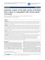
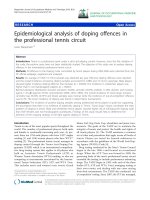
Phylogenetic tree (ML-TreePuzzle) of hantaviruses based on the complete coding region of the S segmentFigure 1
Phylogenetic tree (ML-TreePuzzle) of hantaviruses based on the complete coding region of the S segment. Only bootstrap sup-
port values greater than 70% are shown. Complete S-segment sequences:Thottapalayam virus (TPLV) (GeneBank accession no.
AY526097
); Seoul virus (SEOV), strain SR11 (M34881); Thailand virus (THAIV), strain 741 (AB186420); Dobrava virus
(DOBV), strain Dobrava (L41916
); Saaremaa virus (SAAV), strain Saaremaa/160v (AJ009773); Hantaan virus (HTNV), strain
76–118 (M14626
); Amur virus (AMRV), strain Solovey/AP63/1999 (AB071184); Soochong virus, strain SC-1 (AY675349); Muju
virus, strain Muju99-28 (DQ138142
); Puumala virus (PUUV), strain Sotkamo (X61035); Hokkaido virus (HOKV), strain Kami-
iso-8-Cr-95 (AB010730
); Topografov virus (TOPV), strain Ls136V (AJ011646); Khabarovsk virus (KHAV), strain MF-43
(U35255
); Tula virus (TULV), strain Moravia/02v (Z69991); Isla Vista virus (ISLAV), strain MC-SB-47 (U19302); Prospect Hill
virus (PHV), strain PH-1 (Z49098
); Bloodland lake virus (BLLV), strain MO46 (U19303); Bayou virus (BAYV), strain Louisiana
(L36929
); Black Creek Canal (BCCV) (L39949); Muleshoe virus (MULV), strain SH-Tx-339 (U54575); Maporal virus, strain HV-
97021050 (AY267347
); Choclo virus (DQ285046); Maciel virus (MCLV), strain 13796 (AF482716); Pergamino virus (PRGV),
strain 14403 (AF482717
); Oran virus (ORNV), strain 22996 (AF482715); Hu39694 virus (AF482711); Lechiguanas virus
(LECV), strain 22819 (AF482714
); Bermejo virus (BMJV), strain Oc22531 (AF482713); Andes virus (ANDV), strain AH-1
(AF324902
); Araucaria virus, strain HPR/02-72 (AY740625); Rio Mamore virus (RIOMV), strain Om-556 (U52136); Laguna
Negra virus (LANV), strain 510B (AF005727
); Rio Segundo virus (RIOSV), strain RMx-Costa-1 (U18100); El Moro Canyon
(ELMCV), strain RM-97 (U11427
); Sin Nombre virus (SNV), strain NM H10 (L25784); Monongahela virus (MGLV), strain
Monongahela-1 (U32591
); and New York virus (NYV), strain RI-1 (U09488).
0.1
TPLV
SEOV
THAIV (Thai 741)
THAIV (Thai 0017)
99
96
DOBV
SAAV
95
HTNV
AMRV
Soochong
99
90
100
Muju
PUUV
HOKV
100
100
TOPV
KHAV
99
96
TULV
ISLAV
PHV
BLLV
94
76
98
BAYV
BCCV
MULV
74
96
Maporal
Choclo
MCLV
PRGV
95
ORNV
Hu39694
LECV
BMJV
96
75
ANDV
Araucaria
RIOMV
LANV
74
RIOSV
ELMCV
90
SNV
MGLV
NYV
90
73
98
99
Virology Journal 2006, 3:72 />Page 6 of 9
(page number not for citation purposes)
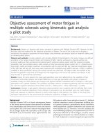
Phylogenetic tree (ML-TreePuzzle) of hantaviruses based on partial sequence (nt 389–946) of the S segmentFigure 2
Phylogenetic tree (ML-TreePuzzle) of hantaviruses based on partial sequence (nt 389–946) of the S segment. Only bootstrap
support values greater than 70% are shown. Partial S-segment sequences:PUUV, strain Sotkamo (X61035
); TULV, strain Moravia/
02v (Z69991
); SEOV, strains Gou3 (AF184988), Gou3v9 (AB027522), Hb8610 (AF288643), R22 (AF288295), L99 (AF288299),
Z37 (AF187082
), zy27 (AF406965), Pf26 (AY006465), IR461 (AF329388), SR11 (M34881), Tchoupitoulas (AF329389),
Jakarta137 (AJ620583
), Cambodia (Camb)41 (AJ427501), Camb32 (AJ427508), Camb58 (AJ427510), Camb180 (AJ427506),
Camb174 (AJ427513
), Camb96 (AJ427512), and Camb117 (AJ427511); THAIV virus, strain 741 (AB186420); SAAV, strain
Saaremaa/160v (AJ009773
); DOBV, strain Dobrava (L41916); Da Bie Shan virus (DBSV), strains NC167 (AB027523), AH211
(AF288647
), and AH09 (AF285264); Amur virus (AMRV), strains Solovey/AP63/1999 (AB071184), and Solovey/AP61/1999
(AB071183
); and HTNV, strains A16 (AB027099), A9 (AF329390), Maaji (AF321095), and 76–118 (M14626).
0.1
Z37
zy27
Pf26
IR461
SR11
Cambodia 41
Cambodia 32
Cambodia 58
Cambodia 180
Cambodia 174
Cambodia 96
Cambodia 117
SAAV
DOBV
AH211
AH09
NC167
100
100
85
99
100
99
96
96
100
100
92
94
98
94
86
71
81
100
100
90
87
100
90
75
PUUV
TULV
Gou3
Gou3v9
Hb8610
R22
L99
Tchoupitoulas
Jakarta 137
Thai 741
Thai 0067
Solovey/AP61
Solovey/AP63
A16
A9
Maaji
76-118
SEOV
THAIV
DBSV
AMRV
HTNV
Thai 0016
Thai 0024
Thai 0017
Virology Journal 2006, 3:72 />Page 7 of 9
(page number not for citation purposes)
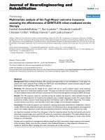
Phylogenetic tree (Fitch-Margoiliash) of hantaviruses based on partial sequence (nt 2021–2303) of the M segmentFigure 3
Phylogenetic tree (Fitch-Margoiliash) of hantaviruses based on partial sequence (nt 2021–2303) of the M segment. Only boot-
strap support values greater than 70% are shown. Partial M-segment sequences:PUUV, strain Sotkamo (X61034
); TULV, strain
Moravia/02v (Z69993
); DOBV, strain Dobrava (L33685); SAAV, strain Saaremaa/160v (AJ009774); DBSV, strain NC167
(AB027115
); HTNV, strains 76–118 (M14627), HoJo (D00376), Lee (D00377), HV114 (L08753), and A9 (AF035831); THAIV,
strain 749 (L08756
); and SEOV, strains Gou3 (AB027521), SR11 (M34882), Tchoupitoulas (U00473), Hubei-1 (S72343), 80–39
(S47716), Girard Point (U00464
), Egypt (U00463), SD227 (AB027091), CD10 (AB027092), Z37 (AF187081), Hebei4
(AB027089
), c3 (AB027088), IR461 (AF458104), Brazil (U00460), Baltimore (U00151), B1 (X53861), France-Rn90 (AJ878418),
Jakarta137 (AJ620583
), Beijing-Rn (AB027087), HN71-L (AB027085), Houston (U00465), Shanxi (AB027084), Henan
(AB027083
), Wan (AB027081), NM39 (AB027080), and J12 (AB027082).
0.1
TULV
PUUV
SAAV
DOBV
94
DBSV
76-118
HoJo
Lee
96
98
HV114
A9
97
93
90
80
Thai 749
Thai 0016
Thai 0024
Thai 0017
95
92
Gou3
SR11
Tchoupitoulas
Hubei-1
80-39
82
Girard Point
Egypt
81
SD227
CD10
92
Z37
Hebei4
c3
81
IR461
Brazil
Baltimore
78
B1
France-Rn90
Jakarta137
Beijing-Rn
HN71-L
Houston
92
Shanxi
Henan
80
Wan
NM39
J12
70
73
77
79
71
93
HTNV
THAIV
SEOV
Virology Journal 2006, 3:72 />Page 8 of 9
(page number not for citation purposes)
expected to be discovered in the future in Southeastern
Asia where Muridae rodents are endemic and highly diver-
sified and where the human population is regularly
exposed to them. The recent discovery of a new hantavirus
in Guinea [28] demonstrate that hantaviruses have to be
tracked wherever Muridae rodents are living. Further stud-
ies are needed to assess the reality of an endemic South-
east Asian group of hantaviruses and to understand their
particularities, their current distribution among rodents in
different areas and in different landscapes and finally their
potential dangerousness for humans. This also supposes
the improvement of our knowledge of the ecology and
biogeography of the hantavirus natural reservoirs in
Southeast Asia. Thailand, which health system is strongly
organized and possesses important and detailed archives
has all the necessary resources to organize such a program.
The results may be of interest for all the surrounding
countries and give rise to a regional cooperation in this
field of study.
Most recently we became aware of the manuscript of S.
Pattamadilok and co-authors [29] in which they charac-
terized the S segment sequence recovered from the THAIV
isolate and also performed antigenic cross-reactivity stud-
ies of rodent and human sera collected in Thailand. Their
observations on THAIV-positive bandicoot rats as well as
results of the phylogenetic analyses are nicely in line with
our data reported here. Most interestingly, the serum of
one patient with the HFRS symptoms showed high titers
of THAIV-neutralisiung antibodies suggesting that this
hantavirus is a human pathogen.
Authors' contributions
JPH participated in the study design and coordination,
trapping and screening of rodents, and drafting the man-
uscript. AngP participated in the screening of the rodent
samples, performed RNA isolation, RT-PCR and sequenc-
ing, participated also in the genetic analysis and drafting
the manuscript. VH participated in the study design, trap-
ping and screening of rodents, and drafting the manu-
script. KN participated in (phylo)genetis analyses and
drafting the manuscript. JL participated in the study coor-
dination and screening of rodents. ÅL participated in the
study coordination and drafting the manuscript. YS par-
ticipated in the study coordination and trapping and
screening of rodents. HH participated in the study design
and coordination and drafting the manuscript. AP partic-
ipated in the study design and coordination,
(phylo)genetic analyses and drafted the manuscript. All
authors read and approved the final manuscript.
Acknowledgements
This work received financial support from the Academy of Finland and the
French program "ANR- Santé-Environnement" (no. 00121 0505). Nucle-
otide sequences described in this paper have been deposited to the data-
bases under accession numbers AM397664-71. The authors are greatful to
Dr. S. Pittamadilok and Dr. J. Arikawa for sharing their data before publica-
tion.
References
1. Nichol ST, Beaty BJ, Elliott RM, Goldbach R, Plyusnin A, Schmaljohn
CS, Tesh RB: Bunyaviridae. In Virus taxonomy. VIIIth report of the
International Committee on Taxonomy of Viruses Edited by: Fauquet CM,
Mayo MA, Maniloff J, Desselberger U, Ball LA. Amsterdam: Elsevier
Academic Press; 2005:695-716.
2. Vapalahti O, Mustonen J, Lundkvist A, Henttonen H, Plyusnin A,
Vaheri A: Hantavirus infections in Europe. Lancet Infect Dis 2003,
3:653-661.
3. Lundkvist Å, Plyusnin A: Molecular epidemiology of hantavirus
infections. In The Molecular Epidemiology of Human Viruses Edited by:
Leitner T. Kluwer Academic Publishers; 2002:351-384.
4. Plyusnin A, Morzunov S: Virus evolution and genetic diversity of
hantaviruses and their rodent hosts. Curr Top Microbiol Immunol
2001, 256:47-75.
5. Nemirov K, Vaheri A, Plyusnin A: Hantaviruses:co-evolution with
natural hosts. Recent Res Devel Virol 2004, 6:201-228.
6. Carey DE, Reuben R, Panicker KN, Shope RE, Myers RM: Thotta-
palayam virus: a presumptive arbovirus isolated from a
shrew in India. J Med Res 1971, 59:1758-1760.
7. Boonsong L, McNeely JA, Marshall JT: Mammals of Thailand. Asso-
ciation for the Conservation of Wildlife, Bangkok 1988:748.
8. Xiao SY, LeDuc JW, Chu YK, Schmaljohn CS: Phylogenetic analy-
sis of virus isolates in the genus Hantavirus, family Bunyaviri-
dae. Virology 1994, 198:205-217.
9. Reynes JM, Soares JL, Hue T, Bouloy M, Sun S, Kruy SL, Flye Sainte
Marie F, Zeller H: Evidence of the presence of Seoul virus in
Cambodia. Microbes Infect 2003, 5:769-73.
10. Elwell MR, Ward GS, Tingpalapong M, Leduc JW: Serologic evi-
dence of Hantaan-like virus in rodents and man in Thailand.
Southeast Asian Journal of Tropical Medicine and Public Health 1985,
16:349-354.
11. Nitatpattana N, Chauvency G, Dardaine J, Poblap T, Jumronsawat K,
Tangkanakul W, Poonsuksombat D, Yoksan S, Gonzalez JP: Serolog-
ical study of Hantavirus in the rodent population of Nakhon
Pathom and Nakhon Ratchasima provinces in Thailand.
Southeast Asian Journal of Tropical Medicine and Public Health 2000,
31:277-282.
12. Sawasdikol S, Tamura M, Jamjit P: Antibody to hemoragic fever
with renal syndrome in man and rat in Thailand. Bulletin of the
Department of Medical Sciences 1989, 31:125-130.
13. Suputtamongkol Y, Nitatpattana N, Chyakulkeree M, Palabodeewat S,
Yoksan S, Gonzalez JP: Hantavirus infection in Thailand: first
clinical case report. Southeast Asian Journal of Tropical Medicine and
Public Health 2005, 36:217-220.
14. Herbreteau V, Gonzalez JP, Hugot JP: Phylogenetic Systematics
of Rodent-Borne Hantaviruses Allows Understanding their
Distribution. Annals of the New-York Academy of Sciences in press.
15. Mills JN, Childs JE, Ksiazek TG, Peters CJ: Methods for trapping
and sampling small mammals for virologic testing. Atlanta:
U.S. Department of Health and Human Services, Centers for Disease
Control and Prevention; 1995.
16. Plyusnin A, Cheng Y, Vapalahti O, Pejcoch M, Unar J, Jelinkova Z, Leh-
väslaiho H, Lundkvist Å, Vaheri A: Genetic variation in Tula
hantaviruses:; sequence analysis of the S and M segments of
strains from Central Europe. Virus Res 1995, 39:237-250.
17. Plyusnina A, Ibrahim IN, Winoto I, Porter KR, Gotama IBI, Lundkvist
Å, Vaheri A, Plyusnin A: Identification of Seoulhantavirus in Rat-
tus norvegicus in Indonesia. Scand J Inf Dis 2004, 36:356-359.
18. Plyusnin A, Vapalahti O, Ulfves K, Lehväslaiho H, Apekina N,
Gavrilovskaya I, Blinov V, Vaheri A: Sequences of wild Puumala
virus genes show a correlation of genetic variation with geo-
graphic origin of the strains. J Gen Virol 1994, 75:405-409.
19. Nemirov K, Vapalahti O, Lundkvist Å, Vasilenko V, Golovljova I, Ply-
usnina A, Niemimaa J, Laakkonen J, Henttonen H, Vaheri A, Plyusnin
A: Isolation and characterization of Dobrava hantavirus car-
ried by the striped field mouse (Apodemus agrarius) in Esto-
nia. J Gen Virol 1999, 80:371-379.
20. Felsenstein J: PHYLIP [Phylogeny Inference Package].
1999:3.5c.
Publish with Bio Med Central and every
scientist can read your work free of charge
"BioMed Central will be the most significant development for
disseminating the results of biomedical research in our lifetime."
Sir Paul Nurse, Cancer Research UK
Your research papers will be:
available free of charge to the entire biomedical community
peer reviewed and published immediately upon acceptance
cited in PubMed and archived on PubMed Central
yours — you keep the copyright
Submit your manuscript here:
/>BioMedcentral
Virology Journal 2006, 3:72 />Page 9 of 9
(page number not for citation purposes)
21. Schmidt HA, Strimmer K, Vingron M, von Haesseler A: TREE-PUZ-
ZLE: maximum likelihood phylogenetic analysis using quar-
tets and parallel computing. Bioinformatics 2002, 18:502-504.
22. Sironen T, Vaheri A, Plyusnin A: Molecular evolution of Puumala
hantavirus. J Virol 2001, 75:11803-11810.
23. Kaukinen P, Vaheri A, Plyusnin A: Hantavirus nucleocapsid pro-
tein: a multifunctional molecule with both housekeeping and
ambassadorial duties. Arch Virol 2005, 150:1693-1713.
24. Wang H, Yoshimatsu K, Ebihara H, Ogino M, Araki K, Kariwa H:
Genetic diversity of hantaviruses isolated in China andchar-
acterization of novel hantaviruses isolated from Niviventer
confucianus and Rattus rattus. Virology 2000, 278:332-345.
25. LeDuc JW, Smith GA, Childs JE, Pinheiro FP, Maiztegui JI, Niklasson
B, Antoniades A, Robinson DM, Khin M, Shortridge KF, Wooster MT,
Elwell MR, Ilbery PLT, Koech D, Rosa EST, Rosen L: Global survey
of antibody to Hantaan-related viruses among peridomestic
rodents. Bull WHO 1986, 64:139-144.
26. Nitatpattana N, Henrich T, Palabodeewat S, Tangkanakul W, Poonsu-
ksombat D, Chauvancy G, Barbazan P, Yoksan S, Gonzalez JP:
Hantaan virus antibody prevalence in rodent populations of
several provinces of north-eastern Thailand. Trop Med Int
Health 2002, 7:1-6.
27. Tantivanich S, Ayuthaya PI, Usawattanakul W, Imphand P: Hantaan-
virus among urban rats from a slum area in Bangkok. South-
east Asian J Trop Med Public Health 1992, 23:504-509.
28. Klempa B, Fichet-Calvet E, Lecompte E, Auste B, Aniskin V, Meisel H,
Denys C, Koivogui L, ter Meulen J, Krüger DH: Hantavirus in Afri-
can Wood Mouse, Guinea. Emerging Infectious Diseases 2006,
12:838-840.
29. Pattamadilok S, Lee B-H, Kumperasart S, Yoshimatsu K, Okumura M,
Nakamura I, Araki K, Khopraser Y, Dangsupa P, Panlar P, Jandrig B,
Krüger DH, Klempa B, Jäkel T, Schmidt J, Ulrich R, Kariwa H, Arikawa
J: Geographical distribution of hantaviruses in Thailand and
potential human health significance of Thailand virus. Amer J
Trop Med Hyg 2006 in press.