báo cáo hóa học:" High ERCC1 expression predicts cisplatin-based chemotherapy resistance and poor outcome in unresectable squamous cell carcinoma of head and neck in a betel-chewing area" pdf
Bạn đang xem bản rút gọn của tài liệu. Xem và tải ngay bản đầy đủ của tài liệu tại đây (475.69 KB, 8 trang )
RESEARCH Open Access
High ERCC1 expression predicts cisplatin-based
chemotherapy resistance and poor outcome in
unresectable squamous cell carcinoma of head
and neck in a betel-chewing area
Tai-Jan Chiu
4,6†
, Chang-Han Chen
2,4,5†
, Chih-Yen Chien
2,4
, Shau-Hsuan Li
1,4
, Hsin-Ting Tsai
2,4
and Yi-Ju Chen
3*
Abstract
Background: This study was to evaluate the effect of excision repair cross-complementation group 1(ERCC1)
expression on response to cisplatin-based induction chemotherapy (IC) followed by concurrent chemoradiation
(CCRT) in locally advanced unresectable head and neck squamous cell carcinoma (HNSCC) patients.
Methods: Fifty-seven patients with locally advanced unresectable HNSCC who received cisplatin-based IC followed
by CCRT from January 1, 2006 through January 1, 2008. Eligibility criteria included presence of biopsy-proven
HNSCC without a prior history of chemotherapy or radiotherapy. Immunohistochemistr y was used to assess ERCC1
expression in pretreatment biopsy specimens from paraffin blocks. Clinical parameters, including smoking, alcohol
consumption and betel nuts chewing, were obtained from the medical records.
Results: The 12-month progression-free survival (PFS) and 2-year overall survival (OS) rates of fifty-seven patients
were 61.1% and 61.0%, respectively. Among these patients, thirty-one patients had low ERCC1 expression and
forty-one patients responded to IC followed by CCRT. Univariate analyses showed that patients with low expression
of ERCC1 had a significantly higher 12-month PFS rates (73.3% vs. 42.3%, p < 0.001) and 2-year OS (74.2 vs. 44.4%,
p = 0.023) rates. Multivariate analysis showed that for patients who did not chew betel nuts and had low
expression of ERCC1 were independent predictors for prolonged survival.
Conclusions: Our study suggest that a high expression of ERCC1 predict a poor response and survival to cisplatin-
based IC followed by CCRT in patients with locally advanced unresectable HNSCC in betel nut chewing area.
Background
Squamous cell carcinoma of the head and neck
(HNSCC) is the sixth most common cancer i n the
world[1]andtwo-thirdsofthese patients initially pre-
sent with locally advanced disease [2]. In Taiwan,
HNSCC rates 4
th
in male cancer-related deaths [3]
among middle-aged male patients between 25 and 45
years old [4]. Most HNSCC patients in Taiwan d iag-
nosed w ith advanced disease are young men. The main
risk factors of this unique patient population are t he
habitual consumption of cigarettes, alcohol, and b etel
nuts [5,6].
Although patients with locally advanced HNSCC receive
surgery and radiotherapy, less than 30% will be cured, and
locoregional recurrences or distant metastases develop in
40% to 60% patients [7,8], which occurs with a median
survival rate of no more than 6 months [9]. Some studies
have demonstrated improved locoregion al control and
overall survival by adding chemotherapy to radiotherapy
concurrently [10]. The Meta-Analysis of Chemotherapy in
Head and Neck Cancer (MACH-NC) study showed that
concomitant chemoradiation is superior to RT alone for
patients with advanced HNSCC and chemoradiotherapy
(radiotherapy plus concurrent chemotherapy) has become
the standard of care for patients with unresectable
* Correspondence:
† Contributed equally
3
Department of Pathology, E-Da hospital, Kaohsiung, Taiwan
Full list of author information is available at the end of the article
Chiu et al. Journal of Translational Medicine 2011, 9:31
/>© 2011 Chiu et al; licens ee BioMed Central Ltd. This is an Open Access article distributed under the t erms of the Creative Commons
Attribution License ( /by/2.0), which permits unrestricted use, distribution, and reproduction in
any me dium, provided the or iginal work is properly cited.
HNSCC [11,12]. However, the best chemotherapeutic regi-
men combined with RT in HNSCC has yet to be defined;
the concomitant administration of cisplatin represents a
widely accepted choice. It has been reported that induc-
tion chemotherapy (IC) with cisplatin and fluorouracil
(PF) benefits this disease [12-14] and results in a signifi-
cantly improved 5-year survival rate in patients with
locally advanced disease compared to surgery and stan-
dard radiotherapy alone [12].
In Taiwan, for public healthy insurance, cisplatin is the
backbone of the chemotherapy regimen as a component
of IC and CCRT in the treatment of loca lly advanced
HNSCC. Its m ain cytotoxic activity is based on the for-
mation of DNA adducts, which cause inter- and intras-
trand cross-linking. These DNA cross-links are
recognized and removed by the nucleotide excision repair
pathway which arms to guard the integrity of the genome
[15,16]. The enzyme excision repair cross-complementa-
tiongroup1(ERCC1)playsaratelimitingroleinthe
nucleotide excision repair pathway, and its expression
has been associated with survival in patients with various
malignancies [17-19]. The relation between ERCC1
expression and resistance to platinum compounds had
been found by some clinical studies in patients with
advanced-stage gastric, ovarian, c olorectal, esophageal,
and non-small-cell lung cancers [15,17,19-21]. However,
there are only few studies to elucidate the relationship
between ERCC1 expression and prognosis in patients
with locally advanced HNSCC treated with CCRT. The
purpose of this study was to evaluate whether the immu-
nohistochemi cal expression status of ERCC1 can predict
the treatment response and survival in patients with
unresectable HNSCC being treated with cisplatin-based
IC followed by CCRT.
Methods
Patients and treatment
A total of 57 patients with pathologically proven locally
advanced inoperable HNSCC were treated with IC fol-
lowed by CCRT between January 1, 2006 and January 1,
2008 at Kaohsiung Chang-Gung Medical Center (Tai-
wan). To be included, all the patients had to have a
biopsy-proven previously untreated IV (M0) unresectable
squamous cell carcinoma of the head and neck region,
have no synchronous primary tumors, and be ≥18 years
old. In addition, the patients had to have a performance
status (PS) of ≤2ontheEasternCooperativeOncology
Group(ECOG)scale,adequatebonemarrow,hepatic
and renal function (creatinine clearance >60 ml/min),
and a compute d tomography or magnetic reson ance
image scan of the head and neck region within t hree
weeks prior to the initiation of treatment. The clinico-
pathological information including age, gender, t umor
(T) stage, nodal (N) status, TNM stage, and survival was
obtained from the clinical records. The histories of betel
nuts chewing, alcohol and tobacco use were obtained by
our detailed questioning at the patients’ first visit to the
otolaryngology clinic of the hospital.
TheICconsistedof2cyclesofcisplatin75mg/m2
and fluorouracil (5-FU) (1000 mg/m2) given as a contin-
uous 24-h infusion for four days. The two cycles of IC
were administered every four weeks. After IC, all
patients received CCRT. During the CCRT, cisplatin
was administered weekly at a dose of 40 mg/m2. RT
was delivered 3-4 weeks after the completion of the IC
with a linear accelerator. Ondansentron ± dexametha-
sone was used as ant iemetic treatment. The response to
IC followed by CCRT was assessed according to the
World Health Organization (WHO) criteria. Surgery
was performed six to twelve weeks after completion of
IC followed by CCRT regimen for patients who had
residual disease. Surgery was also allowed for patients
who did not complete chemoradiation and had resect-
able residual disease at the primary site or in the neck.
Patients were evaluated by CT scan or MRI of the head
and neck every three months. Informed consent was
obtained from study participants and protocol for this
study was approved by the Institutional Review Boards
of Chang-Gung Medical Center (Taiwan).
Immunohistochemical staining for ERCC1
Adjacent non-cancerous and tumor HNSCC tissue sam-
ples were selected by a pathologist based on diagnosis
and microscopic morphology. Adjacent non-cancerous
tissue and tumor tissues were fixed with 10% buffered
formalin embedded in paraffin and decalcified in 10%
EDTA solution. Representative blocks of the formalin-
fixed, paraffin-embedded tissues were cut to 4 mm and
deparaffinized with xylene and rehydrated in a series of
ethanol w ashes (100, 90, 80, and 70%). Slides were
washed with phosphate-buffered saline (PBS) and trea-
ted with 3% H
2
O
2
for 30 minutes to block endogenous
peroxi dase activity. Next, the sections were microwaved
in 10 mM citrate buffer, pH 6.0, to unmask the epitopes.
After antigen retrieval, the sections were incubated with
diluted anti-ERCC1 antibody (monoclonal; 8F1; Thermo
scientific, Fremont, CA, USA; 1:100), for 3 h followed by
washing with PBS. Horseradish peroxidase/Fab polymer
conjugate (PicTure™-Plus k it; Zymed, South San Fran-
cisco, CA, USA) was then applied to the sections for 30
min f ollowed by washing with PBS. Finally, the sections
were incubated with diaminobenzidine for 5 min to
develop the signals. A negative control was run simulta-
neously by omitting the primary antibody.
Evaluation of ERCC1 expression
Two pathologists, who were unaware of the clinical data,
evaluated the ERCC1 staining independently under a
Chiu et al. Journal of Translational Medicine 2011, 9:31
/>Page 2 of 8
light microscope at a magnification of × 400. The
pathologists recorded whether tumor or stromal cells
exp ressed ERCC1 . The staining intensity was graded on
a scale of 0-3, using adjacent nonmalignant cells as a
reference (intensity 2). Five images of representative
areas were acquired for each specimen. The percentage
of positive nuclei was calculated for each specimen, and
a proportion score was assigned (0 if 0%, 0.1 if 1-9%, 0.5
if 10-49%, and 1.0 if ≧ 50%). The proportion score was
multiplied by the staining intensity to obtain a final
semi-quantitat ive H score. The median value of the H
score was chosen as the cutoff point for separating low
and high levels of ERCC1 expression [22].
Statistical analysis
Statistical analyses of 2 × 2 tables of categorical variables
were performed using Pearson’s x
2
test or Fisher’s exact
test, where appropriate. Survival probability analyses were
performed using the Kaplan-Meier method. Survival was
calculated from the date of start of chemotherapy to the
date of death or most recent follow-up. Progression free
survival (PFS) was defined as the time from the date o f
first chemotherapy to the date of first observation of dis-
ease progression, or relapse, or death due to any cause.
Significance between group differences was assessed by
the log-rank test. Multivariate analyses were performed
using a logistic regression model for response and Cox
regression models for PFS and overall survival (OS). Fac-
tors with p-values < 0.05 in univariate analyses were exam-
ined with multivariate regression models. All statistical
tests were two-sided, with significance defined as p < 0.05.
Analyses were performed using SPSS version 13.
Result
Patient characteristics
The median age of the patients was 53 years ( range 36-
72 years), and fifty-five (96.5%) out of 57 were men. Ten
patients had IVA and 47 had stage IVB disease. The
most common sites were the oral cavity (24/57, 42.1%),
followed by the oropharynx (21/57, 36.8%) (Table 1).
The median radiation they received was 6600 cGy.
Nineteen patients received more than 70 Gy of radiation
Table 1 Correlation between expression of ERCC1 and clinicopathological factors of HNSCC
ERCC1 P Multivariates analysis P
No. of patients Low expression High expression OR (95% CI)
Age
≦50 22 (38.6%) 11 (50.0%) 11 (50.0%) 0.598 1
> 50 35 (61.4%) 20 (57.1%) 15 (42.9%) 0.47 (0.11, 2.11) 0315
Gender
Male 55 (96.5%) 30 (54.5%) 25 (45.5%) 1.000 1
Female 2 (3.5%) 1 (50%) 1 (50%) 48.36 (0.54, 4313.32) 0.090
Tumor Site
oral cavity 24 (42.1%) 11 (45.8%) 13 (54.2%) 0.057 1
oropharynx 21 (36.8%) 10 (47.6%) 11 (52.4%) 1.58 (0.35, 7.07) 0.549
hypopharynx/Larynx 12 (21.1%) 10 (83.3%) 2 (16.7%) 0.096 (0.007, 1.33) 0.081
Stage
IVa 10 (17.5%) 7 (70.0%) 3 (30.0%) 0.319 1
IVb 47(82.5%) 24 (51.1%) 23 (48.9%) 3.33 (0.39, 27.89) 0.276
T stage
1/2 6 (10.5%) 6 (100%) 0 (0%) 0.027* 1
3/4 51 (89.5%) 25 (49.0%) 26 (51.0%) Indeterminate 0.999
N stage
negative 12 (21.1%) 5 (41.7%) 7 (58.3%) 0.503 1
positive 45 (78.9%) 26 (57.8%) 19 (42.2%) 0.75 (0.15, 3.64) 0.727
Alcohol drinking
Never 11 (17.2%) 4 (36.4%) 7 (63.6%) 0.318 1
Yes 46 (82.8%) 27 (58.7%) 19 (41.3%) 2.49 (0.31, 19.88) 0.388
Smoking
Never 9 (15.8%) 3 (33.3%) 6 (66.7%) 0.275 1
Yes 48 (84.2%) 28 (58.3%) 20 (41.7%) 1.77 (010, 30.72) 0.695
Betel nuts
Never 20 (35.1%) 8 (40.0%) 12 (60.0%) 0.109 1
Yes 37 (64.9%) 23 (62.2%) 14 (37.8%) 12.78 (1.28-127.62) 0.030*
Chiu et al. Journal of Translational Medicine 2011, 9:31
/>Page 3 of 8
dose. All patients had received their IC and 53 patients
completed the followed up CCRT.
Clinico-pathologic factors of HNSCC patients with ERCC1
expression
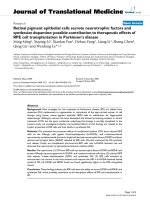
To investigate whether the increased expression of ERCC1
was associated with various prognostic factors, such as
age, gender, and TNM pathologic classification, we classi-
fied the patients into two groups based on their immuno-
histochemical results (low vs. high ERCC1 expression)
(Figure 1A and 1B). The median H score for HNSCC was
1.5. Twenty-six (46%) tumors had an H score of more
than 1.5 and were thus defined as having a high expression
of ERCC1. As can be seen in Table 1 a summary of result
of the ERCC1 immunostaining of the cancer cells and its
correlation with the clinicopathologic variables, the high
and low ERCC1 expression groups did not significantly
with regard to age, gender, TNM tumor stage, and node
metastatic status, alcohol drinking or smoking (Table 1).
The high ERCC1 expression group had a higher T stage
(T3-4) (p = 0.027 ). Thos e with squamous cell carcinoma
of the hypopharynx/larynx were found to have marginal
lower expression of ERCC1. Interestingly, in our multivari-
ate regression model, patients who habitually chewed betel
nuts had a significantly higher expression of ERCC1.
Relationship between treatment response and ERCC1
expression
The overall response rate after CCRT for all patients
was 72% (41/57 with 28 complete responses and 13 par-
tial responses; 9 had stable disease and 7 progressive
disease). Patients with low expression of ERCC1 had a
higher treatment response (28/31, 90.3%) than the high
expression group (13/26, 50%) (p = 0.002, Table 2).
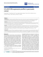
Relationship between survival and ERCC1 expression
The median follow-up was 24.0 months (6 - 46 months).
The overall 12-month PFS rate was 61.1% and the 2-
year OS rate was 61.0%. The 12-month PFS for patients
with low expression of ERCC1 was 73.3% compared
with 42.3% for patients with high expression of ERCC1
(p < 0.001, Figure 2A). The 2-year OS rate was signifi-
cantly higher in patients with low expression of ERCC1
(74.2%) than in those with high expression of ERCC1
(44.4%) (P = 0.023, Figure 2B). Univariate analysis
showed that tumor stage and tumor location were
important factors affecting the OS and PFS (Table 3),
though ERCC1 expression and betel nuts chewing were
theprognosticfactorsinOSbymultivariateanalysis
according to Cox regression model (Table 4).
Discussion
It is of special interest that in our study that specimens
from patients who habitually chewed betel nuts had
high expression of ERCC1. Betel nut chewing is a com-
mon habit among those who live in South Asia, includ-
ing Taiwan [23], and is known as one cause of HNSCC
[24]. There are many compounds in the betel nut that
have been correlated with carcinogenesis; the habit of
chewing betel nut is r elated to p ersistent damage of the
oral mucosa as well as precancerous lesions such as leu-
koplakia and erythroplakia, and oral submucosal fibrosis
[25]. In previous reports, overexpression of epidermal
growth factor receptor (EGFR) was found to be involved
in betel nut-related HNSCC [26,27]. However, the rela-
tionship between betel nut and ERCC1 expressio n has
not been reported before. In this study, we find tissue
samples from patients with habitual consumption of
betel nuts showed significant correlation with hig h
ERCC1 expression. This finding awaits confirmation by
prospective studies with large numbers of patients.
In this study, Forty-six percent of the patients with
inoperable HNSC C had a high expression of ERCC1.
Patients with a high expression of ERCC1 had a lower
treatment response rate to IC followed by CCRT than
those with low expression of ERCC1. In addition, low
Figure 1 Analysis of ERCC1 expression in head and neck squamous cell carcinoma. ERCC1 expression was determined using
immunohistochemistry. A) Low ERCC1 expression (200× magnification). B) High ERCC1 expression (200× magnification).
Chiu et al. Journal of Translational Medicine 2011, 9:31
/>Page 4 of 8
Figure 2 Kaplan-Meier estimates of the probability of survival. (A) PFS according to ERCC1 expression. PFS: progression free survival. (B) OS
according to ERCC1 expression. OS: overall survival.
Table 2 Relationship between treatment response and clinicopathological factors
Treatment response Multi-variates
CR/PR SD/PD P OR
(95%CI)
P
Age
≦50 15 (68.2%) 7 (31.8%) 0.844 1
> 50 26 (74.3%) 9 (25.7%) 0.53 (0.07, 3.65) 0.520
Gender
male 39 (70.9%) 16 (29.1%) 1.000 1
female 2 (100%) 0 (0%) Indeterminate 1.000
Tumor Site
oral cavity 15 (62.5%) 9 (37.5%) 0.049*1
oropharynx 14 (66.7%) 7 (33.3%) 1.29 (0.21, 7.70) 0.778
hypopharynx/larynx 12 (100%) 0 Indeterminate 0.998
Stage
IVa 10 (100%) 0 (0%) 0.048*1
IVb 31 (66.0%) 16 (34.0%) Indeterminate 0.998
T stage
1/2 6 (100%) 0 (0%) 0.170 1
3/4 35 (68.6%) 16 (31.4%) Indeterminate 0.999
N stage
negative 8 (66.7%) 4 (25.0%) 0.732 1
positive 33 (73.3%) 12 (26.7%) 0.75 (0.06, 8.54) 0.818
Radiation
≦6000 cGy 14 (70.0%) 6 (30.0%) 1.000 1
> 6000 cGy 27 (75.7%) 10 (24.3%) 0.22 (0.02, 2.43) 0.222
Alcohol drinking
never 9 (81.8%) 2 (18.2%) 0.710 1 0.120
yes 32 (69.6%) 14 (30.4%) 0.08 (0.004, 1.89)
Smoking
never 7 (77.8%) 2 (22.2%) 1.000 1
yes 34 (70.8%) 14 (29.2%) 2.97 (0.10, 86.59) 0.526
Betel nuts
never 16 (80.0%) 4 (20.0%) 0.491 1
yes 25 (67.6%) 12 (32.4%) 0.38 (0.03, 4.55) 0.452
ERCC1
low expression 28 (90.3%) 3 (9.7%) 0.002* 1
high expression 13 (20.0%) 13 (50.0%) 0.07 (0.009, 055). 0.012*
CR, complete response; PR, partial response; SD, stable disease; PD, disease progression; RTO, radiotherapy; OR, odds ratio; CI, confidence interval.
Chiu et al. Journal of Translational Medicine 2011, 9:31
/>Page 5 of 8
ERCC1 expression was associated with a significantly
longer PFS and OS. Multivariate analysis revealed that
low expression of ERCC1 to be an independent factor
associated with a lower risk of cancer death (HR 0.31,
p = 0.010). Our findings are consistent with previous
report of an increase in tumor response and prolonga-
tion of OS in patients treated by cisplatin based IC fol-
lowed by CCRT for locally advanced HNSCC [28-30].
Moreover, the relationship between the e xpression of
ERCC1 and tumor response or survival has also been
demonstrated in esopha geal cancer patients treated
with chemoradiotherapy [31] and non-small cell lung
cancer treated with cisplatin-based adjuvant che-
motherapy [22].
However, in patients with locally advanced HNSCC
treated with cetuximab-based CCRT, ERCC1 ex pression
has not been found to predict treatment response [32].
In this context, w e assume that pre-therapeutic ERCC1
protein levels within tumor cells might be correlated
with their cisplatin-related DNA damage repair capacity.
A less efficient DNA-repair capacity could affect the cel-
lular response to DNA damage and could thus render
cancer cells more sensitive to cisplatin. In addition, Nix
et al. has reported an association between both ERCC1
and XRCC1 and radioresistance in laryngeal tumors
[33].
Cetuximab is an IgG1 monoclonal antibody against
the ligand-binding domain of EGFR. Cetuximab binds
Table 3 Univariate analyses of prognostic factors for survival
Variables No. of
patients
Cumulative 12-month preogresion free survival
rate
P Cumulative 2-year overall survival
rate
P
Age
< 50 22 49.0% 0.725 50.0% 0.152
≧50 35 68.6% 68.0%
Gender
male 55 61.5% 0.878 65.2% 0.553
female 2 50.0% 50.0%
Site
oral cavity 24 41.7% 45.5% 0.093
oropharynx 21 66.7% 0.101 66.6%
Hypopharynx/
larynx
12 81.8% 0.035* 83.3%
Stage
IVa 10 80.0% 0.119 90.0% 0.049*
IVb 47 57.2% 54.7%
T stage
0-2 6 66.7% 0.396 66.7% 0.694
3-4 51 60.5% 60.3%
N stage
negative 12 50.0% 0.837 72.9% 0.350
positive 45 57.3% 57.8%
Radiation
≦6000 cGy 20 55.0% 0.304 55.0% 0.412
> 6000 cGy 37 64.5% 64.1%
Alcohol
never 11 63.6% 1.000 63.6% 0.754
yes 46 60.6% 64.8%
Smoking
never 9 66.7% 0.614 66.7% 0.679
yes 48 60.1% 60.0%
Betel nuts
never 20 70.0% 0.638 74.0% 0.123
yes 37 56.3% 54.1%
ERCC1
low expression 31 73.7% <
0.001*
74.2% 0.023*
high expression 26 42.3% 44.4%
Chiu et al. Journal of Translational Medicine 2011, 9:31
/>Page 6 of 8
EGFR, sequesters the receptor in the cytoplasm and
eventually targets it for degradation. In vitro studies
have demonstrat ed that this antibody enhances the
radio-sensitivity in HNSCC cells [34,35] through several
processes, such as DNAPK, which are reviewed in
Mukesh et al. [36]. When cetuximab is combined with
radiation, it has been found to inhibit the nuclear trans-
location of the complex between DNA-dependent pro-
tein kinase and EGFR and then delayed the DNA repair
[37-39]. Oxaliplatin induced double-strand breaks [40].
When cetuximab was combined with oxaliplatin, cetuxi-
mab reduced the expression of ERCC-1 and other genes
involved in DNA replication initiation [41,42]. We
might find a subgroup of patients with high ERCC1
expression having poor response to cisplatin-based IC
and CCRT that is particularly benefited from treatments
with cetuximab and other chemotherapeutic agents.
Our study has several limitations. First, the study
was based on a retrospective analysis and only there
were only 57 patients accumulated over a short per-
iod. The primary tumor site was also heterogeneous,
and the pr ognosis of HNSCC is dependent on t he pri-
mary tumor site. In our study, those oral cavity cancer
had the worst prognosis and laryngeal cancer a good
prognosis, although we found no significant diff erence
in our multi-variate analyses. Second, some patients
with IC followed by CCRT had a partial response and
received further salvage surgery. Patients who receive
salvage surgery had signi ficantly longer PFS and OS
rates than those who did not receive such surgery.
The salvage surgery may affect the relationship
between ERCC1 expression and survival. It also sug-
gested that those patients with l ower expression of
ERCC1 would benefit from the potential downstage by
our treatment protocol and become resectable. Our
study was comprised only a small number of patients
for each tumor location, and so w e may need more
homogeneous and a larger number of patients to vali-
date this finding.
Conclusion
This present study suggests that ERCC1 mediated repair
of DNA damage contributes to the clinical outcome in
patients w ith locally advanced inoperable HNSCC trea-
ted with cisplatin-based IC and CCRT. In this context,
it is strongly recommended that tissue be collected to
assess ERCC1 expression before cisplatin-based induc-
tion chemotherapy and concurrent chemoradiotherapy.
If patients with habit of betel nuts chewing may have
higher chance of high ERCC1 expression, they should
consider other treatment approach modalities.
Acknowledgements
Sources of support: Chang Gung Memorial Hospital Grant (CMRPG890471
to Yi-Ju Chen, CMRPG890921 to Chang-Han Chen and CLRPG871342 to
Samuel HH Chan).
Author details
1
Department of Medical Oncology, Chang Gung Memorial Hospital-
Kaohsiung Medical Center, Chang Gung University, College of Medicine,
Kaohsiung, Taiwan.
2
Department of Otolaryngology, Chang Gung Memorial
Hospital-Kaohsiung Medical Center, Chang Gung University College of
Medicine, Kaohsiung, Taiwan.
3
Department of Pathology, E-Da hospital,
Kaohsiung, Taiwan.
4
Kaohsiung Chang Gung Head and Neck Oncology
Group, Cancer Center, Chang Gung Memorial Hospital-Kaohsiung Medical
Center, Kaohsiung, Taiwan.
5
Center for Translational Research in Biomedical
Sciences, Chang Gung Memorial Hospital-Kaohsiung Medical Center.
6
Institute of Clinical Medical Sciences, Chang Gung University, Kaohsiung,
Taiwan.
Authors’ contributions
TJC and CHC conceived the study design, carried out and coordinated
immunohistochemical examinations of tumor specimens and data analysis,
and drafted the manuscript. CYC and HTT participated in the interpretati on
of data and conducted immunohistochemistry analysis. SHL collected the
clinical data of patients and performed statistical data analysis. YJC
coordinated the study and were involved in drafting the manuscript and
revised it critically. All authors read and approved the final manuscript.
Competing interests
The authors declare that they have no competing interests.
Received: 27 December 2010 Accepted: 23 March 2011
Published: 23 March 2011
References
1. Greenlee RT, Hill-Harmon MB, Murray T, Thun M: Cancer statistics, 2001. CA
Cancer J Clin 2001, 51:15-36.
2. Kim ES, Kies M, Herbst RS: Novel therapeutics for head and neck cancer.
Curr Opin Oncol 2002, 14:334-342.
3. Chien CY, Su CY, Chuang HC, Fang FM, Huang HY, Chen CH, Chen CM,
Huang CC: Comprehensive study on the prognostic role of osteopontin
expression in oral squamous cell carcinoma. Oral Oncol 2009, 45:798-802.
4. Department of Health EY, R.O.C.: Cancer Registry Annual Report in Taiwan
Area. Taipei. Department of Health, Executive Yuan, ROC 2006.
5. Ko YC, Huang YL, Lee CH, Chen MJ, Lin LM, Tsai CC: Betel quid chewing,
cigarette smoking and alcohol consumption related to oral cancer in
Taiwan. J Oral Pathol Med 1995, 24:450-453.
6. Hsieh LL, Wang PF, Chen IH, Liao CT, Wang HM, Chen MC, Chang JT,
Cheng AJ: Characteristics of mutations in the p53 gene in oral
squamous cell carcinoma associated with betel quid chewing and
cigarette smoking in Taiwanese. Carcinogenesis 2001, 22:1497-1503.
7. Adelstein DJ, Li Y, Adams GL, Wagner H, Kish JA, Ensley JF, Schuller DE,
Forastiere AA: An intergroup phase III comparison of standard radiation
therapy and two schedules of concurrent chemoradiotherapy in
patients with unresectable squamous cell head and neck cancer. J Clin
Oncol 2003, 21:92-98.
8. Forastiere AAMM, Weber RS, et al: Long-term results of Intergroup RTOG
91-11: a phase III trial to preserve the larynx – induction cisplatin/5-FU
and radiation therapy versus concurrent cisplatin and radiation therapy
versus radiation therapy. J Clin Oncol 2006, 24(Suppl):284s, abstract.
Table 4 Risk factors affecting 1-year disease free survival
and 2-year overall survival rate determined by Cox
regression analysis
Variables PFS OS
HR (95%CI) P HR (95%CI) P
ERCC1 expression
Low vs High 0.27 (0.12-0.61) 0.001 0.31 (0.13-0.75) 0.010
Betel nuts
Never vs Used NE 0.647 0.35 (0.13-0.98) 0.045
CI, confidence interval; HR, Hazard ratio.
Chiu et al. Journal of Translational Medicine 2011, 9:31
/>Page 7 of 8
9. Vokes EE, Weichselbaum RR, Lippman SM, Hong WK: Head and neck
cancer. N Engl J Med 1993, 328:184-194.
10. Forastiere AA, Goepfert H, Maor M, Pajak TF, Weber R, Morrison W,
Glisson B, Trotti A, Ridge JA, Chao C, et al: Concurrent chemotherapy and
radiotherapy for organ preservation in advanced laryngeal cancer. N
Engl J Med 2003, 349:2091-2098.
11. Argiris A: Update on chemoradiotherapy for head and neck cancer. Curr
Opin Oncol 2002, 14:323-329.
12. Pignon JP, Bourhis J, Domenge C, Designe L: Chemotherapy added to
locoregional treatment for head and neck squamous-cell carcinoma:
three meta-analyses of updated individual data. MACH-NC Collaborative
Group. Meta-Analysis of Chemotherapy on Head and Neck Cancer.
Lancet 2000, 355:949-955.
13. Domenge C, Hill C, Lefebvre JL, De Raucourt D, Rhein B, Wibault P,
Marandas P, Coche-Dequeant B, Stromboni-Luboinski M, Sancho-Garnier H,
Luboinski B: Randomized trial of neoadjuvant chemotherapy in
oropharyngeal carcinoma. French Groupe d’Etude des Tumeurs de la
Tete et du Cou (GETTEC). Br J Cancer 2000, 83:1594-1598.
14. Paccagnella A, Orlando A, Marchiori C, Zorat PL, Cavaniglia G, Sileni VC,
Jirillo A, Tomio L, Fila G, Fede A, et al: Phase III trial of initial
chemotherapy in stage III or IV head and neck cancers: a study by the
Gruppo di Studio sui Tumori della Testa e del Collo. J Natl Cancer Inst
1994, 86:265-272.
15. Dabholkar M, Vionnet J, Bostick-Bruton F, Yu JJ, Reed E: Messenger RNA
levels of XPAC and ERCC1 in ovarian cancer tissue correlate with
response to platinum-based chemotherapy. J Clin Invest 1994, 94:703-708.
16. Murray D, Rosenberg E: The importance of the ERCC1/ERCC4[XPF]
complex for hypoxic-cell radioresistance does not appear to derive from
its participation in the nucleotide excision repair pathway. Mutat Res
1996, 364:217-226.
17. Lord RV, Brabender J, Gandara D, Alberola V, Camps C, Domine M,
Cardenal F, Sanchez JM, Gumerlock PH, Taron M, et al: Low ERCC1
expression correlates with prolonged survival after cisplatin plus
gemcitabine chemotherapy in non-small cell lung cancer. Clin Cancer Res
2002, 8:2286-2291.
18. Kwon HC, Roh MS, Oh SY, Kim SH, Kim MC, Kim JS, Kim HJ: Prognostic
value of expression of ERCC1, thymidylate synthase, and glutathione S-
transferase P1 for 5-fluorouracil/oxaliplatin chemotherapy in advanced
gastric cancer. Ann Oncol 2007, 18:504-509.
19. Joshi MB, Shirota Y, Danenberg KD, Conlon DH, Salonga DS, Herndon JE,
Danenberg PV, Harpole DH Jr: High gene expression of TS1, GSTP1, and
ERCC1 are risk factors for survival in patients treated with trimodality
therapy for esophageal cancer. Clin Cancer Res 2005, 11:2215-2221.
20. Metzger R, Leichman CG, Danenberg KD, Danenberg PV, Lenz HJ,
Hayashi K, Groshen S, Salonga D, Cohen H, Laine L, et al: ERCC1 mRNA
levels complement thymidylate synthase mRNA levels in predicting
response and survival for gastric cancer patients receiving combination
cisplatin and fluorouracil chemotherapy. J Clin Oncol 1998, 16:309-316.
21. Shirota Y, Stoehlmacher J, Brabender J, Xiong YP, Uetake H, Danenberg KD,
Groshen S, Tsao-Wei DD, Danenberg PV, Lenz HJ: ERCC1 and thymidylate
synthase mRNA levels predict survival for colorectal cancer patients
receiving combination oxaliplatin and fluorouracil chemotherapy. J Clin
Oncol 2001, 19:4298-4304.
22. Olaussen KA, Dunant A, Fouret P, Brambilla E, Andre F, Haddad V,
Taranchon E, Filipits M, Pirker R, Popper HH, et al: DNA repair by ERCC1 in
non-small-cell lung cancer and cisplatin-based adjuvant chemotherapy.
N Engl J Med 2006, 355:983-991.
23. Lee JM: The synergistic effect of cigarette taxes on the consumption of
cigarettes, alcohol and betel nuts. BMC Public Health 2007, 7:121.
24. Thomas SJ, Bain CJ, Battistutta D, Ness AR, Paissat D, Maclennan R: Betel
quid not containing tobacco and oral cancer: a report on a case-control
study in Papua New Guinea and a meta-analysis of current evidence. Int
J Cancer 2007, 120:1318-1323.
25. Nair U, Bartsch H, Nair J: Alert for an epidemic of oral cancer due to use
of the betel quid substitutes gutkha and pan masala: a review of agents
and causative mechanisms. Mutagenesis 2004, 19:251-262.
26. Chiang WF, Liu SY, Yen CY, Lin CN, Chen YC, Lin SC, Chang KW:
Association of epidermal growth factor receptor (EGFR) gene copy
number amplification with neck lymph node metastasis in areca-
associated oral carcinomas. Oral Oncol 2008, 44:270-276.
27. Chen IH, Chang JT, Liao CT, Wang HM, Hsieh LL, Cheng AJ: Prognostic
significance of EGFR and Her-2 in oral cavity cancer in betel quid
prevalent area cancer prognosis. Br J Cancer 2003, 89:681-686.
28. Handra-Luca A, Hernandez J, Mountzios G, Taranchon E, Lacau-St-Guily J,
Soria JC, Fouret P: Excision repair cross complementation group 1
immunohistochemical expression predicts objective response and
cancer-specific survival in patients treated by Cisplatin-based induction
chemotherapy for locally advanced head and neck squamous cell
carcinoma. Clin Cancer Res 2007, 13:3855-3859.
29. Jun HJ, Ahn MJ, Kim HS, Yi SY, Han J, Lee SK, Ahn YC, Jeong HS, Son YI,
Baek JH, Park K: ERCC1 expression as a predictive marker of squamous
cell carcinoma of the head and neck treated with cisplatin-based
concurrent chemoradiation. Br J Cancer 2008, 99:167-172.
30. Fountzilas G, Bamias A, Kalogera-Fountzila A, Karayannopoulou G, Bobos M,
Athanassiou E, Kalogeras KT, Tolis C, Tsekeris P, Papakostas P, et al: Induction
chemotherapy with docetaxel and cisplatin followed by concomitant
chemoradiotherapy in patients with inoperable non-nasopharyngeal
carcinoma of the head and neck. Anticancer Res 2009, 29:529-538.
31. Kim MK, Cho KJ, Kwon GY, Park SI, Kim YH, Kim JH, Song HY, Shin JH,
Jung HY, Lee GH, et al: Patients with ERCC1-negative locally advanced
esophageal cancers may benefit from preoperative chemoradiotherapy.
Clin Cancer Res 2008, 14:4225-4231.
32. Fountzilas G, Kalogera-Fountzila A, Lambaki S, Wirtz RM, Nikolaou A,
Karayannopoulou G, Bobos M, Kotoula V, Murray S, Lambropoulos A, et al:
MMP9 but Not EGFR, MET, ERCC1, P16, and P-53 Is Associated with
Response to Concomitant Radiotherapy, Cetuximab, and Weekly
Cisplatin in Patients with Locally Advanced Head and Neck Cancer. J
Oncol 2009, 2009:305908.
33. Nix PG, Stafford N, Cawkwell L: Expression of XRCC1 and ERCC1 proteins
in radioresistant and radiosensitive laryngeal cancer. Cancer Therapy
2004, 2:47-53.
34. Huang SM, Bock JM, Harari PM: Epidermal growth factor receptor
blockade with C225 modulates proliferation, apoptosis, and
radiosensitivity in squamous cell carcinomas of the head and neck.
Cancer Res 1999, 59:1935-1940.
35. Huang SM, Harari PM: Modulation of radiation response after epidermal
growth factor receptor blockade in squamous cell carcinomas: inhibition
of damage repair, cell cycle kinetics, and tumor angiogenesis. Clin Cancer
Res 2000, 6:2166-2174.
36. Nyati MK, Morgan MA, Feng FY, Lawrence TS: Integration of EGFR
inhibitors with radiochemotherapy. Nat Rev Cancer 2006, 6:876-885.
37. Wanner G, Mayer C, Kehlbach R, Rodemann HP, Dittmann K: Activation of
protein kinase Cepsilon stimulates DNA-repair via epidermal growth
factor receptor nuclear accumulation. Radiother Oncol 2008, 86:383-390.
38. Dittmann KH, Mayer C, Ohneseit PA, Raju U, Andratschke NH, Milas L,
Rodemann HP: Celecoxib induced tumor cell radiosensitization by inhibiting
radiation induced nuclear EGFR transport and DNA-repair: a COX-2
independent mechanism. Int J Radiat Oncol Biol Phys 2008, 70:203-212.
39. Lo HW, Hsu SC, Ali-Seyed M, Gunduz M, Xia W, Wei Y, Bartholomeusz G,
Shih JY, Hung MC: Nuclear interaction of EGFR and STAT3 in the
activation of the iNOS/NO pathway. Cancer Cell 2005, 7:575-589.
40. Rabik CA, Dolan ME: Molecular mechanisms of resistance and toxicity
associated with platinating agents. Cancer Treat Rev 2007, 33:9-23.
41. Balin-Gauthier D, Delord JP, Pillaire MJ, Rochaix P, Hoffman JS, Bugat R,
Cazaux C, Canal P, Allal BC: Cetuximab potentiates oxaliplatin cytotoxic
effect through a defect in NER and DNA replication initiation. Br J Cancer
2008, 98:120-128.
42. Prewett M, Deevi DS, Bassi R, Fan F, Ellis LM, Hicklin DJ, Tonra JR: Tumors
established with cell lines selected for oxaliplatin resistance respond to
oxaliplatin if combined with cetuximab. Clin Cancer Res 2007,
13:7432-7440.
doi:10.1186/1479-5876-9-31
Cite this article as: Chiu et al.: High ERCC1 expression predicts cisplatin-
based chemotherapy resistance and poor outcome in unresectable
squamous cell carcinoma of head and neck in a betel-chewing area.
Journal of Translational Medicine 2011 9:31.
Chiu et al. Journal of Translational Medicine 2011, 9:31
/>Page 8 of 8