báo cáo hóa học:" Genetically distant American Canine distemper virus lineages have recently caused epizootics with somewhat different characteristics in raccoons living around a large suburban zoo in the USA" pot
Bạn đang xem bản rút gọn của tài liệu. Xem và tải ngay bản đầy đủ của tài liệu tại đây (1.08 MB, 14 trang )
Virology Journal
BioMed Central
Open Access
Research
Genetically distant American Canine distemper virus lineages have
recently caused epizootics with somewhat different characteristics
in raccoons living around a large suburban zoo in the USA
John A Lednicky*1, Jean Dubach2, Michael J Kinsel3, Thomas P Meehan4,
Maurizio Bocchetta5, Laura L Hungerford6, Nicolene A Sarich1,
Kelley E Witecki1, Michael D Braid1, Casandra Pedrak1 and
Christiane M Houde1
Address: 1Department of Pathology, Loyola University Medical Center, Maywood, Illinois 60153, USA, 2Animal Molecular Genetics, Brookfield
Zoo, Brookfield, Illinois 60513, USA, 3Zoological Pathology Program, University of Illinois at Urbana-Champaign, Loyola University Medical
Center, Maywood, Illinois 60513, USA, 4Department of Animal Health, Veterinary Services, Brookfield Zoo, Brookfield, Illinois 60513, USA,
5Cancer Immunology Program, Cardinal Bernardin Cancer Center, Department of Pathology, Loyola University Medical Center, Maywood,
Illinois 60513, USA and 6Department of Epidemiology and Preventive Medicine, University of Maryland School of Medicine, Baltimore, Maryland
21201, USA
Email: John A Lednicky* - ; Jean Dubach - ; Michael J Kinsel - ;
Thomas P Meehan - ; Maurizio Bocchetta - ;
Laura L Hungerford - ; Nicolene A Sarich - ; Kelley E Witecki - ;
Michael D Braid - ; Casandra Pedrak - ; Christiane M Houde -
* Corresponding author
Published: 02 September 2004
Virology Journal 2004, 1:2
doi:10.1186/1743-422X-1-2
Received: 06 July 2004
Accepted: 02 September 2004
This article is available from: />© 2004 Lednicky et al; licensee BioMed Central Ltd.
This is an open-access article distributed under the terms of the Creative Commons Attribution License ( />which permits unrestricted use, distribution, and reproduction in any medium, provided the original work is properly cited.
Abstract
Background: Mortality rates have differed during distemper outbreaks among free-ranging raccoons (Procyon
lotor) living around a large Chicago-area zoo, and appeared higher in year 2001 than in 1998 and 2000. We
hypothesized that a more lethal variant of the local Canine distemper virus (CDV) lineage had emerged in 2001, and
sought the genetic basis that led to increased virulence. However, a more complex model surfaced during
preliminary analyses of CDV genomic sequences in infected tissues and of virus isolated in vitro from the raccoons.
Results: Phylogenetic analyses of subgenomic CDV fusion (F) -, phosphoprotein (P) -, and complete hemagglutinin
(H) – gene sequences indicated that distinct American CDV lineages caused the distemper epizootics. The 1998
outbreak was caused by viruses that are likely from an old CDV lineage that includes CDV Snyder Hill and Lederle,
which are CDV strains from the early 1950's. The 2000 and 2001 viruses appear to stem from the lineage of CDV
A75/17, which was isolated in the mid 1970's. Only the 2001 viruses formed large syncytia in brain and/or lung
tissue, and during primary isolation in-vitro in Vero cells, demonstrating at least one phenotypic property by which
they differed from the other viruses.
Conclusions: Two different American CDV lineages caused the raccoon distemper outbreaks. The 1998 viruses
are genetically distant to the 2000/2001 viruses. Since CDV does not cause persistent infections, the cycling of
different CDV lineages within the same locale suggests multiple reintroductions of the virus to area raccoons.
Our findings establish a precedent for determining whether the perceived differences in mortality rates are actual
and attributable in part to inherent differences between CDV strains arising from different CDV lineages.
Page 1 of 14
(page number not for citation purposes)
Virology Journal 2004, 1:2
Background
Canine distemper virus (CDV) (family Paramyxoviridae,
genus Morbillivirus) is a single-stranded (negative-sense)
enveloped RNA virus that is highly contagious and transmitted predominantly by aerosols [1]. Long known to
cause potentially lethal disease among members of the
Canidae, Mustelidae, and Procyonidae, CDV has recently
been detected as a cause of morbidity and mortality in
large felids [2], fresh-water seals (Phoca sibirica) [3], and
various other animals. CDV killed more than 10,000 Caspian seals (Phoca caspica) in year 2000 [4], and decimated
an African wild dog (an endangered species) breeding
pack [5], demonstrating that CDV epidemics can be catastrophic. It also killed 1/3 of the Serengeti lions (Panthera
leo) in 1994, whereas mortality due to CDV had not been
previously described in large felids [6]. However, CDV is
not uniformly lethal in related species; unlike the situation with lions, house cats (Felis sylvestris catus) can be
infected by CDV wherein pathogenesis is unclear [7,8].
The increased importance of emerging pathogens has
been most commonly attributed to changes in interactions between species or other ecological parameters [9],
though changes in the pathogens or host susceptibility
could also play a role. Closely related genomic variants of
a particular RNA virus can arise within a host, forming a
population of viruses referred to as quasispecies [10,11].
Viral quasispeciation can generate new disease patterns
and broaden host ranges [10-12]. It is possible that CDV
quasispeciation may account for the increasing number of
clinically typical distemper cases in dogs [including those
vaccinated against CDV). This implies the emergence of
CDVs with different antigenic properties from the vaccine
strains [5,13-15,23].
Serological tests of various captive carnivores in 1997
indicated seroconversion to CDV occurred among 28% of
large felids after they were housed in outdoor exhibits at a
large zoo located near Chicago (Illinois, USA) (T. Meehan
and L. Hungerford, unpublished). The animals were CDV
seronegative prior to outdoor display, and had not been
vaccinated against CDV. Seroconversion did not occur
among large felids kept indoors. It was thus apparent that
the large felids acquired CDV infections during outdoor
display. Distemper epizootics occur sporadically among
area raccoons (Procyon lotor), and free-ranging raccoons
were implicated as the source of CDV to the susceptible
animals of the zoo, as large numbers of raccoons from
adjoining forest preserves forage on the zoo grounds. The
raccoons potentially transmit CDV to zoo animals indirectly through droplet infection and perhaps also through
contact infection of nasal and oropharyngeal mucosa,
since they are sometimes caught and consumed by zoo
carnivores. Although CDV can cause high mortality in raccoons [16,17], it can also circulate widely in a population
/>
with many survivors, as documented by seroprevalence
studies [18]. This suggests not only a substantial disease
reservoir, but also the possibility of CDV strains with different levels of virulence. The latter notion cannot be readily resolved by current serology approaches, especially
considering that CDV is presently considered monotypic
by serology. For zoos where free-ranging raccoons can regularly be found, there is concern that CDV carried by raccoons might pose a health risk to susceptible collection
species for two reasons: (a) CDV is highly infectious and
an acknowledged lethal pathogen of many carnivores,
and (b) CDV might mutate into a variant capable of
broad-spectrum lethality. Wild raccoons were previously
incriminated as the source of epizootics in captive carnivores in zoological collections and conservation parks
[2,19]. Also, clinically apparent CDV infections occur in
some omnivores such as Japanese snow monkeys (Macaca
fuscata) [20] and collared peccaries (Tayassu tajacu) [21],
raising the possibility that CDV might also cause lethal
epidemics among non-carnivores.
Live raccoons are trapped on zoo grounds. Those with
clinical neurologic signs are euthanized, necropsied, and
examined for evidence of distemper or other infections.
Dead raccoons found on-site are similarly evaluated
whenever possible [22]. These procedures are routinely
conducted as part of disease surveillance initiatives of the
zoo and local and state agencies, especially because rabies
is a major concern, and neurological signs that occur in
distemper sometimes mimic rabies [22].
Distemper was detected in raccoons on zoo grounds in
years 1998, 2000, and 2001 but not in 1999, 2002, and
2003. A total of 9/25 (36%) of the animals submitted for
necropsy in 1998 and 1/14 (7%) in 2000 had lesions consistent with CDV infection. The number of animals submitted in 2001 was higher (n = 49) than for years 1998
and 2000, as was the percentage positive for CDV: 26/49
(45%). Precise data about the number of animals living
within the forest preserve was not available. It was also
not known whether significantly different numbers of animals utilized the zoo during the time line of this study
(1998–2002). Nevertheless, there appeared to be a surge in
distemper mortality in 2001, and comprehensive
necropsy evaluations (performed by the same pathologist) revealed that the CDV lesions of the 2001 animals
differed somewhat from those seen in the 1998 and 2000
animals. Since phylogenetic analyses suggest that wildtype CDVs differ according to geographical distribution
rather than to host species [6,23], we asked whether a
local CDV strain had mutated into a more virulent variant
in 2001, causing the perceived rise in mortality and differences in histological presentation.
Page 2 of 14
(page number not for citation purposes)
Virology Journal 2004, 1:2
We first sought to identify the local lineage of CDV
through direct sequence analysis of viral RNA (vRNA) in
infected raccoon tissues and also attempted virus isolation
from the specimens. Virus isolation was important not
only to confirm direct sequence analyses but also: (a)
because it was possible that direct sequence analyses
might not work for various technical reasons, and (b) for
future vaccine development in the event that unusual viral
variants were detected for which current vaccines were
ineffective. Following the example of previous investigators, we tried to obtain the identity of the circulating local
CDV by determining the sequence of a subsection of the
CDV phosphoprotein (P) gene, since the P-gene tends to
remain conserved within clades of a given CDV lineage
[24], and is useful for phylogenetic analysis [5,24,25]. To
reduce the risk of bias arising from analysis of only one
section of the CDV genome, we also examined a subsection of the CDV fusion (F) gene sequence that encodes a
protein cleavage site [subtilisin-like endoprotease motif (R-X-K/R-R-)] and the fusion domain [26]. The F-protein is
the most conserved among morbilliviruses [27], and the
F-gene sequence can be used to determine phylogenetic
relationships between different morbillivirus species,
such as the relationship between CDV and the closely
related morbillivirus of salt-water seals called Phocine distemper virus-1 (PDV-1) [28]. F-gene analysis would thus
help establish whether the virus was authentic CDV and
not a related raccoon morbillivirus. Finally, the entire
CDV receptorbinding hemagglutinin (H) gene was analyzed, since the H protein is the major determinant of tropism and cytopathogenicity [29], and is useful for
phylogenetic analyses [6,23].
Whereas all the viruses were related to American CDV
strains, the 1998 and 2001 viruses were clearly resolved by
phylogenetic analyses into two genetically distant CDV
clusters (lineages). The 2000 virus apparently stems from
a sublineage related to the 2001 viruses.
Results
Pathology evaluation
In general, the results obtained from gross and histologic
examinations of the animals were typical for CDVinduced distemper. Major findings included non-suppurative encephalitis and necrotizing bronchointerstitial
pneumonia of variable severity (Table 1). As expected for
wild raccoons of this area, multicentric parasitism was
common, but additional underlying diseases were generally not noted. The presence of Encephalomyocarditis virus
(EMCV) in animals 01-2641 and 01-2690, however, was
unexpected.
Histologic differences in the CDV lesions were apparent.
While lymphoid depletion and characteristic eosinophilic
intracytoplasmic inclusions in various epithelial tissues
/>
were observed in all years, inclusion bodies were more
plentiful in the brain and lung tissues of raccoons examined in year 2001 than those of years 1998 and 2000. Of
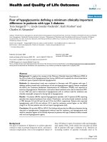
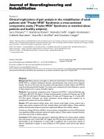
note, small and large (multinucleated) syncytia were
present in the central nervous system and (Fig. 1A) and
lung (Fig. 1B) of some raccoons from year 2001 but not in
animals from 1998 and 2000 (Table 1).
Isolation of virus from infected tissues
Virus was isolated from the tissues of 11/11 animals
(Table 2) [22]. Viral cytopathic effects (CPE) in Vero cells
consisted of the formation of granular-appearing cytoplasm with vacuolization (small vacuoles), followed by
rounding of the cells and detachment, and rare formation
of small stellate syncytia (consisting of 2–3 cells fused
together) for viruses isolated from year 1998 and 2000
specimens or frequent larger rounded syncytia typically
containing >8 nuclei in viruses from year 2001 [22]. Thus,
the 2001 viruses appeared to form large syncytia in vivo
(Table 1) and in vitro [22].
RT-PCR and nucleotide sequence analyses
Where direct comparisons were possible, viral genomic
sequence analyses indicated that the subgenomic viral Fand P- and full-genomic H-gene sequences did not change
during primary isolation in three different cell lines
(MDCK, MV1 Lu, and Vero [22]. Thus, for viruses from
animals 98-2645, 98-2646, and 98-2655, for which direct
RT-PCR from infected tissues failed (Table 2), it was likely
that the sequences obtained were authentic.
The subgenomic F- and P- gene of this study were previously reported [22] and deposited at GenBank (Table 3).
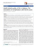
The full-genomic H-gene sequences are available at GenBank (Table 3); since the H-gene sequences are relatively
long (1,824 bp), only the deduced aa sequences are
shown (Fig. 2). As for the P-gene, virus CDV 98-2666 had
two slightly different H-gene sequences that were detected
in vRNA in infected tissues; the same H-gene sequences
were detected in corresponding virus isolates. The dominant H-gene sequence determined directly from infected
tissues is labelled 98-2666 (Fig. 2, and H-gene sequence
98-2666 in Table 3), and is identical to the sequence of
variant 98-2666-1 (Fig. 2, and H-gene sequence 98-26661 in Table 3), whereas the H-gene sequence of the second
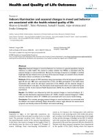
variant is labelled 98-2666-2. An example of RT-PCR for
the CDV H-gene of a primary virus isolate in Vero cells is
shown in figure 3.
Phylogenetic analyses
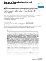
The 70% majority-rule consensus parsimony (Fig. 4) and
neighbor-joining (not shown) cladograms for the P-gene
sequences are almost identical. Both analyses grouped the
1998 sequences together in a single clade with CDV-Lederle and -Snyder Hill with high bootstrap support. These
Page 3 of 14
(page number not for citation purposes)
Virology Journal 2004, 1:2
/>
Table 1: Histologic lesions of CDV-infected raccoons.
Raccoon
Sex
M/Ya
Siteb
Presentation
Encephalitisf
Pneumoniah
Other findings
98-2645
F
8/98
FPc
Euthanized
98-2646
M
8/98
ZGd
Dead
98-2654
M
10/98
ZG
Euthanized
Rare axonal loss
++; Sub-acute to chronic; no
IB
++
98-2655
F
10/98
ZG
Dead
++; IB common
None
98-2666
F
12/98
ZG
Euthanized
++; Chronic; no IB
00-2601
M
1/00
ZG
Euthanized
01-2641
M
5/01
OFPe
Euthanized
++; Axonal loss;
rare neuronal IB
++; Rare neuronal
IB; severe axonal
loss
+; IB; syncytia in
hippocampus
++; Demyelination;
axonal loss; few IBg
-
+++; Chronic; no IB
Lymphoid depletion (LNi); IB
– other sites
IB – other sitesj
-
Ocular discharge; CDV in
footpad ("Hardpad" disease);
lymphoid depletion (LN and
spleen)
Lymphoid depletion (LN and
spleen); IB – other sites
Lymphoid depletion (LN and
spleen); IB – other sites
IB – other sites
-
01-2663
F
6/01
ZG
Euthanized
01-2676
F
7/01
ZG
Euthanized
01-2689
F
8/01
ZG
Euthanized
01-2690
M
8/01
ZG
Euthanized
None
EMCVk
+++ with syncytia; IB
Lymphoid depletion (LN and
spleen); IB – other sites
None
+++ with syncytia; IB
+; Axonal loss;
neuronal necrosis;
IB; syncytia in
hippocampus
+; IB
+++; IB
Lymphoid depletion (LN and
spleen); IB – other sites
Lymphoid depletion (LN); IB –
other sites
Rare neuronal
necrosis; IB
++ with syncytia; IB
None
Lymphoid depletion (LN and
spleen); IB – other sites;
rhinitis; purulent conjunctivitis
Lymphoid depletion (LN); IB –
other sites
-
-
+ brain,
LN,
spleen)
-
-
+
(spleen)
aM/Y;
Month and year animal examined by necropsy and specimens frozen.
Location where animal was trapped or found dead.
cFP; Forest preserve at border of zoo.
dZG; Zoo grounds.
eOFP; Off-site forest preserve
fEncephalitis: -, none; +, mild; ++, moderate.
gIB; Characteristic intracytoplasmic or intranuclear inclusion bodies formed by Canine distemper virus.
hPneumonia: +, mild; ++, moderate; +++, severe.
iLN; Lymph node.
JIB – other sites: Inclusion bodies in other epithelial sites.
kEMCV, Encephalomyocarditis virus.
bSite;
viruses have P-gene sequences similar to those of CDVs
Onderstepoort and Rockport, from S. Africa and Sweden,
respectively. The cluster of the 2001 sequences (01-2663,
-2676, -2689, -2690) was also the same in both cladograms. However, while parsimony joined the 01-2641
sequence from an offsite raccoon to the base, the distance
based tree grouped this sequence with CDV A75/17. The
2000 virus was also not resolved by either method of analysis. Of the 390 bases, 34 were informative. Derivatives of
the 1998 cluster form a distantly related lineage to that of
2001 cluster that is nevertheless rooted in the CDV group
when compared to PDV-1 as an outgroup. CDV Lederle
appears to be more derived than A9224/14b (detected in
1992 in a California (USA) raccoon [6]).
There were a total of 335 nucleotides in the F-gene and 32
of these were parsimony informative. Both parsimony
(Fig. 5) and distance based (not shown) analyses
produced the same topology. The off-site raccoon 012641 failed to group with any other sequences, joining at
the base. The 1998 sequences formed a single cluster
within a clade that included Lederle, Snyder Hill, and vaccine strains Onderstepoort and Bul. 170 (originally isolated from a Bulgarian dog) [30]. This clade also included
the 00-2601 sequence. The remaining 2001 viruses
formed a single clade with high bootstrap support.
The H-gene parsimony (Fig. 6) and neighbor-joining (not
shown) topologies were identical with respect to the
clades that include the raccoon viruses from this study.
Page 4 of 14
(page number not for citation purposes)
Virology Journal 2004, 1:2
/>
B
A
Figure
Panel A 1
Panel A. Hematoxylin and eosin (H & E) – stained thin section of hippocampus tissue from raccoon 01-2676. Syncytia are identified by large arrows. Some CDV inclusion bodies are indicated (small arrows). Original magnification × 200. Panel B. Thin section (H & E-stained) of lung tissue from raccoon 01-2663. Syncytia and CDV inclusion bodies are identified as in panel A.
Table 2: CDV detection by direct RT-PCR of tissue and by virus isolation.
Raccoon
Tissue
Direct RT-PCR of Tissue
Virus isolation
98-2645
98-2646
98-2654
98-2655
98-2666
00-2601
01-2641
brain
brain
brain
brain
brain
brain
brain
lung
lymph node
spleen
brain
lung
lymph node
spleen
lung
lymph node
brain
lymph node
spleen
brain
kidney
liver
lung
spleen
+
+
+
+
+
+
+
+
+
+
+
+
+
+
+
+
-
+
+
+
+
+
+
+
+
+
+
+
+
+
+
+
+
+
+
+
+
+
+
01-2663
01-2676
01-2689
01-2690
Page 5 of 14
(page number not for citation purposes)
Virology Journal 2004, 1:2
/>
Onderst.
98-2645
98-2646
98-2654
98-2654-1
98-2654-2
98-2655
98-2666
98-2666-1
98-2666-2
00-2601
01-2641
01-2676
01-2689
01-2690
1 3 19
31 39 42 50
62 78 83 145
163 176
188
MLS//NSTKLSLVTEEHG//LFVL//LALLAITGVRFHQ//MEKSEA//KVKVNFTNYCESIGIRKAI//SGGRSDIFPPHRC//
... .PS.......... .... ....S.......K ...... ..........DT.....S. ..S.......Y..
... .PS.......... .... ....S.......K ...... ..........DT.....S. ..S.......Y..
... .PS.......... .... ....S...I...K ...... ..........DT.....S. ..S.......Y..
... .PS.......... .... ....S...I...K ...... ..........DT.....S. ..S.......Y..
... .PS.......... .... ....S...I...K ...... ..........DT.....S. ..S.......Y..
... .PS.......... .... ....S.......K ...... ..........DT.....S. ..S.......Y..
... .PS.......... .... ....S.......K ...... ..........DT.....S. ..S.......Y..
... .PS.......... .... ....S.......K ...... ..........DT.....S. ..S.......Y..
... .PS.......... .... ....S.......K ...... ..........DT.....S. ..S.......Y..
... ..SR.......Q. .... M............ ...... .I........DT.....S. ..S.......Y..
... ..S........Q. .... M............ ...... .I........DT.....S. ....G.....YG.
... TPS.......DQE .V.. M............ I...H. .I........DT.....S. ..S.G.....Y..
... ..SR......DQE .... M.......I.... I..... .I.....T..DT.....S. ..S.G.....Y..
... ..SR......DQE .... M.......I.... I..... .I.....T..DT.....S. ..S.G.....Y..
Onderst.
98-2645
98-2646
98-2654
98-2654-1
98-2654-2
98-2655
98-2666
98-2666-1
98-2666-2
00-2601
01-2641
01-2676
01-2689
01-2690
197 203 214 220 238
247 262
281 298
314 323
332
KVFPLSV//SEIINML//DIEREFDTQE//DMPLLQTTNYMVLPENSKAK//EESTVLLYHDSSGSQDG//FWATPMDHIE//
....... .V..... .......... .................... D..........R..... .G........
....... .V..... .......... .................... D..........R..... .G........
....... .V..... .......... .................... D..........R..... .G........
....... .V..... .......... .................... D..........R..... .G........
....... .V..... .......... .................... D..........R..... .G........
....... .V..... .......... .................... D..........R..... .G........
....... .V..... .......... .................... D..........R..... .G........
....... .V..... .......... .................... D..........R..... .G........
....... .V..... .......... .................... D..........R..... .G........
S...... ....... YL.G.....K N................... D..............G. .G.....QV.
R...... P...... YL.G.....K N................... D...I......N..... .G.....QV.
R...... ....S.. YL.G.....K ....F............... D..........N....S .G.....QV.
R...... ....S.. Y..G..V..K ....F..A...........R D..........N....S .G.....QV.
R...... ....S.. Y..G..V..K ....F..A...........R D..........N....S .G.....QV.
Onderst.
98-2645
98-2646
98-2654
98-2654-1
98-2654-2
98-2655
98-2666
98-2666-1
98-2666-2
00-2601
01-2641
01-2676
01-2689
01-2690
339 345 362 368 375
386 401 406 415 420 435
446 459
477//
HPSMEKI//MVPALAS//KGCLESACQRKT//RQLPSY//ASVDLQ//DGMDYYESPLLN//IVGLINKAGRGDQFTVLPH
....... ....... ............ G..... ...... ...V........ .L..............I..
....... ....... ............ G..... ...... ...V........ .L..............I..
....... ....... ............ G..... ...... ...V........ .L..............I..
....... ....... ............ G..... ...... ...V........ .L..............I..
....... ....... ............ G..... ...... ...V........ .L..............I..
....... ....... ............ G..... ...... ...V........ .L..............I..
....... ....... ............ G..... ...... ...V........ .L..............I..
....... ....... ............ G..... ...... ...V........ .L..............I..
....... ....... ............ G..... ...... ...V........ .L..............I..
...V... .....V. .N.......... ...... ..I... ...........D VL......S.......I..
...V... T....V. .N.........S G..... P.I... ...........G VL......S.......T..
...V... .....V. QN.......I.S G..... P.IN.. E.....G....D VL......T.......T..
...V... .....V. .N.......I.S G..... P.IN.. E.....G....D VL......T.......T..
...V... .....V. .N.......I.S G..... P.IN.. E.....G....D VL......T.......T..
484 487 500
519 530 534 542 549 568 572 581 586 603 607 GenBank No.
Onderst. WESS//IDRDVLIESNIVVLPTQSFR//SDHAI//IRTISYTH//VWDDN//FEADIA//NRSNP
AF378705
98-2645
R..G ..........L......... N.... F...F..Y ..... Y..N.. .....
AY445077
98-2646
R..G ..........L......... N.... F...F..Y ..... Y..N.. .....
AY542312
98-2654
R..G ..........L......... N.... F...F..Y ..... Y..N.. .....
AY466011
98-2654-1 R..G ..........L......... N.... F...F..Y ..... Y..N.. .....
AY466011
98-2654-2 R..G ..........L......... N.... F...F..Y ..... Y..N.. .....
AY466011
98-2655
R..G ..........L......... N.... F...F..Y ..... Y..N.. .....
AY548109
98-2666
R..G ..........L......... N.... F...F..Y ..... Y..N.. .....
AY548110[dominant]
98-2666-1 R..G ..........L......... N.... F...F..Y ..... Y..N.. .....
AY548110
98-2666-2 R..G ..........L......... N.... F...F..Y ..... Y..N.. .....
AY548111
00-2601
R..G M.K.......L......N.I G.... ........ ....D ....ST ...K.
AY443350
01-2641
R..G M.K...T...L......D.. G.... ........ ....D ....ST S..K.
AY526496
01-2676
R..G M.K...T...L......N.. R...V ........ A...D ...GST ...K.
AY498692
01-2689
R..G MGK...T...L.G....N.. R...V ........ A...D ...GST ...K.
[same as AY465925]
01-2690
R..G MGK...T...L.G....N.. R...V ........ A...D ...GST ...K.
AY465925
Figure 2
Deduced H-protein amino acid sequences of raccoon CDVs
Deduced H-protein amino acid sequences of raccoon CDVs. Numbers above the sequences identify aa positions in the H-protein of CDV reference strain Onderstepoort.
Page 6 of 14
(page number not for citation purposes)
Virology Journal 2004, 1:2
/>
Table 3: GenBank accession numbers of raccoon CDVsequences.
Virus
98-2645
98-2646
98-2654-1
98-2654-2
98-2655
98-2666-1
98-2666-2
00-2601
01-2641-1
01-2641-2
01-2663
01-2676
01-2689
01-2690
F-gene
AY289612
(AY289612)a
(AY289612)a
(AY289612)a
AY289614
(AY289614)b
AY289615
(AY289615)c
(AY289615)c
(AY289615)c
H-gene
AY445077 (entire genome)
AY542312 (entire genome)
AY466011 (entire genome)
(AY466011)d
AY548109
AY548110
AY548111
AY443350 (entire genome)
AY526496
(AY526496)e
NDf
AY498692
AY465925
(AY465925)g
P-gene
AY286485
AY263373
AY286486
AY286487
AY288310
AY321298
AY288308
AY288309
AY286488
AY264266
aIdentical
to the sequence of AY289612.
to the sequence of AY289614.
cIdentical to the sequence of AY289615.
dIdentical to the sequence of AY466011.
eIdentical to the sequence of AY526496.
fND, Not determined.
gIdentical to the sequence of AY465925.
bIdentical
Out of 1,824 nucleotides, 420 of these were parsimony
informative. As with the previous genes, the 1998 isolates
and the 2000/2001 viruses formed separate clusters. The
1998 sequences joined the tree at a basal position in both
analyses. The 2000 and off-site raccoon 01-2641
sequences grouped with the large felids from another zoo
in Illinois.
Noteworthy, P-, F- and H- gene analyses indicate that the
CDV sequences segregate according to geography and not
to species. Since the H gene had the largest number of
nucleotides, pairwise genetic distances were calculated.
The 1998 isolates were most similar to the Onderstepoort
and Snyder Hill (D = 4% and 1% respectively) while the
2001 isolates were most distant (D = 9% and 10% respectively). Distances within 1998 viruses were low (D ≤
0.2%); within 2001, distances were slightly higher (D =
1%); and comparing years 1998 with 2000 and 2001, distances were highest (D = 7% to 9% respectively).
When the P-, F- and H- genes were combined into a single
linear sequence and analyzed using parsimony and neighbor-joining algorithms with only PDV-1 as an outgroup,
two independent clades are formed, the 1998 clade and
the 2000/2001 clade (data not shown). In the later group,
both methods join the 2000 sequence (00-2601) at a
basal position to the 01-2641 off-site raccoon followed by
the 2001 isolates.
Discussion
This report shows that different CDV sublineages stemming from at least two genetically distant CDV lineages
recently circulated through the local raccoon population.
Our conclusion is based on numerous observations:
differences in the lesions observed in animal tissues, possible dissimilarities of virulence between the viruses, variation in one viral phenotype in tissue culture (formation
of large syncytia by the 2001 viruses), and from the results
of nucleotide sequence and phylogenetic analyses. CDV is
not maintained in hosts that recover from distemper, and
persistent CDV infections do not occur. However, CDV
infects a wide range of genera, and though each individual
population may be small, the number of alternative host
species may be substantial [1]. Forest preserves around the
zoo contain many species susceptible to CDV, and it
appears by inference there are separate reservoirs of different CDV lineages within the area of this study.
Since past studies indicated that wild-type CDVs differed
according to geographical distribution [6,23], we initially
surmised that the local CDV occasionally formed clades of
highly virulent CDV variants, resulting in periodic high
mortality distemper outbreaks. We also speculated that
over time, highly virulent viruses would undergo extinction, and ensuing epizootics would arise from less virulent CDV variants that could affect most of the hosts
without killing them. Thus, there would be an apparent
Page 7 of 14
(page number not for citation purposes)
Virology Journal 2004, 1:2
Figure 3
H-gene RT-PCR amplicons
Ethidium-bromide gel electrophoresis analysis of subgenomic
Ethidium-bromide gel electrophoresis analysis of subgenomic
H-gene RT-PCR amplicons. For CDV-2676, shown are the
1104 bp product (lane 1) using primers CDV-HforD and
CDV-Hrev75, and the 1026 bo product (lane 2) using primers CDVH-forB and CDV-HrevC (29). A 2% agarose gel was
used. Molecular weight markers are loaded in the lane
marked "M". Positive and negative controls were run separately and are not shown.
oscillation (periodicity) of the mortality rates. The situation is not as straightforward, however. As shown in figures 4,5,6, at least two different CDV lineages circulated in
the raccoons from 1998 – 2001. Our findings thus suggest
that the outcomes of distemper might also be influenced
by properties unique to different CDV lineages and their
genetic variants ("strains").
The viruses from year 2001 formed syncytia in vivo and in
vitro. Previously, an inverse relationship between the proficiency of syncytium formation and the level of CDV virulence was reported: the more attenuated a strain is, the
higher its fusogenicity, and fusogenicity was attributed to
the viral H-protein [31-34]. Therefore, the findings of this
study may appear antidogmatic because increased mortality was associated with the 2001 viruses, which formed
large syncytia in vivo and in vitro. However, past notions
concerning the inverse relationship between fusogenicity
and virulence may be imprecise. Indeed, virulent wildtype CDVs that formed syncytia in Vero cells were recently
/>
reported; the same study demonstrated that genetic
changes within the H-gene were not required for CDV
growth in Vero cells [35], as was found in this and our previous study [22]. Also, newer studies indicate that syncytium formation by CDV requires the concerted activities
of both the H- and F- proteins [36-38], and that CDV virulence is the combined affect of various proteins including
the F- and H- proteins [39]. Thus, whereas animal studies
were not performed with the virus isolates of this study to
directly test whether they differ in virulence, the formation of large syncytia does not rule out the possibility that
the 2001 viruses are highly virulent. Noteworthy, the
2001 viruses were detected in the hippocampus and alveoli of the raccoons. Both sites were considered unusual
targets of a CDV variant that was lethal to Serengeti lions,
whereas CDV in dogs was said to most frequently target
the brain stem and bronchi [40,41]. It is possible that tissue localization, especially with regard to the hippocampus, correlates with virus strain. In our experience, CDV in
raccoons does not preferentially target the brain stem but
rather infects all portions of the brain, with the possible
exception of the hippocampus. We will be able to address
the question whether specific CDV strains localize in the
hippocampus of raccoons as we accumulate additional
data from future outbreak, and after we conduct animal
tests with the viruses we isolated. In contrast, CDV targets
epithelial cells, and the presence of CDV in the alveoli of
raccoons with distemper is common.
H-gene phylogenetic analyses (figure 6) suggest that a
viral lineage that includes CDV A75/17 (isolated in 1975)
[32] and the 2000 and 2001 viruses had infected various
species including large felids [Fig. 6 and reference 6] for at
least 28 years on both coasts and a midwestern state (and
thus presumably throughout the continental USA). The
seemingly widespread distribution suggests that viruses
stemming from this lineage may be the dominant "American" CDV currently in circulation in the continental USA.
The F -, H-, and P-gene sequence analyses (figures 4,5,6)
indicate that the 1998 viruses stem from a different CDV
lineage that includes American CDV strains Lederle and
Snyder Hill. A recent phylogenetic analysis of the P-gene
by an independent laboratory that utilized some of our Pgene data generated similar results [42]. Because they were
isolated before CDV Lederle and Snyder Hill were
acquired from the ATCC for this study and have distinguishable F- and H-gene sequences [22], it is certain that
the 1998 CDV isolates are not due to laboratory contamination. Yet, phylogenetic analyses indicate that the CDV
Lederle and Snyder Hill sequences are distant to, and in
the case of the H-gene, ancestral to, those of the 2000 and
2001 viruses, which are as genetically distant from the
1998 viruses as they are from Snyder Hill. The source of
the 1998 viruses is thus intriguing. Prior to 1997, some
area raccoons were trapped, vaccinated against CDV, then
Page 8 of 14
(page number not for citation purposes)
Virology Journal 2004, 1:2
/>
78
(1) Onderstepoort
(2) Rockborn
(3) Lederle
94
(4) Snyder Hill
(5) 98-2655
(6) 98-2645
98
(7) 98-2646
(8) 98-2654-1
(9) 98-2654-2
(10) 98-2666-1
(11) 98-2666-2
(12) 00-2601
99
(13) 01-2641
(14) 01-2690
(15) 01-2689
75
90
(16) 01-2663
(17) 01-2676
81
(18) 5804 (dog)
(19) Bulgarian dog
(20) A75/17 (dog)
(21) Ferret
(22) Siberian seal
(23) Jujo (dog)
(24) A92 (raccoon)
(25) PDV-1
Figure 4
P-gene 70% majority rule parsimony consensus tree
P-gene 70% majority rule parsimony consensus tree. Viruses from this study are high-lighted by a grey background. The animal
source and GenBank numbers from top to bottom are: (1) (South African dog) AF305419, (2) (Swedish dog) AF181446, (3)
(American dog) AY286480, (4) (American dog) AY286481, (5 – 17) Illinois raccoons, GenBank numbers in Table 3, (18) (German dog) AY386315, (19) (Bulgarian dog) AF259549, (20) (American dog) AF164967, (21) (German ferret) AF259550, (22)
(Siberian seal) AF259551, (23) (Japanese dog) AB028916, (24) (Californa raccoon A9224/14b, reference 6), (25) (Phocine distemper virus) D10371.
Page 9 of 14
(page number not for citation purposes)
Virology Journal 2004, 1:2
/>
(1) Lederle
(2) Snyder Hill
(3) 98-2655
(4) 98-2645
86
94
(5) 98-2646
(6) 98-2654-1
(7) 98-2654-2
(8) 98-2666-1
(9) 98-2666-2
(10) 00-2601
(11) Onder.
(12) Bul. 170
(13) 01-2641
(14) 01-2663
99
(15) 01-2676
(16) 01-2689
(17) 01-2690
(18) A75/17 (dog)
(19) PDV-2
86
(20) Danish dog
(21) 5804 (dog)
(22) Hyena
(23) Marten
(24) PDV-1
F-gene 70% majority rule parsimony consensus tree
Figure 5
F-gene 70% majority rule parsimony consensus tree. Viruses from this study are high-lighted by a grey background. GenBank
accession numbers are: (1) CDV Lederle (AY288311); (2) Snyder Hill (AY288312); (3 – 10, Illinois raccoons, Table 3); (11)
Onder., Onderstepoort (AF378705); (12) Bul. 170, Bulgarian dog (AF259549); (13 – 17, Illinois raccoons, Table 3); (18) CDV
A75/17 (AF164967); (19) PDV2, Phocine distemper virus 2 (L07075); (20) Danish dog (AF355188); (21) CDV 5804 (from German dog) (AF026241); (22) Hyena (AF026233); (23) Marten (AF026230); (24) PDV-1 (L07075).
released in an attempt to curtail CDV epidemics within
the local raccoon population. CDV Lederle has been used
as a vaccine strain in the past [3]. The vaccine used for the
raccoons, (Galaxy-D, from Schering-Plough, Kenilworth,
Page 10 of 14
(page number not for citation purposes)
Virology Journal 2004, 1:2
/>
00-2601
(1) 00-2601
Chinese leopard
(2) Chinese leopard
01-2641
(3) 01-2641
Black leopard
(4) Black leopard
Black panther
(5) Black panther
(6) 01-2676
01-2676
01-2689
(7) 01-2689
01-2690
(8) 01-2690
Raccoon (Michigan, USA)
(9) Raccoon (Michigan, USA)
A75/17
(10) A75/17
Dog (Colorado, USA)
(11) Dog (Colorado, USA)
Javelina
(12) Javelina
Raccoon dog T dog Tanu
(13) Raccoon anu (Japan) (Japan)
Dog (T aiwan)
(14) Dog (Taiwan)
Dog Hamam (Japan)
(15) Dog Hamam (Japan)
Dog KDK1 (Japan)
(16) Dog KDK1
Dog Ueno (Japan)
(17) Dog Ueno (Japan)
Dog Yanaka (Japan)
(18) Dog Yanaka (Japan)
Giant panda (China)
(19) Giant panda (China)
Dog 5804 (Germany)
(20) Dog 5804 (Germany)
Dog (Denmark)
(21) Dog (Denmark)
Dog 91a (Denmark)
(22) Dog 91a (Denmark)
Dog isolate A (Denmark)
(23) Dog isolate A (Denmark)
Dog 91b 91b (Denmark)
(24) Dog(Denmark)
Dog 91c 91c (Denmark)
(25) Dog(Denmark)
Dog 91d 91d (Denmark)
(26) Dog(Denmark)
Dog isolate c (Denmark)
(27) Dog isolate C (Denmark)
(28) Dog isolate B (Denmark)
Dog isolate B (Denmark)
Dog isolate D (Denmark)
(29) Dog isolate D
Dog isolate 2544 2544 (Germany)
(30) Dog isolate(Germany)
Dog isolate 404 (Germany)
(31) Dog isolate 404 (Germany)
Dog isolate 4513 4513 (Germany)
(32) Dog isolate(Germany)
Dog (T urkey)
(33) Dog (Turkey)
Ferret (Germany)
(34) Ferret (Germany)
Mink (Denmark)
(35) Mink (Denmark)
Lesser panda (China)
(36) Lesser panda
Siberian seal (Russia)
(37) Siberian seal (Russia)
Dog (China)
(38) Dog (China)
Dog (Greenland)
(39) Dog (Greenland)
Dog 26D 26D (Japan)
(40) Dog (Japan)
Dog 5B (Japan)
(41) Dog 5B (Japan)
Dog SVD (Japan)
(42) Dog 5VD (Japan)
Dog 98002 (Japan)
(43) Dog 98-002 (Japan)
Dog HM3 (Japan)
(44) Dog HM-3 (Japan)
Dog HM6 (Japan)
(45) Dog HM-6 (Japan)
98-2654
(46) 98-2654
(47) 98-2654-1
98-2654-1
98-2654-2
(48) 98-2654-2
98-2655
(49) 98-2655
(50) 98-2666
98-2666
98-2666-1
(51) 98-2666-1
98-2666-2
(52) 98-2666-2
(53) 98-2646
98-2646
98-2645
(54) 98-2645
Snyder Hill
(55) Snyder Hill
Onderstepoort
(56) Onderstepoort
PDV-1
(57) PDV-1
Figure 6
H-gene 70% majority rule parsimony consensus tree
H-gene 70% majority rule parsimony consensus tree. Arrows or boxes demarcate locations of viruses from this study. GenBank accession numbers are: (1) CDV 00-2601 (Illinois raccoon, Table 3); (2) Chinese leopard (Z54156); (3) 01-2641 (Illinois
raccoon, Table 3); (4) black leopard (Z47763); (5) black panther (Z54166); (6 – 8, Illinois raccoons, Table 3); (9) raccoon
(Z47765); (10) A75/17 (AF164967); (11) dog (USA) (Z47762); (12) javelina (Z47764); (13) raccoon dog Tanu (AB016776); (14)
dog (Taiwan) (AY378091); (15) dog Hamam (D85754); (16) dog KDK1 (AB025271); (17) dog Ueno (D85753); (18) dog Yanaka
(D85755); (19) giant panda (AF178038); (20) dog 5804 (AY386315); (21) dog Denmark (Z47761); (22) dog 91A (AF478544);
(23) dog isolate A (AF478543); (24) dog 91B (AF478546); (25) dog 91C (AF478548); (26) dog 91D (AF478550); (27) dog isolate C (AF478547); (28) dog isolate B (AF478545); (29) dog isolate D (AF478549); (30) dog isolate 2544 (Z77672); (31) dog
isolate 404 (Z77671); (32) dog isolate 4513 (Z77673); (33) dog (Turkey) (AY093674); (34) ferret (X84999); (35) mink
(Z47759); (36) lesser panda (AF178039); (37) Siberian seal (X84998); (38) dog (China) (AF172411); (39) dog (Greenland)
(Z47760); (40) dog 26D (AB040766); (41) dog 5B (AY297453); (42) dog 5VD (AY297454); (43) dog 98-002 (AB025270); (44)
dog HM-3 (AB040767); (45) dog HM-6 (AB040768); (46 – 54, Illinois raccoons, Table 3), (55) Snyder Hill (AF259552); (56)
Onderstepoort (AF378705); (57) PDV-1 (AF479274).
Page 11 of 14
(page number not for citation purposes)
Virology Journal 2004, 1:2
NJ), though, was made with CDV Onderstepoort, which is
easily distinguished from the 1998 viruses by F-, H-, and
P-gene analyses. However, we still could not rule out the
possibility that the 1998 viruses are vaccine escape viruses
from a dog vaccinated with CDV Lederle. Dogs and raccoons often frequent the same feeding sites (such as refuse
disposal zones) in urban areas. The possibility of reversion to virulence of attenuated CDV exists [43], and a vaccine escape virus was proposed as a cause of distemper in
a dog in Belfast, Northern Ireland [3]. We could not find
a current manufacturer of anti-CDV vaccine in the USA
that uses CDV Lederle. However, such vaccines were in
distribution overseas around 1998 [22], and the Chicago
area undergoes constant population flux, including translocation of inhabitants (and their pets) from outside of
the continental USA. Related to this, the live attenuated
CDV vaccine (Galaxy-D) used by the zoo up to 1997
caused vaccine-mediated distemper in different species at
the zoo that had been vaccinated. For this reason, use of
that particular vaccine was discontinued; instead,
Purevax™, a recombinant CDV-canary pox virus vaccine
(Merial, Duluth, GA) is used; the CDV insert in the canary
pox virus genome is incomplete and cannot be infectious.
CDV-Lederle was isolated in 1951 from a dog with
encephalitis (information provided by ATCC). An alternative interpretation of our findings is that the CDV lineage
that gave rise to CDV Lederle has stabilized in the local
animals and is still actively circulating; more studies are
needed to resolve this matter.
EMCV has been isolated or detected in raccoons before
[44,45]. However, pathogenesis was uncertain, and it was
thought that raccoons are a dead-end host for this virus
[45]. It is known that mortality during an active case of
distemper is increased in the presence of polymicrobial
disease [46]. For example, a lethal outcome occurs in dogs
co-infected with CDV, Bordetella bronchiseptica, and Toxoplasma gondii. It is possible that the increased mortality in
2001 was due to secondary infections with EMCV; however, no lesions attributable to EMCV were observed in
pathology exams of the animals of this study, and EMCV
was not isolated from all of the 2001 specimens. The significance of isolating EMCV from the brain tissue of animal 01-2641 is thus uncertain.
Our findings are especially useful for the molecular epidemiology of CDV in local wildlife, as they provide a molecular basis for CDV surveillance in area wildlife. Whereas it
is considered difficult to obtain field isolates of CDV, we
succeeded and can now obtain complete viral genomic
sequence data (it would be difficult to do so relying solely
on the limited amount of archived CDV-infected tissues
from the animals of this work). Taken together, we can
now monitor viral genetic drift during a long-term study
of CDV in local raccoons, and will be able to conduct ani-
/>
mal studies with the newly isolated viruses. We can also
clone relevant CDV virulence genes, and express and study
the biochemical properties of their specific products in
vitro. The baseline genetic values established here will be
helpful toward the development of a contemporary fieldbased model (since the animals are free-ranging) for studies on the emergence, evolution, maintenance, and transmission of morbilliviruses, and the efficacy of vaccines
against changing viruses.
Conclusions
The 1998 and 2001 distemper outbreaks were caused by
two genetically distant American CDV lineages. Since
CDV does not cause persistent infections, the cycling of
different CDV lineages within the same locale suggests
multiple reservoirs were responsible for the reintroduction of the virus to area raccoons. Whereas different susceptible species of the forest preserves and perhaps also
some caged animals of the zoo are the most likely reservoirs, our study raises the possibility that vaccines might
also be a source of CDV. The perceived differences in
mortality rates that occur during intermittent distemper
epizootics may be attributed in part to inherent differences between CDV strains.
Methods
Raccoon tissues
The raccoon tissues used in this study were described previously [22]; relevant clinical and histologic findings are
presented in Table 1. Brain tissue was available for animals 98-2645, -2646, -2654, -2655, -2666 (n = 5, each
collected in year 1998) and 00-2601 (n = 1, from year
2000) (Table 1). Additional tissues were available for animals 01-2641, -2663, -2676, -2689, and -2690 (n = 5,
each collected in year 2001) (Table 2).
Virus isolation
Detailed virus isolation procedures were described previously [22]. Briefly, CDV was isolated in vitro in MDCK,
MV1-Lu, and Vero cells, eliminating the need for virus isolation in specific pathogen-free animals or in primary
macrophages or other suitable cells derived thereof [29].
RNA purification and RT-PCR
RNA purification and RT-PCR methods were previously
detailed [22]. Briefly, vRNA was extracted directly from
infected tissues when possible, as well as from CDVinfected tissue culture cells or from liberated CDV virions
in spent cell growth media, using dedicated commercial
kits (Qiagen Inc., Valencia, CA). For the American CDV
strains of this work, many RT-PCR primers based on the
sequence of American CDV isolate A75/17 (GenBank No.
AF164967) were more effective than primers described for
foreign CDV strains [22].
Page 12 of 14
(page number not for citation purposes)
Virology Journal 2004, 1:2
Nucleic acid sequencing
Methods used for nucleic acid sequencing were previously
described [22]. Briefly, all sequences were determined at
least twice, starting from the purification of new RNA
samples from each specimen, and both strands of each
PCR amplicon were sequenced. Slab-gel sequencing utilizing dye-terminator chemistries (LI-COR, Lincoln, NE)
was used at the inception of the project, then replaced by
capillary sequencing using ABI-PRISM technology
(Applied Biosystems, Foster City, CA). The CDV gene
sequences in infected tissues were exactly like those in
matched primary viral isolates [22]. The GenBank accession numbers for all the virus sequences of this work are
given in Table 3.
Phylogenetic analyses
Phylogenetic trees of the P-, F-, and H-gene sequences
were constructed using the maximum-parsimony and
neighbor-joining algorithms in Phylogeny Analysis Using
Parsimony (PAUP) Beta Version 4.0B10 for Macintosh
[47]. Heuristic searches were conducted with "simple"
addition and the tree-bisection-reconnection method of
branch swapping. Distance-based analyses using the minimum-evolution criterion were also conducted within
PAUP using Kimura's-two-parameter model [48]. Phylogenetic tree reliability was estimated with 1000 bootstrap
replications [49,50]. The appropriate Phocine distemper
virus sequence (PDV-1) was included for outgroup
rooting.
P-gene phylogenetic analyses were performed after an
alignment of 25 P-gene sequences. Each P-gene sequence
consisted of 390 ungapped positions (nucleotides 2154 to
2543 of CDV reference strain Onderstepoort) within the
P-gene PCR amplicon. Only the internal 390 bp section of
the P-gene PCR amplicon (432 bp) was analyzed because
many relevant GenBank entries did not include the entire
sequence amplified by the P-gene primers of this study.
An additional P-gene sequence for raccoon A9224/14b
was obtained from published data currently not deposited
at GenBank [6]. Similarly, 24 ungapped F-gene sequences
corresponding to nt 5399–5733 (335 bp) of CDV Onderstepoort were analyzed. Unlike the P- and F-genes, the
entire H-gene was analyzed since many complete H-gene
sequences were available at GenBank. Phocine distemper
virus 1 (PDV1) sequences were included in the analyses
for outgroup rooting.
Competing interests
/>
training of technicians, and drafted the manuscript; JD
performed phylogenetic analyses, interpreted data, and
helped draft the manuscript; MJK performed pathology
examinations, provided tissue specimens, helped draft the
manuscript, and interpreted data; TPM co-conceived the
study, provided serology data, helped draft the manuscript, and interpreted data; MB performed phylogenetic
analyses, interpreted data, and helped draft the manuscript; LLH provided serology data and epidemiology perspectives, and helped draft the manuscript; NAS
participated in virus isolation, molecular genetic studies,
sequence alignment, and proofreading of the manuscript;
KEW participated in virus isolation and molecular genetic
studies, and MDB, CP, and CMH performed molecular
genetic studies. All authors read and approved the final
manuscript
Acknowledgements
The authors thank Chris Anchor and the Wildlife Division of the Forest
Preserve District of Cook County for assisting with sample acquisition.
Andrea Guido provided excellent technical assistance. We thank Dr. K.
MacClatchey for critical review of this manuscript. Partial funding for
necropsy evaluations was obtained from the Department of Animal Control Environmental Impact Program, Cook County, Illinois. Molecular and
viral tests were funded by grant no. 0023 from the Conservation Medicine
Center of Chicago to J.A.L.
References
1.
2.
3.
4.
5.
6.
7.
8.
None declared.
9.
Authors' contributions
10.
JAL co-conceived, designed, and coordinated the study,
isolated virus, participated in the molecular genetic studies and sequence alignment, interpreted data, oversaw the
11.
12.
Appel MJG, Summers B: Pathogenicity of morbilliviruses for terrestrial carnivores. Veterinary Microbiology 1995, 44:187-191.
Appel MJ, Yates RA, Foley GL, Bernstein JJ, Santinelli S, Spelman LH,
Miller LD, Arp LH, Anderson M, Barr M, Pearce-Kelling S, Summers
BA: Canine distemper epizootic in lions, tigers, and leopards
in North America. Journal of Veterinary Diagnostic Investigation 1994,
6:277-288.
Harder TC, Osterhaus ADME: Canine distemper virus – a morbillivirus in search of new hosts? Trends in Microbiology 1997,
5:120-124.
Kennedy S, Kuiken T, Jepson PD, Deaville R, Forsyth M, Barrett T, van
de Bildt MWG, Osterhaus ADME, Eybatov T, Duck C, Kydyrmanov
A, Mitrofanov I, Wilson S: Mass die-off of Caspian seals caused
by Canine distemper virus. Emerging Infectious Diseases 2000,
6:637-639.
van de Bildt MWG, Kuiken T, Visee AM, Lema S, Fitzjohn TR, Osterhaus ADME: Distemper outbreak and its effect on African wild
dog conservation. Emerging Infectious Diseases 2002, 8:211-213.
Harder TC, Kenter M, Vos H, Siebelink K, Huisman W, Van
Amerongen G, Örvell C, Barrett T, Appel MJG, Osterhaus ADME:
Canine distemper virus from diseased large felids: biological
properties and phylogenetic relationships. Journal of General
Virology 1996, 77:397-405.
Appel M, Sheffy BE, Percy DH, Gaskin JM: Canine distemper virus
in domestic cats and pigs. American Journal of Veterinary Research
1974, 35:803-806.
Ikeda Y, Nakamura K, Miyazawa T, Chen M-C, Kuo T-F, Lin JA,
Mikami T, Kai C, Takahashi E: Seroprevalence of canine distemper virus in cats. Clinical and Diagnostic Laboratory Immunology 2001,
8:641-644.
Daszak P, Cunningham AA, Hyatt AD: Emerging infectious diseases of wildlife – threats to biodiversity and human health.
Science 2000, 287:443-449.
Domingo E, Holland JJ: RNA virus mutations and fitness for
survival. Annual Review of Microbiology 1997, 51:151-178.
Steinhauer DA, Holland JJ: Rapid evolution of RNA viruses.
Annual Review of Microbiology 1987, 41:409-433.
Eigen M: Viral quasispecies. Scientific American 1993:42-49.
Page 13 of 14
(page number not for citation purposes)
Virology Journal 2004, 1:2
13.
14.
15.
16.
17.
18.
19.
20.
21.
22.
23.
24.
25.
26.
27.
28.
29.
30.
31.
Blixenkrone-Møller M, Swansson V, Have P, Örvell C, Appel M, Pedersen IR, Dietz HH, Henriksen P: Studies on manifestations of
canine distemper virus infection in an urban dog population.
Veterinary Microbiology 1993, 37:163-173.
Gemma T, Watari T, Akiyama K, Miyashita N, Shin Y-S, Iwatsuki K,
Kai C, Mikami T: Epidemiological observations on recent outbreaks of canine distemper in Tokyo area. Journal of Veterinary
Medicine and Science 1996, 58:547-550.
Shin Y-S, Mori T, Okita M, Gemma T, Kai C, Mikami T: Detection of
canine distemper virus nucleocapsid protein gene in canine
peripheral blood mononuclear cells by RT-PCR. Journal of Veterinary Medicine and Science 1995, 57:439-445.
Hoff GL, Bigler WJ, Proctor SJ, Stallings LP: Epizootic of canine distemper virus infection among urban raccoons and grey
foxes. Journal of Wildlife Diseases 1974, 10:421-428.
Roscoe DE: Epizootiology of canine distemper in New Jersey
raccoons. Journal of Wildlife Diseases 1993, 29:390-395.
Mitchell MA, Hungerford LL, Nixon C, Esker T, Sullivan J, Koerkenmeier R, Dubey JP: Serologic survey for selected infectious disease agents in raccoons in Illinois. Journal of Wildlife Diseases
1999, 35:347-355.
Sedgwick CJ, Young WA: Distemper outbreak in a zoo. Modern
Veterinary Practice 1968, 49:39-44.
Yoshikawa Y, Ochikubo F, Matsubara Y, Tsuruoka H, Ishii M, Shirota
K, Nomura Y, Sugiyama M, Yamanouchi K: Natural infection with
canine distemper virus in a Japanese monkey (Macaca
fuscata). Veterinary Microbiology 1989, 20:193-205.
Appel MJ, Reggiardo C, Summers BA, Pearce-Kelling S, Mare CJ,
Noon TH, Reed RE, Shively JN, Orvell C: Canine distemper virus
infection and encephalitis in javelinas (collared peccaries).
Archives of Virology 1991, 119:147-152.
Lednicky JA, Meehan TP, Kinsel MJ, Dubach J, Hungerford LL, Sarich
NA, Witecki KE, Braid MD, Pedrak C, Houde CM: Effective primary isolation of wild-type Canine distemper virus in MDCK,
MV1 Lu and Vero cells without nucleotide sequence changes
within the entire haemagglutinin protein gene and in subgenomic sections of the fusion and phospho protein genes. Journal of Virological Methods 2004, 118:147-157.
Mochizuki M, Hashimoto M, Hagiwara S, Yoshida Y, Ishiguro S: Genotypes of canine distemper virus determined by analysis of
the hemagglutinin genes of recent isolates from dogs in
Japan. Journal of Clinical Microbiology 1999, 37:2936-2942.
Carpenter MA, Appel MJG, Roelke-Parker ME, Munson L, Hofer H,
East M, O'Brien SJ: Genetic characterization of canine distemper virus in Serengeti carnivores. Veterinary Immunology and
Immunopathology 1998, 65:259-266.
Barrett T, Visser IKG, Mamaev L, Goatley L, van Bressem M-F, Osterhaus ADME: Dolphin and porpoise morbilliviruses are genetically distinct from phocine distemper virus. Virology 1993,
193:1010-1012.
Morrison T, Portner A: Structure, function, and intracellular
processing of the glycoproteins of Paramyxoviridae. In The paramyxoviruses Edited by: Kingsbury D. New York: Plenum Press;
1991:347-382.
Barrett T, Subbarao MS, Belsham GJ, Mahy BWJ: The molecular
biology of the morbilliviruses. In The paramyxoviruses Edited by:
Kingsbury D. New York: Plenum Press; 1991:83-102.
Visser IK, van der Heijden RWJ, van de Bildt MWG, Kenter MJH,
Örvell C, Osterhaus ADME: Fusion protein gene nucleotide
sequence similarities, shared antigenic sites and phylogenetic analysis suggest that phocid distemper virus type 2 and
canine distemper virus belong to the same virus entity. Journal of General Virology 1993, 74:1989-1994.
von Messling V, Zimmer G, Herrler G, Haas L, Cattaneo R: The
hemagglutinin of canine distemper virus determines tropism
and cytopathogenicity. Journal of Virology 2001, 75:6418-6427.
Liermann H, Harder TC, Löchelt M, von Messling V, Baumgärtner W,
Moennig V, Hass L: Genetic analysis of the central untranslated
genome region and the proximal coding part of the F gene
of wildtype and vaccine distemper morbilliviruses. Virus Genes
1998, 17:259-270.
Cosby SL, Lyons C, Fitzgerald SP, Martin SJ, Pressdee S, Allen IV: The
isolation of large and small plaque canine distemper viruses
which differ in their neurovirulence for hamsters. Journal of
General Virology 1981, 52:345-353.
/>
32.
33.
34.
35.
36.
37.
38.
39.
40.
41.
42.
43.
44.
45.
46.
47.
48.
49.
50.
Summers BA, Greisen HA, Appel MJG: Canine distemper encephalomyelitis variation with virus strain. Journal of Comparative
Pathology 1984, 94:65-75.
Tobler LH, Imagawa DT: Mechanism of persistence with canine
distemper virus: difference between a laboratory strain and
an isolate from a dog with chronic neurological disease. Intervirology 1984, 21:77-86.
Zurbriggen A, Vandevelde M, Bollo E: Demyelinating, nondemyelinating and attenuated canine distemper virus strains
induce oligodendroglial cytolysis in vitro. Journal of the Neurological Sciences 1987, 79:33-41.
Nielsen L, Andersen MK, Jensen TD, Blixenkrone-Møller M, Bolt G:
Changes in the receptorbinding haemagglutinin protein of
wild-type morbilliviruses are not required for adaptation to
vero cells. Virus Genes 2003, 27:157-162.
Cattaneo R, Rose JK: Cell fusion by the envelope glycoproteins
of persistent measles viruses which caused lethal human
brain disease. Journal of Virology 1993, 67:1493-1502.
Stern LB, Greenberg M, Gershoni JM, Rozenblatt S: The hemagglutinin envelope protein of canine distemper virus (CDV) confers cell tropism as illustrated by CDV and measles virus
complementation analysis. Virology 1995, 69:1661-1668.
Wild TF, Malvoisin E, Buckland R: Measles virus: both the haemagglutinin and fusion glycoproteins are required for fusion.
Journal of General Virology 1991, 72:439-442.
von Messling V, Springfeld C, Devaux P, Cattaneo R: A ferret model
of canine distemper virus virulence and immunosuppression.
Journal of Virology 2003, 77:12579-12591.
Morell V: New virus variant killed Serengeti cats. Science 1996,
271:596.
Roelke-Parker ME, Munson L, Packer C, Kock R, Cleaveland S, Carpenter M, O'Brien SJ, Pospischil A, Hofmann-Lehmann R, Lutz H,
Mwamengele GLM, Mgasa MN, Machange GA, Summers BA, Appel
MJG: A canine distemper virus epidemic in Serengeti lions
(Panthera leo). Nature 1996, 379:441-445.
Stanton JB, Brown CC, Poet S, Lipscomb TP, Saliki J, Frasca S Jr: Retrospective differentiation of canine distemper virus and phocine distemper virus in phocids. Journal of Wildlife Diseases 2004,
40:53-59.
Appel MJG: Reversion to virulence of attenuated canine distemper virus in vivo and in vitro. Journal of General Virology 1978,
41:385-393.
Gainer JH, Bigler WI: Encephalomyocarditis (EMC) virus recovered from two cotton rats and a raccoon. Bulletin of the Wildlife
Disease Association 1967, 3:47-49.
Zimmerman JJ, Hill RE, Smith KE, Kneeland BL, Platt KB, Hill HT,
Beran GW, Clark WR, Miller LD: Encephalomyocarditis virus
infection in raccoons (Procyon lotor). Journal of Zoo and Wildlife
Medicine 1994, 25:233-239.
Appel MJG: Canine distemper virus. In Virus infections of carnivores.
Virus infections of vertebrates Volume 1. Edited by: Horzinek MC.
Amsterdam: Elsevier; 1987:133-159.
Swofford DL: PAUP*. Phylogenetic Analysis Using Parsimony
(*and Other Methods). [Version 4]. Sunderland, Massachusetts, Sinauer Associates 1998.
Kimura M: A simple method for estimating evolutionary rates
of base substitutions through comparative studies of nucleotide sequences. Journal of Molecular Evolution 1980, 16:111-120.
Felsenstein J: Phylogenies from molecular sequences: inference and reliability. Annual Review of Genetics 1988, 22:521-565.
Nei M: Phylogenetic analysis in molecular evolutionary
genetics. Annual Review of Genetics 1996, 30:371-403.
Page 14 of 14
(page number not for citation purposes)