báo cáo hóa học:" Susceptibility of different leukocyte cell types to Vaccinia virus infection" docx
Bạn đang xem bản rút gọn của tài liệu. Xem và tải ngay bản đầy đủ của tài liệu tại đây (361.69 KB, 7 trang )
BioMed Central
Page 1 of 7
(page number not for citation purposes)
Virology Journal
Open Access
Research
Susceptibility of different leukocyte cell types to Vaccinia virus
infection
Juana M Sánchez-Puig
1
, Laura Sánchez
2
, Garbiñe Roy
2
and Rafael Blasco*
1
Address:
1
Departamento de Biotecnología-I.N.I.A. Ctra. La Coruña km 7.5 E-28040 Spain and
2
Servicio de Inmunología. Hospital Ramón y Cajal.
28034 Madrid, Spain
Email: Juana M Sánchez-Puig - ; Laura Sánchez - ; Garbiñe Roy - ;
Rafael Blasco* -
* Corresponding author
Abstract
Background: Vaccinia virus, the prototype member of the family Poxviridae, was used extensively
in the past as the Smallpox vaccine, and is currently considered as a candidate vector for new
recombinant vaccines. Vaccinia virus has a wide host range, and is known to infect cultures of a
variety of cell lines of mammalian origin. However, little is known about the virus tropism in human
leukocyte populations. We report here that various cell types within leukocyte populations have
widely different susceptibility to infection with vaccinia virus.
Results: We have investigated the ability of vaccinia virus to infect human PBLs by using virus
recombinants expressing green fluorescent protein (GFP), and monoclonal antibodies specific for
PBL subpopulations. Flow cytometry allowed the identification of infected cells within the PBL
mixture 1–5 hours after infection. Antibody labeling revealed that different cell populations had
very different infection rates. Monocytes showed the highest percentage of infected cells, followed
by B lymphocytes and NK cells. In contrast to those cell types, the rate of infection of T
lymphocytes was low. Comparison of vaccinia virus strains WR and MVA showed that both strains
infected efficiently the monocyte population, although producing different expression levels. Our
results suggest that MVA was less efficient than WR in infecting NK cells and B lymphocytes.
Overall, both WR and MVA consistently showed a strong preference for the infection of non-T
cells.
Conclusions: When infecting fresh human PBL preparations, vaccinia virus showed a strong bias
towards the infection of monocytes, followed by B lymphocytes and NK cells. In contrast, very
poor infection of T lymphocytes was detected. These finding may have important implications both
in our understanding of poxvirus pathogenesis and in the development of improved smallpox
vaccines.
Background
Vaccinia virus, the prototype of the Poxviridae, is a large
DNA virus whose replication takes place in the cytoplasm
of the infected cell [1]. Although well characterized in
vitro, little is known about the ability of vaccinia virus to
infect different cell types in vivo. Vaccinia virus host range
in cell culture is known to be determined by several genes.
The importance of host restriction has been highlighted in
Published: 22 November 2004
Virology Journal 2004, 1:10 doi:10.1186/1743-422X-1-10
Received: 11 October 2004
Accepted: 22 November 2004
This article is available from: />© 2004 Sánchez-Puig et al; licensee BioMed Central Ltd.
This is an Open Access article distributed under the terms of the Creative Commons Attribution License ( />),
which permits unrestricted use, distribution, and reproduction in any medium, provided the original work is properly cited.
Virology Journal 2004, 1:10 />Page 2 of 7
(page number not for citation purposes)
recent years by the growing use of the Modified Vaccinia
Ankara (MVA) virus strain, whose replication is severely
impaired in human cells [2-4]. Genes known to influence
the ability of vaccinia virus to infect cells, termed host
range genes, have been identified, and shown to block
productive infection at different points in the replication
cycle. Significantly, MVA replication of non-permissive
cells proceeds through early and late gene expression, but
is blocked at late times in a step of virion morphogenesis
[5].
In addition to host range genes, there are a number of fac-
tors that might influence the infection rate of a given cell
type, such as the accessibility and amount of receptors, the
ability to internalize the virus, and the metabolic state of
the cell. In addition to cellular factors, genetic differences
in the virus might influence the efficiency and fate of the
infection. For instance, cellular nucleotide pools can be
one of the factors that, in conjunction with the expression
of viral thymidine kinase (TK), may influence the rate of
infection.
The above considerations led us to hypothesize that,
although receptors for vaccinia seem to be ubiquitous,
and virus replication is relatively independent from the
host cell, virus tropism in vivo may be determined by
many complex factors that may be dependent on the cell
type and metabolic state.
We have focused here on the differences between two
widely used strains of vaccinia virus (Western Reserve-WR
and MVA), and also to their respective TK(-) mutants, in
their ability to infect different cell types in fresh human
PBLs.
Results
Infection of human PBLs by GFP-expressing vaccinia virus
Previously, we have shown that GFP expression from a
vaccinia virus recombinant can be used to monitor infec-
tion by flow cytometry [6]. Where adequate marker mole-
cules for different cell populations exist, this approach
should facilitate the study of the susceptibility of cell types
to vaccinia virus infection. With this aim, fresh human
PBLs from healthy donors were infected with virus WR-
GFP, and analyzed by flow cytometry at different times
post-infection. The overall rate of infection, measured as
GFP-positive cells, was 4.5%, 7.6% and 10.0% at 1, 3 and
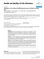
5 h, respectively. Staining with antibodies to CD3, CD14,
CD19 and CD56 was performed on infected cells at 5
h.p.i. (Fig. 1). A marked preference was noted for the
infection of non-T cells, since GFP positive cells
amounted to 19% of non-T lymphocytes, while only 1.9%
of T cells were infected. Among the non-T lymphocytes,
there was a strong bias towards the infection of CD14 pos-
itive cells (monocytes), of which up to 77% showed green
fluorescence, followed by B lymphocytes (CD19
+
, up to
20%) and NK cells (CD56
+
, up to 9%).
Construction of MVA-GFP, WR-TK(-) and MVA-TK(-)
Vaccinia virus MVA and TK-deficient viruses have been
proposed as improved recombinant vaccines. In particu-
lar, the highly attenuated MVA strain has elicited much
interest as a safer vaccine vector. We studied the influence
of the virus strain and the TK phenotype in the infection
of human PBLs. We thus constructed GFP expressing
viruses from vaccinia virus MVA strain, by inserting the
GFP cassette downstream of the F13L gene, using an inter-
genic region for the insertion. Additionally, thymidine
kinase-deficient virus recombinants WR-TK(-) and MVA-
TK(-) were constructed by inserting the GFP cassette
within the viral TK locus. Those viruses grew to high titers
and produced, upon infection of cell lines, bright GFP flu-
orescence (not shown).
Infection of human PBLs with MVA-GFP
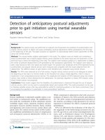
The four GFP-expressing viruses were used to infect fresh
human PBLs from a different individual, and subjected to
flow cytometry analysis at 7 h.p.i. (Fig. 2). The results con-
firmed the above findings with respect to the low infec-
tion rate of T cells in comparison with monocytes, B and
NK cells. Both CD4
+
and CD8
+
cells were poorly infected,
although there was indication of an increased infection of
low-CD8 T lymphocytes in comparison with high-CD8
cells. Notably, this experiment confirmed that most of the
monocytes (CD14+) was infected in our experimental
conditions, and showed a high level of GFP fluorescence.
It was of interest to directly compare the ability of vaccinia
MVA to infect PBLs with that of the standard laboratory
strain WR. A side-by-side comparison of WR-GFP and
MVA-GFP showed that both viruses infected a high per-
centage of CD14
+
monocytes (83 and 70%, respectively),
and a low percentage of T lymphocytes (0.46 and 0.2%,
respectively). No significant differences were noted in the
percentage of CD4 cells infected with both viruses.
Although both virus strains were able to infect the major-
ity of monocytes, MVA-GFP produced a lower level of GFP
fluorescence than WR-GFP in the infected monocytes.
Those differences could be the result of a lower expression
level, or a delay in the course of infection, by the MVA
strain.
In addition to increased GFP expression levels, WR-GFP
was also more efficient than MVA-GFP for the infection of
CD19
+
B lymphocytes (7.1% vs 3.5%) and CD56
+
/CD16
+
NK cells(7.6% vs 4%).
Infection with thymidine kinase-deficient viruses
As stated above, we constructed recombinant viruses from
both WR and MVA by insertion of GFP into the TK locus.
Infection of different populations in human PBLs with
Virology Journal 2004, 1:10 />Page 3 of 7
(page number not for citation purposes)
Analysis of vaccinia infected human PBLsFigure 1
Analysis of vaccinia infected human PBLs. Human PBLs infected with vaccinia WR-GPF for 5 h were subsequently stained
with cell-type specific mAbs, and analyzed by flow cytometry. Plots show the level of GFP fluorescence (recorded in the FL1
channel) versus the amount of labeling with the indicated antibody markers (recorded in the FL2 channel). Numbers inside the
plots indicate the percentage of cells within the respective regions.
CD3
CD56
CD14
CD19
FL1 FLUORESCENCE
F
L
2
F
L
U
O
R
E
S
C
E
N
C
E
10 1
42 47
88 1
6 5
9 2
83 6
8 2
70 20
Virology Journal 2004, 1:10 />Page 4 of 7
(page number not for citation purposes)
Analysis of cell populations infected with different vaccinia virusesFigure 2
Analysis of cell populations infected with different vaccinia viruses. Human PBLs were infected with vaccinia virus
strains WR and MVA, or their respective TK(-) mutants. PBL-subsets were identified by staining with the specific mAbs indi-
cated under each plot. Numbers inside the plots indicate the percentage of GFP-expressing cells within each PBL-subset.
0.46%
W-GFP
CD8
0.8%
2.0%
CD8
0.4%
0.9%
CD8
0.5%
0.32%
0.9%
WR-TK(-)
0.3%
MVA-TK(-)
0.9%
CD3 CD3 CD3
0.6%
CD3
MVA-GFP
0.2%
CD8
G
F
P
-
F
L
1
f
l
u
o
r
e
s
c
e
n
c
e
83%
CD56
7.1%
CD14
CD19
CD14
CD56
4.0%
70%
CD14
80%
6.6%
CD56
CD19
4.2%
CD56
4.4%
70%
CD19
3.4%
CD19
3.5%
CD14
7.6%
CD4
CD4
CD4
CD4
CD16
8.1%
CD16
3.1%
CD16
7.5%
CD16
3.3%
Virology Journal 2004, 1:10 />Page 5 of 7
(page number not for citation purposes)
these viruses was again monitored in paralell using spe-
cific antibodies (Fig. 2). Infection of PBLs with WR-TK(-)
virus resulted in similar percentages of infected CD14 and
CD56/CD16 positive cells, although a slight decrease of
infection rates was noted in CD19 cells. The level of GFP
fluorescence in CD14-positive cells (monocytes) (and to
a lesser extent, in all the WR-TK(-) infected lymphoid sub-
sets) was markedly reduced with respect to the WR-GFP
virus.
Discussion
Detection of cells infected by GFP-expressing vaccinia
viruses provide a fast and sensitive method to measure
virus infection [6]. In this report, we have taken advantage
of this approach to measure infection in freshly prepared
human PBLs. In combination with cell-type specific fluo-
rescent antibodies, we have been able to study the rate of
infection in different cell subset within the PBL
population.
It is to note that the approach used in this work only
allows the detection of viral gene expression derived from
the infection, but does not address whether the infection
results in the production of progeny virus. Early reports
indicated that vaccinia virus cause cythopathic effect in
human leukocytes, although only replicated in mitogen-
stimulated cell populations, indicating that active cell rep-
lication is required for virus replication [7,8]. In this
respect, it has also been reported that vaccinia infection of
dendritic cells and monocytes/macrophages is abortive
[9-11], and that dendritic cells and macrophages die by
apoptosis upon infection [9,12-14]. Less clear is the case
of transformed B lymphocyte cell lines, where virus infec-
tion has been described to be productive [9] and abortive
[15] in different cell lines.
Our results point to a significant preference of vaccinia
virus for certain cell types. In particular, monocytes were
the most susceptible cells, followed by B cells and NK
cells. In contrast, T cells were infected at very low rates.
These observations are in broad agreement with previous
studies, where different infection rates have been noted
between monocytes and lymphocytes [16] and between B
and T lymphocytes [17]. In our analysis, we have detected
different rates of virus infection of different cells but at
this point we cannot relate the differences in infection to
differential virus binding, internalization or gene expres-
sion in different PBL cell lineages. In any event, the conse-
quences of virus tropism in the pathogenicity of
poxviruses remains to be further investigated.
Comparison of the patterns obtained with the two virus
strains and their TK(-) mutants indicate that both the virus
strain and the TK phenotype may determine the amount
of gene expression, as was revealed by the intensity of GFP
fluorescence in infected monocytes. In addition, the abil-
ity of the virus to infect certain cell types (CD19) seems to
be affected to a certain extent by disruption of the TK gene.
While this may be derived from our inability to detect
those infected cells because of decreased gene expression,
we cannot rule out a more direct requirement of TK activ-
ity in those cells.
MVA vaccinia virus strain has elicited much interest
recently because of its safety record. Because clinical com-
plications and side effects of smallpox vaccination are a
critical issue in the event of mass vaccination, understand-
ing the basis of MVA attenuation may lead to the develop-
ment of better vaccine vectors. In this study, a number of
differences were noted between the rates of infection
obtained with WR and MVA virus strains. While both
viruses were able to infect the monocyte population, WR
infected B cells and the NK population (CD56, CD16 pos-
itive cells) more efficiently than MVA. Whether these
observations have implications on the pathogenicity or
immunogenicity of MVA will require further studies.
The fact that both WR and MVA showed a strong prefer-
ence for certain cell populations indicate that, in addition
to host range genes, there are other factors that might
influence the infection rate of PBL cells. Those might
include a variety of such as the accessibility and amount
of receptors, ability to internalize the virus, and the meta-
bolic state of the cell.
Conclusions
Monocytes (CD14+ cells) were the cells in the PBL popu-
lation that showed a greater susceptibility to vaccinia virus
infection, as measured by viral gene expression. On the
other hand, T lymphocytes (CD3+ cells) were infected
with low efficiency. An intermediate susceptibility was
detected in B lymphocytes (CD19+ cells) and NK (CD56+
cells). Both the use of a highly attenuated virus strain
(MVA) or the disruption of the thymidine kinase gene
lead to decreased gene expression in the infected cells.
Those observations highlight the existence of a different
degree of susceptibility to infection if PBL
subpopulations, a fact that may have important implica-
tions in understanding virus pathogenicity and
immunogenicity.
Methods
Cells, plasmids and virus
Vaccinia virus strain WR was grown and titrated in BSC-1
or CV-1 cells, grown in minimal essential medium
(EMEM) supplemented with 5% fetal bovine serum (FBS)
and 2 mM L-Glutamine. MVA virus and recombinants
were grown in BHK-21 cells (ATCC CCL10) cultured in
BHK medium containing 5% FBS, 3 g/ml tryptose phos-
phate broth and 0.01 M hepes. All cells were maintained
Virology Journal 2004, 1:10 />Page 6 of 7
(page number not for citation purposes)
in a 5% CO
2
atmosphere at 37°C. Plasmid pRB21 [18]
contains vaccinia virus gene F13L and flanking sequences,
and a synthetic early/late promoter placed downstream of
the P37 coding sequence.
Construction of recombinant viruses expressing GFP
Plasmid pRBrsGFP, designed to mediate the insertion of
the gene coding an enhanced version of the green fluores-
cent protein gene, rsGFP, (Quantum Biotechnologies,
Inc.) was constructed as follows. rsGFP gene in plasmid
pQBI25 was amplified using oligonucleotides GFP 5'
(AATATAAATGGCTAGC
AAAGGAGAAGAA) and GFPH3
(TTTAAAGCTT
TACTAGTGGATCCTCAG), that include
NheI and HindIII restriction sites, respectively. After diges-
tion with NheI and HindIII, the gene was inserted into the
corresponding sites in plasmid pRB21 [18], downstream
of a synthetic vaccinia early/late promoter.
Plasmid prsGTK, containing the above GFP expression
cassette located between recombination flanks for the TK
locus, was obtained by cloning the rsGFP cassette from
plasmid pRBrsGFP in plasmid pGPTK (Sanchez-Puig and
Blasco, unpublished) after digestion with XhoI and
BamHI.
Viruses WR-GFP and MVA-GFP were obtained by transfec-
tion of plasmid pRBrsGFP in cells infected with WR
mutant vRB12 [19] or MVA, respectively. After plaquing of
the progeny virus, GFP-positive virus plaques were identi-
fied by inspection in a Nikon TE-300 inverted fluores-
cence microscope, plaque-purified three times and
amplified.
Recombinant virus VVrsGFPTK, was isolated after trans-
fection of plasmid prsGTK in cells infected with virus WR.
Recombinant virus plaques were isolated by plaquing on
143B TK(-) cells in the presence of 25 µg/ml bromodoxy-
uridine. GFP positive plaques were identified under the
microscope, and plaque-purified three times before
amplification.
Recombinant virus MVA-GFPTK was isolated by transfec-
tion of plasmid prsGTK in MVA-infected BHK-21 cells.
GFP-positive virus were identified under the microscope,
isolated by three consecutive rounds of plaque purifica-
tion in BHK-21 cells and amplified in BHK-21 cell
cultures.
Finally, virus recombinants were analyzed by Southern
Blot, using digoxigenin-labelled GFP gene sequence as the
probe. The analysis demonstrated that the recombinants
contained the GFP expression cassette in the desired
genome position and that they were stable, double
recombinants.
Isolation of human PBLs
Peripheral blood mononuclear cells from healthy subjects
were obtained by density gradient centrifugation of
heparinized blood on Ficoll-Paque (Pharmacia, Uppsala,
Sweden). Cells obtained from the interface were washed
three times in saline solution and then resuspended in
complete medium (CM) consisting of RPMI 1640 (Gibco,
Life Technologies, Germany) supplemented with 10%
FBS (Gibco), 2 mM L-glutamine (ICN, USA), 100 U/ml
each of penicillin and streptomycin (Laboratorios Nor-
mon, Spain). Viability of the isolated cells always
exceeded 95% as determined by trypan blue exclusion.
Infection of human PBLs was performed as follows: 2 ×
10
5
PBLs were infected with virus recombinants VV-rsGFP,
VVrsGFPTK, MVA-GFP, and MVA-GFPTK, at 10 p.f.u./cell,
in 0.7 ml of RPMI medium containing 2% FBS. After 1 h
adsorption, cells were pelleted and resuspended in 2 ml of
fresh RPMI medium containing 2% FBS. At different infec-
tion times, the cells were sedimented by low-speed centrif-
ugation, resuspended in 100 µl FACS-FLOW, and labeled
with the appropriate conjugated monoclonal antibodies
(mAb) for flow cytometric analysis (FCM) (phycoeryth-
rin, PE- peridinil chlorophyll protein, PerCP- and allophy-
cocianin, APC-conjugated mAb directed against CD3,
CD4, CD8, CD14 and CD16 were obtained from BD;
mAb against CD19 and CD56 from Beckman Coulter).
Cells were incubated with the antibodies for 30 min at
4°C in the dark, washed twice with saline solution and
finally resuspended in 200 µl Cytofix/Cytoperm (BD
Pharmingen). Cells were analyzed in a FACSCalibur (BD
Biosciences, San Diego, CA) and data were processed with
Cell Quest software (BD).
Competing interests
The author(s) declare that they have no competing
interests.
Authors' contributions
JMS carried out the isolation of virus recombinants and
performed viral infections and participated in the drafting
of the manuscript. LS and GR performed the preparation
of PBLs, carried out the flow cytometry and elaborated the
data. GR participated in the interpretation of the data and
helped in the elaboration of the manuscript. RB conceived
the study, designed the virus recombinant constructs,
supervised the experimental work and drafted the manu-
script. All authors read and approved the final
manuscript.
Acknowledgements
This work was supported by contract QLK2-CT2002-01867 from the
European Commission, and grant BMC2002-03047 from Dirección Gen-
eral de Investigación Científica y Técnica, Spain.
Publish with BioMed Central and every
scientist can read your work free of charge
"BioMed Central will be the most significant development for
disseminating the results of biomedical research in our lifetime."
Sir Paul Nurse, Cancer Research UK
Your research papers will be:
available free of charge to the entire biomedical community
peer reviewed and published immediately upon acceptance
cited in PubMed and archived on PubMed Central
yours — you keep the copyright
Submit your manuscript here:
/>BioMedcentral
Virology Journal 2004, 1:10 />Page 7 of 7
(page number not for citation purposes)
References
1. Moss B: Poxviridae: the viruses and their replication. In Fields
Virology Volume 2. 3rd edition. Edited by: Fields BN, Knipe DM and
Howley PM. Philadelphia, Pa, Lippincott-Raven Publishers;
1996:2637-2671.
2. Drexler I, Heller K, Wahren B, Erfle V, Sutter G: Highly attenuated
modified vaccinia virus Ankara replicates in baby hamster
kidney cells, a potential host for virus propagation, but not in
various human transformed and primary cells. J Gen Virol 1998,
79:347-352.
3. Carroll MW, Moss B: Host range and cytopathogenicity of the
highly attenuated MVA strain of vaccinia virus: propagation
and generation of recombinant viruses in a nonhuman mam-
malian cell line. Virology 1997, 238:198-211.
4. Sutter G, Staib C: Vaccinia vectors as candidate vaccines: the
development of modified vaccinia virus Ankara for antigen
delivery. Curr Drug Targets Infect Disord 2003, 3:263-271.
5. Sutter G, Moss B: Nonreplicating vaccinia vector efficiently
expresses recombinant genes. Proc Natl Acad Sci U S A 1992,
89:10847-10851.
6. Dominguez J, Lorenzo MM, Blasco R: Green fluorescent protein
expressed by a recombinant vaccinia virus permits early
detection of infected cells by flow cytometry. J Immunol
Methods 1998, 220:115-121.
7. Miller G, Enders JF: Vaccinia virus replication and cytopathic
effect in cultures in phytohemagglutinin-treated human
peripheral blood leukocytes. J Virol 1968, 2:787-792.
8. Nishmi M, Bernkopf H: The toxic effect of vaccinia virus on leu-
cocytes in vitro. J Immunol 1958, 81:460-466.
9. Engelmayer J, Larsson M, Subklewe M, Chahroudi A, Cox WI, Stein-
man RM, Bhardwaj N: Vaccinia virus inhibits the maturation of
human dendritic cells: a novel mechanism of immune
evasion. J Immunol 1999, 163:6762-6768.
10. Jenne L, Hauser C, Arrighi JF, Saurat JH, Hugin AW: Poxvirus as a
vector to transduce human dendritic cells for immuno-
therapy: abortive infection but reduced APC function. Gene
Ther 2000, 7:1575-1583.
11. Broder CC, Kennedy PE, Michaels F, Berger EA: Expression of for-
eign genes in cultured human primary macrophages using
recombinant vaccinia virus vectors. Gene 1994, 142:167-174.
12. Humlova Z, Vokurka M, Esteban M, Melkova Z: Vaccinia virus
induces apoptosis of infected macrophages. J Gen Virol 2002,
83:2821-2832.
13. Drillien R, Spehner D, Bohbot A, Hanau D: Vaccinia virus-related
events and phenotypic changes after infection of dendritic
cells derived from human monocytes. Virology 2000,
268:471-481.
14. Drillien R, Spehner D, Hanau D: Modified vaccinia virus Ankara
induces moderate activation of human dendritic cells. J Gen
Virol 2004, 85:2167-2175.
15. Baixeras E, Cebrian A, Albar JP, Salas J, Martinez AC, Vinuela E, Revilla
Y: Vaccinia virus-induced apoptosis in immature B lym-
phocytes: role of cellular Bcl-2. Virus Res 1998, 58:107-113.
16. Benda R, Cinatl J, Plaisner V: Reproduction of vaccinia virus
(strain neurolapina) in rabbit blood leucocytes in vitro. J Hyg
Epidemiol Microbiol Immunol 1975, 19:93-104.
17. Alonso JM, Rodriguez J, Vinuela E, Kroemer G, Martinez C: Highly
efficient expression of proteins encoded by recombinant vac-
cinia virus in lymphocytes. Scand J Immunol 1991, 34:619-626.
18. Blasco R, Moss B: Selection of recombinant vaccinia viruses on
the basis of plaque formation. Gene 1995, 158:157-162.
19. Blasco R, Moss B: Role of cell-associated enveloped vaccinia
virus in cell-to-cell virus spread. J Virol 1992, 66:4170-4179.