báo cáo hóa học:" Mid-term results of ponseti method for the treatment of congenital idiopathic clubfoot (A study of 67 clubfeet with mean five year follow-up)" pot
Bạn đang xem bản rút gọn của tài liệu. Xem và tải ngay bản đầy đủ của tài liệu tại đây (2.2 MB, 7 trang )
RESEARCH ARTICLE Open Access
Mid-term results of ponseti method for the
treatment of congenital idiopathic clubfoot -
(A study of 67 clubfeet with mean five year
follow-up)
Milind M Porecha
1*
, Dipak S Parmar
2
, Hiral R Chavda
3
Abstract
Background: Long-term success reports by Dr. Ponseti with the Ponseti method in the treatment of congenital
idiopathic clubfoot have led to a renewed interest in this method among pediatric orthopedists. The purpose of
this study is to evaluate mid-term effectiveness of Ponseti method for the treatment of congenital idiopathic
clubfoot.
Material and Methods: A total of 49 patients (67 clubfeet) were treated by Ponseti method by single orthopedic
surgeon during the period of October 03 to July 07 and were studied prospectively up to July 10 (mean follow up
period 5 years, minimum follow-up period of 3 years). Age at the initiation of the treatment, gender, bilaterality,
severity of the initial clubfoot deformity measured by Pirani Severity Score System, total numbers of Ponseti casts
before the tenotomy, details of tenotomy, compliance with brace and CTEV shoes were examined. Passive range of
movements and look of club foot are evaluated with mean 5 years follow-up.
Results: We followed the functional Ponseti Scoring System and got good to excellent results in 44 patients -
89.79% (58 clubfeet - 86.56%) at mean five year of follow up. Parents of 32 patients (65.30%) accept the look of the
clubfoot nearly normal and parents of 12 patients (24.49%) accept the look of clubfoot as normal. Of the 49
patients who responded to initial Ponseti cast ing, 14 patients - 28.57% (19 clubfeet - 28.35%) had relap se at varying
age; out of which 9 patients - 64.29% (10 clubfeet - 52.63%) were corrected by Ponseti casting method, while 5
patients - 35.71% (9 clubfeet - 47.37%) were resistant to Ponseti method. Poor compliance with the Denis Browne
splint was thought to be the main cause of failure in these patients.
Conclusion: Ponseti method is a safe and satisfactory treatment for congeni tal idiopathic clubfoot with mid- term
effectiveness.
Background
Congenital idiopathic clubfoot is a complex deformity
that is difficul t to correct. The deformit y has four com-
ponents: Ankle Equinus, Hindfoot Varus, F orefoot
Adductus, and Midfoot Cavus. The goal of the treat-
ment is to reduce or eliminate all the components of
clubfoot to obtain painless, plantigrade, pliable and cos-
metically and functionally acceptable foot within the
minimum time duration with least interruption of the
socio-economical life of the parent and child.
There is nearly universal agreement that the initial
treatment of the clubfoot should be non-operative
regardless of the severity of the deformity. Historically,
the treatment consists of forcible serial manipulation
and casting with pressure applied over the calcaneo-
cuboid joint as describe by the Kite [1]. If the deformity
didnotrespondthenmostofthesurgeonsgothrough
Postero-Medial Release of the soft tissue. Although all
of these methods have the potential to be successful
when applied correctly, most of the authors have
* Correspondence:
1
Orthopedic Department, M.P.Shah Medical College, Guru Govind Singh
Hospital, Jamnagar - 361008. Gujarat. India
Full list of author information is available at the end of the article
Porecha et al. Journal of Orthopaedic Surgery and Research 2011, 6:3
/>© 2011 Porecha et al; licensee BioMed Ce ntral Ltd. This is an O pen Ac cess article distributed under the terms of t he Creative Commons
Attribution License ( which permits unrestricted use, distribution, and reproduction in
any medium, provided the original work is properly cited.
reported a long-term success rate of only 15% to 50%
[2,3]. A notable exception is the Ponseti method [4]
which includes serial cor rective manipulation, a specific
technique of the cast application, and a possible percu-
taneus Achilles tenotomy. The method has been
reported to have short-term success rate approaching
90% and mid to long-term results are also equally
impressive [4,5]. Cooper and Dietz, in a review of the
cases of forty-five patients who had bee n treated by
Ponseti and followed for a mean of thirty years, found
that, with the use of pain and functional limitation as
the outcome criteria, thirty-five patients (78%) had
achieved an excellent or good outcome [5].
The unsatisfactory results associated with complete
soft tissue release at 10 to 15 years of follow-up [6-8]
and the long-term success reported with the Ponseti
method have led to a renewed interest in this method
among pediatric orthopedists. Despite this interest,
long-term success with the Ponseti method when it has
been used by ot her orthopedists has not been demon-
strated till recently in world literature.
The purpose of this study was to evaluate the mid-
term effectiveness of the Ponseti method [4] for the
treatment of congenital idiopathic clubfoot.
Materials and methods
A total of 49 patients (67 clubfeet) were treated by Pon-
seti method by single orthopedic surgeon during the
period of October 03 to July 07 and were studied pro-
spectively up to July 10 (mean follow up period 5 ye ars,
minimum follow-up period of 3 years) at our institute
after taking informed consent of parents of patien ts
prior being included into the study and was authorized
by the local ethical committee. The study was per-
formed in accordance with the Ethical standards of the
1964 Declaration of Helsinki as revised in 2000. Club-
foot associated with myelocele, meningomyelocele,
arthrogryposis multiplex congenital and other neuro-
muscular causes were excluded, to avoid the effect of
neuromuscular imbalance on treatment results. Age at
the beginning of the treatment, gender, pattern of invol-
vement of the foot, severity of the foot deformity
according to Piran i Severity Score [9], total number of
the casts applied before tenotomy, details of tenotomy,
details of Denis-Browne Splint and CTEV shoes were
noted.
Clinical assessments included: the incidence of residual
and recurrent deformities, passive range of movement
(measured by goniometer), appearance, muscle power,
calf atrophy, foo t size and other complicat ions. Func-
tional assessments included: gait, functional limitation,
shoe wear, pa in and patient satisfaction. We do not
include radiological assessment in our study. The Ponseti
scoring system [4] for functional results was used, with
100 points indicating a normal foot. This includes a max-
imum score of 30 points for amount of pain; of 20 point s
each for level of activity and patient satisfaction; and of
10 point s each for motion of the ankle and foot, position
of the heel during stance, and gait. For Satisfaction and
Function category, data has been recorded from the
patients’ parents considering patient as minor. (Table 1)
The results were graded as Excellent (90-100 points),
Good (80-89 points), Fair (70-79 points) and Poor (less
than 70 points) [4]. Poor and fair results were consid-
ered failures and needed further management for resi-
dual or recurrent deformity.
Treatment regimen
The Ponseti method is used at our institution according
to following regimen. Treatment is started as soon as
the skin condition permits and consists of gentle manip-
ulation of the foot and the serial application of long leg
plaster casts at weekl y interval without the use of
anesthesia, as described by Ponseti [4].
In all patients, the cavus is corrected first by supinat-
ing the forefoot and dorsiflexing the first metatarsal.
Failure to supinate the forefoot as the f irst step ulti-
mately leads to incomplete correction of the c lubfoot.
To correct the varus and adduction, the foot in supina-
tion is abducted while counter-pressure is a pplied with
the thumb against the head of the talus. Four to eight
long leg casts, changed weekly after proper manipulation
of the foot, are usually sufficient to obtain good correc-
tion. In the last cast, the foot should be markedly
abducted up to 70 degree without Pronation. This posi-
tion is cruc ial in obtaining complete correction and in
helping to prevent early recurrence.
If residual equinus is observed after the adduction of
the foot and the varus deformity of the heel has b een
corrected, a simple percutaneus tenotomy of the
Achilles tendon is performed. We prefer to perform the
tenotomy in the operating room with the patient under
general anesthesia, which allows optimal analgesia for
the infant. This setting also provides the surgeon with
the controlled environment; with hopefully optimize the
safety of this procedure. This approach differs from the
Ponseti [4] who prefers that the Achilles tenotomy
should be done in the clinic with topical and/or local
anesthesia. Tenotomy is performed when 15 degree of
the dorsiflexion is not obtained with the use of casts
after correction of varus and adductus deformities. After
the tenotom y, an additional long leg cas t with knee
flexed in 90 degre e is applied and left in place for three
weeks to allow for healing of the tendon.
A Denis-Browne splint is used to prevent relapse of
the deformity. This is best accomplished with the feet in
well-fitted, open-toed, medial bar, high-top straight -last
shoes attached to Denis-Browne bar of approximately
Porecha et al. Journal of Orthopaedic Surgery and Research 2011, 6:3
/>Page 2 of 7
the length between the child’s shoulders. (Figure 1) The
splint maintains the corrected foot in 70 degree of
external rotation to prevent recurrence of the varus
deformity of the heel, adduction of the foot, and toeing
in [4]. The a nkle should be in dorsiflexion in an attempt
to prevent equinus; and this is accomplished by bending
the bar 10-15 degrees with the convexity of the bar dis-
tally directed. If the deformity is unilateral, the normal
foot is placed in 45 degree of external rotation. The
knees were left free to stretch the gastrocnemius and to
provide a corrective force to the other foot.
The splint were retained until the walking age for
twenty three hours a day, and thereafter worn only at
night until the age of 5 years. By day, shoes with an
open toe b ox, straight medial border, lateral flaring of
the sole and reverse Thomas heels were used until the
age of 5 years. This approach differs from that of the
Table 1 Functional Scoring System According to
Dr. Ponseti [4]
Category Points
Satisfaction (20 points)
I am
1. very satisfied with end results 20
2. satisfied with end results 16
3. neither satisfied nor unsatisfied
with end results
12
4. unsatisfied with end results 08
5. very unsatisfied with end results 04
Function (20 Points)
In my daily living my club foot
1. Does not limit my activities 20
2. Occasionally limit my strenuous
activities
16
3. Usually limits me in strenuous
activities
12
4. Limits me occasionally in routine
activities
08
5. Limits me in walking 04
Pain (30 points)
My club foot
1. Is never painful 30
2. Occasionally causes mild pain
during strenuous activities
24
3. Usually is painful after strenuous
activities only
18
4. Is occasionally painful during
routine activities
12
5. Is painful during walking 06
Position of heel when standing (10 points)
1. Heel varus 0 degree or some heel
valgus
10
2. Heel varus 1-5 degree 5
3. Heel varus 6-10 degree 3
4. Heel varus >10 degree 0
Passive motion (10 Points)
1. Dorsiflexion 1 point per 5 degree
(up to 5 points)
2. Total varus-valgus motion
of heel
1 point per 10 degree
(up to 3 points)
3. Total inversion-eversion
of foot
1 point per 50 degree
(up to 2 points
Gait (10 Points)
1. Normal 6
2. Can toe walk 2
3. Can heel walk 2
4. Limp -2
5. No heel strike -2
6. Abnormal toe off -2
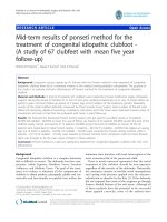
Figure 1 Denis- Browne Splint for bilateral clubfoot.
Porecha et al. Journal of Orthopaedic Surgery and Research 2011, 6:3
/>Page 3 of 7
Ponseti [4] who prefer to apply the Denis-Browne splint
23 ho urs a day for three months and then at night
(12-14 hours) for three years. Non-compliance was
defined as the inability to adhere to the above men-
tioned criteria and also delay in changing the splint and
shoes as the foot size changed.
The parents were instructed to perform range of
motion exercises for the ankle and foot when it was out
of the brace. Two exercises were taught to the parents.
In the first exercise the infant was made to squat on
level ground while being supported by the parents. This
brought the ankle in dorsiflexion and prevents equinus
deformity. In the second exercise the parent uses one
hand to stabilize the leg with knee bent. The other hand
is used to grasp the foot and then place the ankle into
maximum dorsiflexion followed by planter flexion.
The exercises were performed twice a day till the
weight bearing age (when the brace was applied for
twenty three hours a day) and five times daily for the
next three yea rs (when the brace was applied for twelve
hours at night). The parent repeats this exercise twenty
times at a seating.
The patients were followed up on a weekly basis dur-
ing the initial stages of treatment. After orthosis was
applied, the patient was seen on a monthly basis for
three months and then once every three months till
the patients was three years of age. The patient was
also followed up every six moths to one year till
5 years and then after 1-2 years till skeletal maturity is
achieved.
Results
A total number of 49 patients with 67 clubfeet were
treated and followed for mean of five years. Out of 49
patients, 39 patients (79.59%) were male, thus male-
female ratio is 3.9. Out of 49 patients, 18 patients
(36.73%) had bilateral involvement while 31 patients
(63.27%) had unilateral involvement out of which 17
(54.84%) had right foot involvement and 14 (45.16%)
hadleftfootinvolvement.Norelationshiphadbeen
found with birth order or family history.
While beginning of the treatment, 42 patients (85.71%)
are in between 0-12 weeks of the age ( mean 2 weeks),
5 patients (10.20%) are in between 13-24 weeks of age
(mean 15 weeks) while 2 ( 4.08%) patient are in between
25-36 weeks of age (mean 34 weeks). At the commence-
ment of treatment, of the 18 bilateral clubfeet patients
(36 clubfeet) 17 children (34 clubfeet) had Pirani sever-
ity score of six, and one children (2 clubfeet) had a
Pirani score of five. In unilateral group the mean Pirani
score was 5.83 (range 5-6).
The mean Mid Foot Score and Hind Foot Score for the
entire group wa s 2.8 (range 2.5-3) and 2. 76 (range 2-3)
respectively. The mean number of the casts that were
applied to obtain correction was 6.8 (range 6-8). The more
severe the initial deformity and the treatment initiation
after 12 weeks of the age, the more casts were required to
obtain correction. 47 children (95.91%) needed percuta-
neus tenotomy, 18 in the bilateral group and 29 in the uni-
lateral group. The mean Mid Foot Score and Hind Foot
Score for the entire group at the time of tenotomy was 0.5
and 2.5 respectively. There was no delay between final cast
removal and fitting of D-B splint. The mean duration of
the treatment up to application of the D-B Splint was 9.6
weeks. Initial correction was obtained in all 67 clubfeet
(100%) with the Ponseti method.
Fourteen children - 28.57% (19 feet - 28.35%) had a
relapse of the deformity. Patient age at the time of
relapse, bilateralism or unilateralism of the relapse foot,
relapse foot deformity, treatment offered to relapsed
foot, immediate results of the offered treatment accessed
by Pirani Severity Score, and results at mean 5 year fol-
low-up accessed by Ponseti Functional Scoring System
were given. (Table 2)
The original correction was recovered with the use of
repeat application of serial casts in 8 patients (9 clubfeet)
while 5 patients (9 clubfeet) were resistant to Ponseti
serial cast manipulation and were offered surgery in the
form of Postero-medial release; but parents of the
patients were not willing for the surgery and thus had
poor functional outcome at mean five year of follow-up.
All the 8 patients (9 cl ubfeet) who respond well to repeat
application of serial casts were from the 0-12 weeks of
the age group while beginning of the treatment. Out of
5 patients resistant to Ponseti serial cast manipulation
3werefromthe13-24weeksoftheagegroup
while beginning of the treatment, while 2 w as from the
25-36 weeks of the age at the initiation of the treatment.
Thus, relapse is more severe when occurred and not
respond to traditional Ponseti casting method in the
patients whom treatment initiation was done after
12 weeks of the age.
One patient (left clubfeet) developed relapse in the
form of equinus deformity at the age of 18 months for
which repeat percutaneus tenotomy was done and above
knee cast was applied with 15 degree dorsiflexion of
ankle, 60 degree of abduction of f oot and 90 degree
knee bent for 3 weeks. Patient had excellent functional
outcome at final follow-up.
Thus, of 14 relapsed patients, 9 patients - 64.29%
(10 clubfeet - 52.63%) had exc ellent to good functional
outcome and 5 patients - 35.71% (9 clubfeet - 47.37%)
had poor functional outcome according to Ponseti Func-
tional Scoring System [4] at the mean five year follow-
up. The splint compliance was compromised in all the
relapsed cases. In 9 patients the Denis - Browne splint
was used infrequently and it was never used in
5 patients.
Porecha et al. Journal of Orthopaedic Surgery and Research 2011, 6:3
/>Page 4 of 7
At the m ean of five year follow-up, we found nearly
normal passive range of motion in 44 patients - 89.79%
(58 clubfeet - 86.56%). Parents of 32 patients (65.30%)
accept the look of the clubfoot nearly normal and par-
ents of 12 patients (24.49%) accept the look of clubfoot
as normal. We followed the functional Ponseti Scoring
System[4]andgotgoodtoexcellentresultsin
44 patients (89.29%) at mean five year of follow up.
(Figure 2 & Figure 3)
Few complications were encountered. Two children
had a plaster sore on the lateral aspect of the skin over-
lying the talar head. This healed with local dressing
only. The mean time to heal the sore was 7 days (range
6-8 days). The corrective manipulation and cast was not
applied till the sore heal. However, we don ’t encounter
anyallergicreactiontothesoftroll,anytransitorydis-
coloration of the toes following tenotomy and correction
of equinus, serious bleeding following tenotomy or any
wound problems with percutaneus incision.
Discussion
In 1948, Ponseti proposed reducing the idiopathic club-
foot deformity with successive manipulation and casts.
Although treatment with cast is a very old method for
clubfoot, Ponseti’s method is based on strict rules estab-
lished from anatomic evidence.
The major concern with the operative treatment of
congenital clubfoot is functional outcome. Extensive
open surgery like postero-medial release is commonly
associated with long-term stiffness and weakness which
is avoided by the Ponseti technique [6-8 ]. Aronson and
Puskarich studied the disability associated with various
clubfoot treatment options. Their results showed that
patients who underwent casting only and patients who
had additional percutaneus heel cord lengthening had
the least deformity and disability [7].
The Ponseti treatment of clubfoot has three phases:
the corrective phase involves application of casts, the
maintenance phase where splint fitting is emphasized
and the transition phase where the splints are discontin-
ued and regular foot wear allowed. Problems can occur
in any phase due to many causes: incorrect casting tech-
nique, improper tenotomy, under-corrected deformity,
ill-fitting splints, lack of understanding and poor com-
pliance of patients’ parents due to poor socio-economy
can all affect a successful outcome.
The relapse rate in fourteen cases in our study shows
the initial learning curve with this technique . There
Table 2 Details of the Relapse Foot
Patient’s age at
relapse (In months)
Bilateralism/Unilateralism at the
initiation of the treatment
Side of
relapsed
foot
Relapse
deformity
Treatment offer to
correct the deformity
Result of the
Treatment
Result at five
year of follow up
9 1. Bilateral Left Adductus
& Varus
4 Ponseti casts Good Good
2. Bilateral Left Adductus
& varus
3 Ponseti casts Excellent Good
3. Unilateral Right Adductus 2 Ponseti casts Excellent Excellent
12 1. Bilateral Left Adductus
& Varus
3 Ponseti casts Excellent Excellent
2. Unilateral Left Adductus 2 Ponseti casts Excellent Excellent
3. Unilateral Right Adductus
& Varus
3 Ponseti casts Excellent Good
18 1. Bilateral Left Equinus Repeat tenotomy & 3
week cast
Excellent Excellent
2. Bilateral Both Adductus
& Varus
4 Ponseti casts Excellent Good
24 1. Bilateral Both All four
deformities
8 Ponseti casts Poor Poor
30 2. Unilateral Right Adductus
& Varus
3 Ponseti casts Excellent Good
3. Bilateral Both All four
deformities
10 Ponseti casts Poor Poor
36 1. Bilateral Both All four
deformities
8 Ponseti casts Poor Poor
2. Bilateral Both All four
deformities
8 Ponseti casts Poor Poor
3. Unilateral Left All four
deformities
8 Ponseti casts Poor Poor
Porecha et al. Journal of Orthopaedic Surgery and Research 2011, 6:3
/>Page 5 of 7
were more relapse on the left side and this may reflect
righthanddominanceofthetreating surgeon. Thus, a
more abduction force may be required to correct the
left foot when the left hand is the abduction side.
There are three main issues which lead to inferior
results with this technique: splint compliance , splint fit-
ting and under correction of the ankle equinus.
Poor splint compliance was a major issue especially in
children coming from low socio-economic strata and
where the parents education level was poor. Out of 14
relapses, in 9 patients Denis-Browne splint was used
infrequently and it was never used in 5 patients. We
feel that although the f oot morphology improves with
rigid adherence to the casting technique it is the post-
correction phase which needs careful attention and
close follow up to ensure a successful outcome. We
tried to nullify poor splint fitting by prov iding D-B
splint of correct size from a single manufacturer directly
under our observation. We now advocate tenotomy in
every case to achieve at least 15 degrees of ankle dorsi-
flexion. This is a critical step as frequently equinus is
the first sing of recurrence.
Although 92-98% successful short-term results
has been reported for the treatment of idiopathic club-
foot [8,10,11] with Ponseti method, documentation of
the long term results of the technique when it has
been used by other orthopedists are fewer [4,5]. We
tried to evaluate mid-term results for congenital idio-
pathic clubfoot treated by Ponseti method and
are satisfied with the outcome at mean five year of
follow-up.
Acknowledgements
None.
Author details
1
Orthopedic Department, M.P.Shah Medical College, Guru Govind Singh
Hospital, Jamnagar - 361008. Gujarat. India.
2
Department of orthopedics, M.P.
Shah Medical College, Guru Govind Singh Hospital, Jamnagar - 361008.
Gujarat. India.
3
Department of anesthesiology, M.P.Shah Medical College,
Guru Govind Singh Hospital, Jamnagar - 361008. Gujarat. India.
Authors’ contributions
MP is the single orthopedics surgeon who performs the casting technique
in all the patients. DP participate and analysis the study. HC designed and
coordinated and drafted the manuscript. All authors read and approved the
final manuscript.
Figure 2 Front look of bilateral clubfoot at 5 year follow-up. Figure 3 Back look of bilateral clubfoot at 5 year follow-up.
Porecha et al. Journal of Orthopaedic Surgery and Research 2011, 6:3
/>Page 6 of 7
Competing interests
The authors declare that they have no competing interests.
Received: 9 August 2010 Accepted: 12 January 2011
Published: 12 January 2011
References
1. Kite JH: Non-operative treatment of congenital clubfeet. Southern Med J
1930, 23:337.
2. Harrold AJ, Walker CJ: Treatment and prognosis in congenital clubfoot.
Bone Joint Surg Br 1983, 65:8-11.
3. Yamamoto H, Muneta T, Morita S: Non-surgical treatment of congenital
clubfoot with manipulation cast and modified Denis-Browne Splint.
Pediatr Orthop 1998, 18:538-42.
4. Laaveg SJ, Ponseti IV: Long-term results of treatment of congenital club
foot. J Bone Joint Surgery, Am 1980, 62:23-31.
5. Cooper DM, Dietz FR: Treatment of idiopathic clubfoot. A thirty year
follow-up note. J Bone Joint Surg 1995, 77A:1477-89.
6. Hutchins PM, Foster BK, Paterson DC, Cole EA: Long term results of early
surgical release in club feet. J Bone Joint Surgery Br 1985, 67:791-9.
7. Aronson J, Puskarich CL: Deformity and disability form treated clubfoot.
J Pediatr Orthop 1990, 10:109-19.
8. Herzenberg JE, Radler C, Bor N: Ponseti versus traditional methods of
casting for idiopathic clubfoot. J Pediatr Orthop 2002, 22:517-21.
9. Dyer PJ, Davis N: The role of the Pirani scoring system in the
management of club foot by the Ponseti method. J Bone Joint Surg Br
2006, 88-B:1082-1084.
10. Lehman WB, Mohaideen A, Madan S, et al: A method for the early
evaluation of the Ponseti (Iowa) technique for the treatment of
idiopathic clubfoot. J Pediatr Orthop 2003, 12(2):133-40.
11. Goksan SB: Treatment of congenital clubfoot with the Ponseti Method.
Acta Orthop traumatol Turc 2002, 36(4):281-7.
doi:10.1186/1749-799X-6-3
Cite this article as: Porecha et al.: Mid-term results of ponseti method
for the treatment of congenital idiopathic clubfoot - (A study of 67
clubfeet with mean five year follow-up). Journal of Orthopaedic Surgery
and Research 2011 6:3.
Submit your next manuscript to BioMed Central
and take full advantage of:
• Convenient online submission
• Thorough peer review
• No space constraints or color figure charges
• Immediate publication on acceptance
• Inclusion in PubMed, CAS, Scopus and Google Scholar
• Research which is freely available for redistribution
Submit your manuscript at
www.biomedcentral.com/submit
Porecha et al. Journal of Orthopaedic Surgery and Research 2011, 6:3
/>Page 7 of 7