Biomedical Engineering Trends Research and Technologies Part 8 doc
Bạn đang xem bản rút gọn của tài liệu. Xem và tải ngay bản đầy đủ của tài liệu tại đây (1.13 MB, 40 trang )
Biomedical Engineering, Trends, Research and Technologies
270
Marsh, B. J., & Howell, K. E. (2002). The mammalian Golgi complex debates. Nat Rev Mol
Cell Biol, 3. 10. (Oct 2002), 789-795.1471-0072.
Maxfield, F. R., & McGraw, T. E. (2004). Endocytic recycling. Nat Rev Mol Cell Biol, 5. 2. (Feb
2004), 121-132.1471-0072.
Medalia, O., Typke, D., Hegerl, R., Angenitzki, M., Sperling, J., & Sperling, R. (2002).
Cryoelectron microscopy and cryoelectron tomography of the nuclear pre-mRNA
processing machine. J Struct Biol, 138. 1-2. (Apr-May 2002), 74-84.1047-8477.
Mehta, A., Beck, M., Eyskens, F., Feliciani, C., Kantola, I., Ramaswami, U., et al. (2010). Fabry
disease: a review of current management strategies. QJM, 103. 9. (Sep 2010), 641-
659.1460-2393.
Mesika, A., Kiss, V., Brumfeld, V., Ghosh, G., & Reich, Z. (2005). Enhanced intracellular
mobility and nuclear accumulation of DNA plasmids associated with a karyophilic
protein. Hum Gene Ther, 16. 2. (Feb 2005), 200-208.1043-0342.
Minton, A. P. (2006). How can biochemical reactions within cells differ from those in test
tubes? J Cell Sci, 119. Pt 14. (Jul 15 2006), 2863-2869.0021-9533.
Miyata, N., Hosoi, K., Mukai, S., & Fujiki, Y. (2009). In vitro import of peroxisome-targeting
signal type 2 (PTS2) receptor Pex7p into peroxisomes. Biochim Biophys Acta, 1793. 5.
(May 2009), 860-870.0006-3002.
Mukhopadhyay, A., & Weiner, H. (2007). Delivery of drugs and macromolecules to
mitochondria. Adv Drug Deliv Rev, 59. 8. (Aug 10 2007), 729-738.0169-409.
Muratovska, A., Lightowlers, R. N., Taylor, R. W., Turnbull, D. M., Smith, R. A., Wilce, J. A.,
et al. (2001). Targeting peptide nucleic acid (PNA) oligomers to mitochondria
within cells by conjugation to lipophilic cations: implications for mitochondrial
DNA replication, expression and disease. Nucleic Acids Res, 29. 9. (May 1 2001),
1852-1863.1362-4962.
Narla, A., & Ebert, B. L. (2010). Ribosomopathies: human disorders of ribosome dysfunction.
Blood, 115. 16. (Apr 22 2010), 3196-3205.1528-0020.
Nori, A., Jensen, K. D., Tijerina, M., Kopeckova, P., & Kopecek, J. (2003). Tat-conjugated
synthetic macromolecules facilitate cytoplasmic drug delivery to human ovarian
carcinoma cells. Bioconjug Chem, 14. 1. (Jan-Feb 2003), 44-50.1043-1802.
Nori, A., & Kopecek, J. (2005). Intracellular targeting of polymer-bound drugs for cancer
chemotherapy. Adv Drug Deliv Rev, 57. 4. (Feb 28 2005), 609-636.0169-409.
Nunnari, J., & Walter, P. (1996). Regulation of organelle biogenesis. Cell, 84. 3. (Feb 9 1996),
389-394.0092-8674.
Ozcan, U., Cao, Q., Yilmaz, E., Lee, A. H., Iwakoshi, N. N., Ozdelen, E., et al. (2004).
Endoplasmic reticulum stress links obesity, insulin action, and type 2 diabetes.
Science, 306. 5695. (Oct 15 2004), 457-461.1095-9203.
Pante, N., & Kann, M. (2002). Nuclear pore complex is able to transport macromolecules
with diameters of about 39 nm. Mol Biol Cell, 13. 2. (Feb 2002), 425-434.1059-1524.
Panyam, J., Dali, M. M., Sahoo, S. K., Ma, W., Chakravarthi, S. S., Amidon, G. L., et al. (2003).
Polymer degradation and in vitro release of a model protein from poly(D,L-lactide-
co-glycolide) nano- and microparticles. J Control Release, 92. 1-2. (Sep 19 2003), 173-
187.0168-3659.
Parkinson-Lawrence, E. J., Shandala, T., Prodoehl, M., Plew, R., Borlace, G. N., & Brooks, D.
A. (2010). Lysosomal storage disease: revealing lysosomal function and physiology.
Physiology (Bethesda), 25. 2. (Apr 2010), 102-115.1548-9221.
Nanocarriers for Cytosolic Drug and Gene Delivery in Cancer Therapy
271
Pastores, G. M., & Barnett, N. L. (2005). Current and emerging therapies for the lysosomal
storage disorders. Expert Opin Emerg Drugs, 10. 4. (Nov 2005), 891-902.1744-7623.
Petros, R. A., & DeSimone, J. M. (2010). Strategies in the design of nanoparticles for
therapeutic applications. Nat Rev Drug Discov, 9. 8. (Aug 2010), 615-627.1474-1784.
Platta, H. W., & Erdmann, R. (2007). The peroxisomal protein import machinery. FEBS Lett,
581. 15. (Jun 19 2007), 2811-2819.0014-5793.
Poon, I. K., & Jans, D. A. (2005). Regulation of nuclear transport: central role in development
and transformation? Traffic, 6. 3. (Mar 2005), 173-186.1398-9219.
Pouton, C. W., Wagstaff, K. M., Roth, D. M., Moseley, G. W., & Jans, D. A. (2007). Targeted
delivery to the nucleus. Adv Drug Deliv Rev, 59. 8. (Aug 10 2007), 698-717.0169-409.
Rajendran, L., Knolker, H. J., & Simons, K. (2010). Subcellular targeting strategies for drug
design and delivery. Nat Rev Drug Discov, 9. 1. (Jan 2010), 29-42.1474-1784.
Rosenkranz, K., Birschmann, I., Grunau, S., Girzalsky, W., Kunau, W. H., & Erdmann, R.
(2006). Functional association of the AAA complex and the peroxisomal
importomer. FEBS J, 273. 16. (Aug 2006), 3804-3815.1742-464.
Rowe, S. M., Miller, S., & Sorscher, E. J. (2005). Cystic fibrosis. N Engl J Med, 352. 19. (May 12
2005), 1992-2001.1533-4406.
Said Hassane, F., Saleh, A. F., Abes, R., Gait, M. J., & Lebleu, B. (2010). Cell penetrating
peptides: overview and applications to the delivery of oligonucleotides. Cell Mol
Life Sci, 67. 5. (Mar 2010), 715-726.1420-9071.
Samuelson, L. E., Dukes, M. J., Hunt, C. R., Casey, J. D., & Bornhop, D. J. (2009). TSPO
targeted dendrimer imaging agent: synthesis, characterization, and cellular
internalization. Bioconjug Chem, 20. 11. (Nov 2009), 2082-2089.1520-4812.
Sanabria, H., Kubota, Y., & Waxham, M. N. (2007). Multiple diffusion mechanisms due to
nanostructuring in crowded environments. Biophys J, 92. 1. (Jan 1 2007), 313-
322.0006-3495.
Savic, R., Luo, L., Eisenberg, A., & Maysinger, D. (2003). Micellar nanocontainers distribute
to defined cytoplasmic organelles. Science, 300. 5619. (Apr 25 2003), 615-618.1095-
9203.
Schmid, S. L. (1997). Clathrin-coated vesicle formation and protein sorting: an integrated
process. Annu Rev Biochem, 66. 1997), 511-548.0066-4154.
Seksek, O., Biwersi, J., & Verkman, A. S. (1997). Translational diffusion of macromolecule-
sized solutes in cytoplasm and nucleus. J Cell Biol, 138. 1. (Jul 14 1997), 131-
142.0021-9525.
Sheu, S. S., Nauduri, D., & Anders, M. W. (2006). Targeting antioxidants to mitochondria: a
new therapeutic direction. Biochim Biophys Acta, 1762. 2. (Feb 2006), 256-265.0006-
3002.
Tarrago-Trani, M. T., & Storrie, B. (2007). Alternate routes for drug delivery to the cell
interior: pathways to the Golgi apparatus and endoplasmic reticulum. Adv Drug
Deliv Rev, 59. 8. (Aug 10 2007), 782-797.0169-409.
Terlecky, S. R., & Koepke, J. I. (2007). Drug delivery to peroxisomes: employing unique
trafficking mechanisms to target protein therapeutics. Adv Drug Deliv Rev, 59. 8.
(Aug 10 2007), 739-747.0169-409.
Torchilin, V. P. (2005). Fluorescence microscopy to follow the targeting of liposomes and
micelles to cells and their intracellular fate. Adv Drug Deliv Rev, 57. 1. (Jan 2 2005),
95-109.0169-409.
Biomedical Engineering, Trends, Research and Technologies
272
Ungar, D. (2009). Golgi linked protein glycosylation and associated diseases. Semin Cell Dev
Biol, 20. 7. (Sep 2009), 762-769.1096-3634.
Wanders, R. J., & Waterham, H. R. (2006). Biochemistry of mammalian peroxisomes
revisited. Annu Rev Biochem, 75. 2006), 295-332.0066-4154.
Warren, G., & Wickner, W. (1996). Organelle inheritance. Cell, 84. 3. (Feb 9 1996), 395-
400.0092-8674.
Watson, P., Jones, A. T., & Stephens, D. J. (2005). Intracellular trafficking pathways and drug
delivery: fluorescence imaging of living and fixed cells. Adv Drug Deliv Rev, 57. 1.
(Jan 2 2005), 43-61.0169-409.
Weissig, V., D'Souza, G. G., & Torchilin, V. P. (2001). DQAsome/DNA complexes release
DNA upon contact with isolated mouse liver mitochondria. J Control Release, 75. 3.
(Aug 10 2001), 401-408.0168-3659.
Weissig, V., Lizano, C., & Torchilin, V. P. (2000). Selective DNA release from
DQAsome/DNA complexes at mitochondria-like membranes. Drug Deliv, 7. 1.
(Jan-Mar 2000), 1-5.1071-7544.
Wood, C. S., Koepke, J. I., Teng, H., Boucher, K. K., Katz, S., Chang, P., et al. (2006).
Hypocatalasemic fibroblasts accumulate hydrogen peroxide and display age-
associated pathologies. Traffic, 7. 1. (Jan 2006), 97-107.1398-9219.
Xiong, R., Li, Z., Mi, L., Wang, P. N., Chen, J. Y., Wang, L., et al. (2010). Study on the
intracellular fate of Tat peptide-conjugated quantum dots by spectroscopic
investigation. J Fluoresc, 20. 2. (Mar 2010), 551-556.1573-4994.
Xu, C., Xie, J., Kohler, N., Walsh, E. G., Chin, Y. E., & Sun, S. (2008). Monodisperse magnetite
nanoparticles coupled with nuclear localization signal peptide for cell-nucleus
targeting. Chem Asian J, 3. 3. (Mar 7 2008), 548-552.1861-471.
Yan, F. F., Casey, J., & Shyng, S. L. (2006). Sulfonylureas correct trafficking defects of
disease-causing ATP-sensitive potassium channels by binding to the channel
complex. J Biol Chem, 281. 44. (Nov 3 2006), 33403-33413.0021-9258.
Yang, S. R., Lee, H. J., & Kim, J. D. (2006). Histidine-conjugated poly(amino acid) derivatives
for the novel endosomolytic delivery carrier of doxorubicin. J Control Release, 114. 1.
(Aug 10 2006), 60-68.0168-3659.
Yang, Z., Zhang, Y., Yang, Y., Sun, L., Han, D., Li, H., et al. (2010). Pharmacological and
toxicological target organelles and safe use of single-walled carbon nanotubes as
drug carriers in treating Alzheimer disease. Nanomedicine, 6. 3. (Jun 2010), 427-
441.1549-9642.
Yessine, M. A., & Leroux, J. C. (2004). Membrane-destabilizing polyanions: interaction with
lipid bilayers and endosomal escape of biomacromolecules. Adv Drug Deliv Rev, 56.
7. (Apr 23 2004), 999-1021.0169-409.
Part 5
Biomaterials and Medicines
12
Antimicrobial Peptides: Diversity and
Perspectives for Their Biomedical Application
Joel E. López-Meza
1
, Alejandra Ochoa-Zarzosa
1
José A. Aguilar
2
and Pedro D. Loeza-Lara
2
1
Centro Multidisciplinario de Estudios en Biotecnología, CMEB-FMVZ-UMSNH
Morelia, Michoacán
2
Genómica Alimentaria, Universidad de La Ciénega del Estado de Michoacán de Ocampo
UCM, Sahuayo, Michoacán,
1,2
México
1. Introduction
For over fifty years, people have used antibiotics to treat illnesses caused by pathogens.
However, the excessive and inappropriate use of these antibiotics in clinical treatment of
humans and animals has increased pathogen resistance to these compounds, turning them
into less effective agents. There has also been an increase in the generation of multidrug-
resistant pathogens, primarily bacteria and fungi that resist the effects of most currently
available antibiotics (Heuer et al., 2006; Field, 2010).
Until now, the pharmaceutical industry is facing this problem by looking for new antibiotics
or modifying existing ones. However, pathogens have proven to have the ability to quickly
develop and disseminate resistance mechanisms, which compromises this strategy,
becoming it less effective. This clearly shows the need to develop new biomedical
treatments with different action mechanisms from those of conventional antibiotics (Parisien
et al., 2008).
This problem has led that efforts being made on research and development of new
biomedical alternatives, among which antimicrobial peptides (AMPs) are considered one of
the most promising options. AMPs are produced by a wide variety of organisms as part of
their first line of defense (eukaryotes) or as a competition strategy for nutrients and space
(prokaryotes). These molecules are usually short peptides (12-100 amino acid residues);
have a positive charge (+2 to +9), although there are also neutral and negatively charged.
They are amphipathic and have been isolated from bacteria, plants and animals, including
humans; which give us an overview of the enormous structural diversity of these molecules
and their different action mechanisms (Murray & Liu, 2008).
The continuous discovery of new AMPs groups in diverse organisms has turned these natural
antibiotics into the basic elements of a new generation of potential biomedical treatments
against infectious diseases in humans and animals. Besides the above, the broad spectrum of
biological activities reported for these molecules suggests a potential benefit in cancer
treatment, viral and parasitic infections and in the modulation of the immune system, which
reinforces the importance of studying these molecules (Mercado et al., 2005; Schweizer, 2009).
Biomedical Engineering, Trends, Research and Technologies
276
The contents of this chapter shows the importance of AMPs for living organisms, not only
from the antimicrobial point of view, but also in bacterial cell communication processes,
immune response modulation in animals and plant defense mechanisms. It also emphasizes
on AMPs’ biological and structural diversity, as well as their various action mechanisms
and, finally, their possible biotechnological development for the pharmaceutical industry is
discussed.
2. AMPs from Gram positive bacteria and their classification
During their evolution, bacteria have acquired mechanisms that allow them to have success
in competition for nutrients and space in their habitat. These mechanisms include from the
enhancement of chemotaxis systems to the acquisition of defense systems such as the
production of antimicrobial peptides (AMPs), also called bacteriocins (Riley & Wertz, 2002).
AMPs are biologically active molecules that have the ability to inhibit the growth of other
members of the same specie or members of different bacterial genres (Cotter et al., 2005b).
These molecules are synthesized by the vast majority of bacterial groups; in fact, it has been
proposed that 99% of bacteria produce at least one, as they have been found in most
examined species, covering Gram positive and Gram negative bacteria and archaea; in
addition they are used as an important tool in evolutionary and ecological studies
(Klaenhammer, 1988). Also, the successful commercial development of nisin (produced by
Lactococcus lactis) and the use of molecular biology and genetic engineering tools in recent
years have provoked a resurgence in AMPs studies, particularly in relation to their potential
biomedical applications (Cotter et al., 2005a, b; Bierbaum & Sahl, 2009; Field et al., 2010).
AMPs from Gram positive bacteria represent a heterogeneous group of chemical molecules;
nevertheless only three main categories have been established based on their structural
modifications, size, thermostability and action mechanisms (Table 1). Class I (lantibiotics) is
constituted by cationic peptides ranging from 19 to 38 amino acid residues, which undergo
posttranslational modifications and exert their effect at membrane and cell wall levels. Their
posttranslational modifications are diverse; the most important involve dehydration
reactions of serine and threonine residues, resulting in the formation of didehydroalanine
(Dha) and didehydroaminobutyric acid (Dhb), respectively (Cotter et al., 2005b). The
reaction of these amino acids with the thiol group (SH) of a cysteine residue generates a
thioether bond producing lanthionine (in the case of Dha) and β-methyl-lanthionine (in the
case of Dhb). The formation of these bonds within the peptide generates a series of
"globular" structures that are characteristic of lantibiotics. This AMPs class is further divided
into subgroups A and B, having nisin as the representative member of subgroup A, while
mersacidin, produced by bacteria of the Bacillus genus, is a member of subgroup B (Table 1)
(McAuliffe et al., 2001; Cotter et al., 2005a).
On the other hand, class II (non lantibiotics) is formed by AMPs constituted by 30 to 60
amino acid residues; they do not contain lanthionine, are thermostable and induce the
formation of pores in the membrane of target cells. These peptides in turn are divided into
subclasses IIa, IIb, IIc and IId (Table 1). Subclass IIa is the largest and its members posses the
amino terminal motif YGNGVXCXXXXVXV (X indicates any amino acid residue) and have
one or two disulfide bonds. AMPs from this subclass show specific activity against the
bacteria Listeria monocytogenes (Ennahar et al., 2000). Leucocin A from Leuconostoc gelidum is
a representative member of this subclass (Hastings et al., 1991).
Antimicrobial Peptides: Diversity and Perspectives for Their Biomedical Application
277
Class
Subclass
Representative
AMPs
Producing bacteria
I Lantibiotic I A Nisin
Lactococcus lactis
I Lantibiotic I B Mersacidin
Bacillus spp.
II Non lantibiotic IIa Leucocin A
Leuconostoc gelidum
II Non lantibiotic IIb Lactococcin G
L. lactis
II Non lantibiotic IIc AS-48 enterocin
Enterococcus faecalis
II Non lantibiotic
III Proteins
IId
Lactococcin A
Helveticin J
L. lactis
L. helveticus
Table 1. Classification of AMPs found in Gram positive bacteria (Cotter et al., 2005a; Drider
et al., 2006)
Subclass IIb comprises AMPs that require the combined action of two peptides in order to
have activity; these peptides do not show inhibitory activity on an individual basis.
Lactococcin G from L. lactis is a representative member of this subclass (Moll et al., 1996).
The AMPs that make up subclass IIc posses a cyclic structure as a result of the covalent
binding of their carboxyl and amino terminal ends; AS-48 enterocin from Enterococcus
faecalis is one of the main representatives of this subclass (Sánchez et al., 2003). Subclass IId
is formed by a variable group of linear peptides, among which lactococcin A from L. lactis is
found (Holo et al., 1993). Finally, the class III is formed by proteins with molecular masses
higher than 30 kDa, the helveticin J from L. helveticus, is an example (Drider et al., 2006).
2.1 Genes involved in AMPs synthesis and expression regulation from Gram positive
bacteria
The genes encoding AMPs are organized as operons, which could contain several genes
involved in the synthesis and regulation. For example, the enterocin A operon of
Enterococcus faecium contains the entA gene that codifies for pre-enterocin; in addition, this
operon contains the genes that codify for the protein involved in the self-protection of the
producing strain (entI), the AMP synthesis induction gene (entF), genes for proteins
involved in extracellular transport (entT, D), as well as the genes of proteins related to the
AMP synthesis regulation (entK, R) (Nilsen et al., 1998). In the case of lantibiotics, these have
additional genes that codify for AMP modification enzymes (McAuliffe et al., 2001).
AMPs synthesis regulation is mediated by two signal transduction systems constituted by
two or three components. Diverse factors activate these systems, which include: the
presence of other competing bacteria (Maldonado et al., 2004), temperature or pH stress
(Ennahar et al., 2000) and a mechanism of "quorum sensing" (Kuipers et al., 1998). An
Biomedical Engineering, Trends, Research and Technologies
278
interesting example is the three-component system that regulates the synthesis of enterocin
A in E. faecium, which is regulated by the mechanism of quorum sensing. This system
includes: 1) a histidine kinase (HK), located in the cytoplasmic membrane which detects
extracellular signals, and 2) a cytoplasmic response regulator (RR) that mediates an adaptive
response, which usually is a change in the gene expression and an induction factor (IF),
whose presence is detected by the HK protein (Figure 1, stage 1) (Cotter et al., 2005b). In this
case, the system is triggered as a result of an IF excess concentration through a slow
accumulation during cell growth, the HK detects this concentration and initiates the
signaling cascade that activates the transcription of genes involved in enterocin A synthesis
(Figure 1, stages 2 and 3) (Ennahar et al., 2000). Other examples of this type of regulation
include several class II members such as sakacin P and A from Lactobacillus sake (Hühne et
al., 1996). Moreover, some examples of regulation mediated by two-component systems
include numerous lantibiotics, for example, subtilin from Bacillus subtilis and nisin from L.
lactis. In these systems AMPs have a dual function, as they have antimicrobial activity and
also act as a signal molecule by inducing its own synthesis (not shown) (Kleerebezem, 2004).
2.2 AMPs secretion and self-protection mechanisms from Gram positive bacteria
AMPs are synthesized as inactive pre-peptides containing a signal peptide at the N-terminal
region (Figure 1, stage 3). This signal keeps the molecule in an inactive form within the
producing cell facilitating its interaction with the carrier, and in the case of lantibiotics plays
an important role in the pre-peptide recognition by the enzymes that perform
posttranslational modifications. The signal peptide may be proteolytically removed during
transport of the pre-peptide into the periplasmic space by the same transport proteins (ATP-
dependent ABC membrane transporters, which may also contain a proteolytic domain)
(Figure 1, stage 4), or by serine-proteases present on the outside of the cell membrane. Thus,
the carboxyl terminus is separated from the signal peptide and is released into the
extracellular space to produce the biologically active peptide (Figure 1, stage 5) (Ennahar et
al., 2000; Cotter et al., 2005b).
AMPs producing bacteria possess proteins that protect them from the action of their own
peptides. The exact molecular mechanisms by which these proteins confer protection to the
producing bacteria are unknown; however, two protection systems have been proposed,
which, in some cases act in the same bacteria (Kleerebezem, 2004). The protection can be
provided by a specific protein that sequesters and inactivates the AMP, or that binds to the
AMP receptor causing a conformational change in its structure making it inaccessible to the
AMP (Figure 1, stage 6) (Venema et al., 1994). The second system is constituted by the ABC
transport proteins, which in some cases provide the protection mechanism through the
expulsion of the membrane-binding AMPs (Otto et al., 1998).
2.3 AMPs spectrum and action mechanisms from Gram positive bacteria
In general, the antibacterial action spectrum of AMPs of Gram positive bacteria is restricted
to this bacterial group. However, there are several molecules with a wide range of action,
inhibiting the growth of Gram positive (McAuliffe et al., 2001) and Gram negative bacteria
(Motta et al., 2000), human pathogenic fungi (De Lucca & Walsh, 1999) and viruses (Jenssen
et al., 2006). Also, AMPs have activity against various eukaryotic cells, such as human and
bovine erythrocytes (Datta et al., 2005). With regard to their antimicrobial activity, AMPs
possess essential characteristics in order to carry out the activity, regardless of their target
Antimicrobial Peptides: Diversity and Perspectives for Their Biomedical Application
279
Protein sensing,
Histidine-kinase (EntK)
P
Response
regulator
(EntR)
ATP
ADP
AMP active
4
5
1
2
Transport
Proteolytic
proccesing
Protein (EntI)
autoprotection
Signal
IF
6
Pre-AMP
IF
P entA
IF
Carrier ABC
(EntT)
Protein
ancillary EntD
P
P
Gene activation
3
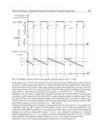
Fig. 1. Regulation of the synthesis of enterocin A from Enterococcus faecium (non-lantibiotic).
Stage 1, the EntK protein detects the presence of the induction factor (IF) and
autophosphorylates. Stages 2 and 3, the phosphate group is transferred to the EntR response
regulator, which activates genes involved in the synthesis of the pre-peptide (pre-enterocin
A) and of the IF. Stages 4 and 5, the pre-enterocin A and the IF are transported to the outside
by the EntT and EntD proteins, and processed by the same system, releasing the active
enterocin A and the IF. Stage 6, the EntI protein protects the producing bacteria from the
effect of enterocin A (Ennahar et al., 2000; Cotter et al., 2005b)
cell. These include, 1) a net positive charge which favors its interaction with the negatively
charged lipopolysaccharide membrane of Gram negative bacteria, or with teichoic and
lipoteichoic acids from the wall of Gram positive bacteria; 2) hydrophobicity, required for
the insertion of the AMP in the cell membrane; and 3) flexibility, which allows a
conformational change from a soluble state to one of membrane interaction. These
characteristics vary from molecule to molecule; however, all are important for antimicrobial
activity (Jenssen et al., 2006).
It has been shown that the action targets of AMPs studied to date are the cell membrane and
wall, as well as some important enzymes for cell metabolism. The action mechanisms
include: i) pore formation in the cell membrane, causing loss of cell contents, this is the
mechanism described for nisin (Enserink, 1999) and lactococcin A from L. lactis (Van Belkum
et al., 1991); ii) cell wall synthesis inhibition, this mechanism has been described for
mersacidin, which involves binding to lipid II, the main transporter of peptidoglycan
subunits (UDP-Mur -Nac-pentapeptide-GlcNAc) (Brotz et al., 1995); and iii) inhibition of the
activity of enzymes such as phospholipase A2, which is involved in membrane repair; this is
the reported mechanism for cinamicin from Streptomyces cinnamoneus (Marki et al., 1991).
Biomedical Engineering, Trends, Research and Technologies
280
Additionally, there have been reports of AMPs that possess a dual action mechanism, such
as nisin (Figure 2) (Breukink et al., 1999; Bierbaum & Sahl, 2009). The most accepted model
showing the dual action mechanism of nisin proposes that it initially binds to the cell wall
by electrostatic attraction, events that are facilitated due to the positive charge of this
peptide and negative charges of cell wall components (Figure 2, stage 1). Subsequently, nisin
binds to lipid II, the main transporter of peptidoglycan subunits, and uses this molecule to
anchor itself to the cell membrane (Figure 2, stage 2). Then, it changes its orientation with
respect to the membrane and inserts itself in it; this involves the translocation of its carboxyl
terminus through the membrane. Finally, the binding of different peptides in the insertion
site leads to the formation of a transmembrane pore that allows the exit of important
molecules such as amino acids and ATP, leading the bacteria to a rapid cell death (Figure 2,
stage 3) ( Wiedemann et al., 2001; Bierbaum & Sahl, 2009).
2.4 AMPs resistance from Gram positive bacteria
Resistance development in pathogenic bacteria that are normally sensitive to AMPs is of
great interest because of their possible use in biomedical therapies, as bacterial resistance
might limit their use. Within a particular bacterial species there may be naturally resistant
members to AMPs or resistance may arise as a result of continuous exposure; which are
known as intrinsic and acquired resistance, respectively (Xue et al., 2005).
Most research in this area has focused on specific AMPs such as nisin and class IIa members.
In the first case, L. monocytogenes, L. innocua, Streptococcus pneumoniae and S. bovis resistant
mutants have been detected, whose resistance has been correlated to changes in the wall and
cell membrane (Gravesen et al., 2002). More specifically the synthesis and incorporation of
various structural components to the membrane (Li et al., 2002) and the cell wall (Mantovani
& Russell, 2001) have been observed in the mutants, which has favored an increase in
positive charges in these cell structures and reduced the antibacterial activity of nisin (which
has a net positive charge). Likewise, changes in the fluidity of cell membrane (Verheul et al.,
1997) and an increase in the thickness of the cell wall of some mutant bacteria have been
observed (Maisnier & Richard, 1996; Murray & Liu, 2008).
The mechanisms of resistance to type II AMPs have been studied in strains of L.
monocytogenes, essentially towards class IIa peptides, in which the resistance is related to
several factors including reduced expression of a permease that acts as a potential receptor
(Dalet et al., 2001), as well as changes in membrane fluidity (Vadyvaloo et al., 2002), and in
cell surface charges (Vadyvaloo et al., 2004). The importance of studying the resistance lies
not only in the possible long term ineffectiveness of AMPs, but also in generating
knowledge that could serve as a basis for strategies to improve the therapeutic potential of
these antimicrobial molecules, i.e. the development of protein engineering strategies to
improve the biological properties of AMPs (Field et al., 2010).
Currently, the existence of natural AMPs variants suggests that there is flexibility in the
location of some important amino acid residues for antimicrobial activity, which indicates
that it is possible to generate mutants with changes that increase this activity. Thus,
additional studies are needed to determine the mechanisms of resistance to AMPs, as well as
the frequency with which it occurs (Cotter et al., 2005a).
2.5 Current and potential Gram positive AMPs applications in biomedical therapies
AMPs null toxicity to humans and animals and activity directed towards pathogenic
bacteria has allowed investigating their potential applications in biomedical therapies. In
particular, the action mechanisms of these peptides and their activity against pathogens
Antimicrobial Peptides: Diversity and Perspectives for Their Biomedical Application
281
(+)
(-)
AMP
Cell
wall
Plasmatic
membrane
Li
p
id II
2
3
Peptidoglycane
subunits
1
Lí
p
id II
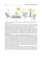
Fig. 2. Model showing the dual action mechanism of nisin from Lactococcus lactis. Stage 1,
nisin has a net positive charge that increases its interaction with the negative charges of the
cell wall components. Stage 2, nisin binds to lipid II, the main transporter of peptidoglycan
subunits from the cytoplasm to the cell wall, interfering with its synthesis, leading the
bacteria to cell death. Stage 3, in addition, several nisin molecules use lipid II to anchor and
insert themselves into the cell membrane and begin the formation of pores, leading the
bacteria to a rapid cell death (Wiedemann et al., 2001; Cotter et al., 2005a)
resistant to conventional antibiotic therapy, making them an attractive option as
antimicrobial agents (Table 2) (Cotter et al., 2005a, b; Piper et al., 2009). Broad spectrum
AMPs or bioengineered AMPs could be used against Gram positive pathogens of humans
and animals. For example, lacticin 3147 from L. lactis has shown in vitro activity against
methicillin-resistant Staphylococcus aureus (MRSA); vancomycin-resistant enterococci (VRE);
vancomycin-intermediate S. aureus (VISA); streptococci, S. pneumoniae, S. pyogenes, S.
agalactiae, S. dysgalactiae, S. uberis, S. mutans; Clostridium botulinum and Propionibacterium
acnes (Galvin et al., 1999; Piper et al., 2009). In the same way, it has been created two nisin
variants by bioengineered (nisin V and nisin T) with enhanced antimicrobial activity against
Gram positive pathogens like MRSA, VRE, VISA, Clostridium difficile, L. monocytogenes and B.
cereus (Field et al., 2010).
Biomedical Engineering, Trends, Research and Technologies
282
AMPs and
producing
strain
Activity Potential biomedical applications
Nisin
L. lactis
Inhibits Gram positive
and Gram negative
bacteria, including
Helicobacter pylori
Bacterial mastitis, oral hygiene, treatment
of methicillin-resistant Staphylococcus,
enterococcal infections, topical
formulations, deodorants and cosmetics,
treatment of peptic ulcers and
enterocolitis
Epidermin
S. epidermidis
Inhibits
Propionibacterium acnes,
staphylococci,
streptococci
Acne, folliculitis, impetigo
Mersacidin
Bacillus spp.
Cinamicin
Streptomyces
cinnamoneus
Inhibits staphylococci
and streptococci strains
Phospholipase A2
inhibitor, angiotensin
and HSV converting
enzyme
Treatment of methicillin-resistant
Staphylococcus aureus and streptococcal
infections
Inflammation reduction, blood pressure
regulation and viral infection treatment
Table 2. A few Gram positive AMPs examples and their potential biomedical use (Cotter et
al., 2005a)
On the other hand, in vivo experiments using animal models have shown positive results
after using lantibiotics, such as mersacidin and nisin in the treatment of respiratory tract
infections caused by S. aureus MRSA (Kruszewska et al., 2004; De Kwaadsteniet et al., 2009),
and Streptococcus pneumonia (Goldstein et al., 1998), in addition to skin care and oral
therapies, such as tooth paste for prevention of teeth loss, bad breath and gingivitis (Howell
et al., 1993; Arauz et al., 2009). Likewise, nisin has showed that has the potential for
treatment of human mastitis (Fernández et al., 2008).
The Oragenics pharmaceutical company has realized extensive preclinical testing on the
lantibiotic mutacin MU1140 of S. mutans, which has demonstrated activity against wide
variety of disease-causing Gram positive bacteria, including MRSA, VRE, Mycobacterium
tuberculosis, and anthrax. For the complete trials, this company has created the synthetic
version MU1140-S, and they expect to conclude the preclinical testing in 2011. Likewise, in
New Zealand, the BLIS K12® dietary supplement is sold as an inhibitor of bacteria
responsible for bad breath, because it contains a strain of S. salivarus that produces
salivaricin A2 and B peptides (Tagg, 2004).
In relation to animal disease, several AMPs have been proposed as potential alternatives to
bovine mastitis control. Nisin has activity against mastitis pathogens and has been
formulated in Wipe Out® and Mast Out®, commercially available products (Ryan et al.,
1998; Wu et al., 2007). Also, AMPs produced by S. aureus, S. epidermidis and Streptococcus
gallolyticus have been tested against strains of both S. aureus and Streptococcus species
Antimicrobial Peptides: Diversity and Perspectives for Their Biomedical Application
283
isolated from bovine mastitis (Varella et al., 2007; Pieterse et al., 2008). Finally, B.
thuringiensis AMPs have showed inhibitory action against S. aureus isolates from bovine
mastitis (Barboza-Corona et al., 2009).
From a non antimicrobial medical perspective, AMPs such as cinamicin may have different
biomedical applications, because this peptide inhibits the function of phospholipase A2 and
the angiotensin converting enzyme, which are involved in the immune system and in
maintaining blood pressure in humans, respectively; so that they could be used in
inflammatory processes and in blood pressure regulation (Ennahar et al., 2000) (Table 2). In
the same way, nisin has shown contraceptive activity (Gupta et al., 2009) and protector
activity in rabbits and mice vaginas in in vitro and in vivo studies (Reddy et al., 2004).
3. AMPs from Gram negative bacteria and their classification
The term "bacteriocinogenicity" is used to describe the ability of Gram negative bacteria to
synthesize and excrete AMPs (Daeschel et al., 1990). These molecules were first detected in
Escherichia coli and were called colicins. Later, they were found in Gram positive bacteria
and have been studied with great interest, especially those produced by lactic acid bacteria,
which can be used in food preservation because its activity against Gram negative bacteria,
the leading cause of food poisoning (Hardy, 1975; Tagg et al., 1976). Colicin V from E. coli
and pyocin from Pseudomonas aeruginosa, are the two best studied peptides in the Gram
negative bacteria group (Table 3) (Jack et al., 1995).
The colicin group has been taken as the representative group of Gram negative AMPs,
although there are differences between them. Pyocins are AMPs of high molecular weight
synthesized by P. aeruginosa strains, which could participate in establishing and protection
of bacteria. There are three types of pyocins: R, F and S, which resemble the tails of
bacteriophages of the Myoviridae family. Type R pyocins show broad similarities with the
fibers of the tails of these phages. Type R pyocins are contractile and not flexible, the F type
are flexible, but are not contractile; and the S type are susceptible to proteases (Michel-
Briand & Baysse, 2002; Waite & Curtis, 2009).
The colicins are proteins between 29 and 90 kDa, which have binding, transport and specific
activity domains, same as those found in pyocins. The secretion of colicins is carried out in
cell lysis, which involves their death (Riley & Wertz, 2002; Sano et al., 1993). Other kind of
AMPs produced by E. coli and other enterobacteria are the microcins, which are a group of
circular peptides, from which microcin J25, produced by E. coli AY25, has been taken as a
model (Craik et al., 2003). Microcins are low molecular weight molecules under 10 kDa,
which play an important role in competition for colonization of the gastrointestinal tract.
They are generally hydrophobic, highly stable in relation to heat, extreme pH and proteases
(Duquesne et al., 2007). Some other Gram negative AMPs are: Serracin P, produced by
Serratia plymithicum J7; mundticin KS, synthesized by Enterococcus mundtii, NFRI 7393 and
caratovoricin, produced by Pectobacterium carotovorum subsp. carotovorum (Jabrane et al.,
2002; Kawamoto et al., 2002; Yamada et al., 2008).
3.1 Genes involved in Gram negative AMPs synthesis
The genes required for colicin synthesis are encoded usually in plasmids, and consist of a
colicin gene, a gene for immunity and a lysis gene. Most of the genes coding for AMPs in
Gram negative bacteria probably derived from recombination of existing AMPs genes.
Colicins contains a central domain (50%) involved in the recognition of the target cell
receptor; a N-terminal domain (> 25%) responsible for the translocation of the peptide to the
Biomedical Engineering, Trends, Research and Technologies
284
AMPs and
producing bacteria
Group Main features
Colicin
Escherichia coli
Group A N-terminal domain rich in glycine (~20-40%)
Group B N-terminal domain rich in glycine (~10-20%)
Microcins
E. coli
Class I
The self-immunity genes are not close to microcin
structural gene
Class IIa Cluster of four genes encoded in plasmids
Class IIb
Chromosomally encoded, have a complex
transcriptional organization
Pyocins
P. aeruginosa
Type R
Resemble the fibers of the tails of bacteriophages of
the Myoviridae family and are contractile but are not
flexible
Type F Flexible, but are not contractile peptides
Type S Susceptible to proteases
Table 3. Principal groups of Gram negative AMPs
target cell, and the rest of the protein has the lethal and immunity activities. Pyocin genes
from P. aeruginosa PAO1 strain are found in the chromosome, are present as a group of 16
open reading frames, of which 12 are analogous to bacteriophage genes (Riley & Wertz,
2003; Williams et al., 2008). Microcins are encoded in plasmids or the chromosome; a typical
gene clusters include the microcin precursor, the self-immunity factor, the secretion proteins
and frequently the post-translational modification enzymes (Duquesne et al., 2007).
3.2 Synthesis and AMPs secretion from Gram negative bacteria
The production of colicins is performed under stress, reason why it is mediated by the SOS
regulon (Gillor et al., 2008). The number of cells producing colicin in culture is very small,
but the proportion increases when cells are exposed to stressors such as mitomycin and UV
light (Jack et al., 1995). Pyocin synthesis in P. aeruginosa PAO1 occurs in a similar way.
Synthesis starts when the stressor (which could cause damage to DNA) stimulates the
expression of the RecA protein, whose main function is the repair of damaged DNA and to
degrade the repressor protein (PRTR) to initiate the expression of the prtN activator gene;
the PrtN protein then activates the expression of genes that codify for pyocins (Waite &
Curtis, 2009). Microcins are also synthesized under stress conditions like a pro-microcin that
is secreted to the medium after suffering a cut of 15 to 37 amino acid residues to release the
active microcin; only the MccC7/C5 AMP from E. coli does not undergo this change
(Duquesne et al., 2007; Novikova et al., 2007).
3.3 Gram negative AMPs action mechanisms
Colicins generally present three action mechanisms: some of them form pores or ion
channels in the membrane, others have nuclease activity (colicin E2 and pyocin S3), others
inhibit the synthesis of macromolecules (colicin E3), or as in the case of microcin, the action
mode depends upon the organism that it is acting on. Microcin J25 acts on E. coli inhibiting
RNA polymerase, while on Salmonella enterica forms pores in the membrane (Pugsley, 1984;
Craik et al., 2003).
Antimicrobial Peptides: Diversity and Perspectives for Their Biomedical Application
285
AMPs whose action is to form pores in the membrane destroy the organism by altering the
membrane permeability, affecting the normal flow of ions like potassium, magnesium,
sodium and chloride, as well as inhibiting ATP synthesis through the dissipation of the
membrane electric potential and of the pH gradient. Examples of these AMPs are: glycinecin
A from Xanthomonas campestris; A, E1, K, Ia and Ib colicins from E. coli; pyocin S5 from P.
aeruginosa and xenocin from Xenorhabdus nematophila (Pham et al., 2004; Cascales et al., 2007;
Singh & Banerjee, 2008; Zhang et al., 2010). Once released, some AMPs are attached to a
membrane receptor present in the target cell, afterwards enter to the cell, usually helped by
Tol-like proteins, and finally they may have access to intracellular targets (Lazaroni et al.,
2002; Singh & Banerjee, 2008).
The AMPs that have nuclease activity enter to the cell and bind to tRNA or rRNA and break
it at specific sites, thus inhibiting protein synthesis. Also, several AMPs can degrade nucleic
acids without any specificity, for example: colicins E5, D and E7, and pyocins S1, S2, S3, S4
and AP41 (Masaki & Ogawa, 2002; Michel-Briand & Baysse, 2002; Hsia et al., 2005).
In the case of microcins, the facts that have a great diversity of post-translational
modifications suggests that also have a great variety of action mechanisms; however, they
show the typical nuclease and pore-formation mechanisms, although the latter is related to
the production of siderophores. This dual mechanism of siderophore production and pore
formation has been found in some microcins such as MccE492, produced by Klebsiella
pneumoniae RYC492. The mechanism works as follows: the bacteria produces the
siderophore to chelate environmental Fe
3+
, thus preventing its use by other microorganisms;
afterwards the siderophore undergoes post-translational modification and creates a
glycopeptide capable of forming pores in the membrane of competing bacteria (Thomas et
al., 2004; Duquesne et al., 2007; Nolan et al., 2007; Mercado et al., 2008).
3.4 AMPs resistance from Gram negative bacteria
Resistant mechanisms for Gram negative AMPs, different to self-immunity, have been
described. It has been found some strains of E. coli resistant to others E. coli colicins, which
have a Tol or Ton mechanisms altered, but is very specific and only works with the specific
colicin. These resistant strains have been used to study the Tol and Ton mechanism (Braun
et al., 1994). The pyocin resistant strains of Neisseria gonorrhoeae and Haemophilus ducreyi,
have been found to be associated with structural differences in the outer membrane
lipooligosaccharides in both species (John et al., 1991; Filiatrault et al., 2001). An E. coli K12
microcin resistant has been found, this strain possess a YojI protein which works as microcin
J25 efflux pump (Socias et al., 2009). These examples show the variety of mechanisms
displayed by bacteria to counteract the AMPs activity.
3.5 Potential Gram negative AMPs applications in biotechnology and biomedical
therapies
The consumption of AMPs producing bacteria or the consumption of the purified peptides
can be useful in establishing probiotic microorganisms in the gastrointestinal tract of
humans and animals, which can lead to health improvements (Gillor et al., 2009). It has been
found that in cystic fibrosis patients with an P. aeruginosa infection this organism produces
pyocins that inhibit the growth of its closest competitors, so it could also be used as a
therapeutic agent in these kind of patients and minimize the effects of the infection, that
besides rooting out other susceptible. P. aeruginosa strains, also has an effect on Haemophilus,
Biomedical Engineering, Trends, Research and Technologies
286
Neisseria and Campylobacter. Regarding the latter, peritonitis treatment in mice has been
successful (Scholl & Martin, 2008; Waite & Curtis, 2009; Williams et al., 2008). In other
studies, colicin E1 has shown to inhibit the growth of E. coli O157:H7 in vitro, and the next
step is to try it in meat and in the feeding of cattle to avoid the growth of E. coli O157:H7 in
the gut (Callaway et al., 2004). The pyocin R-Type is studied as an antibiotic against E. coli,
Salmonella, Yersinia pestis and Pseudomonas species by AvidBiotics Corp., with the name
“Avidocin™ Proteins”, but there is not still commercially available.
4. Animal and plant AMPs
As part of the defense mechanisms of multicellular organisms it can be found the
production of compounds to eliminate invading microorganisms. Among these AMPs stand
out; they are components of the innate immune response in higher eukaryotes. AMPs are
mostly small, amphipathic and cationic peptides that possess diverse functions in addition
to their antimicrobial properties. Currently, there have been over 1500 different AMPs
described (Guaní-Guerra et al., 2010). Because of their great diversity, AMPs classification in
higher eukaryotes has been hampered; however, five groups have been established based on
their amino acid sequence and structural conformation; whereas in plants 10 families have
been classified. Here are some general aspects of AMPs produced by animals and plants,
emphasizing their action mechanism and their therapeutic and biomedical properties.
4.1 Animal AMPs
In animals, AMPs are produced at sites that are in constant contact with microorganisms,
such as mucosal epithelial cells (respiratory, oral, genitourinary, gastrointestinal, etc.) or
skin cells. In the case of insects, they are also produced in the fat body and hemocytes; and
in vertebrates are produced and stored in monocytes, neutrophils, and mast cells, which
constitute some of the non-oxidative effector mechanisms against potential pathogens.
Animal AMPs can be produced constitutively or in response to infection (Brogden, 2005).
4.2 Animal AMPs classification
AMPs diversity is so large that their classification has been held back; however, five main
groups are proposed which consist of those found in plants, vertebrates and invertebrates.
These are described in Table 4, and the main representatives of the groups mentioned.
Briefly, a group comprises anionic peptides including small peptides rich in glutamic and
aspartic acid; a second group contains short cationic peptides (<40 residues) which lack
cysteines and that in some environments adopt certain α-helical structures; a third group
includes cationic peptides rich in various amino acids. There is a fourth group of anionic
and cationic AMPs that present several cysteine residues, and therefore form disulfide
bonds and stable α-sheets. These include most of the AMPs produced by plants as described
below. Finally, there is a fifth group containing anionic and cationic peptides, which are
fragments of larger proteins.
4.3 Plant AMPs
Plant AMPs are part of the defense mechanisms of these, they may be expressed
constitutively or can be induced in response to a pathogen attack, and although lack of the
sophistication of vertebrate adaptive immunity, they offer "fast" protection against
pathogens. Compared with the production and action of secondary metabolites, AMPs can
Antimicrobial Peptides: Diversity and Perspectives for Their Biomedical Application
287
Group Representative AMPs Source
Anionic peptides
Dermacidin
Maximin H5
Human sweat glands
Amphibians
Linear cationic
peptides with α-
helical structures
Melittin
Magainin 2
Cecropin 37
Dermaseptin
Cathelicidin LL37
Bee venom
Amphibian skin
Insects
Amphibian skin
Humans
Cationic peptides
rich in certain amino
acid residues
Histatin-5 (histidin rich)
PR-39 (proline and arginine
rich)
Indolicin (triptophan rich)
Human saliva
Pig neutrophils
Cattle
Anionic and cationic
peptides that contain
cysteine and form
disulfide bonds
Brevinin (1 S-S bond)
Protegrin (2 S-S bonds)
α and β defensins (3 S-S bonds)
Defensins and Thionins (>3 S-S
bonds)
Drosomycin (>3 S-S bonds)
Amphibians
Pigs
Mammals (α and β), avians
(α)
Plants
Drosophila melanogaster
Cationic and anionic
peptides that are
fragments of larger
proteins
Lactoferricin from lactoferrin
Bovine milk
Table 4. Animal and plants AMPs classification based on amino acid composition, net
charge and secondary structure (Epand & Vogel, 1999; Bradshaw, 2003; Brogden, 2005)
be released immediately after the infection is produced; they are expressed by a single gene
and therefore require less biomass and energy expenditure (Thomma et al., 2002; Lay &
Anderson, 2005). Most of characterized plant AMPs to date have a molecular weight in the
range of 2 to 10 kDa; are basic and contain 4, 6, 8 or 12 cysteines that form disulfide bonds,
giving them structural and thermodynamic stability (García-Olmedo et al., 2001; Lay &
Anderson, 2005)
4.4 Plant AMPs classification
Plant AMPs are classified based on the identity of their amino acid sequence and the
number and position of cysteines forming disulfide bonds. So far, 10 families have been
described in plants, these are listed in Table 5 (García-Olmedo et al., 2001; Lay & Anderson,
2005). These include lipid transfer peptides (LTPs), thionins, defensins, hevein and knottin
like proteins, as well as antimicrobial proteins isolated from Macadamia integrifolia (MBP-1)
and Impatiens balsamina (Ib-AMP). All these AMPs exert their effect at the plasma membrane
of the microorganisms that they attack, although their action mechanisms vary depending
on the family. The cyclotides are members of a recently discovered peptide family rich in
cysteine, commonly found in the Rubiaceae, Violaceae and Cucurbitaceae families; they present
antibacterial and antiviral activities, as well as insecticide properties; besides containing a
Biomedical Engineering, Trends, Research and Technologies
288
head-tail cyclic backbone and a knotted arrangement of three conserved disulfide bonds
(Daly et al., 2009).
Family
Amino acid
number
Disulfide bonds Acitivity vs.
LTPs 90–95 3–4 Bacteria and fungi
Snakins 61–70 6 Bacteria and fungi
Defensins 45–54 4 Bacteria and fungi
Thionins 45–47 3–4 Bacteria and fungi
Hevein-like 43 4
Gram (+) bacteria and
fungi
Knottin-like 36–37 3
Gram (+) bacteria and
fungi
Shepherins** 28–38 0 (linear) Bacteria and fungi
MBP-1* 33 2 Bacteria and fungi
Cyclotides 29–31 3
Bacteria, viruses and
insects
Ib-AMP* 20 2
Gram (+) bacteria and
fungi
Table 5. Plant AMPs families (Lay & Anderson, 2005; García-Olmedo et al., 1998; Daly et al.,
2009). * One member family; **two member family, which are derived from a polypeptide
precursor
Thionins were the first AMPs whose antimicrobial activity against plant pathogens was
demonstrated in vitro (García-Olmedo et al., 2001). This class of molecules has been found in
various plant tissues, such as the seed endosperm, the stem and roots; they present a three-
dimensional structure that can be represented by gamma letter (γ), where the vertical
portion consists of a pair of antiparallel α-helices and the short horizontal arm consists of an
antiparallel β-sheet (Thevissen et al., 1996). Thionins belong to a small group of basic
peptides rich in cysteine that are toxic to bacteria and phytopathogenic fungi (Vignutelli et
al., 1998; Zasloff, 2002). It has been suggested that toxicity requires the electrostatic
interaction of the thionins with the negative charges of the membrane, causing the formation
of pores (Thevissen et al., 1996).
Plant defensins are AMPs with an approximate molecular weight of 5 kDa, they are
composed of 45 to 54 amino acids; they are basic and typically have eight cysteines. γ-
purotionina (γ-1P) and γ-hordotionina (γ-1H) were the first isolated defensins, which were
obtained from wheat and barley grains, respectively. These AMPs have been found in all
studied plants, even it is hypothesized that they are ubiquitous in the plant kingdom. They
have been isolated from sorghum, pea, tobacco, potato, petunia, beet, radish and several
members of the Brassicaceae family (García-Olmedo et al., 1998), also from broad beans (Vicia
faba) (Zhang & Lewis, 1997) and maize (Zea mays) (Kushmerick et al., 1998). AMPs have been
detected in various tissues, mainly in those that are most exposed to contact with pathogens
such as leaf primordia, the cells adjacent to the substomatal cavity, epidermis and stomata;
in addition to seeds, leaves, pods, tubers, fruit, roots and bark (García-Olmedo et al., 1998;
Lay & Anderson, 2005).
In relation to shepherins, they have been isolated from Capsella bursa-pastoris, they are rich in
glycine and histidine and show activity against Gram negative bacteria and fungi (Park et
Antimicrobial Peptides: Diversity and Perspectives for Their Biomedical Application
289
al., 2000). The snakins are peptides containing 12 cysteines, 6 disulfide bonds and have been
isolated from potato. They present activity against plant pathogenic fungi and bacteria
(Berrocal-Lobo et al., 2002).
4.5 Animal and plant AMPs genes
With regard to the genes that codify for animals and plants AMPs, they can be found in one
or more copies with a variable intron number. In animals, it has been found that many of the
genes that codify for AMPs have κB regulatory sequences, and therefore many of them are
activated by NF-κB transcription factors, although it has also been reported that in higher
eukaryotes there are other expression regulatory factors, such as the hypoxia-inducible
factor (HIF), which regulates the expression of cathelicidins in mammals (Zarember &
Malech, 2005; Hölzl et al., 2008), and the activator protein 1 (AP-1) transcription factor that
regulates the expression of mammalian defensins (Wehkamp, 2004).
4.6 Animal and plant AMPs posttranslational modifications
Most studied AMPs are the product of larger proteins that contain a signal peptide, a pre-
domain and a region corresponding to the mature peptide. The presence, length and relative
position of these three regions varies among the different AMPs families, and only the mature
peptide is the one that interacts with microorganisms (Lay & Anderson, 2005). They can also
show modifications such as glycosylation, circularization, amidation of the ends and amino
acid modification including D-amino acids (Boman, 1995; Nissen-Meyer & Nes, 1997).
4.7 Animal and plant AMPs action mechanisms
The nature of AMPs, based on their amino acid composition, charge and size allows them to
be easily inserted into the lipid bilayer membranes of microorganisms. The general
mechanism by which AMPs damage plasma membranes is considered universal for all
described peptides, and is based on electrostatic interactions. In the case of bacteria, the
interaction of cationic AMPs with anionic membrane phospholipids (phosphatidylglycerol
and cardiolipin), and with the phosphate groups of Gram negative lipopolysaccharide
(LPS), as well as the interaction with teichoic acids in Gram positive bacteria, occurs through
electrostatic mechanisms, constituting the first step of action. Subsequently, peptides that
are in close contact with the bacterial cell must pass through the capsular polysaccharide or
teichoic and lipoteichoic acids to interact with the plasma membrane. Once the peptides
have contacted it they can interact with the lipid bilayer. The second step is the
permeabilizaton of the membrane; this mechanism is given by the formation of pores in the
membrane due to interactions and arrangements of the AMPs. This leads to cell lysis by
osmotic shock (Ogata et al., 1992; Boman, 1995, 2003).
These mechanisms may vary depending on different AMPs types, their concentration and
the organism with which they interact. Besides, recently novel action mechanisms have been
described that include the synthesis inhibition of nucleic acids, proteins, the cell wall, as well
as the activity inhibition of some other enzymes (Bradshaw, 2003; Murray & Liu, 2008). The
mechanisms related to cell membrane disruption and to intracellular target interactions are
described below.
The AMPs interaction with membranes has been studied mainly in cationic peptides with α-
helical structures. Although the interaction mechanism may be different for each type of
peptide, their main action involves the instability of the outer membrane, translocating it
Biomedical Engineering, Trends, Research and Technologies
290
through the outer bilayer (Bradshaw, 2003; Téllez & Castaño, 2010); these mechanisms are
explained in the "barrel-stave", "toroidal pore", "carpet", "molecular electroporation” and
perforation of the" lipid raft” models, which are described below.
4.7.1 “Barrel-stave” model
This model proposes that initially, a group of cationic AMPs molecules with α-helical
structures interact with each other on the surface of the plasma membrane to form a
complex. Subsequently, the peptides are oriented perpendicular to the plane of the
membrane allowing the hydrophobic region of the peptide to interact with the hydrophobic
region of the bilayer, while the hydrophilic surface of the peptide is oriented inwards,
forming a hydrophilic channel that expands along the membrane. In this way, the formed
protein complex behaves as a pore inserted into the membrane. The formation of these
channels causes alterations in the membrane potential, provokes the output of solutes and
eventually results in cell lysis (Zhao et al., 2003) (Figure 3).
4.7.2 “Carpet” model
In this model it is proposed that cationic AMPs bind to the phospholipids in the outer layer
of the membrane covering the bilayer as a "carpet", but without inserting themselves in it. At
the beginning of the interaction, the peptides orient themselves parallel to the membrane.
When the peptide reaches a certain critical concentration, the monomers rotate and reorient
towards the hydrophobic core of the membrane causing the formation of micelles and the
collapse of the membrane (Shai, 1995). The early stages of the AMP interface with the
membrane are based on electrostatic interactions between the peptide positive charges and
the negative charges of the membrane phospholipid heads (Shai, 1995, 1999); while pore
formation is mainly governed by interactions between the hydrophobic region of the AMP
and the hydrophobic center of the bilayer (Papo & Shai, 2003). The peptides that are
characterized by having a "carpet"-type action mechanism have a low affinity for
zwitterionic lipids in comparison with acidic lipids (Zhao et al., 2003). This model describes
the action mechanism of most cationic AMPs, including dermaseptin from the skin of
amphibians and insect cecropin. The "carpet" model (Figure 4) may explains the action
mechanism of peptides with a size of less than 23 or 24 amino acid residues that do not cross
the plasma membrane and whose action mechanism cannot be explained by the “barrel-
stave” model (Zhao et al., 2003).
4.7.3 “Toroidal pore” model
The "toroidal pore" model explains the action mechanism of cationic peptides with α-helical
structures and from those that form disulfide bonds. Initially, the peptide orients itself
parallel to the plane of the plasma membrane and binds to the region of the phospholipid
polar heads in a functionally inactive state. When the threshold of a peptide-lipid molar
ratio is exceeded (e.g., 1:30 for magainin 2), the peptides are reoriented perpendicular to the
plane of the bilayer, and in conjunction with several surrounding lipids they invert
themselves towards the interior of the membrane’s hydrophobic region. This forms a
"dynamic supramolecular peptide-lipid complex”, which causes the irreversible rupture of
the membrane. The transition between the inactive and active state of the peptide bound to
the membrane depends on AMPs concentration and the phospholipid composition of the
bilayer (Huang, 2000).
Antimicrobial Peptides: Diversity and Perspectives for Their Biomedical Application
291
Fig. 3. Schematic representation of the "barrel stave" model explaining the interaction of
antimicrobial peptides with bacterial membranes. In a first step (recruitment), the peptide
monomers are joined together on the surface of the outer membrane of the bilayer. This
process is governed primarily by the interaction of the peptide hydrophilic regions (shown
in black), the recruited peptides are oriented parallel to the plane of the bilayer (panel A),
when sufficient peptides are recruited (at least three of them) the peptide complex
undergoes a perpendicular re-orientation to the plasma membrane (panel B), and finally the
complex enters through the hydrophobic region of the bilayer (inset), forming a channel
(panel C). Modified from Zhao et al., 2003
According to this model, the pores are formed by rows of lipids interposed to the peptides,
which are oriented perpendicularly to the surface of the membrane, allowing the interaction
of the hydrophilic regions of the pore with the polar heads of the phospholipids; which
causes the lipid heads and the polar face of the α-helix, in the case of cationic peptides, to
become oriented towards the pore’s interior. As a result, the outside and interior faces of the
bilayer become a continuous layer that delimits the interior of the pore. The newly formed
pore allows for a coupled lipid and peptide transport across the bilayer with an increase of
transmembrane movement of phospholipids ("flip-flop") and the orientation of the peptide
monomers towards the interior of the bilayer. This arrangement differs from the classical
channel depicted in the “barrel-stave” model (Figure 5); where interactions occur mainly
between the hydrophobic face of the pore and the acyl chains of the bilayer’s lipid core
(Zhao et al., 2003). The magnitude, duration and required concentration for pore formation
depends on the peptide, but is generally considered that the multipore state is the most
stable structure and is formed when high concentrations of the peptide exist. However,
individual pores may have a short lifetime and allow ion diffusion (Matsuzaki et al., 1997).
4.7.4 “Molecular electroporation” model
In this model, cationic AMPs are associated to the bacterial membrane generating an electric
potential difference across it. The pore is generated when the potential difference reaches 0.2
V (Murray et al., 2008).
4.7.5 “Lipid raft” perforation model
This model proposes that the binding of an amphipathic AMP causes a mass imbalance and
therefore an increase in the curvature of the membrane, which provides sufficient force to it
to translocate through itself. Since AMPs self-associate, in this model they would sink into
the membrane, generating a transient pore in which the peptides would be in both sides of it
Biomedical Engineering, Trends, Research and Technologies
292
(Murray & Liu, 2008). Moreover, there is growing evidence that indicates that AMPs have
intracellular targets in addition to their plasma membrane interactions, because targets have
been identified within microbial cells, and also because this mechanism explains why AMPs
can enter the microbial cell without affecting its outer structure by passive transport
(Nicolas, 2009).
Fig. 4. Schematic representation of the “carpet” model explaining the interaction between
antimicrobial peptides and bacterial membranes. This model describes the interaction that
occurs between the positive charges of the -helical cationic peptides and negatively
charged polar phospholipid heads, which are oriented towards the outside of the
membranes. Bound peptides remain parallel to the outer membrane of the bilayer (panel A),
when they reach a critical concentration, the peptides rotate on their axis, causing the
phospholipids bound to them to redirect (panel B), this shift produces the collapse of the
structure of the plasma membrane and the formation of micelles with a hydrophobic core,
forming a pore in the membrane (panel C). Modified from Zhao et al., 2003
Fig. 5. Schematic representation of the "toroidal pore" model describing the interaction of
antimicrobial peptides with bacterial membranes. This model, also known as a "two stage"
model, describes the transition of the peptide from an inactive state to an active state. At low
concentrations (inactive state), the peptides are oriented parallel to the plane of the bilayer
(panel A). When they reaches a critical concentration, the peptide molecules are reoriented
perpendicularly penetrating the hydrophobic region of the bilayer (active state) and
together with some lipid molecules they adopt a multipore transitional state, known as a
supramolecular peptide-lipid dynamic complex (panel B '), this produces the irreversible
rupture of the plasma membrane and an increase in the "transmembranal movement" of
lipids (two-headed arrow) (panel B). As a result of this increased "transmembranal
movement" of lipids an orientation of the peptide molecules towards the inner layer of the
bilayer may occur (panel C). Modified from Zhao et al., 2003
Antimicrobial Peptides: Diversity and Perspectives for Their Biomedical Application
293
Two general mechanisms have been proposed to describe the process by which AMPs enter
microbial cells: 1) spontaneous assisted translocation by lipids, and 2) a stereospecific
receptor-mediated endocytosis. These internalization mechanisms vary depending on the
peptide type and the target cell. In addition, the AMPs amino acid composition plays a
crucial role in the internalization, since they are composed mainly of basic amino acids
(principally arginine), AMPs can interact in a better way with membrane lipids allowing
them to pass inside (Nicolas, 2009).
Once AMPs access the interior of the microbial cells, they interfere in metabolic functions
such as: cytoplasm alteration, intracellular content agglutination, signaling pathways
modification, regulation of transcription and inhibition of the transcription process, cell wall
synthesis, nucleic acid synthesis, protein synthesis or enzyme activity (Brogden, 2005).
4.7.6 Other plant and animal AMPs action mechanisms
It has been reported that some AMPs from plants and insects carry out their effects through
specific receptors localized in the membranes of some fungi. Such is the case of plant
defensins RsAFP2 and DmAMP1 from Raphanus sativus and Dahlia merckii respectively, and
the insect defensin heliomicin from Heliothis virescens; which interact with specific
sphingolipids of plant and animal pathogenic fungi (Thevissen et al., 2007).
Many antimicrobial peptides are ineffective in normal mammalian cells. This seems to be
related mainly to the lipid composition of target membrane (i.e. fluidity, negative charge
density and the presence or absence of cholesterol), and to present a highly negative
transmembrane electric potential (Nicolas, 2009). In tumor cells, AMPs interact with the
membrane of cancer cells, which contain a small amount of phosphatidylserine giving them
a greater negative charge compared to normal cells. In addition, cancer cells contain O-
glycosylated mucins that attract serines and threonines from the AMPs. Another possible
explanation for the peptide interaction with cancer cells is the high number of microvilli
present in them, compared to normal cells, which increases the bonding surface of cancer
cell membranes for AMPs (Papo & Shai, 2005).
The action mechanism of AMPs may also vary depending on their concentration, for
example, at high concentrations the peptides can “carpet” the plasma membrane quickly
generating micelles, causing cell lysis. On the other hand, at low concentrations, AMPs can
slowly form pores in the membrane, they can also insert their polar region between
phospholipids through the membrane from side to side causing the thinning of it, or they
can cross the cell membrane without causing damage and attack or block an intracellular
target (Hancock & Rozek, 2002; Brogden, 2005). It has also been shown that some AMPs
regulate diverse functions of innate immunity such as neutrophil, mast cell or monocyte
chemotaxis; they induce phagocytosis, are involved in tissue repair and angiogenesis, they
can show anti-inflammatory properties and in some cases stimulate the production of
cytokines and increase vascular permeability (Nicolas, 2009; Téllez & Castaño, 2010; Hölzl,
2008).
4.8 Resistance mechanisms towards animal and plant AMPs
Although AMPs production is an essential component of the plant and animal immunity,
microorganisms, particularly bacteria, have developed various resistance mechanisms to
them. These include mechanisms against AMP adhesion and insertion, as well as
mechanisms that modify membrane permeability. In this sense, some bacteria have