báo cáo hóa học:" High energy tibial plateau fractures treated with hybrid external fixation" pptx
Bạn đang xem bản rút gọn của tài liệu. Xem và tải ngay bản đầy đủ của tài liệu tại đây (601.25 KB, 7 trang )
RESEARCH ARTICLE Open Access
High energy tibial plateau fractures treated with
hybrid external fixation
George C Babis
1
, Dimitrios S Evangelopoulos
2*
, Panagiotis Kontovazenitis
1
, Konstantinos Nikolopoulos
3
and
Panagiotis N Soucacos
1
Abstract
Management of high energy intra-articular fractures of the proximal tibia, associated with marked soft-tissue
trauma, can be challenging, requiring the combination of accurate reduction and minimal invasive techniques. The
purpose of this study was to evaluate whether minimal intervention and hybrid external fixation of such fractures
using the Orthofix system provide an acceptable treatment outcome with less complications. Between 2002 and
2006, 33 patients with a median ISS of 14.3 were admitted to our hospital, a level I trauma centre, with a
bicondylar tibial plateau fracture. Five of them sustained an open fracture. All patients were treated with a hybrid
external fixator. In 19 of them, minimal open reduction and stabilization, by means of cannulated screws, was
performed. Mean follow-up was 27 months (range 24 to 36 months). Radiographic evidence of union was
observed at 3.4 months (range 3 to 7 months). Time for union was different in patients with closed and grade I
open fractures compared to patients with grade II and III open fractures. One non-union (septic) was observed
(3.0%), requiring revision surgery. Pin track infection was observed in 3 patients (9.1%).
Compared to previously reported series of conventional open reduction and internal fixation, hybrid external
fixation with or without open reduction and minimal internal fixation with the Orthofix system, was associated
with satisfactory clinical and radiographic results and limited complications.
Introduction
Intra-articular fractures of the proximal end of the tibia,
the so-called ‘plat eau fractures’ , are serious, complex
injuri es difficult to treat [1]. The mechanism of injury is
based on the presence of an initial axial load, which
fractures the tibial articular surface resulting in impac-
tion. In most of the cases the initial load is combined
with angular forces, leading to comminution not only of
the articular surface, but of the metaphysis as well. The
medial compartment is split in a medio-lateral direction
with a postero-medial main fragment, combined with
various amounts of multifragmental lateral compartment
depression [2].
According to Schatzker’ s classification [3,4], these
fractures are divided into six groups: S-I to S-VI. Of
these types, those involving both condyles (S-V) and
those separating tibial metaphysis from diaphysis (S -VI)
are the most challenging fractures for the Orthopaedic
Surgeon to treat not only for the osseous damage but
for the restoration of the soft tissue envelope as well.
Standard radiographic imaging includes anteroposter-
ior and lateral views. Suspicion of distal extension of the
fracture mandates that full-length tibia and fibula x-rays
should be obtained. The CT scan is becoming more and
more useful in the evaluation of the size, comminution
and orientation of the articular fragme nts, allowing
proper classification and preoperative planning, thus
facilitating reduction, especially for the less invasive
techniques of treatment [5].
Over the years, many treatment modalities have been
proposed for th ese complex fractures. All of them, from
simple traction to demanding surgery, presented fair
results but also serious complications.
Traction, in terms of ligamentotaxis and casting, do
not properly reduce the articular surface and lack the
necessary stability, leading to unacceptable rate of varus/
valgus deformity, collapsed articular surface and post-
immobilization stiffness [6-9]. On th e other hand, open
surgical procedures, despite their good reduction results,
do not protect the already damaged “ soft-tissue
* Correspondence:
2
C’ Orthopaedic Department, University of Athens, KAT Accidents’ Hospital,
Athens, Greece
Full list of author information is available at the end of the article
Babis et al. Journal of Orthopaedic Surgery and Research 2011, 6:35
/>© 2011 Babis et al; licensee BioMed Central Ltd. This is an Open Access article distributed under the terms of the Creative Commons
Attribution License ( which permits unrestricted use, distribution, and reproduction in
any medium, provided the origina l work is prop erly cited.
envelope” , leading to skin or muscle necrosis and to
high rates of infection [10,11].
The use of a “minim al invasive technique” , an external
fixator, in the t reatment of S-V and S-VI fractures may
provide fair reduction results without endangering the
soft-tissue elements. Moreover, it facilitates the access
to any endangered soft tissue elements requiring inter-
ventions along the tr eatment period. The addition of
minimal internal fixation with cannulated screws and k-
wires prior to an external fixator application provides
minimum soft tissue striping and greater fixation stabi-
lity, allowing for early mobilization and greater range of
motion [12-17].
Thepurposeofthecurrentstudywastotestthe
hypothesis whether minimal intervention and hybrid
external fixation using the Orthofix system can provide
a fair outcome with less complications and to compare
our results and complications with previously reported
data of inte rnal and external fixation for types V and VI
high energy tibial plateau fractures.
Materials and methods
After receiving approval from our Institutional Review
Board, we retrospectively examined a consecutive series
of 33 patients (33 bicondylar tibial plateau fractures
(Schatzker type V, VI) admitted at our level I trauma
centre between 2002 and 2006. Fractures were identified
through our trauma database and were cross-matched
with operating room records . Median ISS was 14.3, ran-
ging from 9 to 33. Inclusion criteria were the presence
of a bicondylar tibial plateau fracture Schatzker type V-
VI, patients’ age over 18 years and the ability to walk
without assistance before injury. Polytrauma patients
with tibial plateau fractures requiring prolonged ICU
care (AIS>3 for head and chest) and patients with bilat-
eral plateau fractures, were excl uded from the study. All
patients were followed according to a protocol. All frac-
tures were treated with either closed reduction and
hybrid external fixation (14 fxs/36.6%) or with minimal
open reduction and a hybrid system (19 fxs/63.4%). The
study group was consisted of 20 males (60.6%) and 13
females (39.4%) with an average age for males of 40.3
years (range 30 - 62 years) and for females 49 years
(range 17 - 86 years). In 27 patients (81.8%) the
mechanism of injury was high energy trauma (motor
vehicle accident or fall from height greater than 3 m).
All patients had anteroposterior and lateral radiographs
as well as a CT-scan for proper preoperative evaluation
of their fracture.
The preoperative radiographs were used to classify the
fractures according to Schatzker’s classification system.
There were 16 S-V (48.5%), and 17 S-VI ( 51.5%) frac-
tures. Twenty eight (28) were closed (84.8%) and five (5)
were open fractures (15.2%). Of those, one (1) fracture
was type I, two (2) type II and two (2) were type IIIA
open fractures according to Gusti lo-Anderson classifica-
tion. Peroneal nerve injury occurred in one (1 ) patient
(3.0%), at the time o f the injury. Two patients (6.0%)
had major knee instability with rupture of ACL and
LCL.
Nineteen (19) patients (57.5%) were submitted to
minimal open reduction by means of cannulated screws
prior to the application of an external fixator . In seven-
teen (17) of these patients (51.5%), cortical allografts
were used. All patients were available for follow up
(average 27 months, range 24 - 36 months) with
repeated anteroposterior and lateral radiographs at 1.5,
3, 6, 12, 18, 24 and 36months.
Soft tissue condition had a crucial importance on our
planning for the time of the operation. All pat ients with
open fractures (5) (15.2%) were operated immediately
with irrigation, debridement, intravenous antibiotics. 18
(54.5%) closed fractures were treated in the first day
after the accident while 7 fractures (21.2%) were treated
with an average o f 5 days delay (range 3 - 9 days) in
order to allow soft tissue edema to subside. For the lat-
ter group a posterior long leg splint was placed to the
affected limb.
Prophylactic antibiotics were administered intrave-
nously in all cases. In the open fracture cases, antibiotics
were prescribed as necessary for the first days and sub-
sequently replaced according to the cultures results. All
open fractures received initially a combination of a 2
nd
generation cephalosporin with a n aminoglycoside. Both
open and closed fractures received preoperatively a sin-
gle dose of teicoplanin.
Surgical technique
We used the Orhtofix hybrid external fixation system.
Surgery was performed under general or spinal anesthe-
sia with the patient positioned on the operating table
with the knee flexed at 30°. A tourniquet is not a signifi-
cant advantage in closed reduction, but if used, should
be deflated as soon as possible. The fracture reduction
was visualized with an image intensifier. Through a
small incision over the antero-m edial aspect of the tibial
metaphysis, a small “window” was made in the tibial
cortex.Ablunttippedcurved3mmk-wireorasimple
pusher was inserted through the hole, up to the articular
fragments, which were e levated under image intensifier
control. In most of the cases, more than one k-wire was
required to reduce the articular fracture. Bone grafts
were applied to feel osseous gaps. Through a small lat-
eral incision, a Kirschner wire w as inserted a cross the
tibial plateau to stabilize the reduced fragments and a
cannulated screw was introduced over it. After closed or
minimal open reduction of fracture fragments, an
Orthofix hybrid external fixator was applied. A ring of
Babis et al. Journal of Orthopaedic Surgery and Research 2011, 6:35
/>Page 2 of 7
appropriate size was positioned at the level of the fibular
head. All wires were applied in the transverse plane, 2
from lateral to medial and the remaining 2 from antero-
lateral to postero-medial. Each wire was tensioned to
1,400 N and locked to the frame. The metaphyseal frac-
ture was reduced accurately and the body of the exter-
nal fixator was applied on the ring on the antero-medial
aspect of the tibia. Two pin guides were inserted down
to the skin which was then incised. Pin holes were pre-
drilledwitha4.8mmdrillbitandthree5/6mm
tapered self-tapping cortical pins were inserted. The
fixator was clamped to the screws. It was of crucial
importance that the fracture was reduced before the
permanent fixation of the hybrid system. After achieving
adequate reduction, the system was locked and secured.
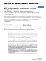
The reduction was then confirmed by C-arm. If align-
ment was not satisfactory, a minimal exposure of the
fracture site was performed to enable the desirable
reduction (Figure 1, 2).
For mini open exposures, wound was closed primarily
for close fractures. For open fractures, we preferred
either to leave the wound open for surgical debridement
or to proceed to a delaye d primary closure 72 h post-
operatively. Skin graft coverage was needed only for one
patient (S-V(G-IIIA).
Post-operative care consisted of daily performed thor-
ough pin care, from the first postoperative day, with
hydrogen peroxide and betadine as well as immediate
passiverangeofmotionoftheknee.Forhighlycom-
minuted fractures, a posterior splint was applied a nd
after 48 hours the patient was encouraged to s tart con-
trolled knee movement as soon as possible. Patients
were discharged from the hospital between the 5th and
15th postoperative day, depending on their general
condition. Patients with Gustilo grade II and III open
fractures were checked weekly in the outpatient depart-
ment. All the other patients were checked monthly.
They were instructed not to bear weight on the oper-
ated limb and to regularly perform pin site care. Pro-
gressive weight bearing was allowed between the 8th
and 12th week depending on the radiographic appear-
ance of callus. The weight bearing started with 10 kg
and, based on the clinical and radiographic signs of
union, advanced to 30 kg after one month. In most of
Figure 1 Postoperative AP x-ray of a male patient
demonstrating a Schatzker’s VI tibial plateau fracture of the
left lower limb.
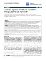
Figure 2 Postoperative lateral x-ray of the same patient.
Babis et al. Journal of Orthopaedic Surgery and Research 2011, 6:35
/>Page 3 of 7
our cases, the external fixator was removed at 3.4
months after surgery depending on the radiological
appearance of union.
Results
Patient resu lts are given in Table 1. All fractures in this
series except one (3.0%) healed. Uni on was de termined
by the presence of a bridging callus on the follow-up
radiographs and by the clinical i mpression of stabilit y.
Follow-up evaluation was available for all fractures.
Based on the parameters considered at the follow-up
(radiological results, knee ROM, pain, ability to perform
sport activities, and patient’ s satisfaction), according to
KSS criteria [18], the results were evaluated as excel lent
in 18 patients (55%), good in 10 patients (30%), fair in 4
patient (12%), and poor in 1 (3%) (Table 1).
There were no systemic complications attributable to
our s urgical treatments. All associated ligamentous and
meniscal lesions w ere repaired a t a second stage after
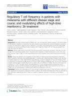
fracture healing. All fractures healed, with an average
time of treatment with the frame of 3.4 months
(Figure 3, 4). The external fixator was tolerated for the
entire treatment period in all cases. Two fractures
(6.0%) took longer than 6 months to heal.
In our series only one (1) fracture was complicated
with deep infection leading to septic non-union (3.0%).
It was treated with surgical debridement and i.v. antibio-
tics until CRP and ESR reached normal values. Later on,
open reduction and internal fixation with plate and
autologus bone grafting was performed. Deep venous
thrombosis was detected in one patient (3.0%) and was
treated successfully with low molecular weight heparin.
There were 3 pin track infections (9.1%). These
infections were superficial or limited to the soft tissue
and did not extend to the bone. None of the patients
required hospital admission. There were treated with
oral antibiotic and local pin care. All pin track infec-
tions healed without requiring wire or half-pin removal
that could compromise frame’s stability. Two fractures
(6.0%) resulted in malunion (10° of valgus, < 5° procur-
vatum), but faced no symptoms. In one case of an
open fracture, local skin ne crosis occurred requiring a
skin graft.
A total of 26 (78.8%) patients regained functional use
of the knee joint, good axis, without pain or instability.
Patients’ knee ROM was gradually increasing at conse-
cutive clinical evaluations. Patients were discharged
from the hybrid fixator after an average time of 3.4
months (range 3 - 7 months). At the one year follow-
up, range of motion averaged 115° of flexion (range 75°
to 125°) and 5° lack of extension (range 0°- 8°). During
the radiographic follow-up evaluation, early osteoar-
thritic changes at the knee joint were noticed in one (1)
patient (3.0%) (SVI/GII fracture).
Overall, 5 patients (15.1%) faced with at least 1 minor
complication such as pin track infection, stiffness, malu-
nion and 1 patient (3.0%) came up with at least one (1)
major complication including septic-nonunion and
osteomyelitis. No amputation was performed.
Table 1 Fractures’ characteristics, complications and
results of our study group.
No
of
pts
Schatzker
type
Open/
closed
Results Complications
1 V closed excellent None
2 V closed excellent None
3 V closed good pin track infection-per os
antibiotics
4 V closed excellent None
5 V closed excellent None
6 V open: G-
I
excellent None
7 V closed excellent None
8 V closed good None
9 V closed excellent deep venous thrombosis
10 V closed excellent None
11 V closed good None
12 V closed excellent None
13 V closed excellent None
14 V closed excellent None
15 V closed excellent None
16 V closed excellent None
17 VI closed good None
18 VI closed excellent pin track infection- per os
antibiotics
19 VI open:G-
III
fair local skin necrosis
20 VI closed excellent None
21 VI open: G-
II
good pin track infection- per os
antibiotics
22 VI closed good None
23 VI closed good None
24 VI open:G-II fair malunion, 10° valgus
25 VI closed excellent None
26 VI open:G-
III
fair traumatic peroneal nerve palsy
27 VI closed good None
28 VI closed fair malunion, 5° procurvatum
29 VI closed excellent None
30 VI closed excellent None
31 VI open: G-
III
poor deep infection- septic
pseudarthrosis
32 VI closed good None
33 VI closed good None
Babis et al. Journal of Orthopaedic Surgery and Research 2011, 6:35
/>Page 4 of 7
Discussion
The importance of the soft-tissue envelope in the heal-
ing of plateau f ractures has been analyzed in the l itera-
ture and a correlation of poor results with severely
damaged soft-tissues has already been established [19].
High energy trauma is considered as a major cause of
poor results in the treatment of tibial plateau fractures.
Different methods for treating these complex injuries
have been proposed, including limited open reduction
and stabilizatio n with percutaneous screws, open reduc-
tion and internal fixation [4,20-23] and indirect reduc-
tion and application of a hybrid [24-26] or a circular
external fixation device [27,28].
Internal fixation, despite the advantages of direct
visualization, proper and stable reduction of the articular
surface as well as the acute repair of soft tissue inju ries,
presents also serious disadvantages, including skin or
soft-tissue necrosis caused by surgical manipulations on
an already damaged soft-tissueenvelopeandthehigh
rate of infection, which may compromise the final result.
Tscherne et al, comparing the r esults of surgical versus
conservative treatm ent for ti bial plateau fractures,
reported im proved range of motion, decreased
percentage of malunion and 5% reoperation rate for the
surgical group [29]. Stevens et al, presented several
transoperative - postoperative complicatio ns [30], while
Young and Barrack, in their series of dual plating for
complex bicondylar tibial plateau fractures reported an
88% deep infection rate [31,32]. Certain authors have
treated bicondylar tibial fractures by means of a l ateral
fixed angular plate (FAP) t hrough a single lateral
approach, thus avoiding medial periosteal striping
[33,34]. Jiang R et al, in their prospective study compar-
ing locked plates, to classic double plates (DP), for the
repair of bicondylar tibial plateau fractures reported
similar results for the two groups [35]. Nevertheless, as
presented by Higgins et al., bicondylar fractures stabi-
lized by means of a FAP present a higher rate of subsi-
dence compared to dual plating stabilized fractures [36].
The external fixation as a definite treatment for the
polytrauma patient with multiple osseous and soft tissue
Figure 3 AP x-ray of the same patient after hybrid external
fixator removal.
Figure 4 Lateral x-ray of the same patient after hybrid external
fixator removal.
Babis et al. Journal of Orthopaedic Surgery and Research 2011, 6:35
/>Page 5 of 7
injuries has been described in the literature [37,38].
Certain a uthors believe that external fixation should be
limited to bicondylar tibial fractures with a compro-
mised soft-tissue envelope, as a temporary stabilizing
technique, prior to definite treatment [39]. In the last 2
decades, the evolution of devices and techniques of
external fixation has led many surgeons to apply the
principles of biologic osteosynthesis and minimally inva-
sive surgery for the treatment of comminuted tibial pla-
teau fractures [4,28,32,39]. The development of circular
and hybrid frames, the capability of axial, lateral com-
pression and dynamization, the development of olive
wir es have offered new possi bilities to the external fixa-
tors for the treatment of complex fractures [40]. Maha-
dena et al, comparing external to internal fixation,
concluded that hybrid external fixation possesses theore-
tical advantages in terms of the soft tissues protection;
however the benefit over internal fixation is modest as
far as accuracy of reduction is concerned [41]. Chin et
al presented 38.9% good/excellent, and 6 1.1% fair/poor
results in his type V and VI fracture series [42]. Cat-
agni et al, in their series of high-energy Schatzker V
and VI tibial plateau fractures treated with circular
external fixator, reported excellent and good results in
30 (50.85%) and 27 (45.76%) patients respe ctively [23].
In a similar study on type V and VI tibial plateau frac-
tures, Katsenis et al recorded excellent or good final
clinical results in 36 patients (76%) [24]. In 2009, the
Canadian Orthopaedic Trauma Association, in a multi-
center, prospective, randomized clinical trial of 83 S-V,
VI tibial plateau fractures treated with internal or
external fixation, repo rted similar quality of osseo us
reduction and ROM for both groups but lower rate of
early postoperative complications and improved HSS
scores for the external fixation group at the six
months’ follow up. However, at the two years’ follow
up, no significant difference in ROM, HSS scores,
WOMAC and SF-36 was observed between the two
groups [43].
In our series, we used the Orthofix hybrid external
fixator as a definite treatment for Schatzker V, VI closed
fractures as well as for some open tibial plateau frac-
tures. When necessary, open reduction and m inimal
internal fixation by means of k-wires or screws were
performed prior to external fixation application. Overall,
we had an incidence of infection of 12.1%. This rate
compares favorably with historical controls as seen in
table 1. The rate drops to 3.0% (1 pt) if we look only at
deep infections. All the other cases (3 pts), were superfi-
cial pin tract infections that resolved with proper care
and oral antibiotics. Malunion (valgus-procurvatum) was
observed in two patients. It is important to note that
the case o f deep infection as well as the two cases of
malunion occurred in the group of Schatzker VI-open
fractures. In many older articles, authors do not break
down their complications according to the type of the
tibial plateau fracture [4,22,44]. Our cohort by contrast
is essentially a homogeneous group composed of Schatz-
ker V and VI fractures secondary to a high-energy
mechanism. A similar homogenous group was presented
by Covall et al. The authors treated 32 bicondylar tibial
plateau fractures during a 7-year period an d reported a
42% deep infection rate in the cases treated acutely wit h
internal fixation [15].
As far as minor complications are c oncerned, Hutson
et al, in a meta-analysis of 16 studies with a total of 568
patients found pin site infection rates of 10% for tibial
plateau fractures [45]. This number is similar to the rate
of pin tract infection (9.1%) observed i n our series.
Moreover, the two cases of malunion (6.1%) represent
an acceptable rate as compared with other series [23].
Complications concerning th e external fixation device
such as intolerance or pin loosening were not observed
in our study.
Limitations
As limitations of this study, one should consider its ret-
rospective nature. Additionally, since our study group is
composed of high energy plateau fractures w ith a high
complication rate, the average follow up of 27 months
can be considered as inadequate to draw safe conclu-
sions for the development of post-traumatic osteoarthri-
tis. This report may be the basis for a new study
examining the development of post- traumatic arthritis
in patients with high energy plateau fractures.
Conclusions
Schatzker’s type V and VI tibial plateau fractures repre-
sent serious injuries with substantial residual limb-speci-
fic a nd general health deficits [43]. We believe that the
use of Orthofix external fixation, as a definite treatment,
for high-energy proximal tibia bicondylar fractures
proved to be beneficial. While conf ronting such limb-
threatening injuries, external fixation successfully pro-
vided continuous access on the surrounding tissues as
well as proper osseous stabilization without compromis-
ing the sensitive soft tissue envelope.
Author details
1
A’ Orthopaedic Department University of Athens, Attikon Universi ty
Hospital, Athens, Greece.
2
C’ Orthopaedic Department, University of Athens,
KAT Accidents’ Hospital, Athens, Greece.
3
Associate Professor, C’ Orthopaedic
Department, University of Athens, KAT Accidents’ Hospital, Athens, Greece.
Authors’ contributions
All authors contributed equally to this work. BGC, DSE, PK, KN and PNS
participated in the design of the study and drafted the manuscript. BGC and
DSE participated in the design of the study. BGC and KN conceived of the
study, participated in its design and coordination and helped to draft the
manuscript. All authors read and approved the final manuscript.
Babis et al. Journal of Orthopaedic Surgery and Research 2011, 6:35
/>Page 6 of 7
Competing interests
The authors declare that they have no competing interests.
Received: 1 August 2010 Accepted: 14 July 2011 Published: 14 July 2011
References
1. Papagelopoulos PJ, Partsinevelos AA, Themistocleous GS, Mavrogenis AF,
Korres DS, Soucacos PN: Complications after tibia plateau fracture
surgery. Injury 2006, 37:475-84.
2. Eggli S, Hartel MJ, MD S, Haupt U, Exadaktylos AK, Roder C: Unstable
Bicondylar Tibial Plateau Fractures: A Clinical Investigation. J Orthop
Trauma 2008, 22:673-679.
3. Schatzker J: Fractures of the tibial plateau.Edited by: Schatzker J, Tile M.
The Rationale of Operative Orthopaedic Care, Springer-Verlag, New York;
1988:279-295.
4. Schatzker J, McBroom R, Bruce D: The tibial plateau fracture: The Toronto
experience (1968-1975). Clin Orthop Relat Res 1979, 138:94-104.
5. Hackl W, Riedl J, Reichkendler M, Benedetto KP, Freund M, Bale R:
Preoperative computerized tomography diagnosis of fractures of the
tibial plateau. Unfallchirurg 2001, 104:519-523.
6. Appley AG: Fractures of the tibial plateau. Orthop Clin North Am 1979,
10:61-74.
7. Delamarter R, Hohl M: The cast brace and tibial plateau fractures. Clin
Orthop Relat Res 1989, 242:26-31.
8. Scotland T, Wardlaw D: The use of cast-bracing as treatment for fractures
of the tibial plateau. J Bone Joint Surg Br 1981, 63:575-578.
9. Oestern HJ, Tscherne H: Pathophysiology and Classification of Soft Tissue
Injuries Associated with Fractures. In Fractures with Soft Tissue Injuries,
Springer-Verlag. Edited by: Tscherne H, Gotzen L. Berlin; 1984:1-9.
10. Barei DP, Nork SE, Mills WJ, Henley MB, Benirschke SK: Complications
Associated With Internal Fixation of High-Energy Bicondylar Tibial
Plateau Fractures Utilizing a Two-Incision Technique. J Orthop Trauma
2004, 18:649-657.
11. Mallik AR, Coval DJ, Whitelaw GP: Internal versus external fixation
of bicondylar tibial plateau fractures. Orthop Rev 1992,
21:1433-1436.
12. Murphy CP, D’Ambrosia R, Dabezies EJ: The small pin circular fixator for
proximal tibial fractures with soft tissue compromise. Orthopedics 1991,
14:273-280.
13. Swiontkowski MF, Bucholz RW: Knee and Leg Trauma: Bone Trauma. In
Orthopaedic Knowledge Update 4, American Academy of Orthopaedic
Surgeons. Edited by: Frymoyer JW. Rosemont; 1993:579-592.
14. Coval DJ, Fowble CD, Foster TE, Whitelaw GP: Bicondylar tibial plateau
fractures: Principles of treatment. Contemp Orthop 1994, 28:115-122.
15. Duwelius PJ, Conolly JF: Closed reduction of tibial plateau fractures. A
comparison of functional and roentgenographic end results. Clin Orthop
Rel Res
1988, 230:116-26.
16. Duwelius PJ, Rangitsch MR, Colville MR, Woll TS: Treatment of tibial
plateau fractures by limited internal fixation. Clin Orthop Relat Res 1997,
339:47-57.
17. Giannoudis PV: Surgical priorities in damage control in polytrauma. J
Bone Joint Surg Br 2003, 85:478-483.
18. Insall JN, Dorr LD, Scott RD, Scott WN: Rationale of the knee Society
clinical rating system. Clin Orthop Relat Res 1989, 248:13-4.
19. Delamarter RB, Hohl M, Hopp E: Ligament injuries associated with tibial
plateau fractures. Clin Orthop Relat Res 1990, 250:226-233.
20. Blokker CP, Rorabeck CH, Bourne RB: Tibial plateau fractures. An analysis
of the results of treatment in 60 patients. Clin Orthop Relat Res 1984,
182:193-199.
21. Reichard AK, Seligson D, Alt V: External fixation of tibial plateau fractures:
a retrospective evaluation and case report. Osteo Trauma Care 2004,
12:33-36.
22. Waddell JP, Johnston DW, Neidre A: Fractures of the tibial plateau: a
review of ninety-five patients and comparison of treatment methods. J
Trauma 1981, 21:376-381.
23. Catagni MA, Ottaviani G, Maggioni M: Treatment strategies for complex
fractures of the tibial plateau with external circular fixation and limited
internal fixation. J Trauma 2007, 63:1043-1053.
24. Katsenis D, Athanasiou V, Megas P, Tillianakis M, Lambiris E: Minimal
internal fixation augmented by small wire transfixion frames for high-
energy tibial plateau fractures. J Orthop Trauma 2005, 19:241-248.
25. Weiner LS, Kelley M, Yang E, Steuer J, Watnick N, Evans M, Bergman M: The
use of combination internal fixation and hybrid external fixation in
severe proximal tibial fractures. J Orthop Trauma 1995, 9:244-250.
26. Weigel DP, Marsh JL: High-energy fractures of the tibial plateau. Knee
function after longer follow-up. J Bone Joint Surg Am 2002, 84:1541-1551.
27. Watson JT, Coufal C: Treatment of complex lateral plateau fractures using
Ilizarov techniques. Clin Orthop Relat Res 1998, 353:97-106.
28. Dendrinos GK, Kontos S, Katsenis D, Dalas A: Treatment of high energy
tibial plateau fractures by the Ilizarov circular fixator. J Bone Joint Surg Br
1996, 78:710-717.
29. Tscherne H, Obenhofferm P: Tibia1 plateau fractures. Management and
expected results. Clin Orthop Relat Res 1993,
292:87-100.
30. Stevens DG, Beharry R, McKee MD, Waddell PJ, Schemitsch EH: The long-
term functional outcome of operatively treated tibial plateau fractures. J
Orthop Trauma 2001, 15:312-320.
31. Young MJ, Barrack RL: Complications of internal fixation of tibial plateau
fractures. Orthop Rev 1994, 23:149-54.
32. Keogh P, Kelly C, Cashman WF, McGuinness AJ, O’Rourke SK: Percutaneous
screw fixation of tibial plateau fractures. Injury 1992, 23:387-389.
33. Partenheimer A, Gösling T, Müller M, Schirmer C, Kääb M, Matschke S, Ryf C,
Renner N, Wiebking U, Krettek C: Management of bicondylar fractures of
the tibial plateau with unilateral fixed-angle plate fixation. Unfallchirurg
2007, 110:675-83.
34. Gosling T, Schandelmaier P, Muller M, Hankemeier S, Wagner M, Krettek C:
Single lateral locked screw plating of bicondylar tibial plateau fractures.
Clin Orthop Relat Res 2005, 439:207-14.
35. Jiang R, Luo CF, Wang MC, Yang TY, Zeng BF: A comparative study of Less
Invasive Stabilization System (LISS) fixation and two-incision double
plating for the treatment of bicondylar tibial plateau fractures. Knee
2008, 15:139-143.
36. Higgins TF, Klatt J, Bachus KN: Biomechanical Analysis of Bicondylar Tibial
Plateau Fixation: How Does Lateral Locking Plate Fixation Compare to
Dual Plate Fixation? J Orthop Trauma 2007, 21:301-306.
37. Scalea TM, Boswell SA, Scott JD: External fixation as a bridge to
intramedullary nailing for patients with multiple injuries and with femur
fractures: damage control orthopaedics. J Trauma 2000, 48:613-621.
38. Sirkin M, Sanders R, Di Pasquale T, Herscovici D Jr: A staged protocol for
soft tissue management in the treatment of complex pilon fractures. J
Orthop Trauma 1999, 13:78-84.
39. Lemon RA, Bartlett DH: Arthroscopic assisted internal fixation of certain
fractures about the knee. J Trauma 1985, 25:355-358.
40. Schwartsman V, Martin SN, Ronquist RA, Schwartsman R: Tibial fractures.
Clin Orthop Relat Res 1992, 278:207-216.
41. Mahadeva D, Costa ML, Gafrey A: Open reduction and internal fixation
versus hybrid fixation for bicondylar/severe tibial plateau fractures: a
systematic review of the literature. Arch Orthop Trauma Surg 2008,
128:1169-75.
42. Chin TYP, Bardana D, Bailey M, Williamson OD, Miller R, Edwards ER,
Esser MP: Functional outcome of tibial plateau fractures treated with the
fine-wire fixator. Injury 2005, 36:1467-1475.
43. Hall JA, Beuerlein MJ, McKee MD, Canadian Orthopaedic Trauma Society:
Open reduction and internal fixation compared with circular fixator
application for bicondylar tibial plateau fractures. Surgical technique. J
Bone Joint Surg Am
2009, 91:74-88.
44. Anglen JO, Healey WV: Tibial plateau fractures. Orthopedics 1988,
11:1527-1534.
45. Hutson JJ, Zych GA: Infections in periarticular fractures of the lower
extremity treated with tensioned wire hybrid fixators. J Orthop Trauma
1998, 12:214-8.
doi:10.1186/1749-799X-6-35
Cite this article as: Babis et al.: High energy tibial plateau fractures
treated with hybrid external fixation. Journal of Orthopaedic Surgery and
Research 2011 6:35.
Babis et al. Journal of Orthopaedic Surgery and Research 2011, 6:35
/>Page 7 of 7