báo cáo hóa học:" Reverse shoulder arthroplasty leads to significant biomechanical changes in the remaining rotator cuff" doc
Bạn đang xem bản rút gọn của tài liệu. Xem và tải ngay bản đầy đủ của tài liệu tại đây (890.51 KB, 7 trang )
RESEARC H ARTIC L E Open Access
Reverse shoulder arthroplasty leads to significant
biomechanical changes in the remaining rotator
cuff
Sebastian Herrmann
1*
, Christian König
2
, Markus Heller
2
, Carsten Perka
1
and Stefan Greiner
1
Abstract
Objective: After reverse shoulder arthroplasty (RSA) external and internal rotation will often remain restricted. A
postoperative alteration of the biomechanics in the remaining cuff is discussed as a contributing factor to these
functional deficits.
Methods: In this study, muscle moment arms as well as origin-to-insertion distance (OID) were calculated using
three-dimensional models of the shoulder derived from CT scans of seven cadaveric specimens.
Results: Moment arms for humeral rotation are significantly smaller for the cranial segments of SSC and all
segments of TMIN in abduction angles of 30 degrees and above (p ≤ 0.05). Abduction moment arms were
significantly decreased for all segments (p ≤ 0.002). OID was significantly smaller for all muscles at the 15 degree
position (p ≤ 0.005), apart from the cranial SSC segment.
Conclusions: Reduced rotational moment arms in conjunction with the decrease of OID may be a possible
explanation for the clinically observed impaired external and internal rotation.
Keywords: shoulder arthroplasty, cuff tear arthropathy, reverse shoulder prosthesis, biomechanics shoulder,
moment arms, rotator cuff
Background
Promising early functional results can be achieved with
reverse shoulder arthroplasty (RSA), especially in
patients with severe cuff tear arthropathy[1-3]. It also is
a salvage procedure for fract ure sequelae[4-7] and revi-
sion of failed hemiarthroplasty[8,9], even though out-
come is less predictable in these patients.
Patients suffering from the above conditions experi-
ence severe restrictions in their activities of daily living
by either loss of function due to the insufficient rotator
cuff or pain. E ven though functional impairment can be
extensive and all parts of the cuff can be affected, M.
supra-and M. infraspinatus seem to be the most com-
monly involved, whereas teres minor and subsca pularis
often remains intact [10].
One mechanism by which RSA improves function is
the increase of the deltoids moment arm by shifting the
centre of rotation medially. Additionally the deltoid’ s
proportion, contributing to active elevation, is enlarged
and the hemispheric design provides stability and con-
straint. These changes result in a significantly improved
ability to actively abduct and forward-flex the arm[11],
while internal and external rotation often remains
impaired or even decreases postoperatively[12].
Previous studies have given a thorough insight into
the biomechanics of the shoulder joint after RSA includ-
ing joint forces and deltoid function[13], transfer proce-
dures[14] and strategies to avoid inferior impingement
[15,16]. However, so far it remains unclear why func-
tional deficits in internal/external rotation can occur,
even though the muscles mainly responsible for this
function remain intact.
We hypothesised that RSA reduces the moment arms
and the origin-to-insertion distance (OID) of
* Correspondence: sebastian.herrmann@char ite.de
1
Center for Musculosceletal Surgery, Charité-Universitätsmedizin Berlin,
Charitéplatz 1, D-10117 Berlin, Germany
Full list of author information is available at the end of the article
Herrmann et al. Journal of Orthopaedic Surgery and Research 2011, 6:42
/>© 2011 Herrmann et al; licensee BioMed Central Ltd. This is an Open Access a rticle distributed under the terms of the Creative
Commons Attribution License (http://creativeco mmons.org/licenses/by/2.0), which permits u nrestricted use, distribution, and
reproduction in any medium, pro vided the original work is properly cite d.
subscapularis (SSC) and teres minor (TMI), which in
healthy shoulders are responsible for internal/external
rotation.
The aim of this study was therefore to analyse how
RSA changes the moment arms and the OID of the SSC
and TMI during glenohumeral abduction before and
after RSA using a combined in-vitro/in-silico approach,
where in silico refers to a virtual, computational model.
Methods
Specimens
Sho ulder specimens of seven fresh frozen human speci-
mens (mean age 77 years, range 63-84 years) were
tested. All donors have consented participation in the
institutional body donor program, which is approved by
local authorities. None of these shoulders showed signs
of previous surgery, trauma, deformities or distinct
osteoarthritis. There were five right and two left
shoulders. Image data of the left specimens were mir-
rored with respect to the sagittal plane, so definitions of
right shoulder were applicable.
Specimen preparation
After thawing, careful dissection of all specimens was
undertaken. Excessive soft tissue was removed so muscle
origins and insertions of subscapularis and teres minor
could be visualised. To mark the bony insertion sites of
both muscles their outermost limits were marked with
radio opaque markers. Bony landmarks including the
medial and l ateral epicondyle, angulus acromialis, trigo-
num scapulae and angulus inferior were also marked
with markers to ensure an accurate repetitive l andmark
acquisition.
Thin sliced computed tomography (Aquilion 64,
Toshiba Medical Systems) with a resolution of 512 ×
512 and a slice thickness of 0.5 mm was performed.
Using a 3D data visualization, analysis and modelling
software (AMIRA; Mercury Computer Systems, Chelms-
ford, MA, USA), the spatial position of a ll previously
marked landmarks was determined and 3D models of
the humerus and the scapula were created for each
specimen.
Thereafter a polycarbonate resin model of a reverse
prosthesis (Mathys AG, Bettlach, Switzerland) was
implanted by an experienced orthopaedic surgeon fol-
lowing the standard surgical protocol. The humeral
component was implanted in ten degrees of retroversion
as measured by the forearm axis, according to our clini-
cal practice to avoid anterior or posterior impingement.
The glenoid component was implanted so a slight infer-
ior overhang could be observed. Height of the humeral
component was adjusted so substantial deltoid tension
and therefore sufficient joint stab ility was g ained. The
prosthesis resembles the company’s reverse prosthesis
model (Affinis Inverse
®
). The advantage of the polycar-
bonate material was the prevention of radiologic arte-
facts, which allowed reconstruction of the proximal
humerus anatomy with high accuracy. The same implant
size was used for all specimens (glenoid component 39
mm, humeral component stem 6/110 mm).
After the implantation the CT scans were repeated
and the position of the prostheses components relative
to the bones determined in each specimen.
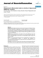
Definition of the joint coordinate systems
In the 3D surface models of each specimen joint coordi-
nate systems (CS) were defined in the scapula and the
humerus according to the recommendations of the
International Society of Biomechanics[17] (Figure 1). In
brief, the scapula CS originates at the angulus acromialis
and is defined by three bony, scapular landmarks. The
coordinate system’sx-axisispointinganteriorly;they-
axis cranially and the z-axis laterally. The humeral CS is
defined by two bony landmarks, the medial and the lat-
eral epicondyle and the centre of the humeral head. The
anatomical direction of the axes was equivalent to the
scapula CS. To determine the centre of the humeral
head a sphere was fitted into the computer mode l of the
humeral articular surface, using a least-square fit algo-
rithm[18]. In the post operative condition after RSA, the
centre of the articula r surface of the glenoid component
was determined to define the centre of rotation in post-
operative shoulders. For easy and distinct interpretation
in line with clinical practice the following definition for
functional moments was used: a positive moment arm
in regard to the scapular z-axis, describes the potential
of anteflexion. Respectively negative values describe the
muscles potential of retroversion. The humeral y-axi s is
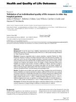
Figure 1 Three-Dimens ional shoulder model creat ed from CT-
scans after implantation of a polycarbonate-resin inverse
shoulder prosthesis. Two coordinate systems (Scapula (S);
Humerus (H)) were defined according to the recommendations of
the Society of Biomechanics.
Herrmann et al. Journal of Orthopaedic Surgery and Research 2011, 6:42
/>Page 2 of 7
the r otational axis. A positive moment arm around this
axis stands for capability to internally rotate the
humerus, negative values result in potential external
rotation. Finally the x-axis of the scapular coordinate
system is considered the axis for abduction and adduc-
tion. Positive values indicating abduction capability;
negative values adduction potential.
Humeral position was expressed in the scapula CS.
Analysis of moment arms
All moment arms and the origin-to-insertion distance
were calculated in the three -dimensional, virtual model
derived from the CT scans.
Since the relative position of the humerus to the sca-
pula could not be accurately set during the CT scan, the
rotations were calculated that transformed the humerus
tothescapulaCS,definingazerodegreeposition.To
analyse a representative range of motion (ROM) in gle-
nohumeral abduction, conditions of 15, 30, 45 and 60°
abduction were simulated by virtually rotating the
humerus around the humeral anterior-posterior (x) axis.
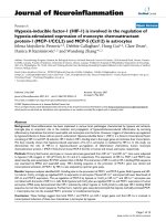
To calculate moment arms for M. subscapularis and
M. teres mino r the radio opaque markers which were
placed in the specimen were identified in the CT scan
and the muscles modelled as lines betwee n the muscles’
origin and insertion. Since the markers only represented
the outermost boundaries of the muscles, a third line
was defined in the middle of these two lines (Figure 2).
Wrapping of these muscles was not considered.
Moment arms for abductio n/adduction, anteflexion/
retroversion and external/internal rotation for these
three segments of each muscle were acquired using the
origin-to-insertion method which is described elsewhere
in more detail[19]. In b rief, to c alculate the moments
for the individual rotations, the total moment is multi-
plied with t he unit vector pointing in the direction of
the axis of that specific rotation.
M
rot axis
=
(
r ×
F
)
•
e
rot axi
s
The moment arms for each rotation (l
rot_axis
)were
then obtained by dividing the calculated moment by the
absolute value of the acting force.
l
rot axis
=
(
r ×
F)
F
•
e
rot axi
s
l
hum
rot
=(
r
hum
×
u
hum
) •
e
hum
y
= r
hum
z
u
hum
x
− r
hum
x
u
hu
m
z
Simplification of the formula allowed using the unit
vector of the acting force
u
instead of specific muscle
forces. The moment arms are therefore dependent on
the vector (
r
) pointing from the centre of rotation to
the point of muscle force application and the direction
of the force (
u
). Moment arms for external/internal rota-
tion were calculated with respect to the y-axis of the
humerus coordinate system, while the abduction/adduc-
tion and anteflexion/retroversion moment arms were
calculated with respect to the x- and the z-axis of the
scapula co ordinate s ystem respectively. These calcula-
tions were repeated for each abduction position.
To estimate how the muscle tension may be influ-
enced by RSA, the length of the previously defined mus-
cle lines was determined pre- and postoperatively. A
shorter distance postoperatively is indicative of a
decreased muscle tension.
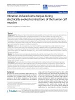
Figure 2 Three Muscle-Segments were defined by virtual lines from its origintoitsinsertionfora:M.subscapularisandb:M.teres
minor.
Herrmann et al. Journal of Orthopaedic Surgery and Research 2011, 6:42
/>Page 3 of 7
Pre- and postoperative moment arms as well as origin-
to-insertion distance for subscapularis and teres minor
were analysed for statistical differences using the inde-
pendent, two-sided Student’s t-test.
Results
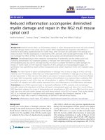
Subscapularis
There was a significa nt change of abduction moment
arm values for all three muscle segments in all tested
positions after reverse arthroplasty (p ≤ 0. 0012), except
for the most cranial segment at 60 degree abduction (p
= 0.86)(F igur e 3). In the pre-operati ve group, the calcu-
lated moment arms resu lted in small abduction capacity
as observed in the cranial segments, to small adductive
moment arms in the distal segments. In postoperative
shoulders all segments had significant bigger adduction
moment arms (p ≤ 0.05), indicating an increased poten-
tial in generating adductive forces, whereas the abduc-
tion-potential will be lost.
Postoperative rotational moment arms of the two
more cranial segments were significantly smaller at all
position s (p ≤ 0.05), whereas no difference could be
seen for the distal segment (p ≥ 0.45).
Origin-to-insertion distances of the two distal seg-
ments decreased significantly after RSA at the 15 degree
position (p ≤ 0.005) and of most distal segment only at
the 30 degree position (p = 0.003). No difference in
length was seen for the other positions (Figure 4).
Teres minor
In t he postoperative group, significantly bigger negative
values for abduction/adduction (x-axis) moment arm
could be seen (p ≤ 0.0005), indicating a higher potential
of generating an adduct ion force ( Figure 5). Contrary to
the postoperative group, positive values for one or two
cranial segments could be seen at the 45 and 60 degree
position in pre-operati ve shoulders indicating an abdu c-
tive potential of these segments.
While no difference was seen for rotational moment
arms at the 15 degree position, values were significantly
smaller with increasing abduction angle in the post-
operative group (p ≤ 0.05).
Small negative values for flexion/extension moment
arm could be seen, with no statistical differences
between the two groups.
Origin to insertion distance was significantly smaller
for all segments at 15 and 30 degrees abduction (p ≤
0.005). The overall differences ranged from 7 to 20 mm.
At 45 degrees this differences could only be observed
for the two distal segments. At 60 degrees abduction no
difference in muscle length was determined (Figure 4).
Discussion
This study aimed to analyse moment arms and origin-
to-insertion distance of the subscapularis and teres
minor before and after reverse shoulder arthroplasty
using a combined in-vitro/in-silico approach. Even
though the functionally deficient infraspinatus may con-
tribute to a loss of external rotation, the aim of this
study was to investigate the effect RSA has on the intact
muscles and their capability to perform rotational move-
ments. Therefore the function of the infraspinatus was
not specifically analysed in this study. This is the first
study to characterise these properties after RSA. Knowl-
edge of the functional properties of these muscles is of
enormous importance for clinical practice and possible
further improvement on prosthesis design or surgical
technique.
Our pre-operative group consists of healthy shoulders,
in which the humeral head is centered in the glenoid
cavity. This might not be the case in shoulders with cuff
tear arthropathy, but as the position of the humerus and
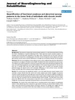
Figure 3 Moment Arms for Abduction/Adduction, Rotation and Flex ion/Extens ion for all three segments of subscapu laris before and
after RSA.
Herrmann et al. Journal of Orthopaedic Surgery and Research 2011, 6:42
/>Page 4 of 7
the refor e the center of rotation is highly variable in this
pathology, we assumed this not practicable in terms of
reproducibility. However, we assume in cases with a sig-
nificantly cranialised humeral head the overall distalisa-
tion will be even more pronounced, leading to even
more substantial changes in the joint’s biomechanics.
The humeral component was implanted in ten degrees
of retroversion in our entire specimens. Varying the
humeral components’ rotational alignment will likely
have an impact on muscle tension. However in our opi-
nion it is not an option to decrease muscle slackening
as, for example, tensioning the posterior cuff will result
in reduced tension of the anterior segments and vice
versa. Also an increased retroversion might result in
increased prosthetic impingent in neutral rotation or
even increase the risk of prosthetic dislocation.
The methodology used is based on three-dimensional
models derived from CT specimens’ data. While CT
scans allow reconstruction of the osseous anatomy with
high precision, accuracy for identification of muscle ori-
gins and insertions was assumed not to be high enough.
Therefore we marked muscle origins and insertions
after preparation and visualisation using radio-opaque
markers. Muscle wrapping was not included in this
model, as it was considered negligible in the tested posi-
tions. Nonetheless we are aware of its possible impact to
the overall value of our results. However, in our study
weaimedtoanalysethechangeofmuscleproperties
rather than to obtain absolute values. The possible inac-
curacy was therefore assumed acceptable. The pre-
operative moment arm values calculated using this
method are comparable to data from previous studies
concerning normal shoulders [20,21].
One of the major drawbacks of RSA is its lacking
potential to improve active external and internal rota-
tion. While in healthy shoulders external rotation is
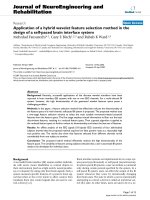
Figure 4 Origin-to-Insertion distance for all segments of subscapularis and teres minor before and after RSA.
Figure 5 Moment Arms for Abduction/Adduction, Rotation and Flex ion/Extension fo r all three segments of teres minor before and
after RSA.
Herrmann et al. Journal of Orthopaedic Surgery and Research 2011, 6:42
/>Page 5 of 7
dependent on teres minor integrity, after RSA potential
of external rotation remains small irrespective of its pre-
operative status. Even in patients with only mild fatty
degeneration preoperatively, the gain in active external
rotation remains small. Patients with higher grade fatty
infil tration pre-opera tively, might even experience a loss
in external rotation[22]. While Boileau et al. [23] pro-
pose several reasons, such as prosthesis design and
altered biomechanical properties of the deltoi d, as being
responsible for this, postoperative changes to t he teres
minor’s rotational moment arms and origin-to -insertion
distance, as shown in our study might be another,
important contributing factor. Rotational moment arms
are significantly smaller for all but the 15 degrees posi-
tion, even though a corresponding trend in this posi tion
can be seen as well. Additionally muscle slackening
might further reduce its efficiency, as origin-to-insertion
distance is significantly smaller, especially in the 15
degrees position, reaching up to 20 mm for the distal
segment.
Accordingly internal rotation, which in healthy
shoulders depends on intact subscapularis function,
often is compromised after RSA as well[24]. The subsca-
pularis muscle tendon unit is t he main internal rotator
and contributes considerably to active stabilisation of
the glenohumeral joint. In this study the two more cra-
nial segments had significant smaller rotational moment
arms after RSA, while no difference could be seen for
the distal s egment. No defin ite rational can be given to
explain this d ifference. Further mathematical analysis
might therefore be necessary.
While failed or non-performed reconstruction of the
subscapularis has shown to have an influence on clinical
outcome[25] in anatomical shoulder arthroplasty, no dif-
ference was seen after RSA at this stage[26]. Even
though Edwards et al. [27] identified impaired subscapu-
laris integrity at the time of surgery as the most impor-
tant risk factor for dislocations in shoulders where
rec onstruction was impossible due to insuffi cien t proxi-
mal humerus bone stock, no higher risk was seen in
patients with cuff tear arthropathy as aetiology. Unfa-
vourable biomechanical properties after RSA, as shown
in this study, with a decreased moment arm in conjunc-
tion with the decreased muscle tension might impede
better results, no matter if the subscapularis is recon-
structed or not. On the other hand, its integrity might
have been irreversibly impaired pre-operatively or sec-
ondary to the surgical approach.
Differences of the origin-to-inser tion distances were
most pronounced for the cranial segments in the 15 and
30 degrees abduction positions for both muscles. With
increasing abduction this diff erence decreases and for
some segments and positions no significant difference
can be seen. We assume that with implantation of the
RSA and distalisation of the humerus an increased dis-
tance of the tendon insertions to the rotational center
arises. This results in a more eccentric motion of t hese
landmarks and might explains the decrease of the or i-
gin-to-insertion distance with increasing abduction.
In both m uscles some seg ments had posit ive abduc-
tion moment arms preoperatively, which in healthy
shoulders is essential for their function as dynamic sta-
bilisers of the shoulder joint. The loss of this function
will lead to a small er joint compression force and as a
result increase subluxation forces[28]. These increased
forces might abet glenoid loosening and instability. No
beneficial effect can be seen for the increased postopera-
tive adduction moment arms as adduction is usually not
impaired in patients with cuff arthropathy, neither pre-
nor postoperatively.
Scapular notching is one major complication in
reverse shoulder arthroplasty[29]. Mechanical impinge-
ment as well as secondary bone erosion due to polyethy-
lene wear is believed to contribute to this phenomenon.
In our study, inferior impingement between the humeral
component and the scapular neck was only observed in
the zero degree reference position, which, however, is
not the neutral thoraco-humeral position , but rather an
adduction position which is not of high clinical rele-
vance. Even though scapular notching was not the speci-
fic focus of this study, these findings are in agreement
with the observations of other authors[30] on this
subject.
Conclusion
In conclusion, this study is the first to analyse the
moment arms and the change in the distance between
muscle insertion sites of the rema ining rotator cuff after
RSA. During glenohumeral abduction, significant
changes were seen in both, the teres minor and the sub-
scapularis moment arms. These changes may contribute
to the clinically observed functional deficits.
Acknowledgements
The authors would like to thank the Robert Mathys Research foundation for
financially supports.
Author details
1
Center for Musculosceletal Surgery, Charité-Universitätsmedizin Berlin,
Charitéplatz 1, D-10117 Berlin, Germany.
2
Julius Wolff Institute, Charité -
Universitätsmedizin Berlin, Center for Sports Science and Sports Medicine
Berlin (CSSB) Philippstr. 13, 10115 Berlin, Germany.
Authors’ contributions
SH, CK, and SG contributed to conception and design of the study,
acquisition of data, analysis and interpretation of data, and drafting the
manuscript. CK and MH derived the mathematical model. SG and CP helped
to draft the manuscript and supervised the whole study. All authors read
and approved the final manuscript.
Competing interests
The authors declare that they have no competing interests.
Herrmann et al. Journal of Orthopaedic Surgery and Research 2011, 6:42
/>Page 6 of 7
Received: 20 January 2011 Accepted: 16 August 2011
Published: 16 August 2011
References
1. Cuff D, Pupello D, Virani N, Levy J, Frankle M: Reverse shoulder
arthroplasty for the treatment of rotator cuff deficiency. J Bone Joint Surg
Am 2008, 90:1244-1251.
2. Sirveaux F, Favard L, Oudet D, Huquet D, Walch G, Mole D: Grammont
inverted total shoulder arthroplasty in the treatment of glenohumeral
osteoarthritis with massive rupture of the cuff. Results of a multicentre
study of 80 shoulders. J Bone Joint Surg Br 2004, 86:388-395.
3. Werner CM, Steinmann PA, Gilbart M, Gerber C: Treatment of painful
pseudoparesis due to irreparable rotator cuff dysfunction with the Delta
III reverse-ball-and-socket total shoulder prosthesis. J Bone Joint Surg Am
2005, 87:1476-1486.
4. Boileau P, Watkinson D, Hatzidakis AM, Hovorka I: Neer Award 2005: The
Grammont reverse shoulder prosthesis: results in cuff tear arthritis,
fracture sequelae, and revision arthroplasty. J Shoulder Elbow Surg 2006,
15:527-540.
5. Klein M, Juschka M, Hinkenjann B, Scherger B, Ostermann PA: Treatment of
comminuted fractures of the proximal humerus in elderly patients with
the Delta III reverse shoulder prosthesis. J Orthop Trauma 2008,
22:698-704.
6. Kontakis G, Tosounidis T, Galanakis I, Megas P: Prosthetic replacement for
proximal humeral fractures. Injury 2008, 39:1345-1358.
7. Wall B, Walch G: Reverse shoulder arthroplasty for the treatment of
proximal humeral fractures. Hand Clin 2007, 23:425-4vi.
8. Flury MP, Frey P, Goldhahn J, Schwyzer HK, Simmen BR: Reverse shoulder
arthroplasty as a salvage procedure for failed conventional shoulder
replacement due to cuff failure–midterm results. Int Orthop 2011,
35:53-60.
9. Levy JC, Virani N, Pupello D, Frankle M: Use of the reverse shoulder
prosthesis for the treatment of failed hemiarthroplasty in patients with
glenohumeral arthritis and rotator cuff deficiency. J Bone Joint Surg Br
2007, 89:189-195.
10. Simovitch RW, Helmy N, Zumstein MA, Gerber C: Impact of fatty
infiltration of the teres minor muscle on the outcome of reverse total
shoulder arthroplasty. J Bone Joint Surg Am 2007, 89:934-939.
11. Boileau P, Watkinson DJ, Hatzidakis AM, Balg F: Grammont reverse
prosthesis: design, rationale, and biomechanics. J Shoulder Elbow Surg
2005, 14:147S-161S.
12. Werner CM, Steinmann PA, Gilbart M, Gerber C: Treatment of painful
pseudoparesis due to irreparable rotator cuff dysfunction with the Delta
III reverse-ball-and-socket total shoulder prosthesis. J Bone Joint Surg Am
2005, 87:1476-1486.
13. Terrier A, Reist A, Merlini F, Farron A: Simulated joint and muscle forces in
reversed and anatomic shoulder prostheses. J Bone Joint Surg Br 2008,
90:751-756.
14. Favre P, Loeb MD, Helmy N, Gerber C: Latissimus dorsi transfer to restore
external rotation with reverse shoulder arthroplasty: a biomechanical
study. J Shoulder Elbow Surg 2008, 17:650-658.
15. Gutierrez S, Levy JC, Frankle MA, Cuff D, Keller TS, Pupello DR, Lee WE III:
Evaluation of abduction range of motion and avoidance of inferior
scapular impingement in a reverse shoulder model. J Shoulder Elbow
Surg 2008, 17:608-615.
16. Roche C, Flurin PH, Wright T, Crosby LA, Mauldin M, Zuckerman JD: An
evaluation of the relationships between reverse shoulder design
parameters and range of motion, impingement, and stability. J Shoulder
Elbow Surg 2009, 18:734-741.
17. Wu G, van der Helm FC, Veeger HE, Makhsous M, Van Roy P, Anglin C,
Nagels J, Karduna AR, McQuade K, Wang X, Werner FW, Buchholz B: ISB
recommendation on definitions of joint coordinate systems of various
joints for the reporting of human joint motion–Part II: shoulder, elbow,
wrist and hand. J Biomech 2005, 38:981-992.
18. Schneider P, Eberly D: Least Squares Fitting. Geometric Tools for Computer
Graphics Morgan Kaufmann Publishers; 2003, 882.
19. Hughes RE, Niebur G, Liu J, An KN: Comparison of two methods for
computing abduction moment arms of the rotator cuff. J Biomech 1998,
31:157-160.
20. Favre P, Sheikh R, Fucentese SF, Jacob HA: An algorithm for estimation of
shoulder muscle forces for clinical use. Clin Biomech (Bristol, Avon) 2005,
20:822-833.
21. Gatti CJ, Dickerson CR, Chadwick EK, Mell AG, Hughes RE: Comparison of
model-predicted and measured moment arms for the rotator cuff
muscles. Clin Biomech (Bristol, Avon) 2007, 22:639-644.
22. Simovitch RW, Helmy N, Zumstein MA, Gerber C: Impact of fatty
infiltration of the teres minor muscle on the outcome of reverse total
shoulder arthroplasty. J Bone Joint Surg Am 2007, 89:934-939.
23. Boileau P, Watkinson DJ, Hatzidakis AM, Balg F: Grammont reverse
prosthesis: design, rationale, and biomechanics. J Shoulder Elbow Surg
2005, 14:147S-161S.
24. Werner CM, Steinmann PA, Gilbart M, Gerber C: Treatment of painful
pseudoparesis due to irreparable rotator cuff dysfunction with the Delta
III reverse-ball-and-socket total shoulder prosthesis. J Bone Joint Surg Am
2005, 87:1476-1486.
25. Gerber C, Yian EH, Pfirrmann CA, Zumstein MA, Werner CM: Subscapularis
muscle function and structure after total shoulder replacement with
lesser tuberosity osteotomy and repair. J Bone Joint Surg Am 2005,
87:1739-1745.
26. Boulahia A, Edwards TB, Walch G, Baratta RV: Early results of a reverse
design prosthesis in the treatment of arthritis of the shoulder in elderly
patients with a large rotator cuff tear. Orthopedics 2002, 25:129-133.
27. Edwards TB, Williams MD, Labriola JE, Elkousy HA, Gartsman GM,
O’Connor DP: Subscapularis insufficiency and the risk of shoulder
dislocation after reverse shoulder arthroplasty. J Shoulder Elbow Surg
2009, 18:892-896.
28. Oosterom R, Herder JL, van der Helm FC, Swieszkowski W, Bersee HE:
Translational stiffness of the replaced shoulder joint. J Biomech 2003,
36:1897-1907.
29. Farshad M, Gerber C: Reverse total shoulder arthroplasty-from the most
to the least common complication. Int Orthop 2010, 34:1075-1082.
30. Simovitch RW, Zumstein MA, Lohri E, Helmy N, Gerber C: Predictors of
scapular notching in patients managed with the Delta III reverse total
shoulder replacement. J Bone Joint Surg Am 2007, 89:588-600.
doi:10.1186/1749-799X-6-42
Cite this article as: Herrmann et al.: Reverse shoulder arthroplasty leads
to significant biomechanical changes in the remaining rotator cuff.
Journal of Orthopaedic Surgery and Research 2011 6:42.
Submit your next manuscript to BioMed Central
and take full advantage of:
• Convenient online submission
• Thorough peer review
• No space constraints or color figure charges
• Immediate publication on acceptance
• Inclusion in PubMed, CAS, Scopus and Google Scholar
• Research which is freely available for redistribution
Submit your manuscript at
www.biomedcentral.com/submit
Herrmann et al. Journal of Orthopaedic Surgery and Research 2011, 6:42
/>Page 7 of 7