báo cáo hóa học:" Biomechanical analysis of a synthetic femoral spiral fracture model: Do end caps improve retrograde flexible intramedullary nail fixation?" ppt
Bạn đang xem bản rút gọn của tài liệu. Xem và tải ngay bản đầy đủ của tài liệu tại đây (2.96 MB, 7 trang )
RESEARCH ARTICLE Open Access
Biomechanical analysis of a synthetic femoral
spiral fracture model: Do end caps improve
retrograde flexible intramedullary nail fixation?
Martin M Kaiser
1*
, Gregor Zachert
2
, Robert Wendlandt
2
, Marion Rapp
3
, Rebecca Eggert
1
, Christine Stratmann
1
,
Lucas M Wessel
4
, Arndt P Schulz
2,5
and Benjamin J Kienast
5
Abstract
Background: Elastic Stable intramedullary Nailing (ESIN) of dislocated diaphyseal femur fractures has become an
accepted method for the treatment in children and adolescents with open physis. Studies focused on
complications of this technique showed problems regarding stability, usually in complex fracture types such as
spiral fractures and in older children weighing > 40 kg. Biomechanical in vitro testing was performed to evaluate
the stability of simulated spiral femoral fractures after retrograde flexible titanium intramedullary nail fixation with
and without End caps.
Methods: Eight synthetic adolescent-size femoral bone models (Sawbones
®
with a medu llar canal of 10 mm and a
spiral fracture of 100 mm length identically sawn by the manufacturer) were used for each group. Both groups
underwent retrograde fixation with two 3.5 mm Titanium C-shaped nails inserted from medial and lateral entry
portals. In the End Cap group the ends of the nails of the eight specimens were covered with End Caps (Synthes
Company, Oberdorf, Switzerland) at the distal entry.
Results: Beside posterior-anterior stress (4.11 Nm/mm vs. 1.78 Nm/mm, p < 0.001), the use of End Caps
demonstrated no higher stability in 4-point bending compared to the group without End Caps (anterior-posterior
bending 0.27 Nm/mm vs. 0.77 Nm/mm, p < 0.001; medial-lateral bending 0.8 Nm/mm vs. 1.10 Nm/mm, p < 0.01;
lateral-medial bending 0.53 Nm/mm vs. 0.86 Nm/mm, p < 0.001) as well as during internal rotation (0.11 Nm/° vs.
0.14 Nm/°, p < 0.05). During compression in 9°- positi on and external rotation there was no statistical significant
difference (0.37 Nm/° vs. 0.32 Nm/°, p = 0.13 and 1.29 mm vs. 2.18 mm, p = 0.20, respectively) compared to the
“classic” 2-C-shaped osteosynthesis without End Caps.
Conclusion: In this biomechanical study the use of End Caps did not improve the stability of the intramedullary
flexible nail osteosynthesis.
Keywords: Elastic stable intramedullary nailing, ESIN, Flexible intramedullary nails, biomechanical testing, femoral
shaft fracture, End Caps, Adolescents, Children
Background
Several treatment options for femoral shaft fractures in
children and adolescents have been described. Children
below the age of 3 can be treated with cast or extensional
devices. In the past two decades the management of
displaced femoral shaft fractures in older children has
gradually evolved toward a more operative approach due
to a more rapid recovery, faster reintegration of the
patients and possible negative effects of immobilisation
even in children [1,2]. Published complications of external
fixation include rotational malalignment, secondary varus
deformity as well as Re-Fractures or fractures in the area
of the Pin entry [3-6]. Therefore, elastic stable intramedul-
lary nail fixation (ESIN) of diaphyseal femoral fractures
has become the most accepted method of treatment for
children older than 3 years [7]. Contradictory information
* Correspondence:
1
Department of Paediatric Surgery, Medical Faculty of the University of
Luebeck, Ratzeburger Allee 160, Luebeck, 23562, Germany
Full list of author information is available at the end of the article
Kaiser et al. Journal of Orthopaedic Surgery and Research 2011, 6:46
/>© 2011 Kaiser et al; lic ensee BioMed Central Ltd. This is an Open Access article distributed under the terms of the Creative Commons
Attribution License ( which permits unrestricted use, distribu tion, and reproduction in
any medium, provided the original work is properly cited.
regarding the results can be fo und. Several retrospective
studies report about a few or no complications [8-11].
Some authors report about skin problems and soft tissue
irritation [12,13], while studies focused on complicati ons
following ESIN demonstrate problems between 10
and 50% [13-19]. In Ho’s publication (94 fractures) the
complication rate was 17% with 8 patients (significantly
higher for patients aged 10 years or older) requiring an
unplanned revision; average time to full weight bearing
was 10 weeks and time to return to preope rative level of
activity averaged 4.9 months [20]. Narayanan reported 41
soft-tissue problems, eight malalignments, two re-fractures
and nine reoperations in 78 patients [18]. The highest
number of complications is observed in complex fracture
types and older children weighing more than 40 kg
[17,20,21]. Due to instability some authors use an addi-
tional immob ilization, additional screws or an additional
external Fixateur [2,12,14,22-27]. Sink et al. changed their
treatment concept towards submuscular plating, Kraus
et al. recommend the external Fixateur for these fractures
[28,29]. Our own retrospective data [30] revealed 43 chil-
dren with closed fractures of the femur sha ft between
March 2002 and April 2007. 31 of these patients were
treated with elastic stable intramedullary nailing (including
three additional casts). Besides three cases of additional
secondary immobilization eight of them needed reopera-
tion: four patients due to varus deformity and four patients
due to shortening of the fracture ("telescoping”).
Due to our own mediocre results and the complica-
tions described in the literature we searched for an
improvement of the method. Thus, the aim of our project
was to determine, i f the stabil ity of the C-shaped osteo-
synthesis would be improved by different modifications
[31]. The German guidelines for paediatric surgery also
recommend the use of End Caps. They should improve
stability in cases of instability following elastic stable
intramedullary nailing [32] by interlocking the nails and
preventing the “backing out”. Despite that, very little clin-
ical research has been published and proved the advan-
tage of using these Caps [33]. In this second part of our
project, we present the results of additional End Caps in
composite bones using a spiral fracture type.
Methods
Mechanical testing was performed using 16 synthetic ado-
lescent-sized composite femoral models (4
th
generation,
Sawbones
®
, Vashon, Washington, USA, European depart-
ment in Sweden) that simulated both cortical and cancel-
lous bone. The femoral model measured 45 cm in length,
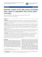
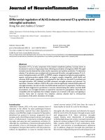
with a central canal diameter of 10 mm. A standard spiral
fracture was created on Sawbones
®
with a length of
100 mm (Figure 1). Due to the reason that paediatric
Sawbone
®
models are not available we decided to use
this specimen as this Sawbone
®
is corresponding to an
adolescent sized femur and the approached question is
most relevant for children weighing more than 40 kg and
adolescents [17,20,21]. We used an established procedure
to create the spiral fractures: Each standard mid-shaft
spiral fracture was industrially sawen by Sawbone
®
.
The fractures were identical: fracture length 100 mm with
almost identical spiral and fragment angles. The para-
meters of the fracture were measured before the
Sawbones
®
were used in the biomechanical model [31].
All further details of this setting are described in our pub-
lication concerning the influence of different nail materials
[31]. According to the literat ure the entry portals medial
and lateral at the distal femoral physis were created by
drilling a hole in the femur 2 to 3 cm proximal to the phy-
sis [34]. All nails were equally prebent 40 degrees, which
brought the curve of the bending in contact with the frac-
ture zone [10,34]. Eight femur models underwent retro-
grade intramedullary fixation (2 C-shaped ESIN pattern =
“classical configuration” = “2E” ) with two 3.5-mm Tita-
nium nails (Santech Nord
®
, Germany) placed through two
drill holes (5-mm drill) at the distal femoral metaphysis 2
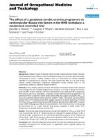
cm above the virtual physis. The nails ended at the proxi-
mal end of the canal, just inferior to the greater Trochan-
ter (Figure 2). Fluoroscopic imaging was performed on
each specimen to confirm the correct configuration and
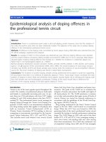
position. The osteosynthesis of the other eight models
were created in a similar fashion with 3.5 mm 40° prebent
Titanium Nails (Santec h Nord
®
, Germany) and cylindric
hollow-threaded End Caps ("2EEC” )wereapplied(Fa.
Synthes, Oberdorf, Switzerland, Figure 3). The specimens
were tested using the UTM (Universal Testing Machi ne)
Zwick 1465 testing machine (Zwick
®
Company, Ulm,
Germany). Custom-fit moulds were produced to secure
the head of the femur and the femoral condyles in the
testing machine. Each specimen was placed in the
machine for a 4-point bending test, a torsional test and
Figure 1 Standard Sawbone
®
Spiral Fracture.
Kaiser et al. Journal of Orthopaedic Surgery and Research 2011, 6:46
/>Page 2 of 7
finally a compression test in 9°-position. The first cycle of
the four individual tests was used as preconditioning; data
for evaluation was collected from three subsequent cycles.
After the last cycle of testing (9°-position) all specimens
were again tested during anterior-posterior stress to check
for possible destructive changes which could have influ-
enced the results (Figure 4). The results of these cycles
confirmed that all tests were performed without destruc-
tion of the osteosynthesis and the specimens.
The 4-point bendin g (Figure 5) wa s performed according
to the ASTM F383-73 and F1264-03 description. With an
incremental linear encoder bending was measured at a
maximum of 5 Nm. Measurement took place at the mid-
point of the two lower force bars, speed was set at 0,05
mm/s. Maximum bending was defined at 2 mm. After this
was reached, tests were halted. The specimens were tested
in the following order: anterior-posterior (AP), posterior-
anterior (PA), lateral-medial (LM) a nd finally medial-lateral
(ML). We chose fixed order to exclude any possible influ-
ence of random order on the results. For torsional testing
the following criteria were set: The maximum allowed tor-
sion during testing was 10°, the maximum torque was set
at 10 Nm. Speed was set at 20°/min. With two angular
encoders t h e torsion was m easured. The femoral head are a
was gimbals-mounted. For compression testing the femur
was positioned in 9° with a calibrated wedge ("AX9”). Fixa-
tion proximal and distal was performed with polymethyl-
metacrylate (PMMA, Technovit 4006) moulds for both
sides. Acompression load up to 100 N was applied at a
speed of 0.05 mm/s. Lateral shiftin g was measured at the
Trochanter major, ventral shifting at the Crista intertr o-
chanterica. Reduction of the fracture gap was measured
using two incremental linear encoders (Product ID: MS30-
1-LD-2, Megatron, Putzbrunn, Germany). Data ( shortening
in 9°-position, torsional stiffness in IR/ER and bending
moments in 4-point bending) were analysed with SPSS
17.0 (SPSS Inc., Chicago, USA). Distributions were checked
for normality (Shapir o-Wilk-Test) before stati s tical analysis
was p erformed. Where significant departure from a normal
distribution occurred a comparison of configurations
regarding the evaluated parameters was performed with
the Mann-Whitney-Test. If no significant departure from
normal distribution was found, the F-Test and analyses of
variance (ANOVA) were used. For adjusting significance
levels to account for multiple comparisons post hoc pair
comparison of homogenous distribution according to
Scheffé and of inhomogeneous variances testing according
to Games-Howell were parts of the control. All values are
presented as mean values. Significance was se t at p < 0.05.
Results
All results of the stiffness of the two different configura-
tions (2E = “classical configuration” vs. 2EEC = “classical
configuration” with End Caps) are shown in Table 1.
The 4-point bending tests from anterior-posterior
showed mean values of the stiffness for the 2-C shaped
ESIN configuration of 0.27 Nm/mm with End Caps
(2EEC) compared to 0.77 Nm/mm for 2 Nails without
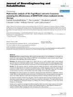
Figure 2 Lateral Fluoroscopic image of a Sawbone
®
composite
graft with a long spiral fracture after implantation of two
elastic stable intramedullary nails; the endings of the nails (2
C-configuration) are inferior to the greater Trochanter.
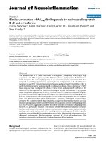
Figure 3 AP Fluoroscopic image of a Sawbone
®
composite graft
with a long spiral fracture after implantation of two elastic
stable intramedullary nails with End Caps.
Figure 4 Control cycle of testing to check for possible
destructive changes which could have influenced the results.
Kaiser et al. Journal of Orthopaedic Surgery and Research 2011, 6:46
/>Page 3 of 7
Figure 5 Biomechanical testing of a Sawbone
®
with spiral fracture in 4-point bending.
Table 1 Summary of the results 2 ESIN vs. 2 ESIN with End Caps
2 Titanium Nails (2E) 2 Titanium Nails with End Caps (2EEC) p-value
Mean value (SD) Mean value (SD)
2 ESIN with End Caps more stable than 2 ESIN
Posterior-anterior 1.78 (1.31) Nm/mm < 4.11 (2.24) Nm/mm < 0.001
2 ESIN with End Caps less stable than 2 ESIN
Anterior-posterior 0.77 (0.29) Nm/mm > 0.27 (0.08) Nm/mm < 0.001
Medial-lateral 1.10 (0.40) Nm/mm > 0.80 (0.35) Nm/mm < 0.01
Lateral-medial 0.86 (0.33) Nm/mm > 0.53 (0.13) Nm/mm < 0.001
Internal Rotation 0.14 (0.04) Nm/° > 0.11 (0.01) Nm/° < 0.05
No statistical significant difference
External Rotation 0.32 (0.18) Nm/° ~ 0,37 (0.11) Nm/° 0.13
Compression in 9°-Position 2.18 (1.37) mm ~ 1.29 (1.61) mm 0.20
Kaiser et al. Journal of Orthopaedic Surgery and Research 2011, 6:46
/>Page 4 of 7
End Caps (2E). Two nails were significantly more stable
than the configuration with End Caps (p < 0.001). Dur-
ing the 4-point bending tests from posterior-anterior
mean values of the stiffness for the 2-C shaped ESIN
configuration of 4 .11 Nm/mm with End Caps (2EEC)
and 1.78 Nm/mm without End Caps (2E). In this testing
ESIN with End Caps was significantly more stable t han
the classical setting (p < 0.001). During varus stress test-
ing (medial-lateral direction) mean values were lower
with End Caps (2EEC) than without (0.80 Nm/mm
2EECvs.1.10Nm/mm2E,p<0.01).Acomparable
results was found for the 4-point bending tests from lat-
eral-medial: mean values for the 2-C shaped ESIN con-
figuration were 0.53 Nm/mm with End Caps (2EEC)
and 0.86 Nm/mm without End Caps (p < 0.001). During
torsional testing, the distal part of the femur was rotated
10° against the proximal part. As this occurred, the tor-
que was determined. The internal rotation testing
showed mean values of stiffness for the 2-C shape d
ESIN configurati on of 0.11 Nm/° with End Caps (2EEC)
and 0.14 Nm/° without End Caps (2E). Thus, ESIN with
End Caps was significantly less stable (p < 0.05) than
the classical 2-C-shaped configuration. During external
rotation testing no significant difference could be
detected (0.37 Nm/° 2EEC vs. 0.32 Nm/° 2C; p = 0.14).
Finally axial compression in 9°-position was measured in
mm the level of the greater trochanter. Mean value was
1.29 mm with End Caps (2EEC) and 2.18 mm without
End Caps (2C). By this, there was also no significant dif-
ference (p = 0.20).
After the complete testing a second circle of anterior-
posterior testing was done as a control.
Results of the first cycle compared to the control ser-
ies showed no significant difference for 2-Nail-setup
(p = 0.71) and the 2-Nail-configuration with End Caps
(p = 0.78).
Summary of Tests
With t he use of End Caps (2EEC) a significantly higher
stability could only be gained in stress tests from poster-
ior-anterior. The classical setting with two elastic stabl e
nails alone (2E) was more stable in bending from ante-
rior-posterior, medial-lateral (Varus stress) as well as
from lateral-medial (Valgus stress) and Internal rotation.
No statistical significant difference could be found for
External rotation and the compression in 9°-position.
Discussion
This biomechanical study is the fir st published survey to
deal with the influence of End Caps in the use of flexible
nails for femoral shaft spiral fractures. Limitations of thi s
study include the use of a synthetic bone model that pos-
sibly cannot precisely reproduce all in-vivo conditions.
However, the synthetic bone model has been used
successfully in previous biomechanical studies and
provides more consistency among specimens than cada-
ver ic bones [35-38]. Due to the configuration, the end of
the nails could not be placed as proximal as it would b e
aspired at the operation in humans. This should be
equalized as both configurations were establi shed identi-
cally. During setup, the focus was on an identical surgical
technique with an exact and even pre-bending and intro-
duction of the nails. Improper location of the bends in
the nails or the nails themselves may create an imbalance
in the bending forces, which will result in an angular
deformity. This technical mistake has been reported in
the literature [10]. By this means the proper configura-
tion of the nails was achieved more precisely than in a
real surgical situation. Despite that, we saw some differ-
ence between the eight nail configurations of each group.
We believe that this is due to slight differences at the
fracture site despite industrial production. In oblique
fractures these differences are expected to be much smal-
ler, because even during industrial production a trans-
verse or an oblique fracture is much easier created than a
more complex spir oid type fracture. The biomechanical
proper ties of retrograde C-shaped flexible intramedullary
nailing have been described in the literature [39-46].
Most of the authors studied oblique or transverse frac-
tures; only two studies examined the spiral type fracture
[45,46]. More or less comparable data of biomechanical
testing is thereby only available in these studies. In an
evaluation of spiral fractures in 10 canine bones Benz et
al showed that stabilization with intramedullary flexible
nails was only possible in 3 cases. In the other cases the
osteosynthesis did not even gain sufficient stability to
make testing setup possible. Gwyn et al performed
biomechanical testing with different fracture types in
synthetic bone models using 2 titanium elastic nails of
4 mm diameter to evaluate the femoral stability with
intramedullary nails. Only external and internal rotation
forces were tested. In this study, transverse and commin-
uted fractures were the least stable. For spiral fracture
types, stability was much lower in internal rotation (our
data: 0.11 Nm/°) compared to external rotation (our data:
0.37 Nm/°). The reason for this difference is the direction
of the spiral fracture - one direction will lead to a slipping
of the fracture edges while during movement in the other
direction the edges will be ca ught. In t ransverse or obli-
que fractures the internal and external rotational forces
are more or less equal. These results show that a stabili-
zation of complex fractures is possible- but very unpre-
dictable in terms of the stability gained with different
fracture types and acting forces. It is an interesting point
that other study groups decided to test only one or two
allocation levels. In all of these studies no rational was
given for this [39,42,44,45]. In co ntrast, we are certain
that the complex structure of a spiral-fracture requires
Kaiser et al. Journal of Orthopaedic Surgery and Research 2011, 6:46
/>Page 5 of 7
testing in all levels. We detected different results con-
cerning stability: more stability in the posterior-anterior
bending with End Caps vs. less stability in anterior-pos-
terior-/medial-lateral- and lateral-medial-bending as well
as during Internal rotation.
In summary, we could not find a benefit in adding
End Caps to the classical way of elastic stable intrame-
dullary nailing in our in vitro synthetic model of spiral
femoral fractures. The technique could not provide a
more stable fixation to maintain length and rotational
control of these spiral midshaft fractures. The only
advantage was seen in posterior-anterior bending.
This is in contrast to the published data of Anastaso-
poulos et al, were 7 patients with diaphyseal femoral frac-
tures (classified as “o blique or comminutive” , without
explicit data on age and body weight) and three patients
with tibia fractures were operated with the use of End
Caps. Concerning only the femoral fractures, difficulties
were encountered in two patients while inserting the End
Caps: in one case it was impossible to screw the End Cap
into the bone cortex and in the second the caps were
held rather loosely in t he bone. In conclusion, fitting of
the End Caps was quot ed as “fair”, because in 6 cases the
end of the nail was not 100% in contact with the end cap.
They described only one 5-10 mm shortening, one
10-mm leg shortening in another patient i n whom the
end caps could not be properly inserted and one Internal
rotation greater than 10°. One patient gained an addi-
tional immobil isation due to pain, another due to impor-
tant knee instability with a patellar fracture. No weight
bearing was allowed for at least three we eks. The authors
pointed out, that removing the implants was eased by the
use of the End Caps after bone healing [ 33]. The solution
might be less than 100% contact of the nails in the End
Caps: too close contact might lead to a small, almost invi-
sible distraction at th e fracture site with consecutive loss
of stiffness in a model without surrounding periosteum
and other soft tissue.
For the future further biomechanical research is
required to improve this type of osteosynthesis and to
make it more feasible for different types of fractures.
Also tra nsverse and oblique fractures need to be tested
with the combination of elastic stable intramedullary
nailing and End Caps.
Author details
1
Department of Paediatric Surgery, Medical Faculty of the University of
Luebeck, Ratzeburger Allee 160, Luebeck, 23562, Germany.
2
Department of
Biomechatronics and Academic Orthopaedics, Medical Faculty of the
University of Luebeck, Ratzeburger Allee 160, Luebeck, 23562, Germany.
3
Department of Child and Adolescent Health, Medical Faculty of the
University of Luebeck, Ratzeburger Allee 160, Luebeck, 23562, Germany.
4
Department of Paediatric Surgery, University of Mannheim, Theodor-Kutzer-
Ufer 1-3, Mannheim, 68167, Germany.
5
Department of Traumatology,
Orthopaedics and Sports Medicine, Trauma Center Hamburg, Bergedorfer Str.
10, Hamburg, 21033, Germany.
Authors’ contributions
MMK is the responsible author and the head of the study group. GZ and
RW are responsible for all testings in the laboratory and edited/reviewed the
manuscript. RE, CS and APS did the testings and edited/reviewed the
manuscript. LMW was responsible for the statistics. MR was responsib le for
translation and proof-reading of the manuscript. BJK was responsible for
translation, proof-reading, and supervision of all versions of the manuscript.
All authors read and approved the final manuscript.
Competing interests
All authors declare that no benefits in any form have been received or will
be received from a commercial party related directly or indirectly to the
subject of this article. The elastic stable nails used in our testings were
sponsored by Santech Nord Company, Schneverdingen, Germany.
Received: 24 December 2010 Accepted: 18 September 2011
Published: 18 September 2011
References
1. Heinrich SD, Drvaric DM, Darr K, MacEwen GD: The operative stabilization
of pediatric diaphyseal femur fractures with flexible intramedullary nails:
a prospective analysis. J Pediatr Orthop 1994, 14:501-507.
2. Carey TP, Galpin RD: Flexible intramedullary nail fixation of pediatric
femoral fractures. Clin Orthop Relat Res 1996, 110-118.
3. Domb BG, Sponseller PD, Ain M, Miller NH: Comparison of dynamic versus
static external fixation for pediatric femur fractures. J Pediatr Orthop 2002,
22:428-430.
4. Galpin RD, Willis RB, Sabano N: Intramedullary nailing of pediatric femoral
fractures. J Pediatr Orthop 1994, 14:184-189.
5. Kirschenbaum D, Albert MC, Robertson WW Jr, Davidson RS: Complex
femur fractures in children: treatment with external fixation. J Pediatr
Orthop 1990, 10:588-591.
6. Probe R, Lindsey RW, Hadley NA, Barnes DA: Refracture of adolescent
femoral shaft fractures: a complication of external fixation. A report of
two cases. J Pediatr Orthop 1993, 13:102-105.
7. Guidelines of the AWMF: Guideline of the german association of pediatric
surgeons: Femur shaft fractures. 2008 [ />detail/ll/006-016.html].
8. Townsend DR, Hoffinger S: Intramedullary nailing of femoral shaft
fractures in children via the trochanter tip. Clin Orthop Relat Res 2000,
113-118.
9. Metaizeau JP: Stable elastic intramedullary nailing for fractures of the
femur in children. J Bone Joint Surg Br 2004, 86:954-957.
10. Slongo TF: Complications and failures of the ESIN technique. Injury 2005,
36(Suppl 1):A78-85.
11. Anastasopoulos J, Petratos D, Konstantoulakis C, Plakogiannis C, Matsinos G:
Flexible intramedullary nailing in paediatric femoral shaft fractures. Injury
2009.
12. Oh CW, Park BC, Kim PT, Kyung HS, Kim SJ, Ihn JC: Retrograde flexible
intramedullary nailing in children’s femoral fractures. Int Orthop 2002,
26:52-55.
13. Sink EL, Gralla J, Repine M: Complications of pediatric femur fractures
treated with titanium elastic nails: a comparison of fracture types. J
Pediatr Orthop 2005, 25:577-580.
14. Flynn JM, Hresko T, Reynolds RA, Blasier RD, Davidson R, Kasser J: Titanium
elastic nails for pediatric femur fractures: a multicenter study of early
results with analysis of complications. J Pediatr Orthop 2001, 21:4-8.
15. Jubel A, Andermahr J, Prokop A, Bergmann H, Isenberg J, Rehm KE: [Pitfalls
and complications of elastic stable intramedullary nailing (ESIN) of
femoral fractures in infancy]. Unfallchirurg 2004, 107:744-749.
16. Luhmann SJ, Schootman M, Schoenecker PL, Dobbs MB, Gordon JE:
Complications of titanium elastic nails for pediatric femoral shaft
fractures. J
Pediatr Orthop 2003, 23:443-447.
17. Moroz LA, Launay F, Kocher MS, Newton PO, Frick SL, Sponseller PD,
Flynn JM: Titanium elastic nailing of fractures of the femur in children.
Predictors of complications and poor outcome. J Bone Joint Surg Br 2006,
88:1361-1366.
18. Narayanan UG, Hyman JE, Wainwright AM, Rang M, Alman BA:
Complications of elastic stable intramedullary nail fixation of pediatric
femoral fractures, and how to avoid them. J Pediatr Orthop 2004,
24:363-369.
Kaiser et al. Journal of Orthopaedic Surgery and Research 2011, 6:46
/>Page 6 of 7
19. Flynn JM, Schwend RM: Management of pediatric femoral shaft fractures.
J Am Acad Orthop Surg 2004, 12:347-359.
20. Ho CA, Skaggs DL, Tang CW, Kay RM: Use of flexible intramedullary nails
in pediatric femur fractures. J Pediatr Orthop 2006, 26:497-504.
21. Leet AI, Pichard CP, Ain MC: Surgical treatment of femoral fractures in
obese children: does excessive body weight increase the rate of
complications? J Bone Joint Surg Am 2005, 87:2609-2613.
22. Pankovich AM, Goldflies ML, Pearson RL: Closed Ender nailing of femoral-
shaft fractures. J Bone Joint Surg Am 1979, 61:222-232.
23. Fein LH, Pankovich AM, Spero CM, Baruch HM: Closed flexible
intramedullary nailing of adolescent femoral shaft fractures. J Orthop
Trauma 1989, 3:133-141.
24. Buckley SL: Current trends in the treatment of femoral shaft fractures in
children and adolescents. Clin Orthop Relat Res 1997, 60-73.
25. Linhart WE, Roposch A: Elastic stable intramedullary nailing for unstable
femoral fractures in children: preliminary results of a new method. J
Trauma 1999, 47:372-378.
26. Ozdemir HM, Yensel U, Senaran H, Mutlu M, Kutlu A: Immediate
percutaneous intramedullary fixation and functional bracing for the
treatment of pediatric femoral shaft fracture. J Pediatr Orthop 2003,
23:453-457.
27. Caird MS, Mueller KA, Puryear A, Farley FA: Compression plating of
pediatric femoral shaft fractures. J Pediatr Orthop 2003, 23:448-452.
28. Kraus R, Schiefer U, Schafer C, Meyer C, Schnettler R: Elastic stable
intramedullary nailing in pediatric femur and lower leg shaft fractures:
intraoperative radiation load. J Pediatr Orthop 2008, 28:14-16.
29. Sink EL, Hedequist D, Morgan SJ, Hresko T: Results and technique of
unstable pediatric femoral fractures treated with submuscular bridge
plating. J Pediatr Orthop 2006, 26:177-181.
30. Rapp M, Albers K, Kaiser MM: Corrective procedures after operation of
femoral shaft fractures in children. Chir Praxis 2011, 73:499-512.
31. Kaiser MM, Wessel LM, Zachert G, Stratmann C, Eggert R, Gros N, Schulze-
Hessing M, Kienast B, Rapp M: Biomechanical analysis of a synthetic
femur spiral fracture model: Influence of different materials on the
stiffness in flexible intramedullary nailing. Clin Biomech (Bristol, Avon)
2011, 26:592-597.
32. Guideline K of the AWMF: 2008 [].
33. Nectoux E, Giacomelli MC, Karger C, Gicquel P, Clavert JM: Use of end caps
in elastic stable intramedullary nailing of femoral and tibial unstable
fractures in children: preliminary results in 11 fractures. Journal of
children’s orthopaedics
2008, 2:309-314.
34. Dietz HG, Schmittenbecher PP, Illing P: Intramedullary osteosynthesis in
adolescence. Urban+Schwarzenberg München Wien Baltimore 1997.
35. Chong AC, Friis EA, Ballard GP, Czuwala PJ, Cooke FW: Fatigue
performance of composite analogue femur constructs under high
activity loading. Ann Biomed Eng 2007, 35:1196-1205.
36. Chong AC, Miller F, Buxton M, Friis EA: Fracture toughness and fatigue
crack propagation rate of short fiber reinforced epoxy composites for
analogue cortical bone. J Biomech Eng 2007, 129:487-493.
37. Cristofolini L, Viceconti M: Mechanical validation of whole bone
composite tibia models. J Biomech 2000, 33:279-288.
38. Cristofolini L, Viceconti M, Cappello A, Toni A: Mechanical validation of
whole bone composite femur models. J Biomech 1996, 29:525-535.
39. Fricka KB, Mahar AT, Lee SS, Newton PO: Biomechanical analysis of
antegrade and retrograde flexible intramedullary nail fixation of
pediatric femoral fractures using a synthetic bone model. J Pediatr
Orthop 2004, 24:167-171.
40. Kiely N: Mechanical properties of different combinations of flexible nails
in a model of a pediatric femoral fracture. J Pediatr Orthop 2002,
22:424-427.
41. Lee SS, Mahar AT, Newton PO: Ender nail fixation of pediatric femur
fractures: a biomechanical analysis. J Pediatr Orthop 2001, 21:442-445.
42. Mahar AT, Lee SS, Lalonde FD, Impelluso T, Newton PO: Biomechanical
comparison of stainless steel and titanium nails for fixation of simulated
femoral fractures. J Pediatr Orthop 2004, 24:638-641.
43. Mani US, Sabatino CT, Sabharwal S, Svach DJ, Suslak A, Behrens FF:
Biomechanical comparison of flexible stainless steel and titanium nails
with external fixation using a femur fracture model. J Pediatr Orthop
2006, 26:182-187.
44. Green JK, Werner FW, Dhawan R, Evans PJ, Kelley S, Webster DA: A
biomechanical study on flexible intramedullary nails used to treat
pediatric femoral fractures. J Orthop Res 2005, 23:1315-1320.
45. Gwyn DT, Olney BW, Dart BR, Czuwala PJ: Rotational control of various
pediatric femur fractures stabilized with titanium elastic intramedullary
nails. J Pediatr Orthop 2004, 24:172-177.
46. Benz G, Kallieris S, Blume U: Biomechanics of the experimental produced
bending and torsional fracture before and after treatment with Nancy-
Nails. Zentralbl Kinderchir 2000, 9:104-109.
doi:10.1186/1749-799X-6-46
Cite this article as: Kaiser et al.: Biomechanical analysis of a synthetic
femoral spiral fracture model: Do end caps improve retrograde flexible
intramedullary nail fixation? Journal of Orthopaedic Surgery and Research
2011 6:46.
Submit your next manuscript to BioMed Central
and take full advantage of:
• Convenient online submission
• Thorough peer review
• No space constraints or color figure charges
• Immediate publication on acceptance
• Inclusion in PubMed, CAS, Scopus and Google Scholar
• Research which is freely available for redistribution
Submit your manuscript at
www.biomedcentral.com/submit
Kaiser et al. Journal of Orthopaedic Surgery and Research 2011, 6:46
/>Page 7 of 7