báo cáo hóa học:" Progressive obesity leads to altered ovarian gene expression in the Lethal Yellow mouse: a microarray study" pptx
Bạn đang xem bản rút gọn của tài liệu. Xem và tải ngay bản đầy đủ của tài liệu tại đây (401.49 KB, 9 trang )
BioMed Central
Page 1 of 9
(page number not for citation purposes)
Journal of Ovarian Research
Open Access
Research
Progressive obesity leads to altered ovarian gene expression in the
Lethal Yellow mouse: a microarray study
John Brannian*
1,2,3
, Kathleen Eyster
1,2
, Mandi Greenway
4
, Cody Henriksen
4
,
Kim TeSlaa
4
and Maureen Diggins
4
Address:
1
Department of Obstetrics & Gynecology, Sanford School of Medicine, University of South Dakota, Sioux Falls, SD 57105, USA,
2
Division
of Basic Biomedical Sciences Sanford School of Medicine, University of South Dakota, Vermillion, SD, USA,
3
Sanford Research USD, Sioux Falls,
SD, USA and
4
Department of Biology, Augustana College, Sioux Falls, SD, USA
Email: John Brannian* - ; Kathleen Eyster - ; Mandi Greenway - ;
Cody Henriksen - ; Kim TeSlaa - ; Maureen Diggins -
* Corresponding author
Abstract
Background: Lethal yellow (LY; C57BL/6J A
y
/a) mice exhibit adult-onset obesity, altered
metabolic regulation, and early reproductive senescence. The present study was designed to test
the hypothesis that obese LY mice possess differences in expression of ovarian genes relative to
age-matched lean mice.
Methods: 90- and 180-day-old LY and lean black (C57BL/6J a/a) mice were suppressed with GnRH
antagonist (Antide
®
), then stimulated with 5 IU eCG. cRNA derived from RNA extracts of whole
ovarian homogenates collected 36 h post-eCG were run individually on Codelink Mouse Whole
Genome Bioarrays (GE Healthcare Life Sciences).
Results: Fifty-two genes showed ≥ 2-fold differential (p < 0.05) expression between 180-day-old
obese LY and lean black mice. LY mice exhibited elevated ovarian expression of agouti (350×),
leptin (6.5×), and numerous genes involved in cholesterol/lipid transport and metabolism, e.g.
lanosterol synthase, Cyp51, and steroidogenic acute regulatory protein (Star). Fewer genes showed
lower expression in LY mice, e.g. angiotensinogen. In contrast, none of these genes showed
differential expression in 90-day-old LY and black mice, which are of similar body weight.
Interestingly, 180-day-old LY mice had a 2-fold greater expression of 11beta-hydroxysteroid
dehydrogenase type 1 (Hsd11b1) and a 2-fold lesser expression of 11beta-hydroxysteroid
dehydrogenase type 2 (Hsd11b2), differences not seen in 90-day-old mice. Consistent with altered
Hsd11b gene expression, ovarian concentrations of corticosterone (C) were elevated in aging LY
mice relative to black mice, but C levels were similar in young LY and black mice.
Conclusion: The data suggest that reproductive dysfunction in aging obese mice is related to
modified intraovarian gene expression that is directly related to acquired obesity.
Background
The negative impact of obesity on fertility is well recog-
nized [1-3]. Moreover, obesity leads to progressive health
disorders associated with the metabolic syndrome. These
include polycystic ovary syndrome (PCOS), which is the
most prevalent endocrinopathy of reproductive age
Published: 3 August 2009
Journal of Ovarian Research 2009, 2:10 doi:10.1186/1757-2215-2-10
Received: 29 June 2009
Accepted: 3 August 2009
This article is available from: />© 2009 Brannian et al; licensee BioMed Central Ltd.
This is an Open Access article distributed under the terms of the Creative Commons Attribution License ( />),
which permits unrestricted use, distribution, and reproduction in any medium, provided the original work is properly cited.
Journal of Ovarian Research 2009, 2:10 />Page 2 of 9
(page number not for citation purposes)
women and a major cause of infertility. Numerous animal
models of obesity have been studied, including the ob/ob
and db/db mutant mouse strains. However, these mouse
models do not mimic typical human obesity. The ob/ob
mouse, for example, lacks bioactive leptin [4] whereas the
db/db mouse possesses a dysfunctional leptin receptor [5].
These types of mutations resulting in complete dysregula-
tion of body weight control are rarely found in the human
population.
The lethal yellow (LY) mouse (C57BL/6J A
y
/a) possesses a
gene deletion in the promoter and first exon region of the
agouti protein gene locus that brings an upstream pro-
moter into place, resulting in the inappropriate constitu-
tive expression of the agouti gene [6]. In the
hypothalamus, the over-expressed agouti protein acts as
an antagonist of melanocortin-4 receptors (MCR4) [7],
which play a critical role in central appetite and metabo-
lism regulation [8]. This interferes with normal satiety
control resulting in hyperphagia [9]. As a consequence, LY
mice exhibit progressive adult-onset obesity, and gradu-
ally develop insulin resistance [10], hyperleptinemia
[11,12], central leptin resistance [13].
Early reproductive senescence is also a hallmark feature of
the A
y
/a genotype [12,14-18]. Granholm and co-workers
[14] found that LY mice over 120 days old exhibited
abnormal estrous cyclicity and decreased mating success
relative to age-matched black mice lacking the agouti
mutation (C57BL/6J a/a), although ovulation rate did not
differ. Based on vaginal smears, younger (< 120 days) LY
mice had estrous cycles of 4–5 days in length and were
indistinguishable from cycles of age-matched black mice
[15]. With advancing age and progressively increasing
obesity, the estrous cycles of yellow mice lengthened and
prematurely ceased between 200–250 days [15]. Ovarian
function could be maintained in aged LY mice stimulated
with eCG/hCG, although fewer developing embryos
tended to be recovered than from identically-treated black
mice [14].
To elucidate whether impaired fertility in aging LY mice
was due to intrinsic ovarian defects or to extraovarian fac-
tors, Granholm and Dickens [16] performed reciprocal
ovarian transplantation between young (70–90 days old)
LY (A
y
/a) and black (a/a) mice and followed reproductive
function as the animals aged. Black mice with trans-
planted ovaries from LY mice exhibited normal fertility. In
contrast, LY mice with transplanted ovaries from black
mice experienced diminished reproductive function simi-
lar to intact LY mice [16]. These authors concluded that
there was no underlying intrinsic defect in the ovaries of
LY mice, but rather impaired fertility must result from
either abnormal hypothalamic-pituitary control or from
extraovarian factors that altered the function of ovarian
cells.
The loss of reproductive function in LY mice is directly
related to obesity. LY mice maintained on a fat-restricted
diet that kept their body weight under 30 g, continued to
cycle normally as they aged, but LY mice weighing more
than 30 g acquired irregular and lengthened cycles [17]. In
addition, 270-day old LY mice fed a low-fat diet had sim-
ilar ovarian histology and equivalent number of antral
follicles on proestrus as age-matched black mice [18]. Pre-
mature cessation of ovulation in aging LY mice correlated
with increasing body weight and circulating leptin con-
centrations [12]. Moreover, in vitro blastocyst develop-
ment of embryos from 180-day LY mice was impaired
compared with embryos from black mice, and this corre-
lated negatively with leptin levels [12]. Collectively these
results suggest that early loss of fertility in LY mice is the
result of progressive obesity, which is mediated by altered
ovarian function as the result of either modified gonado-
tropic control and/or extraovarian factors arising from
obesity. The present study was designed to test the
hypothesis that progressive obesity in LY mice alters ovar-
ian gene expression independently of altered hypotha-
lamic-pituitary control.
Methods
Animals
The study was approved by the Augustana College Animal
Care and Use Committee. Black (C57BL/6J a/a) and LY
(C57BL/6 A
y
/a) mice from the Augustana College Biology
Department breeding colony were used for the study.
Founder mice were originally obtained from The Jackson
Laboratory (Bar Harbor, ME, USA). Mice were fed mainte-
nance diet (Harlan Teklad, Madison, WI, USA) and fresh
water ad libitum, and housed in groups of three mice per
cage on a 14:10 light/dark cycle with lights on at 0600
[12].
To exclude gonadotropin-mediated effects, 90- and 180-
day old LY and black female mice were suppressed with
GnRH antagonist (Antide
©
, Bachem, Torrance, CA) prior
to administration of eCG (Sigma) to stimulate coordi-
nated follicle development. The ovarian suppression pro-
tocol was validated in preliminary studies by suppression
(> 80%) of serum FSH, cessation of cyclicity based on vag-
inal cytology, and absence of large follicles and corpora
lutea on ovarian histology (unpublished data). Late
estrus/metestrus mice were given Antide (10 μg/g BW,
i.p.) on the morning of day 1 of treatment, and again on
the morning of day 4. On the evening of day 5, mice were
injected i.p. with 1 IU/5 g BW eCG. The mice were sacri-
ficed 36 hours after eCG injection and ovaries immedi-
ately removed and trimmed of surrounding fat and
Journal of Ovarian Research 2009, 2:10 />Page 3 of 9
(page number not for citation purposes)
connective tissue. Ovaries were placed in RNA Later
(Ambion, Austin, TX) for subsequent RNA extraction.
RNA Extraction
RNA was extracted as described [19]. Each ovary was
homogenized in 1 ml TRI reagent (Molecular Research
Center, Cincinnati, OH). Sodium acetate and bromochlo-
ropropane were mixed with the homogenate, the sample
was incubated on ice for 15 min, and then centrifuged to
separate the phases. The aqueous phase containing RNA
was removed and purified on an RNeasy column (Qiagen,
Valencia, CA). The sample was treated with an on-column
RNase-free DNase to remove any potentially contaminat-
ing genomic DNA. Total RNA was eluted from the col-
umn. The RNA concentration and purity were calculated
using the RNA 6000 Nano LabChip in an Agilent Bioana-
lyzer. The RNA was stored at -70°C prior to processing for
DNA microarray analysis.
DNA Microarrays
CodeLink Whole Mouse Genome Bioarrays (GE/Amer-
sham, Piscataway, NJ, now Applied Microarrays, Tempe,
AZ) were used for the analysis of differential gene expres-
sion. These microarrays contain 3.3 × 10
4
single-stranded
30-mer oligonucleotide probes for mouse genes and tran-
scribed sequences. Biotinylated cRNA probes were synthe-
sized from the extracted RNA samples per supplier's
directions as previously described [19] using CodeLink
Expression Assay Reagent Kit (GE-Amersham Bio-
sciences). Individual samples were run on separate micro-
arrays (90-day LY n = 3, 90-day black n = 3, 180-day LY n
= 3, 180-day black n = 3); no samples were pooled. The
biotinylated cRNA was fragmented and hybridized with
the DNA microarray slides for 18 hours at 37°C. The
hybridized slides were washed and incubated with
streptavidin-Alexa Fluor 647 (Molecular Probes/Invitro-
gen) to label the cRNA and washed again. An Axon Gene-
Pix Scanner was used to scan the microarrays. GenePix Pro
software (MDS, Inc., Toronto, ON) was used to acquire
and align the microarray image. CodeLink software
(Applied Microarrays, Tempe, AZ) applied the back-
ground correction. GeneSpring 7.0 software (Agilent,
Santa Clara, CA) was used to normalize the expression of
each gene to the median gene expression and to normal-
ize each slide to the 50
th
percentile of gene expression. Sta-
tistical analysis of the data was performed using
GeneSpring 7.0 (Agilent), with the p value set at 0.05 for
the t-test. Multiple testing correction used the Benjamini
and Hochberg False Discovery Rate. Approximately 5% of
the genes would be expected to pass this restriction by
chance with this test. The data set for these DNA microar-
rays has been deposited at the National Center for Bio-
technology Information Gene Expression Omnibus
[GEO; />] as recom-
mended by Minimum Information About a Microarray
Experiment [MIAME] standards and can be accessed
through accession number GSE14937.
Real Time RT-PCR
Pre-designed primers and fluorescent (FAM) labeled
minor groove binding probe were obtained from Applied
Biosystems (Foster City, CA). Real time RT-PCR was car-
ried out with TaqMan Gold RT-PCR reagents (Applied
Biosystems) as described [19]. Changes in expression of
genes of interest were calculated relative to an endog-
enous control (GAPDH). An RNA concentration-response
validation curve was carried out to determine the concen-
tration of RNA to add to the RT-PCR reaction. All samples
were run in duplicate, n = 3 animals. The Relative Expres-
sion Software Tool (REST
©
) [20] was used to analyze the
data from the real time RT-PCR reaction.
Radioimmunoassay and Tissue Extraction for
Corticosterone Measurement
An additional set of 90- and 180-day old LY and black
mice (n = 5 per group) was GnRH antagonist-suppressed
and eCG-stimulated as described earlier. Both trimmed
ovaries from each animal were combined, weighed and
homogenized in a 200 μL of methanol to extract the ster-
oids, yielding ~90% recovery efficiency. Corticosterone
concentrations in ovarian extracts were measured using a
competitive RIA for mouse and rat corticosterone (MP
Biomedicals, Orangeburg, NY). All samples were run in a
single assay run. Intra-assay CV was ~7.5%. Tissue concen-
trations were expressed as ng/mg wet weight, and were
compared among groups by ANOVA with Fisher's LSD
test.
Results
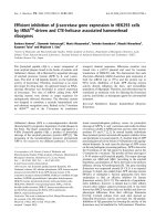
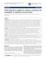
There was no difference in body weight between 90-day
old LY and black mice, but 180-day old LY mice were sig-
nificantly heavier than black mice (Figure 1). Initial DNA
microarray experiments were conducted using 180-day
old mice to determine whether there were differences in
ovarian gene expression between obese LY mice and lean
black mice. Unidentified genes and expressed sequence
tags (EST) were removed from analysis, as were those
genes whose expression was less than 0.2 relative intensity
units (the limit of sensitivity) in both control and treat-
ment groups. To limit analysis to those genes most likely
to be physiologically relevant, only those identified genes
with at least a 2 ± 0.1-fold difference in expression were
included in the final data set. After these exclusions, 52 of
the roughly 3.3 × 10
4
genes analyzed with the microarrays
showed statistically significant differential expression.
Twenty-eight of the differentially expressed genes have
indentified protein products (Table 1). Two of these
genes, agouti and Raly (hnRNP-associated with lethal yel-
low), a gene located in the deleted segment responsible
for the LY syndrome, exhibited the expected differences in
Journal of Ovarian Research 2009, 2:10 />Page 4 of 9
(page number not for citation purposes)
relative expression, i.e. agouti was 350-fold greater in LY
mice and Raly expression was half that in black mice.
Several genes involved in steroid synthesis and metabo-
lism were up-regulated in LY mice, including steroidog-
enic acute regulatory protein (Star) and aldo-keto
reductase family 1, member B7 (Akr1b7). Notably, aged
LY mice had two-fold greater expression of 11beta-
hydroxysteroid dehydrogenase type 1 (Hsd11b1) and a
two-fold lesser expression of 11beta-hydroxysteroid dehy-
drogenase type 2 (Hsd11b2). Numerous differentially
expressed genes are involved in cholesterol biosynthesis,
e.g. isopentenyl-diphosphate delta isomerase (Idd1),
Cyp51, lanosterol synthase, mevalonate (diphospho)
decarboxylase, and sterol-C4-methyl oxidase-like
(Sc4mol). In each case, LY mice exhibited an approxi-
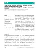
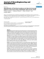
mately 2-fold greater expression than black mice. Further
examination of the microarray data revealed that genes
representing nearly every step in the cholesterol biosyn-
thetic pathway were expressed at a significantly higher
level in LY mice (Figure 2). Other differentially expressed
genes included angiotensinogen, leptin, and fibroblast
growth factor 12.
Subsequently DNA microarray experiments were per-
formed using 90-day old LY and black mice in the same
manner as described to determine whether the gene
expression differences in 180-day old mice were evident
in younger mice that exhibited no difference in body
weight. Expression levels of agouti and Raly served as
internal controls, (agouti: 350- and 330-fold, and Raly:
0.5- and 0.5-fold, 180-day versus 90-day, respectively),
confirming the comparability of the two sets of microar-
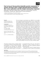
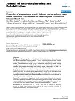
ray data. Other than agouti and Raly, of the genes with dif-
ferential expression in 180-day old mice, only leptin
showed a significant difference in 90-day old mice (Figure
3). However, leptin expression was only 2.5-fold greater
in 90-day old LY mice, as compared to 6.5-fold greater in
180-day old LY mice. None of the genes involved in sterol
synthesis and metabolism that were elevated in 180-day
old LY mice were differently expressed in 90-day old mice.
However, there were other genes that differed in expres-
sion between 90-day old LY and black mice that did not
differ in aged mice. These data will be presented in a sep-
arate communication.
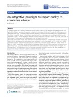
To confirm DNA microarray data, gene expression of
selected genes, i.e. angiotensinogen, Cyp51, 3-hydroxy-3-
methylglutaryl-Coenzyme A-reductase (HMG-CoA
reductase) (Hmgcr), Star, Hsd11b1 and Hsd11b2, was
determined by Real time RT-PCR. Relative expression of
these genes as demonstrated by RT-PCR was very similar
to microarray results (Figure 4).
Hsd11b1 and Hsd11b2 were of particular interest due to
the opposing action of their protein products and diver-
gence in their expression in obese LY mice. These enzymes
interconvert corticosterone to its inactive metabolite 11-
dehydrocorticosterone. RIA analysis of ovarian steroid
extracts showed that aged LY mice had approximately
twice the amount of corticosterone present in ovarian tis-
sue as compared to age-matched black mice and young LY
and black mice (Figure 5B), consistent with the shift in
enzyme expression.
Discussion
This is the first report of differences in the levels of ovarian
gene expression in an obese mouse model. The most
important finding of this study is that modified gene
expression in the ovaries of aging LY mice occurs as a
direct consequence of acquired obesity and is not due to
an altered gonadotropic state. Since all mice were GnRH-
suppressed and stimulated with exogenous gonadotropin,
differences in gene expression were not due to alterations
in hypothalamic-pituitary control in older mice, or to dif-
ferences in estrous cycle state. Stimulation of 180-day old
LY mice with exogenous gonadotropin results in similar
ovarian histology and leads to the same number of preo-
vulatory follicles and ovulated oocytes as in age-matched
black mice (unpublished data). Since progressive obesity
in LY mice is accompanied by the development of insulin
and leptin resistance, changes in gene expression may be
related to altered metabolic state. Albeit a caveat of the
present study is that only whole ovarian gene expression
was determined, and therefore cellular localization can-
not be determined.
Body weight (g) of 90 and 180-day black and LY mice (mean ± SEM; n = 3 for each group)Figure 1
Body weight (g) of 90 and 180-day black and LY mice
(mean ± SEM; n = 3 for each group).
Journal of Ovarian Research 2009, 2:10 />Page 5 of 9
(page number not for citation purposes)
The A
y
mutation is a large deletion that encompasses the
promoter region of the agouti gene as well as a large por-
tion of the coding region of the adjacent upstream Raly
gene, which is constitutively expressed in all somatic cells
[9]. The Raly promoter is thus brought into juxtaposition
with the agouti gene resulting in the ubiquitous over-
expression of agouti [9]. The similar level of expression of
agouti gene in both young and aging LY mice relative to
black mice serves as an intrinsic control confirming the
comparability of the two sets of microarrays. Conversely,
the A
y
mutation leads to a reduced expression of the Raly
gene which was expressed at half the level of black mice in
both 90- and 180-day old animals. This is predicted since
the LY mice are heterozygous for the agouti mutation, i.e.
they possess a single normal allele.
Other than agouti and Raly, leptin was the only other gene
that was significantly altered in both 90- and 180-day old
mice, although the difference in expression was much
greater in the 180-day old obese mice than in the younger
mice. Leptin is primarily produced by adipocytes, and cir-
culating leptin levels increase dramatically in aging LY
mice in proportion to body weight [12]. Leptin may also
be produced by theca and granulosa cells of maturing fol-
licles [21,22]. It has been proposed that leptin resistance
develops in the ovaries of obese animals [12], and increas-
ing ovarian leptin production in obese mice may be
related. Although great care was taken to remove all
adhering fat tissue from the ovaries before RNA extrac-
tion, the possibility that adherant fat may be the source of
the disparate leptin gene expression cannot be excluded.
A major finding of this study was the consistent enhanced
ovarian expression of genes involved in cholesterol bio-
synthesis in obese LY mice. Aging LY mice become insu-
lin-resistant and hyperleptinemic with increasing obesity
[10,11]. It's been long recognized that hepatic cholesterol
synthesis is elevated in obesity [23], and is exacerbated in
diabetes [24]. Moreover, adipokines such as leptin, play a
regulatory role in cholesterol metabolism. Cholesterol
biosynthetic enzymes were among the hepatic genes
whose expression was reduced by leptin in ob/ob mice
[25]. Hepatic HMG-CoA-reductase activity was elevated in
obese Zucker rats, which are resistant to leptin, but leptin
infusion reduced HMG-CoA-reductase activity in both
lean and obese rats [26]. Elevated cholesterol synthetic
enzymes in the face of high leptin levels is consistent with
a state of leptin resistance in the ovaries of obese LY mice.
Table 1: Genes with differential (2.0 ± 0.1-fold; p < 0.05) ovarian expression in 180-day LY mice compared to age-matched black mice.
Accession Number Relative Expression Name
NM_028744.1 0.4 phosphatidylinositol 4-kinase type 2 beta
AK041828.1 0.4 SH3-domain kinase binding protein 1
NM_023130.1 0.5 hnRNP-associated with lethal yellow (Raly)
NM_018867.3 0.5 carboxypeptidase × 2 (M14 family)
NM_008289.1 0.5 hydroxysteroid 11-beta dehydrogenase 2 (Hsd11b2)
AW411692.1 0.55 BCL2-like 11 (apoptosis facilitator)
NM_010350.1 0.55 glutamate receptor, ionotropic, NMDA2C (epsilon 3)
NM_007428.2 0.55 angiotensinogen
NM_009338.1 1.9 acetyl-CoA-acetyl transferase
NM_145942.2 1.9 3-hydroxy-3-methylglutaryl-CoenzymeA-synthase (Hmgcs1)
BI246566.1 1.9 hepatic lipase
NM_026784.1 1.9 phosphomevalonate kinase
BM945729.1 1.9 NAD(P) dependent steroid dehydrogenase-like
NM_011485.3 1.9 steroidogenic acute regulatory protein (Star)
NM_053245.1 1.9 aryl hydrocarbon receptor-interacting protein-like 1
NM_030210.1 2 acetoacetyl-CoA synthetase
NM_008288.1 2 hydroxysteroid 11-beta dehydrogenase 1 (Hsd11b1)
NM_025436.1 2 sterol-C4-methyl oxidase-like (Sc4mol)
BY616448.1 2.1 fibroblast growth factor 12
NM_138656.1 2.1 mevalonate (diphospho) decarboxylase (Mvd)
NM_146006.1 2.2 lanosterol synthase
NM_010742.1 2.3 lymphocyte antigen 6 complex, locus D
NM_009714.1 2.4 asialoglycoprotein receptor 1 (Asgr1)
NM_020010.1 2.5 cytochrome P450, 51 (Cyp51)
NM_145360.1 2.6 isopentenyl-diphosphate delta isomerase (Idd1)
NM_009731.1 5.6 aldo-keto reductase family 1, member B7 (Akr1b7)
NM_008493.3 6.5 leptin
NM_015770.2 350 agouti
Genes structurally associated with the LY mutation are shown in bold. N = 3 animals per group.
Journal of Ovarian Research 2009, 2:10 />Page 6 of 9
(page number not for citation purposes)
Genes (in bold type) involved in cholesterol biosynthesis exhibiting elevated expression (p < 0.05) in 180-day LY mice as com-pared to black miceFigure 2
Genes (in bold type) involved in cholesterol biosynthesis exhibiting elevated expression (p < 0.05) in 180-day
LY mice as compared to black mice. Numbers in boxes indicate fold difference (RU) in gene expression compared to
black mice. Other genes in the pathway tended to be elevated (normal type with fold difference in parentheses).
Journal of Ovarian Research 2009, 2:10 />Page 7 of 9
(page number not for citation purposes)
Collectively, greater expression of cholesterol synthetic
genes would suggest enhanced ovarian steroid produc-
tion. Other than the glucocorticoid measurements
described, ovarian extracts were insufficient to further
assess steroid production in the current study. However,
naturally-cycling 120- and 180-day old LY mice six days
post-mating had higher intraovarian progesterone con-
centrations than black counterparts [Diggins and Bran-
nian, unpublished data]. The enhanced gene expression
of Akr1b7, whose protein product is an enzyme that
metabolizes isocaproaldehyde, a by-product of pregne-
nolone synthesis, further implies an augmentation of ster-
oid synthesis in the ovaries of obese LY mice.
One cholesterol synthetic gene over-expressed in obese LY
mice that is of particular interest is Cyp51. Cyp51 catalyzes
an intermediate step in the conversion of lanosterol to
cholesterol, and is highly expressed in ovary and testis
[27]. Specifically Cyp51 is responsible for the C14-
demethylation of lanosterol. Regulation of Cyp51 expres-
sion in the gonads is gonadotropin-dependent [27,28].
Unlike other cholesterol synthetic genes, the promoter
region of the Cyp51 gene contains both steroid- (SRE) and
cAMP-response elements (CRE) [27]. The product of this
reaction has been identified as meiosis-activating steroid
(MAS), which induces resumption of meiosis in cumulus-
enclosed oocytes [29]. In eCG-stimulated rats, Cyp51
expression and MAS concentrations increased in preovu-
latory follicles, and further increased after hCG adminis-
tration [28]. Although insulin plays a critical role in
regulation of hepatic Cyp51 expression [30], it does not
appear to regulate ovarian Cyp51 expression [28].
Not only was there greater expression of genes involved in
cholesterol synthesis, but the expression of other genes
Ovarian gene expression in 90- and 180-day LY mice relative to black mice analyzed by microarray (n = 3 for each group)Figure 3
Ovarian gene expression in 90- and 180-day LY mice
relative to black mice analyzed by microarray (n = 3
for each group). Solid line indicates 1:1 expression ratio.
Ovarian expression of selected genes in 90- and 180-day LY and black mice analyzed by Real Time RT-PCRFigure 4
Ovarian expression of selected genes in 90- and 180-
day LY and black mice analyzed by Real Time RT-
PCR. Bars represent mean ratios (LY:black) of expression in
90- (black bars) and 180-day (gray bars) old mice. All samples
were run in duplicate (n = 3 for each group).
Corticosterone concentrations in whole ovarian homoge-nates from 90- (black bars) and 180-day (gray bars) old black and LY miceFigure 5
Corticosterone concentrations in whole ovarian
homogenates from 90- (black bars) and 180-day (gray
bars) old black and LY mice. Bars represent mean ± SEM
(n = 5 mice per group). Different letters denote statistical
significance (P < 0.05) by ANOVA with Fisher's LSD test.
Journal of Ovarian Research 2009, 2:10 />Page 8 of 9
(page number not for citation purposes)
related to sterol metabolism were also elevated in obese
LY mice, e.g. hepatic lipase, Star, and Akr1b7, also known
as mouse vas deferens protein (MVDP). Hepatic lipase,
Star, and Akr1b7 are all gonadotropin-regulated genes in
the ovary [31-33]. Furthermore, hepatic lipase and Star
expression can be modulated by insulin [34,35] and lep-
tin [34,36]. The hyperinsulinemia/insulin-resistance of
the obese LY mice may contribute to the elevated expres-
sion of these genes. Hepatic lipase is elevated in diabetics
[34], and leptin enhanced hepatic lipase expression when
given to ob/ob mice [25]. Star expression was increased in
theca cells from follicles of women with PCOS, a syn-
drome characterized by hyperinsulinemia/insulin resist-
ance [37]. Moreover, leptin bi-phasically modulates
granulosa cell Star expression [36].
An interesting and unexpected finding was the reciprocal
shift in Hsd11b1 and Hsd11b2 expression in aging obese
LY mice. These enzymes catalyze the interconversion of
bioactive and bio-inactive glucocorticoids, which is an
important mechanism of regulating glucocorticoid action
in many target tissues. In rodents, the major bioactive glu-
cocorticoid is corticosterone, which is converted to inac-
tive 11-dehydrocorticosterone by 11beta-hydroxysteroid
dehydrogenase type 2 [38]. Conversely, 11-dehydro-corti-
costerone is converted to corticosterone by 11beta-
hydroxysteroid dehydrogenase type 1. In humans, cortisol
and cortisone are the major active and inactive forms,
respectively. Glucocorticoids are important in the patho-
genesis of obesity and insulin resistance, and expression
and activity of 11beta-hydroxysteroid dehydrogenases can
be altered in obesity and diabetes in a tissue-specific man-
ner [39,40]. For example, 11beta-hydroxysteroid dehy-
drogenase type 1 activity was enhanced in obese rat [41]
and human [39] adipose tissue, but reduced in liver. An
increase in type 1 and a decrease in type 2 in the ovaries of
obese LY mice would predict an overall increase in ovar-
ian corticosterone as observed. Although the ovary does
not synthesize glucocorticoids de novo, modulation of glu-
cocorticoid action by interconversion of corticosterone
and 11-dehydrocorticosterone likely plays an important
role in regulating ovarian function. That glucocorticoids
alter ovarian steroidogenesis has long been recognized
[42]. Furthermore, an up-regulation of Hsd11b1 and
down regulation of Hsd11b2 occurs in response to gona-
dotropins, particularly as associated with the LH surge
[43-45]. The shift in 11beta-hydroxysteroid dehydroge-
nase activity leads to an increase in the ratio of active to
inactive glucocortiocoid around the time of ovulation
[46]. Interestingly, a higher cortisol:cortisone ratio is asso-
ciated with a higher clinical pregnancy rate in IVF patients
[47-49]. In addition, 11beta-hydroxysteroid dehydroge-
nases may be important in ovarian metabolism of miner-
alocorticoids [45], progestins [50], and androgens [51],
which may alter ovarian function.
Conclusion
Altered ovarian gene expression in aging LY mice is
directly related to progressive obesity and is not due to an
altered gonadotropic state. There was a universal up-regu-
lation of major genes of the cholesterol synthetic path-
way, as well as certain key genes involved in steroid
synthesis and metabolism. Notably, obesity was associ-
ated with a regulatory shift in ovarian glucocorticoid
metabolism. These results suggest that obesity impacts
reproductive function in LY mice at least partly via direct
modification of ovarian gene expression. Modulation of
ovarian gene expression may involve altered insulin and/
or leptin exposure or sensitivity, which is closely related to
progressive obesity. The mechanisms by which the altered
ovarian gene expression observed in obese mice affects
ovarian function and fertility remains to be elucidated.
Competing interests
The authors declare that they have no competing interests.
Authors' contributions
JB and MD conceived and designed the study. MG, CH,
and KT carried out the treatments and tissue collection,
prepared preliminary data summaries, and participated in
microarray analyses. KE performed RNA extractions and
microarray analyses, and performed statistical analyses on
microarray data. JB performed final data analysis and
drafted the manuscript. All authors read and approved the
final manuscript.
Acknowledgements
Grant Support: NIH INBRE 2P20RR016479, NIH R15 HD044438, and San-
ford Research USD Women's Health Research Center
References
1. Wang JX, Davies M, Norman RJ: Body mass and probability of
pregnancy during assisted reproduction treatment: retro-
spective study. Brit Med J 2000, 321:1320-1321.
2. Norman RJ, Clark A: Obesity and reproductive disorders: a
review. Reprod Fert Dev 1998, 10:55-63.
3. Fedorcsák P, Storeng R, Dale PO, Tanbo T, Abyholm T: Obesity is
a risk factor for early pregnancy loss after IVF or ICSI. Acta
Obstet Gyn Scand 2000, 79:43-48.
4. Zhang Y, Proenca M, Maffei M, Barrone M, Leopold L, Friedman JM:
Positional cloning of the mouse obese gene and its human
homologue. Nature 1994, 372:425-432.
5. Tartaglia L, Dembski M, Weng X, et al.: Identification and expres-
sion cloning of a leptin receptor, OB-R. Cell 1995,
83:1263-1271.
6. Bultman S, Michaud E, Woychik R: Molecular characterization of
the mouse agouti locus. Cell 1992, 71:1195-1204.
7. Lu D, Willard D, Patel IR, Kadwell S, Overton L, Kost T, Luther M,
Chen W, Woychik RP, Wilkison WO: Agouti protein is an antag-
onist of the melanocyte-stimulating hormone receptor.
Nature 1994, 371:799-802.
8. Ollmann MM, Wilson BD, Yang YK, Kerns JA, Chen Y, Gantz I, Barsh
GS: Antagonism of central melanocortin receptors in vitro
and in vivo by agouti-related protein. Science 1997,
278:135-138.
9. Wolff GL, Roberts DW, Mountjoy KG: Physiological conse-
quences of ectopic agouti gene expression: the yellow obese
mouse syndrome. Physiol Genomics 1999, 1:151-163.
10. Klebig M, Wilkinson J, Geisler J, Woychik R: Ectopic expression of
the agouti gene in transgenic mice causes obesity, features
Journal of Ovarian Research 2009, 2:10 />Page 9 of 9
(page number not for citation purposes)
of type II diabetes, and yellow fur. Proc Natl Acad Sci USA 1995,
92:4728-4732.
11. Zemel M, Jones B, Moore J: Agouti regulation of leptin in adi-
pocytes. FASEB J 1997, 11:A352.
12. Brannian JD, Furman GM, Diggins M: Declining fertility in the
Lethal Yellow mouse is related to progressive hyperleptine-
mia and leptin resistance. Reprod Nutr Dev 2005, 45:143-150.
13. Halaas J, Boozer C, Blair-West J, Fidahusein N, Denton D, Friedman
J: Physiological response to long-term peripheral and central
leptin infusion in lean and obese mice. Proc Natl Acad Sci USA
1997, 94:8878-8883.
14. Granholm N, Jeppesen K, Japs R: Progressive infertility in female
lethal yellow mice (A
y
/a; strain C57BL/6J). J Reprod Fertil 1986,
76:279-287.
15. Hogan C, Sehr H, Diggins M: Premature lengthening and cessa-
tion of estrous cycles in the lethal yellow mouse. Proc SD Acad
Sci 1991, 70:249.
16. Granholm N, Dickens G: Effects of reciprocal ovary transplan-
tation on reproductive performance of lethal yellow mice
(A
y
/a, C57BL/6J). J Reprod Fertil 1986, 78:749-753.
17. Swier N, Diggins M, Dillavou G: The relationship between levels
of follicle stimulating hormone and mating/ovulation rates in
the lethal yellow mouse. Proc SD Acad Sci 1993, 72:326-327.
18. Diggins M, Christopher R: Body weight and ovarian function in
Ay/a mice. Proc SD Acad Sci 1999, 78:215-216.
19. Eyster KM, Klinkova O, Kennedy V, Hansen KA: Whole genome
deoxyribonucleic acid microarray analysis of gene expres-
sion in ectopic versus eutopic endometrium. Fertil Steril 2007,
88:1505-1533.
20. Pfaffl MW, Horgan GW, Dempfle L: Relative expression software
tool (REST
©
) for group-wise comparison and statistical anal-
ysis of relative expression Results in real-time PCR. Nucleic
Acids Research 2002, 30:E36.
21. Cioffi JA, Van Blerkom J, Antczak M, Shafer A, Wittmer S, Snodgrass
HR: The expression of leptin and its receptors in pre-ovula-
tory human follicles. Mol Hum Reprod 1997, 3:467-72.
22. Löffler S, Aust G, Köhler U, Spanel-Borowski K: Evidence of leptin
expression in normal and polycystic human ovaries. Mol Hum
Reprod 2001, 7:1143-1149.
23. Miettinen TA: Cholesterol Production in Obesity. Circulation
1971, 44:842-850.
24. Simonen PP, Gylling HK, Miettinen TA: Diabetes contributes to
cholesterol metabolism regardless of obesity. Diabetes Care
2002, 25:1511-1515.
25. Liang C-P, Tall AR: Transcriptional profiling reveals global
defects in energy metabolism, lipoprotein, and bile acid syn-
thesis and transport with reversal by leptin treatment in ob/
ob mouse liver. J Biol Chem 2001, 53:49066-49076.
26. VanPatten S, Ranginan N, Shefer S, Nguyen LB, Rossetti L, Cohen DE:
Impaired biliary lipid secretion in obese Zucker rats: leptin
promotes hepatic cholesterol clearance. Am J Physiol Gastroin-
test Liver Physiol 2001, 281:G393-G404.
27. Rozman D: Lanosterol 14alpha-demethylase (CYP51)-a cho-
lesterol biosynthetic enzyme involved in production of mei-
osis activating sterols in ooocytes and testis-a minireview.
Pflugers Archiv-Eur J Physiol 2000, 439(Suppl 3):R56-R57.
28. Yamashita C, Aoyama Y, Noshiro M, Yoshida Y: Gonadotropin-
dependent expression of sterol 14-demethylase (CYP51) in
rat ovaries and its contribution to the production of a meio-
sis-activating steroid. J Biochem 2001, 130:849-856.
29. Byskov AG, Andersen CY, Nordholm L, Thogersen H, Guoliang X,
Wassman O, Andersen JV, Guddal E, Roed T: Chemical structure
of sterols that activate oocyte meiosis. Nature 2002,
374:559-562.
30. Yamashita C, Kudo M, Noshiro M, Aoyama Y: Insulin is the essen-
tial factor maintaining the constitutive expression of hepatic
sterol 14-P450 (CYP51). J Biochem 2000, 128:93-99.
31. Vieira-van Bruggen D, Verhoeven AJM, Heuveling M, Kalkman C, de
Greef WJ, Jansen H: Hepatic lipase gene expression is tran-
siently induced by gonadotropic hormones in rat ovaries.
Mol Cell Endocr 1997, 126:35-40.
32. Kiriakidou M, McAllister JM, Sugawara T, Strauss JF III: Expression
of steroidogenic acute regulatory protein (Star) in the
human ovary. J Clin Endocr Metab 1996, 81:4122-4128.
33. Brockstedt E, Peters-Kottig M, Badock V, Hegele-Hartung C, Lessl M:
Luteinizing hormone induces mouse vas deferens protein
expression in the murine ovary. Endocrinology 2000,
141:2574-2581.
34. Perret B, Mabile L, Martinez L, Terce F, Barbaras R, Collet X:
Hepatic lipase: structure/function relationship, synthesis,
and regulation. J Lipid Res 2002, 43:1163-1169.
35. Devoto L, Christenson LK, McAllister JM, Makrigiannakis A, Strauss
JF III: Insulin and insulin-like growth factor-I and -Iimodulate
human granulosa-lutein cell steroidogenesis: enhancement
of steroidogenic acute regulatory protein (Star) expression.
Mol Hum Reprod 1999, 11:1003-1010.
36. Ruiz-Cortes ZT, Martel-Kennes Y, Gevry NY, Downey BR, Palin M-
F, Murphy BD: Biphasic effects of leptin in porcine granulosa
cells. Biol Reprod 2003, 68:789-796.
37. Jakimiuk AJ, Weitsman SR, Navab A, Magoffin DA: Luteinizing hor-
mone receptor, steroidogenic acute regulatory protein, and
steroidogenic enzyme messenger ribonucleic acids are over-
expressed in thecal and granulosa cells from polycystic ova-
ries. J Clin Endocr Metab 2001, 86:1318-1323.
38. Krozowski Z, Li KX, Koyama K, Smith RE, Obeyesekere VR, Stein-
Oakley A, Sasano H, Coulter C, Cole T, Sheppard KE: The type I
and type II 11-beta-hydroxysteroid dehydrogenase enzymes.
J Steroid Biochem Mol Biol 1999, 69:391-401.
39. Rask E, Olsson T, Söderberg S, Andrew R, Livingstone DE, Johnson
O, Walker BR: Tissue-specific dysregulation of cortisol metab-
olism in human obesity. J Clin Endocr Metab 2001, 86:1418-1421.
40. Valsamakis G, Anwar A, Tomlinson JW, Shackleton CH, McTernan
PG, Chetty R, Wood PJ, Banerjee AK, Holder G, Barnett AH, Stewart
PM, Kumar S: 11beta-hydroxysteroid dehydrogenase type I
activity in lean and obese males with type II diabetes melli-
tus. J Clin Endocr Metab 2004, 89:4755-4761.
41. Livingstone DE, Jones GC, Smith K, Jamieson PM, Andrew R, Kenyon
CJ, Walker BR: Understanding the role of glucocorticoids in
obesity: tissue-specific alterations of corticosterone metabo-
lism in obese Zucker rats. Endocrinology 2000, 141:560-563.
42. Adashi EY, Jones PBC, Hsueh AJW: Synergistic effect of glucocor-
ticoids on the stimulation of progesterone production by fol-
licle-stimulating hormone in cultured rat granulosa cells.
Endocrinology 1981, 109:1888-1894.
43. Tetsuka M, Thomas FJ, Thomas MJ, Anderson RA, Mason JI, Hillier
SG: Differential expression of messenger ribonucleic acids
encoding 11beta-hydroxysteroid dehydrogenase types 1 and
2 in human granulosa cells. J Clin Endocr Metab 1997,
82:2006-2009.
44. Tetsuka M, Milne M, Simpson GE, Hillier SG: Expression of 11β-
hydroxysteroid dehydrogenase, glucocorticoid receptor,
and mineralocorticoid receptor genes in rat ovary. Biol Reprod
1999, 60:330-335.
45. Fru KN, Voort CA Vande, Chaffin CL: Mineralocorticoid synthe-
sis during the preovulatory interval in macaques. Biol Reprod
2006, 75:568-574.
46. Harlow CR, Jenkins JM, Winston RML: Increased follicular fluid
total and free cortisol levels during the luteinizing hormone
surge. Fertil Steril 1997, 68:48-53.
47. Keay SD, Harlow CR, Wood PJ, Jenkins JM, Cahill DJ: Higher corti-
sol:corticosterone ratios in the preovulatory follicle of com-
pletely unstimulated IVF cycles indicate oocytes with
increased pregnancy potential. Hum Reprod 2002,
17:2410-2414.
48. Lewicka S, von Hagens C, Hettinger U, Grunwald K, Vecsei P, Run-
nebaum B, Rabe T:
Cortisol and cortisone in human follicular
fluid and serum and the outcome of IVF treatment. Hum
Reprod 2003, 18:1613-1617.
49. Thurston LM, Norgate DP, Jonas KC, Gregory L, Wood PJ, Cooke
BA, Michael AE: Ovarian modulators of type 1 11β-hydroxys-
teroid dehydrogenase (11βHSD) activity and intra-follicular
cortisol:cortisone ratios correlate with the clinical outcome
of IVF. Hum Reprod 2003, 18:1603-1612.
50. Murphy BEP: Specificity of human 11β-hydroxysteroid dehy-
drogenase. J Steroid Biochem 1981, 14:807-809.
51. Owen EJ, Holownia P, Conway GS, Jacobs HS, Honour JW: 11 beta-
hydroxyandrostenedione in plasma, follicular fluid, and gran-
ulosa cells of women with normal and polycystic ovaries. Fer-
til Steril 1992, 58:713-718.