báo cáo hóa học:" The prosurvival activity of ascites against TRAIL is associated with a shorter disease-free interval in patients with ovarian cancer" docx
Bạn đang xem bản rút gọn của tài liệu. Xem và tải ngay bản đầy đủ của tài liệu tại đây (669.63 KB, 10 trang )
RESEARC H Open Access
The prosurvival activity of ascites against TRAIL is
associated with a shorter disease-free interval in
patients with ovarian cancer
Denis Lane, Isabelle Matte, Claudine Rancourt, Alain Piché
*
Abstract
Background: The production of ascites is a common complication of ovarian cancer. Ascites constitute a unique
tumor microenvironment that may affect disease progression. In this context, we recently showed that ovarian
cancer ascites may protect tumor cells from TRAIL-induced apoptosis. In this study, we sought to determine
whether the prosurvival effect of ascites affects disease-free intervals.
Methods: Peritoneal fluids were obtained from 54 wome n undergoing intra-abdominal surgery for suspected
ovarian cancer (44 cancers and 10 benign diseases). The ability of peritoneal fluids to protect from TRAIL was
assessed in the ovarian cancer cell line CaOV3, and IC
50
were determined. The anti-apoptotic activity of 6 ascites
against cisplatin, paclitaxel, doxorubicin, etoposide and vinorelbine was also assessed in CaOV3 cells, and the
prosurvival activity of two ascites was assessed in 9 primary ovarian cancer cultures.
Results: Among the 54 peritoneal fluids tested, inhibition of TRAIL cytotoxicity was variable. Fluids originating from
ovarian cancer were generally more protective than fluids from non-malignant diseases. Most of the 44 ovarian
cancer ascites increased TRAIL IC
50
and this inhibitory effect did not correlate strongly with the protein
concentration in these ascites or the levels of serum CA125, a tumor antigen which is used in the clinic as a
marker of tumor burden. The effect of ascites on cisplatin- and paclitaxel-induced cell death was assessed with 4
ascites having inhibitory effect on TRAIL-induced cell death and 2 that do not. The four ascites with prosurvival
activity against TRAIL had some inhibitory on cisplatin and/or paclitaxel. Two ovarian cancer ascites, OVC346 and
OVC509, also inhibited TRAIL cytotoxicity in 9 primary cultures of ovarian tumor and induced Akt activation in
three of these primary cultures. Among a cohort of 35 patients with ascites, a threshold of TRAIL IC
50
with ascites/
IC
50
without ascites > 2 was associated with shorter disease-free interval.
Conclusions: The prosurvival activity of ascites against TRAIL is associated with shorter disease-fre e interval, which
may be explained, at least in part, by ascites-induced cisplatin/paclitaxel resistance. Our findings suggest that
ascites may contain prosurvival factors that protect against TRAIL and chemotherapy and consequently affect
disease progression.
Introduction
Ovarian cancer is the fifth cause of cancer-related
deaths in women, the second most common gynecologi-
cal cancer , and the leading cause of death from gyneco-
logical malignancies [1]. Ovarian cancer is lethal
because of invasiveness, insidious progression, and rapid
development of resistance to chemotherapy. The
incidence of ascites in women presenting with ovarian
cancer ranges from 45% to 75% depending on the
tumor type [2]. This exudative fluid contains ovarian
cancer, lymphoid and mesothelial cells. Ascites fluids
also harbour growth factors [3,4], bioactive lipids such
as lysophosphatidic acid (LPA)[5],cytokines[6,7]and
extracellular matrix constituents [8]. Individually, these
factors may promote cell growth [4,5,8], invasion [9],
and survival [10] suggesting that ascites play an active
role in ovarian cancer progression rather than a passive
one. We recently demonstrated that some ovarian
* Correspondence:
Département de Microbiologie et Infectiologie, Faculté de Médecine,
Université de Sherbrooke, 3001, 12ième Avenue Nord, Sherbrooke, J1H 5N4,
Canada
Lane et al. Journal of Ovarian Research 2010, 3:1
/>© 2010 Lane et al; licensee BioMed Central Ltd. This is an Open Access article distributed under the terms of the Creative Commons
Attribution License ( which permits unrestricted use, distribution, and reproduction in
any medium, provided the original work is properl y cited.
cancer ascites inhibit TRAIL- and FasL-induced apopto-
sis in vitro [10]. In that study, six ovarian cancer ascites
were tested and five out of six inhibited TRAIL-induced
cell death, albeit to different degree. Using the COV2
ascites, we showed that the prosurvival activity w as
dependent upon the activation of Akt [10]. Given the
relatively small number of ascites tested in this study, it
was difficult to appreciate whether the prosurvival activ-
ity against TRAIL i s a commo n property of ascites or
whether it is associated with a specific sub-type of ovar-
ian cancer. In addition, the effect of ascites on primary
tumor cells and most importantly the clinical signifi-
cance of the prosurvival activity of ascites have not been
assessed.
The extrinsic apoptotic pathway is activated by death
receptor ligand stimulation such as TRAIL. TRAIL
binds to its death receptors, TRAIL-R1 and -R2 to acti-
vate caspase-8 [11-13]. TRAIL may also interact with
two decoy receptors (TRAIL-R3 and -R4) that are
unable to transduce death signals [14,15]. Upon TRAIL
binding, activated TRAIL-R1 and -R2 recruit FADD
(Fas-associated death domain). FADD via its death effec-
tor domain (DED) recruits procaspases-8/10, which
assemble into a DISC (death-inducing signaling com-
plex) [16]. When recruited to the DISC, procaspases-8 is
activated through a series of proteolytic cleavages.
Active caspase-8 can directly activate procaspase-3 to
execute apoptosis (type I cells) or cleave Bid to produce
a truncated form (tBid), which induces release of cyto-
chrome C (cyto C) from the mitochondria and leads to
procaspase-9 and subsequently procaspase-3 activation
(type II cells) [17]. T RAIL holds great promise as an
anti-cancer therapy due to its selective apoptosis-indu-
cing action on tumor cells versus normal cells [18].
TRAIL-based therapies are now in phase I/II clinical
trials but resistance to
TRAIL by tumor cells, including ovarian c ancer, may
limit its therapeutic use [19-21]. Consequently, to fully
exploit the potential of TRAIL, it is essential to under-
stand how the tumor microenvironment may i mpact on
the sensitivity of tumor cells to TRAIL.
In this study, we characterized the effect of a large
number of peritoneal fluids isolated from women under-
going intra-abdominal surgery for suspected neoplasia
for their ability to inhibit TRAIL-induced cell death in
the CaOV3 cell line. These ascites originated from var-
ious sub-types of ovarian cancer including serous, endo-
metrioid, mucinous and others. We establish that most
ovarian cancer ascites have some inhibitory effect on
TRAIL-i nduced cell death. We also evaluated the antia-
poptotic effect of two ovarian cancer ascites in vitro on
primary cultures of ovarian tumor cells establishe d from
ascites (n = 8) or tissues (n = 1). The effect of having
ascites with prosurvival activi ty against TRAIL on
disease-free intervals in a cohort of 35 patients was
determined.
Materials and methods
Primary cultures, ascites samples and human subjects
Informed consent was obtained from women that
undergone surgery by the gynecolo gic oncology service
at the Centre Hospitalier Universitaire de Sherbrooke
for this institutional review board approved protocol.
Peritoneal fluids were obtained at the time of initial
cytoreductive surgery for allpatients.Allfluidswere
supplied by the Banque de tissus et de données of the
Réseau de Recherche en Cancer of the Fonds de la
Recherche en Santé du Québec. Histopathology and
tumor grade were assigned according to International
Federation of Gynecology and Obstetrics (FIGO) cri-
teria. Peritoneal fluids were centrifuged at 10 00 rpm for
15 min and supernatants were stored at -20°C until
assayed for protein content or XTT. Primary tumor
cells were isolated as follow: ovarian cancer ascites were
centrifuged at 1000 rpm for 15 min and cells were
washed twice with OSE medium (Wisent, St-Bruno,
Québec, Canada). Cells were then resuspended in OSE
medium supplemented with 10% FBS and b-estradiol
(10
-8
M) and plated into 75 cm
2
flasks. All fl oating cells
were removed the next day. All tumor cell samples were
used at low passage (< 10). All patients with advanced
ovarian cancer in this study were treated with primary
cytoreductive surgery followed by platinum-based che-
motherapy. Clinical data were obtained from the medi-
cal record. The disease-free interval was defined as the
interval between the surgery and the date of progression
of the disease. Disease progression was defined by
CA125 ≥ 2 X nadir value on two occasions, documenta-
tion of increase or new lesions or death [22]. The ovar-
ian cancer cell line CaOV3 was obtained from American
Type Culture Collection (Manassas, VA) and maintained
in D MEM/F12 (Wisent) supplemented with 10% FBS, 2
mM glutamine and antibiotics at 37°C in 5% CO
2
.
Reagents
Recombinant human TRAIL was purchased from Pepro-
Tech. (Rocky Hill, NJ). Anti-Akt, HRP-conjugated anti-
mouse and -rabbit antibo dies were purchased from Cell
Signaling (Beverly, MA). Anti -phospho-Akt (Ser-473)
was from Invitrogen (Biosource, Carlsbad, CA). XTT
reagent (2,3-bis-(2-methoxy-4-nitro-5-sulfo-phenyl)2H-
tetrazolium-5-carboxonilide) was from Invitrogen. Cis-
platin, paclitaxel, doxorubicin, vinorelbine and etoposide
were obtained from the hospital pharmacy.
Cell viability assays
Cell viability in the presence or absence of TRAIL or
drugs w as determined by XTT assay. Briefly, cells were
plated at 20,000 cells /well in 96-well plates in compl ete
medium. The next day, cells (confluence 60-70%) were
Lane et al. Journal of Ovarian Research 2010, 3:1
/>Page 2 of 10
pre-treated for 2 hrs with or without ascites and then
treated with human TRAIL or cisplatin and incubated
for 48 h. At the termination of the experiment, the cul-
ture media was removed a nd a mixture of PBS and
fresh media (without phenol red) containing phenazine
methosulfate and XTT was added for 30 min at room
temperature. The O.D. was determined using a micro-
plate reader at 450 nm (TecanSunrise, Research Triangle
Park, NC). The percentage of cell viability was defined
as the relative absorbance of untreated (no TRAIL, no
ascites) versus TRAIL/drugs treated cells in the presence
or absence of a specific ascites.
Immunoblot analysis
Cells were harvested and washed with ic e-cold PBS.
Whole cell extracts were prepared in lysing buffer (gly-
cerol 10%, Triton X-100 1%, TRIS 10 mM pH 7.4, NaCl
100 mM, EGTA 1 mM, EDTA 1 mM, Na
4
P
2
O
7
20 mM,
NaF 1 mM, Na
3
VO
4
2 mM, SDS 0.1%) containing pro-
tease inhibitors (0.1 mM AEBSF, 5 μg/ml pepstatin, 0.5
μg/ml leupeptin and 2 μg/ml aprotinin) and cyto solic
proteins were separated by 12% SDS-PAGE gels. Lysates
for phosphorylated proteins were done in the presence
of phosphatase inhibitors (100 mM sodium fluoride, 100
μM sodium pyrophosphate, 250 μM sodium orthovana-
date). Proteins were transferred to PVDF membranes
(Roche, Laval, Québec, Canada) by electroblotting, and
immunoblot analysis was performed as previously
described [20]. All primary antibodies w ere incubated
overnight at 4°C. Proteins were visualized by enhanced
chemiluminescence (GE Healthcare, Baie d’ Urfé, Qué-
bec, Canada). Densitometric quantification of phos-
phorylated Akt was performed from three separate
experiments normalized to total Akt.
Statistical analysis
Statistical comparisons be tween two groups were per-
formed using the Student’ s t-test and with ANOVA
when comparing the data with more than two treat-
ments groups. Clinical categorical variables were com-
pared between the two groups with Fisher’sexacttest.
The Pearson’s correlation coefficient test was used to
estimate the correlation between the protein concentra-
tions or the CA125 levels and TRAIL sensitivity. Pro-
gression-free disease analysis was compared using
Kaplan-Meier curves coupled with the log rank test. For
these analyses, the TRAIL IC
50
with ascites/TRAIL IC
50
without ascites were group as having a threshold ≥ 2or
< 2 based on median values. Statistical significance was
indicated by P < 0.05. Statistical analyses were per-
formed with SPSS software (SPSS Inc., Chicago, IL).
Results
Effect of ascites on TRAIL sensitivity
We have previously demonstrated that TRAIL-induced
apoptosis was inhibited by the presence of ascites in
ovarian cancer cell lines CaOV3 and OVCAR3 as a con-
sequence of Akt activation and up-regulation of c-FLIP
S
,
an inhib itor of TRAIL-induced caspase-8 activation [10].
To determine whether the inhibitory effect on TRAIL is
a common property of asci tes, we analyzed 54 peritoneal
fluids. From June 2003 to December 2008, peritoneal
fluids from patients undergoing surgery by the gynecolo-
gic oncology service at the Centre Hospitalier Universi-
taire de Sherbrooke for suspected neoplasia were
obtained. Tissue biopsies were available for a ll patients
and diseases were classified as benign or malignant
according to the histology. To characterize the prosurvi-
val activity of the peritoneal fluids against TRAIL, we
assessed the cell via bility in the presence or absence of
peritoneal fluids at increasing concentrations of TRAIL.
Fluids were added to ovarian cancer cell line CaOV3 at
10% of the total assay volume based on our previous
study[10].Thecharacteristicsofascitesareshownin
Additional file 1, Table S1. Forty four fluids originated
from patients with ovarian cancer and 10 were consid-
ered benign. Among malignant ascites, most were from
patients with serous adenocarcinoma (60%). The protec-
tion against T RAIL-induced cell death varied according
to peritoneal fluids and examples with OVC509 and
OVC 361 ascites are shown in Fig. 1A. OVC509 signifi-
cantly inhibited TRAIL-induced cell death in CaOV3
cells whereas OVC361 did not. TRAIL IC
50
was deter-
mined from these cell viability curves done w ith the
CaOV3 cell line. The anti-apoptotic activity of ovarian
cancer ascites and benign fluids was expressed as TRAIL
IC
50
with ascites/IC
50
withoutascitesandisshownin
Fig. 1B. Ovarian cancer ascites were generally more pro-
tective than fluids from non-malignant diseases (mean
IC
50
increase 2.0 versus 1.25; P = 0.02). Most of the 44
ovarian cancer ascites (82%) led to some degree of inhibi-
tion of TRAIL-induced apoptosis as demonstrated by an
increase of TRAIL IC
50
with ascites > 1.25 fold while the
few remaining did not affect the TRAIL sensitivity of
CaOV3 cells (neutral effect). By comparison, 60% of
benign fluids displayed an increase of TRAIL IC
50
>1.25
fold. It should be noted that we have previously shown
that the presence of FBS 10% or conditioned medium
from ovarian cancer cells do not affect TRAIL-induced
cell death [10]. Furthermore, the anti-apoptotic effect of
ascites was almost completely abolished by Akt inhibition
in CaOV3 cells [10]. All together, these data demonstrate
that most ovarian cancer ascites have an inhibito ry effe ct
on TRAIL-induced cell death. The magnitude of this
effect however was heterogeneous among ascites. The
prosurvival activity of asc ites against TRAIL was not
associated with a specific tumor sub-type.
Protein concentration in ascites and serum CA125 levels
The protein concentration was measured in the 54 peri-
toneal fluids. The mean protein concentration was
Lane et al. Journal of Ovarian Research 2010, 3:1
/>Page 3 of 10
significantly higher in ovarian cancer ascites than in
non-malignant fluids with P < 0,001 (data not shown).
However, among ovarian cancer ascites, the ability to
inhibit TRAIL-induced cell death did not strongly corre-
late (by Pearson’s correlation coefficient test) with the
protein content of each ascites (r = 0.673; P = 0.01) (Fig.
2A).
The CA125 tumor antigen is detected in the majority
of serous ovarian carcinoma [23]. It is a mucin-like
transmembrane glycoprotein of high molecular weight
which is used in the clinic as a marker of tumor burden.
There is indeed a strong correlation between rising and
falling levels of serum CA125 with progression and
regression of the disease [24,25]. CA125 serum levels at
presentation reflect to some extent the i nitial tumor
burden. We therefore assessed the baseline serum
CA125 levels, which likely reflect the levels in ascites, in
our 44 patients with ovarian cancer to determine
whether CA125 levels were associated with the anti-
apoptotic activity of ascites. As shown in Fig. 2B, the
baseline serum CA125 levels did not correlate (r =
0.103; P = 0,14) with the anti-apoptotic activity of
ascites.
Effect of ascites on drug sensitivity
The sensitivity o f CaOV3 cells to 5 chemotherapeutic
drugs was compared to that of TRAIL in the presence
or absence of ascites. Some ascites had anti-apoptotic
activity against all drugs (OVC34 6, OVC509), some
against a few drugs only (OVC508, OVC488, OVC551)
and some (OVC432) were mostly ineffective (Table 1).
All these ascites were obtained from chemotherapy
naïve patients (Additional file 1, Table S1). Fig. 3 shows
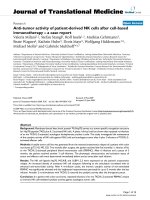
Figure 1 Effect of peritoneal fluids on TRAIL-induced cell death in CaOV3 cells. (a) CaOV3 cells were pre-incubated for 2 h with OVC509
and OVC361 ascites (10% v/v) obtained from women with advanced serous ovarian cancer and treated with TRAIL (10 ng/ml) for 48 h. Cell
viability was measured by XTT assay. Data are shown as the percent cell viability relative to untreated (no TRAIL, no ascites) cells. Results are
from three independent experiments done in triplicate and express as mean ± SEM. (b) TRAIL IC
50
was determined by XTT assay and defined as
the concentration of TRAIL required to kill 50% of CaOV3 cells in the presence or absence of a specific ascites. The prosurvival activity of ovarian
cancer ascites and benign fluids was determined by their ability to increase TRAIL IC
50
after 48 h compared to the TRAIL IC
50
of CaOV3 cells not
exposed to peritoneal fluids. A value of 1 indicates a neutral effect of ascites on TRAIL-induced cytoxicity.
Lane et al. Journal of Ovarian Research 2010, 3:1
/>Page 4 of 10
the effect of ascites on TRAIL, cisplatin and paclitaxel-
induced cell death, cisplatin and paclitaxel being two
drugs that are usually part of the initial treatment for
ovarian cancer. Cisplatin IC
50
was i ncreased by ascites
OVC346, OVC508 and OVC509 whereas the other
ascites tested had a more limited effect. These three
ascites also had an inhibitory on TRAIL-induced cell
death. The increase of paclitaxel IC
50
was observed only
with OVC346, OVC488 and OVC509 ascites. Ovarian
cancer a scites OVC432 had little anti-apoptotic activity
against cisplatin, paclitaxel and TRAIL. These data
demonstrate that the inhibitory effect of ascites against
drug cytotoxicity is heterogeneous. However, ascites that
have a protective effect on TRAIL cyt otoxicity are often
protective against chemotherapeutic drugs.
Ascites decrease TRAIL cytotoxicity in primary cultures of
ovarian tumor cells and activate Akt in these cells
The prosurvival activity of ascites against TRAIL cyto-
toxicity has been shown in ovarian cancer cell lines [10]
but has never been demonstrated in primary ovarian
cancer cultures. Cell-free ovarian cancer ascites
OVC509 were added to primary cultures of tumor cells
isolat ed from ascites obtained from advanced (stage III)
serous ovarian cancer patients. TRAIL cytotoxicity was
significantly reduced in the presence of OVC509 ascites
in primary cultures of tumor cells (346, 327, 318 cells)
tested with P < 0.001 (Fig. 4A). We ex tended these data
by testing OVC346 and OVC509 ascites in 9 primary
cultures. The clinicopat hologic data of the 9 primary
cultures is shown in Additional file 2, Table S2. TRAIL
IC
50
was determined in the presence or absence of
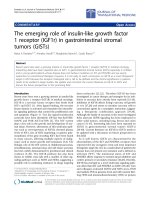
Figure 2 Protein concentration of peritoneal fluids and baseline serum CA125 levels. (a) Protein concentration of the 44 ova rian cancer
ascites was determined and correlated with TRAIL IC
50
fold increased mediated by ascites. (b) Baseline serum CA125 levels were obtained for all
except one patient and correlated with TRAIL IC
50
fold increased mediated by ascites. Correlation coefficients (r) were determined by Pearson’s
correlation coefficient test.
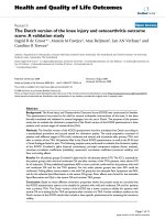
346
432
488 508 509 551
Drug IC50 fold increased
Ascites
Figure 3 Effect of ovarian cancer ascit es on T RAIL-, cisplatin-
and paclitaxel-induced cell death in CaOV3 cells. CaOV3 cells
were pre-incubated for 2 h with various fluids (10% v/v) obtained
from women with advanced ovarian cancer and treated with
increasing concentrations of TRAIL for 48 h or with cisplatin or
paclitaxel for 72 h. Cell viability was assessed by XTT assays. TRAIL,
cisplatin and paclitaxel IC
50
were determined in the presence of
ascites and expressed as fold increased relative to IC
50
in the
absence of ascites. A value of 1 indicates a neutral effect of ascites
on these drugs. Results are from three independent experiments
done in triplicate.
Lane et al. Journal of Ovarian Research 2010, 3:1
/>Page 5 of 10
OVC346 and OVC509 ascites in the 9 primary cultures
of ovarian tumor cells (Table 2). When expressed as
TRAIL IC
50
fold increased, OVC346 and OVC509 dis-
played anti-apoptotic activity, albeit at different degree
in all 9 primary cultures of ovarian cancer (Fig. 4B).
OVC509 had stronger anti-apoptotic activity c ompared
to OVC346.
Consistent with our previous findings in CaOV3 cell line
[10], we found that both OVC346 and OVC509 ascites
induced Akt act ivation in primary tumor samp les as
determined by increased Akt phosphorylation on Wes-
tern blot (Fig. 5). There was a 2 fold increased of Akt
phosphorylation mediated by these ascites (P < 0.001).
Prosurvival activity of ovarian cancer ascites and disease-
free intervals
Among the 44 patients for which we characterized their
ascites with regards to TRAIL sensitivity, 35 had follow
up > 1 year. We therefore used this cohort of 35
patients to assess the prognosis potential of having pro-
tective ascites against TRAIL-induced CaOV3 cell death.
Protecti ve ascites were arbitrarily defined as TRAIL IC
50
with ascites/IC
50
without ascites > 2-fold. Clinical follow
up ranges from 14 months to over 10 years for these 35
patients. The patients were divided into two groups
based on whether the ascites isolated from these
patients were protective or not against TRAIL-induced
cell death. The clini cal characteristics of the patients are
shown in Table 3. There was no difference between the
two groups for age, optimal debulking, tumor histology,
stage of disease or grade. Most patients (80%) had
advanced disease (stage III or IV). Of note, baseline
CA125 levels were similar between the two groups (P =
0,064), which suggest that the tumor burden at presen-
tation was not significantly different between the two
groups. Kaplan Meier analysis showed that women in
the group with TRAIL IC
50
with ascites/TRAIL IC
50
without ascites threshold > 2 h ad significantly shorter
time from baseline to first relapse (mean time 12 vs 15
months, P = 0.014 log rank) (Fig. 6).
Discussion
In this study, using a cell viability-based assay, we evalu-
ated a large number of peritoneal fluids (n = 54) and
showed that fluids originating from malignant diseases
were generally more protective than fluids from non-
malignant diseases against TRAIL-induced cell d eath.
Most of ovarian cancer ascit es (82%) led to some degree
of inhibition of TRAIL-induced apoptosis as demon-
stratedbyanincreaseofTRAILIC
50
with ascites while
the few remaining did not affect the TRAIL sensitivity
of CaOV3 cells (neutral effect). The ability of ascites to
inhibit TRAIL-induced cell death did not correlate
strongly with the protein content of e ach ascites (r =
0.673) or with serum CA125 levels at baseline (r =
0.103) . Importantly, ovarian cancer ascites also inhib ited
TRAIL cytotoxicity i n primary cultures of tumor cells
originating either from ascites (n = 8) or from a meta-
static ovarian tumor (n = 1).
We have previously shown that the antiapoptotic
activity of ascites was not simply due to the prese nce of
Table 1 Effect ovarian cancer ascites on drug-induced cell death
Ovarian cancer
ascites
Cisplatin
IC
50
(ng/ml)
Paclitaxel
IC
50
(ng/ml)
Doxorubicin
IC
50
(ng/ml)
Etoposide
IC
50
(ng/ml)
Vinorelbine
IC
50
(ng/ml)
TRAIL
IC
50
(ng/ml)
fluids - + - + - + - + - + - +
346 746 ±
38
1400 ±
24
16 ± 7 100 ±
9
86 ± 8 250 ±
15
2183 ±
147
7500 ±
245
4.1 ±
0.25
10 ± 1 8.4 ±
2.7
28.6 ± 4
432 746 ±
38
900 ± 43 16 ± 7 16 ± 3 86 ± 8 70 ± 10 2183 ±
147
2000 ± 87 4.1 ±
0.25
4.4 ±
0.4
8.4 ±
2.7
8.1 ±
2.8
488 746 ±
38
900 ± 23 16 ± 7 37 ± 5 86 ± 8 107 ±
13
2183 ±
147
6000 ±
184
4.1 ±
0.25
6.3 ± 1 8.4 ±
2.7
9.0 ± 3
508 746 ±
38
2300 ±
16
16 ± 7 16 ± 4 86 ± 8 145 ± 5 2183 ±
147
>50000 4.1 ±
0.25
>1000 8.4 ±
2.7
38 ± 4.2
509 746 ±
38
3000 ±
54
16 ± 7 80 ± 3 86 ± 8 750 ± 8 2183 ±
147
>50000 4.1 ±
0.25
>1000 8.4 ±
2.7
32 ± 3.1
551 746 ±
38
820 ± 47 16 ± 7 13 ± 4 86 ± 8 112 ±
12
2183 ±
147
4600 ±
231
4.1 ±
0.25
6.6 ±
0.2
8.4 ±
2.7
15 ± 5.4
Table 2 Effect ovarian cancer ascites OVC346 and
OVC509 on TRAIL IC
50
in primary samples of ovarian
cancer cells
Primary samples ascites OVC346 ascites OVC509
ascites - + - +
218A 7.2 ± 1.3 12 ± 1.1 7 ± 0.7 14.5 ± 0.8
231A 4.8 ± 0.7 5.5 ± 0.7 4.4 ± 0.8 7.5 ± 0.6
238A 16 ± 1.8 19.5 ± 0.9 15 ± 0.4 > 30
285A 12 ± 2.1 18.8 ± 1.4 14 ± 1.1 > 30
318A 5.5 ± 0.5 7.5 ± 0.7 5.7 ± 0.6 12 ± 0.9
327A 5.3 ± 0.7 7.5 ± 0.5 5.2 ± 0.7 9.7 ± 0.8
339A 7.8 ± 1.1 14.5 ± 0.7 7.5 ± 1.2 19.5 ± 0.6
341T 10.8 ± 1.3 16.5 ± 1.2 10 ± 0.4 19.5 ± 0.9
346A 6.5 ± 0.9 9.2 ± 0.6 6.5 ± 0.6 16 ± 1
Lane et al. Journal of Ovarian Research 2010, 3:1
/>Page 6 of 10
molecules that bind to TRAIL or its receptor and pre-
vent TRAIL binding [10]. Instead, the antiapoptotic
activity of ascites was, for the most part, related to the
activation of the intracellular survival pathways such as
theAktpathway.ThefindingsthatOVC346and
OVC509 ascites activate Akt in primary culture of
tumor cells are therefore consistent with our previous
observations. Furthermore, proteomic analysis of ovarian
cancer ascites demonstrated that malignant cells from
ascites have higher levels of activated Akt and discrimi-
nated malignant asci tes and poor survival outcomes
[26]. This is consistent with the fact that PI3K/Akt path-
way promotes cell survival by reducing TRAIL-induced
apoptosis [10]. The PI3K/Akt pathway is activated in a
significant number of ovarian cancers (~70%) and is
thought to play an important role in the growth and
346A cells
327A cells
318A cells
TRAIL (ng/ml)
0 5 10 25
Cell viability (%)
Cell viability (%)
A
Without ascites
With ascites
Without ascites
With ascites
Without ascites
With ascites
TRAIL IC50 fold increased
Primary cultures of ovarian tumor cells
B
*
*
*
*
*
*
*
*
*
TRAIL (ng/ml)
051025
TRAIL (ng/ml)
051025
*
Figure 4 Effect of ovarian cancer ascites on TRAIL-induced cell death in primary ovarian tumor samples. (A) Primary cultures ovarian
tumor cells (samples 346, 327, 318) were pre-incubated for 2 h with OVC509 (10% v/v) and treated with increasing TRAIL concentrations for 48
h. Cell viability was measured by XTT assay. Data are shown as the percent cell viability relative to TRAIL and ascites untreated cells. Results are
from three independent experiments done in triplicate and express as mean ± SEM. *, indicates P < 0,001. (b) TRAIL IC
50
were determined in the
presence of OVC346 or OVC509 ascites and expressed as fold increased relative to IC
50
in the absence of ascites for 9 primary cultures of ovarian
tumor cells. Cells were isolated either from ascites (A) or from tissues (T). A value of 1 indicates a neutral effect of ascites on TRAIL cytotoxicity.
Lane et al. Journal of Ovarian Research 2010, 3:1
/>Page 7 of 10
invasion of ovarian tumor s [27]. Activation of this path-
way has been associated with cisplatin resistance in
ovarian cancer [28]. In addition, the inhibition of Akt
prevents the growth of ovarian cancer xenografts [29].
Thus, Akt activation by ascites may promot e tumo r cell
survival and consequently may accelerate relapses.
In CaOV3 cells, although most ascites inhibited
TRAIL-induced cell death to some degree, this effect
was variable with some ascites increasing TRAIL IC50
by 1.5 to 2-fold whereas others by > 3-fold (Fig. 1B).
Furthermore, the specific anti-apoptotic activity of
ascites OVC346 and OVC509 differed among primary
cultures of ovarian tumor cells (Fig. 4). Similarly, some
ascites were effective for inhibiting cisplatin-induced cell
death but not paclitaxel-induced cell death and vice
versa (Fig. 3). Some were effective to inhibit both drugs.
These results suggest that the presence or concentration
of prosurvival factors differ in different ovarian cancer
ascites. However, ascites that have a protective effect on
TRAIL cytotoxicity are often protective against cisplatin.
Whether this is related to Akt activation by some ascites
in CaOV3 cells is unclear at this point but Akt activa-
tion has been associa ted with the inhibition of cisplatin-
induced apoptosis [28].
The present study suggests the importance of ascites
as a tumor microenvironment in promoting tumor cell
survival. Ovarian cancer is a highly metastatic disease
characterized by widespread intraperitoneal dissemina-
tion of tumor cells and ascites formation. The intraperi-
toneal dissemination of ovarian tumor cells involves
different processes including migration, survival in peri-
toneal fluids, invasion and proliferation. Our data show
that the prosurvival activity of ascites against TRAIL is
associated with a shorter disease-free interval. In pre-
vious studies, death receptors or ligands have been
repo rted to be associated with outcome in patients with
ovarian cancer. In a study by Conner and Felder the
inhibitory effect of ovarian cancer ascites was associ ated
with platinum resistance [30]. Lancaster et al.reported
that low expression of TRAIL by epithe lial ovarian can-
cer was correlated with a favourable outcome [31]. Sev-
eral mechanisms underlying the association between
ascites inhibitory effect on TRAIL cytotoxicity and
shorter disease-free survival may be proposed. Our in
vitro data demonstrate that the ascites inhibitory effect
on TRAIL is of ten associated with decreased sensitivity
to chemotherapeutic drugs. Activation of apoptosis by
death receptor ligands is an important mechanism used
by the immune system to eliminate floating tumor cells.
The functional expression of TRAIL by immune cells in
ascite s may contribute to the destructi on of TRAIL-sen-
sitive cells and limit tumor proliferation and metastasis
[32,33]. Inhibition of this process c ould potentially
impact on progression-free survival. Although clinical
Figure 5 Ovarian cancer ascites OVC346 and OVC509 were
incubated with primary cultures from sample 346A for 90 min.
Lysates were obtained and Western blot analysis was performed
with phospho-Ser473 Akt (p-Akt) and Akt antibody (Akt).
Densitometric quantification of phosphorylated Akt from three
separate experiments normalized to total Akt. Data are expressed as
Akt phosphorylation fold increased relative to 349A cells not treated
with ascites.
Figure 6 Impact of having protective ascites on time to first
relapse. Kaplan-Meier curve for 35 patients with ovarian cancer
ascites showing the association between protective or non-
protective ascites and disease-free interval. Log-rank test was used
to verify the significance of the difference (P = 0.014).
Lane et al. Journal of Ovarian Research 2010, 3:1
/>Page 8 of 10
presentation with stage III or IV and suboptimal surgery
are poor prognostic factors, there was no statistical dif-
ference between the two groups for these variables. In
addition, baseline serum CA125 levels, a surrog ate mar-
ker for tumor burden, did not correlate with the apopto-
tic activity of ascites suggesting that the two groups had
initial similar tumor burden. Our data raise also the
possibility that EOC cells survive in the peritoneal cavity
despite active therapy, at least in part, due to the action
of anti-apoptotic factors and/or growth factors in ascites
that favour tumor c ells to re-populate causing tumor
relapse.
Our data emphasize the need to continue and expand
our understanding of the cross-talk between tumor cells
and their microenvironment. The identification of sig-
naling molecules in ovarian cancer ascites and the pro-
filing of activated pathways in tumor cells will be critical
for this understanding. Mapping apoptosis-blocking
related events may help improve therapi es for advanced
ovarian cancers.
Additional file 1: Table S1: Description of ascites samples. Table S1
describes the characteristics of the 54 peritoneal fluids used in this study.
Click here for file
[ />S1.DOC ]
Additional file 2: Table S1: Clinicopathologic data of primary
cultures. Table S2 describes the characteristics of the 9 primary cultures
of ovarian tumor used in the study.
Click here for file
[ />S2.DOC ]
Acknowledgements
We are very grateful to the patients for providing the samples. We also wish
to thank the nurses and doctors on the gynecological and pathological
service and department for their excellent collaboration. The authors are
grateful to Nathalie Carrier for statistical expertise. This work was supported
by a grant from the Cancer Research Society (AP). We thank the Banque de
tissus et de données of the Réseau de recherche sur le cancer of the Fonds
de la Recherche en Santé du Québec (FRSQ, affiliated with the Canadian
Tumor Repository Network (CTRNet).
Authors’ contributions
AP conceived and designed the study, and drafted the manuscript. Cr
participated in substantial contribution in revising the manuscript. DL carried
out all in vitro studies with ascites. IM performed patient’s data collection
and ascites samples collection. All authors read and approved the final
manuscript.
Competing interests
The authors declare that they have no competing interests.
Received: 17 October 2009
Accepted: 18 January 2010 Published: 18 January 2010
References
1. Jemal A, Siegel R, Ward E, Hao Y, Xu J, Murray T, Thun MJ: Cancer statistics,
2008. CA Cancer J Clin 2008, 49:8-31.
2. Auersperg N, Ota T, Mitchell GW: Early events in ovarian epithelial
carcinogenesis: progress and problems in experimental approaches. Int J
Gynecol Cancer 2002, 12:691-703.
3. Mills GB, May C, McGill M, Roifman CM, Mellors A: A putative new growth
factor in ascitic fluid from ovarian cancer patients: identification,
characterization, and mechanism of action. Cancer Res 1988, 48:1066-
1071.
4. Mills GB, May C, Hill M, Campbell S, Shaw P, Marks A: Ascitic fluid from
human ovarian cancer patients contains growth factors necessary for
intraperitoneal growth of human ovarian adenocarcinoma cells. J Clin
Invest 1990, 86:851-855.
5. Xu Y, Gaudette DC, Boynton JD, Frankel A, Fang XJ, Sharma A, Hurteau J,
Casey G, Goodbody A, Mellors A, Holub BJ, Mills GB: Characterization of an
ovarian cancer activating factor in ascites of ovarian cancer patients. Clin
Cancer Res 1995, 1:1223-1232.
6. Abdollahi T, Robertson NM, Abdollahi A, Litwack G: Identification of
interleukin 8 as an inhibitor of tumor necrosis factor-related apoptosis-
inducing ligand-induced apoptosis in the ovarian carcinoma cell line
OVCAR3. Cancer Res 2003, 63:4521-4526.
7. Radke J, Schmidt D, Bohme M, Schmidt U, Weise W, Morenz J: Cytokine
level in malignant ascites and peripheral blood of patients with
advanced ovarian carcinoma. Geburtshilfe Frauenheilkd 1996, 56:83-87.
8. Ahmed N, Riley C, Oliva K, Rice G, Quinn M: Ascites induces modulation of
a6b1 integrin and urokinase plasminogen activator receptor expression
and associated functions in ovarian carcinoma. Br J Cancer 2005, 92:1475-
1485.
9. Puiffe ML, Le Page C, Filali-Mouhim A, Zietarska M, Ouellet V, Tonin PN,
Chevrette M, Provencher DM, Mes-Masson AM: Characterization of ovarian
cancer ascites on cell invasion, proliferation, spheroid formation, and
gene expression in an in vitro model of epithelial ovarian cancer.
Neoplasia 2007, 9:820-829.
10. Lane D, Robert V, Grondin R, Rancourt C, Piché A: Malignant ascites
protect against TRAIL-induced apoptosis by activating the PI3K/Akt
pathway in human ovarian carcinoma cells. Int J Cancer 2007, 121:1227-
37.
11. Wiley SR, Schooley K, Smolak PJ, Din WS, Huang CP, Nicholl JK,
Sutherland GR, Smith TD, Rauch C, Smith CA, Goodwin RG: Identification
and characterization of a new member of the TNF family that induces
apoptosis. Immunity 1995, 3:673-682.
12. Degli-Esposti MA, Smolak PJ, Walczak H, Waugh J, Huang CP, DuBose RF,
Goodwin RG, Smith CA: Cloning and characterization of TRAIL-R3, a novel
member of the emerging TRAIL receptor family. J Exp Med 1997,
186
:1165-1170.
13. Bodmer JL, Holler N, Reynard S, Vinciguerra P, Schneider P, Juo P, Blenis J,
Tschopp J: TRAIL receptor-2 signals apoptosis through FADD and
caspase-8. Nat Cell Biol 2000, 2:241-243.
14. Marsters SA, Sheridan JP, Pitti RM, Huang A, Skubatch M, Baldwin V,
Yuang J, Gurney A, Goddard AD, Godowski P, Ashkenazi A: A novel
Table 3 Baseline characteristics of the patients
Characteristics Non-protective ascites n =
17
Protective
ascites
n=18
P
Age (yrs, median) 62 (27-85) 57 (36-88) NS
Histopathology
Serous 12 (71%) 15 (83%) NS
Other 5 (29%) 3 (17%)
Grade
1-2 10 (59%) 12 (67%) NS
3 7 (41%) 6 (33%)
Stage
I or II 4 (24%) 3 (17%) NS
III or IV 13 (76%) 15 (83%)
Optimal surgery
Yes 9 (53%) 10 (56%) NS
No 8 (47%) 8 (44%)
Lane et al. Journal of Ovarian Research 2010, 3:1
/>Page 9 of 10
receptor for Apo2L/TRAIL contains a truncated death domain. Curr Biol
1997, 7:1003-1006.
15. Degli-Esposti MA, Dougall WC, Smolak PJ, Waugh JY, Smith CA,
Goodwin RG: The novel receptor TRAIL-R4 induces NF-KappaB and
protects against TRAIL-mediated apoptosis, yet retains an incomplete
death domain. Immunity 1997, 7:813-820.
16. Kischkel FC, Hellbardt S, Behrmann I, Germer M, Pawlita M, Krammer PH,
Peter ME: Cytotoxicity-dependent APO-1 (Fas/CD95)-associated proteins
form a death-inducing signaling complex (DISC) with the receptor. EMBO
J 1995, 14:5579-5588.
17. Scaffidi C, Schmitz I, Zha J, Korsmeyer SJ, Krammer PH, Peter ME:
Differential modulation of apoptosis sensitivity in CD95 type I and type
II cells. J Biol Che 1999, 274:22532-38.
18. Newsom-Davis T, Prieske S, Walczak H: Is TRAIL the holy grail of cancer
therapy?. Apoptosis 2009, 14:607-623.
19. LeBlanc H, Lawrence D, Varfolomeev E, Totpal K, Morlan J, Schow P, Fong S,
Schwall R, Sinicropi D, Ashkenazi A: Tumor cell resistance to death
receptor-induced apoptosis through mutational inactivation of the
proapoptotic Bcl-2 homolog Bax. Nat Med 2002, 8:274-81.
20. Lane D, Cartier A, L’Espérance S, Côté M, Rancourt C, Piché A: Differential
induction of apoptosis by tumor necrosis factor-related apoptosis-
inducing ligand (TRAIL) in human ovarian carcinoma cells. Gynecol Oncol
2004, 93:594-604.
21. Zhang L, Fang B: Mechanisms of resistance to TRAIL-induced apoptosis
in cancer. Cancer Gene Ther 2005, 12:228-237.
22. Rustin GJ, Timmers P, Nelstrop A, Shreeves G, Bentzen SM, Baron B,
Piccart MJ, Bertelsen K, Stuart G, Cassidy J, Eisenhauer E: Comparison of
CA-125 and standard definitions of progression of ovarian cancer in the
intergroup trial of cisplatin and paclitaxel versus cisplatin and
cyclophosphamide. J Clin Oncol 2006, 24:45-51.
23. Kabawat SE, Bast RC, Welch WR, Knapp RC, Colvin RB: Immunopathologic
characterization of a monoclonal antibody that recognizes common
surface antigens of human ovarian tumors of serous, endometrioid, and
clear cell types. Am J Clin Pathol 1983, 79:98-104.
24. Bast RC Jr, Klug TL, St-John E, Jenison E, Niloff JM, Lazarus H, Berkowitz RS,
Leavitt T, Griffiths CT, Parker L, Zurawski VR Jr, Knapp RC: A
radioimmunoassay using a monoclonal antibody to monitor the course
of epithelial ovarian cancer. NEngJMed1983, 309:883-887.
25. Hogdall EV, Christensen L, Kjaer SK, Blakaer J, Kjaerby-Thygesen A,
Gayther S: CA125 expression pattern, prognosis and correlation with
serum CA125 in ovarian tumor patients from the Danish “MALOVA”
Ovarian Cancer Study. Gynecol Oncol 2007, 104:506-513.
26. Davidson B, Espina V, Steinberg SM, Florenes VA, Liotta LA, Kristensen GB,
Tropé CG, Berner A, Kohn EC: Proteomic analysis of malignant ovarian
cancer effusions as a tool for biologic and prognostic profiling. Clin
Cancer Res 2006, 12:791-799.
27. Bast RC Jr, Hennessy B, Mills GB: The biology of ovarian cancer: new
opportunities for translation. Nat Rev Cancer 2009, 9:415-428.
28. Liu L-Z, Zhou X-D, Qian G, Shi X, Fang J, Jiang B-U: AKT1 amplification
regulates cisplatin resistance in human lung cancer cells through the
mammalian target of rapamycin/p70S6K1 pathway. Cancer Res 2007,
67:6325-6332.
29. Hu L, Hofmann J, Lu Y, Mills GB, Jaffe RB: Inhibition of phophatidylinositol
3’kinase increases efficacy of paclitaxel in in vitro and in vivo ovarian
cancer models. Cancer Res 2002, 62:1087-1092.
30. Connor JP, Felder M: Ascites from epithelial ovarian cancer contain high
levels of functional decoy receptor 3 (DcR3) and is associated with
platinum resistance. Gynecol Oncol 2008, 111:330-335.
31. Lancaster JM, Sayer R, Blanchette C, Calingaert B, Whitaker R, Schildkraut J,
Marks J, Berchuck A: High expression of tumor necrosis factor-related
apoptosis-inducing ligand is associated with favourable ovarian cancer
survival. Clin Cancer Res 2003, 9:762-766.
32. Kayagaki N, Yamaguchi N, Nakayama M, Takeda K, Akiba H, Tsutsui H,
Okamura H, Nakanishi K, Okumura K, Yagita H: Expression and function of
TNF-related apoptosis-inducing ligand on murine activated NK cells. J
Immunol 1999, 163:1906-1913.
33. Koyama S, Koike N, Adachi S: Expression of TNF-related apoptosis-ligand
(TRAIL) and its receptors in gastric carcinoma and tumor-infiltrating
lymphocytes: a possible mechanism of immune evasion of the tumor. J
Cancer Res Clin Oncol 2002, 128:73-79.
doi:10.1186/1757-2215-3-1
Cite this article as: Lane et al.: The prosurvival activity of ascites against
TRAIL is associated with a shorter disease-free interval in patients with
ovarian cancer. Journal of Ovarian Research 2010 3:1.
Publish with BioMed Central and every
scientist can read your work free of charge
"BioMed Central will be the most significant development for
disseminating the results of biomedical research in our lifetime."
Sir Paul Nurse, Cancer Research UK
Your research papers will be:
available free of charge to the entire biomedical community
peer reviewed and published immediately upon acceptance
cited in PubMed and archived on PubMed Central
yours — you keep the copyright
Submit your manuscript here:
/>BioMedcentral
Lane et al. Journal of Ovarian Research 2010, 3:1
/>Page 10 of 10