báo cáo hóa học:" Augmented inhibition of angiogenesis by combination of HER2 antibody chA21 and trastuzumab in human ovarian carcinoma xenograft" pptx
Bạn đang xem bản rút gọn của tài liệu. Xem và tải ngay bản đầy đủ của tài liệu tại đây (1001.59 KB, 8 trang )
RESEA R C H Open Access
Augmented inhibition of angiogenesis by
combination of HER2 antibody chA21 and
trastuzumab in human ovarian carcinoma
xenograft
Anli Zhang
1†
, Guodong Shen
2,3†
, Ting Zhao
4
, Guihong Zhang
1
, Jing Liu
5
, Lihua Song
2,4
, Wei Wei
2
, Ling Bing
3
,
Zhengsheng Wu
1
, Qiang Wu
1*
Abstract
Background: chA21 is a novel tumor-inhibitory antibody which recognized subdomain I of HER2 extracellular
domain with an epitope distinct from other HER2 antibodies. Previously, we demonstrated that chA21 inhibits
human ovarian carcinoma cell line SKO V-3 growth in vitro and in vivo study. In this study, we further investigated
the anti-angiogenic efficacy combination of chA21 with trastuzu mab in SKOV-3 xenograft model.
Methods: Nude mice were s.c. challenged with SKOV-3 cells and received treatment of chA21 alone, trastuzumab
alone or both antibodies together twice a week for 21 days. Tumor volume and microvessel density (MVD) were
evaluated. The effect of chA21 plus trastuzumab treament on vascular endothelial growth factor (VEGF) secretion,
endothelial cells proliferation and migration, and the status of HER2 downstream pathway AKT/phosphorylated AKT
(pAKT) were evaluated in vitro.
Results: In vivo study combination of chA 21 with trastuzumab resulted in reduce tumor growth and angiogenesis
than each monotherapy. In vitro study, the combination of chA21 with trastuzumab inhibits VEGF secretion,
endothelial cells proliferation and migration. Furthermore, the combination treatment inhibits pAKT expression.
Conclusion: Our findings suggested that the combination of chA21 with trastuzumab can cause augmented
inhibition of angiogenesis in SKOV-3 xenograft model. Inhibition of agniogenesis may through suppression of AKT
pathway. The therapeutic bene fits of combination chA21 with trastuzumab warrant further study in an attempt to
make the translation into the clinic.
Introduction
Epithelial ovarian carcinoma is the most lethal gynecolo-
gic malignancy and resulting in high mortality rates
among women patients [1]. Despite th e advances in sur-
gery, chemotherapy and radiotherapy, the average time
of clinical remission is 2.5 years and approximately 20%
of patients never achieve r emission [2]. Thus it under-
scores the need for new therapeutic strategies that can
be translated to the clinical treatment.
HER2, also named ErbB2/p185
her2/neu
,isakeymem-
ber of the epidermal growth factor receptor (EGFR)
family. Overexpression of HER2 is associated with
tumor metastasis and poor prognosis [3]. HER2 overex-
pression has been reported to in 15% to 30% of ovarian
carcinoma patients [4,5]. HER2-targeted therapy with
monoclonal antibodies (mAbs) is a promising strategy
for the ovarian carcinoma, although trastuzumab (trade-
mark: herceptin, Genetech, Roche) has not got such
great success in o varian carcinoma as in breast or gas-
tric cancer [6,7].
Previously we have developed a new HER2 mAb A21.
This new antibody is a single-chai n chime ric derivati ves
of chA21, which recognizes a conformational epitope
* Correspondence:
† Contributed equally
1
Department of Pathology, Anhui Medical University, Meishan Road, Hefei,
China
Full list of author information is available at the end of the article
Zhang et al. Journal of Ovarian Research 2010, 3:20
/>© 2010 Zhang et al; licensee BioMed Central Ltd. This is an Open Access article distribute d under the terms of the Creative Commons
Attribution License (http://creativecomm ons.org/licenses/by/2.0), which permits unrestricted use, distribution, and reproduction in
any medium, provided the original work is properly cited.
distinct from trastuzumab and other HER2 therapeutic
antibodies, thus i t may represents a novel target site for
HER2 therapeutics [8-11].
It is well accepted that angiogenesis plays a key role in
tumor growth and metastasis. Research has shown that
HER2 signaling i s invovled in angiogenesis [12,13].
HER2 antibody trastuzumab have been shown to inhibit
angiogenesis in HER2-overe xpressing tumor cells [14].
The HER2 phosphorylates downstream substrates and
activates a variety of signaling cascades, including the
phosphatidylinositol-3 kinase (PI3K)/serine/threonine-
specific protein kinase (AKT), and it regulates various
cell functions especially in tumor growth, and angiogen-
esis [15].
In a previous study, we had found chA21 monother-
apy could inhibit human ova rian carcinoma cell line
SKOV-3 growth in vitro and in vivo [16]. In t hi s study,
we further investigated if more effective inhibition of
angiogenesis is one of the underlying causes of the bet-
ter therapeutic efficacy of the chA21 with trastuzumab
combination in SKOV-3 xenograft model.
Materials and methods
Humanized monoclonal antibodies and cell lines
HER2 antibody chA21 was prepared as described in pre-
vious study [8]. Trastuzumab was purchased from Roche
company (Shanghai, China).
Human ovarian carcinoma cell line SKOV-3 and
human umbilical vein endothelial cells (HUVECs) were
obtained from the American Type Culture Collection.
SKOV-3 cells were cultured in RPMI 1640 (Gibco,
USA) supplemented with 10% fetal bovine serum
(Gibco, USA). HUVECs were maintained in F-12 nutri-
ent mixture (Invitrogen, USA) enriched with 10% new-
born calf serum (Invitrogen, USA).
Mice xenograft model
Female BALB/c nude mice at 6-8 weeks of age were
purchased from Nanjing Laboratory Animal Center of
China. The experimental animal study protocols were
approved by the Committee for Ethics in Animal
Experimentation in University of Science and Technol-
ogy of China. For tumor xenograft model, mice were
subcutaneously injected with 5×10
6
SKOV-3 cells into
the left flank. After inoculation, animals were weighed
and tumor sizes were measured twice a week with cali-
pers. Tumor volumes were calculated by the formula:
(smaller diameter)
2
× larger diameter × 0.5. When
tumor volume reached about 70 mm
3
, the mice beari ng
xenografts were randomly assigned into four groups (n
= 8): normal saline control, chA21 alone (30 mg/kg),
trastuzumab alone (20 mg/kg), and chA21 plus trastuzu-
mab (30 mg/kg + 20 mg/kg). Drug were delivered t wice
a week via caudal vein. All animals were killed after
treatment for 21 days. The tumors were r emoved,
weighedandfixedin10%neutralbufferedformalinfor
pathological study. The tumor inhibition ratio (TIR) was
calculated as previous study: (1-experimental tumor
mean weight/control tumor mean weight) × 100% [17].
Immunohistochemistry examination
The sections of paraffin-embedded tiss ue from SKOV-3
nude mice xenografts were dewaxed and rehydrated.
Immunohistochemistry procedure was performed using
DAKO Envision Plus kit (DAKO) according to the man-
ufacturer’ s instructions. After antigen retrieval with
autoclaving in citric acid, and inactivating endogenous
peroxidase with 3% H
2
O
2
, the slides were incubated
with the rabbit anti-mouse antibody CD34 (1: 200,
Bioss, China) or the r abbit anti-human antibody VEGF
(working solutio n, ZhongShan, China) overnight at 4°C.
Second antibody conjugated with peroxidase labeled
polymer was applied for 30 min at room temperature.
The sections were developed in 3,3-diamino benzidine
and counterstained with hematoxylin. As a negative
control, sections were stained normal human serum
instead of the primary antibody. The mean optical den-
sity (MOD) was quantitatively analyzed using Image-pro
Plus 5.02 (Media Cybernetics Inc, USA) for VEGF
expression. MVD was determined by counting the num-
ber of microvessels (marked by CD34 staining) per
high-power field (200×) in the sections as previously
described [18].
ELISA VEGF secretion
SKOV-3 cells (8×10
3
per well) were seeded in 96-well
plates and cultured overnight. The next day, medium
was replaced with fresh RPMI 1640 or medium contain-
ing chA21 (5 μg/ml), trastuzumab (5 μg/ml), or chA21
plus tr astuzumab (5 + 5 μg/ml) for 12 h. After the
supernatant was collected, the c oncentration of VEGF
were measured using an ELISA kit for human VEGF
(R&D Systems, USA) according to the manufacturer’s
instructions. The amount of VEGF in the supernatant
was extrapolated from the VEGF standard curve and
expressed in pg/ml. The levels of VEGF that could be
detected in this assay ranged from 30-1200 pg/ml.
HUVECs proliferation
SKOV-3 cells (4×10
3
per well) were seeded in 96-well
plates. The next day cel ls were treated with chA21 (5
μg/ml), trastuzumab (5 μg/ml), or chA21 plus trastuzu-
mab (5 + 5 μg/ml) for 48 h. The supernatant was col-
lected and frozen at -20°C for the HUVECs proliferation
assay. HUVE Cs were seeded in 96-well plates at a den-
sity of 5×10
3
per 100 μl and allowed to adhere over-
night. Next, 100 μl of SKOV-3 supernatant was added
to each well and HUVECs were cultured for 72 h. The
Zhang et al. Journal of Ovarian Research 2010, 3:20
/>Page 2 of 8
number of HUVECs was measured by the MTS assay
according to the manufactor’s introduction (Promega,
USA).
HUVECs Migration assay
The HUVECs migration was assessed by Transwell assay
(8 μm, Millipore, USA) in a double chamber co-culture
system. Briefly, SK OV-3 cells (1.5×10
4
) were plated into
24-well plates (bottom chambers) and cultured with
medium or medium supplemented with chA21 (5 μg/
ml), trastuzumab (5 μg/ml), or chA21 (5 μ g/ml) plus
trastuzumab (5 μg/ml) for 24 h. HUVECs (8×10
3
per
well) were seeded in Matrigel pre-coated Transwell
chamber (top chamber), then the Transwell chambers
were incubated into the 24-well plates. After co-cultured
for 48 h, t he top surfaces of the Transwell chambers
were wiped with c otton swab. The migrated cells were
fixed and stained with hematoxylin. Migration cells
adhering to the u ndersurface of the filter were counted
using an optical microscope (×400). Data was shown as
the mean of the number of migrated HUVECs in five
representative fields.
Western blotting analysis
SKOV-3 cells were grown in 6-well dishes and treated
with chA21 (5 μg/ml), trastuzumab (5 μg/ml), or both
agents together (5 + 5 μg/ml) for 12 h. After t he med-
ium was removed, cells were washed twice with cold
PBS and lysed in a 0.3 ml of radioimmunoprecipitation
assay (RIPA) lysis buffer (20 mM sodium phosphate, pH
7.4, 150 mM NaCl, 1% Triton X-100, 5 mM EDTA, 5
mM phenylmethylsulfonyl fluoride, 10 mg/ml aprotinin,
10 mg/ml leupeptin, 250 mg/ml sodium vanadate) on
ice. After removal o f cell debris by centrifugation, pro-
tein concentration was determined by Lowry assay (Bio-
Rad, USA). Cell lysates were subjected to 8% sodium
dodecyl sulfate-polyacrylamide gel electrophoresis (SDS-
PAGE) and then electrotransferred into the nitrocellu-
lose membrane. After blocking with 5% defatted milk,
the membrane was incubated separately with antibody
against VEGF (1:500, Neomarkers), AKT (1:1000, Cell
signaling Technology) or phospho-AKT (pAKT) at
Ser473 (1:1000, Cell signaling Technology) for 2 h at
room temperature. Sequently, the membrane was
probed with horseradish peroxidase ( HRP)-conjugated
secondary goat anti-mouse antibody (1:10,000, Sigma)
for 2 h at room temperature. Immunoreactive bands
were developed with chemiluminescence (ECL) reagents
(Pierce). The band were scanned for densitometric ana-
lysis using ImageJ 1.42 software (NIH, USA).
Statistical analysis
Data are shown as means ± standard deviation (SD).
Statistical analyses of the data were performed using
one-way ANOVA test by SPSS 13.0. Value of P <0.05
was considered statistically significant.
Results
Enhanced tumor growth inhibition by combination of
chA21 with trastuzumab
Initially, we evalua ted whether the chA21 plus trastuzu-
mab treatment leads t o better tumor inhibition in
SKOV-3xenografts.FemaleBALB/cnudemicewere
subcutaneously inoculated with human ovari an cancer
cells SKOV-3 (5×10
6
) into th e left flank o f mice. Mice
were randomized and injected twice weekly via i.v with
either normal saline control, chA21 (30 mg/kg), trastu-
zumab (20 mg/kg), or chA21 plus trastuzum ab (30 + 20
mg/kg) for 21 days. Either chA21 or trastuzumab alone
treatment resulted in an effective suppression of tumor
volume (Fig. 1A) and tumor weight (Fig. 1B) at day 21.
The tumor inhibition ratios by chA21 or trastuzumab
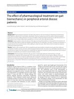
Figure 1 The tumor volume and the weight of SKOV-3
xenograft in the different treatment groups. (A) Either chA21 or
trastuzumab treatment cause a marked growth inhibition in SKOV-3
xenograft compared with the control (P < 0.01), and the
combination of chA21 with trastuzumab treatment induced a more
efficient efficacy than the each antibody alone (P < 0.05). (B) When
the experiment ended, all tumors were removed and weighted.
Results are representative of the mean ± SD of 8 animals in each
group. *, P < 0.01 compared with control. **, P < 0.01 compared
with chA21 or trastuzumab alone.
Zhang et al. Journal of Ovarian Research 2010, 3:20
/>Page 3 of 8
were 37% and 58%, respectively (P <0.01).Moreover,
the combination of chA21 and trastuzumab resulted in
an 81% inhibition in tumor weight compared with the
control (P < 0.001), which is greater than single treat-
ment (P < 0.01). In addition, complete tumor eradica-
tion was seen in one mice from the combination
treatment group.
Increased anti-angiogenesis efficacy by combination of
chA21 with trastuzumab
Angiogenesis pla ys an important role in cancer growth,
we then examined whether the chA21 plus trastuzumab
treatment leads to a more effect ive inhibition of
angiogenesis than either treatment alone. MVD values
were assessed by staining these with CD34 in tumor tis-
sues that were removed from SKOV-3 xenografts. The
most highly vascularized area of ea ch tumor was identi-
fied on five high-powered fields were counted in this
area of greatest vessel density. As shown in Fig. 2A and
2B, The number of MVD was 31% of the control in
chA21 p lus trastuzumab group, while this number was
56% in chA21 alone group and 54% in trastu zumab
alone group. So chA21 combined with trastuzumab
treatment resulted in a marked inhibition of tumor
MVD compared with the control (P < 0.001) and either
of chA21 or trastuzumab alone treatment (P < 0.01).
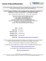
Figure 2 Tumor microvessel density (MVD) and VEGF expression. (A) The VEGF and CD34 (marker for MVD) expression in SKOV-3 xenografts
were detected by immunohistochemistry. (B) Tumor MVD and MOD of VEGF expression were calculated and the values were shown as percents
of the control treatment. *, P < 0.01 compared with control. **, P < 0.01 compared with chA21 or trastuzumab alone.
Zhang et al. Journal of Ovarian Research 2010, 3:20
/>Page 4 of 8
Similarly, the tumor mean optical density (MOD) values
of VEGF in t he chA21 plus trastuzumab treatment
group were 60% of the co ntrol, lower than those of 80%
and 77% in individual treatment groups of chA21 and
trastuzumab, respectively (P < 0.01).
Augmented down-regulation of VEGF expression induced
by combination of chA21 with trastuzumab
Previouly, we found HER2 antibody inhibits angiogen-
esis and downregulats VEGF expression. Hence, we
explored the effect of antibody synergy on VEGF secera-
tion by ELISA test. We examined the amount of VEGF
secreted into the medium f rom SKOV-3 cells that were
treated with chA21, trastuzumab, or chA21 plus trastu-
zumab for 12 h. Compared with 1255.6 ± 153.6 pg/ml
in the control group, the level of secreted VEGF
decreased 918.7 ± 109.8 pg/ml in chA21 group (P <
0.01), 839.1 ± 137.8 pg/ml in trastuzumab group (P <
0.01), and 583.5 ± 87.7 pg/ml in t he chA21 plus trastu-
zumab group (P < 0.001) (Fig. 3A).
We determined VEGF protein expression by Western
blot upon HER2 antibody treatment for 12 h in SKOV-3
cells. In cosistent with ELISA data, we found treatment
with chA21 or trastuzumab resulted in similar reduction
of VEGF expression compared with the control. How-
ever, the combination of chA21 and trastuzumab
induced a further inhibition of VEGF protein expression
(Fig. 3B).
Enhanced suppression of HUVECs proliferation and
migration by combination of chA21 with trastuzumab
To further investigate the influence on the function of
typical endothelial cells such as HUVECs, the effect of
supernatant from SKOV-3 cells treated with antibodies
on HUVECs prolifera tion was de termined by the MTS
assay. Compared with control group, HUVECs prolifera-
tion was inhibited by 33% in SKOV-3 supernatants trea-
ted with chA21 plus trastuzumab treatment group, 14%
in chA21 group (P < 0.05) and 1 6% trastuzumab group
(P < 0.05) (Fig. 4A).
HUVECs were co-cultured with SKOV-3 supernatants
for 48 h. Migrated cells were stain ed and counted. As
shown in Fig. 4B and 4C, the migrating capability of
HUVECs was inhibited by SKOV-3 cell supernatant
treated with chA21 p lus trastuzumab compared with
untreated cells (P < 0.001), chA21-treated cells ( P <
0.01) or trastu zumab-treated cells (P < 0.01). The num-
ber of migrated cells (40×10 magnification) were 73%,
70% or 48% in chA21, trastuzumab, or combined treat-
ment group compared with the control (Fig. 4C). Our
data demonstrated that chA21 or trastuzumab treatment
suppressed HUVECs migration and c ombination of
chA21 and trastuzumab induced a further suppression.
Potent inhibition of pAKT activity by combination of
chA21 with trastuzumab
To detect the underlying molecular mechanism of
angiogenesis, we investigated the effect of chA21 plus
trastuzumab treatment on AKT activity, which is a cru-
cial pathway in angiogenesis [19] . After SKOV-3 cells
were treated with antibodies for 12 h, the cell lysates
were analyzed by Western blotting assay. As shown in
Fig. 5A, pAKT (Ser
473
) expression were reduced by
ChA21 or t rastuzumab treatment, but more dramatic
reduction was observed in the ChA21 plus trastuzumab
treatment group. Total AKT protein were not altered
significantly upon various interventions. As shown in
Fig. 5B, The ratios of p-AKT/AKT were 0.61, 0.65 and
0.45 in chA21, trastuzumab and chA21 plus trastuzu-
mab group. These data indicate that the ChA21 plus
trastuzumab treatment cause greater inhibition of AKT
expression compared with either treatment alone.
Discussion
It’s well known that HER2-overexpressing tumors confer
enhanced metastasis -related properties and resistance to
Figure 3 Detection on secretion of VEGF from SKOV-3 cells.(A)
After co-culture of SKOV-3 cells with chA21 (5 μg/ml), trastuzumab
(5 μg/ml) or chA21(5 μg/ml) plus trastuzumab (5 μ g/ml) for 12 h,
secreted content of VEGF in the medium was detected by ELISA. *,
P < 0.01 compared with control. **, P < 0.01 compared with chA21
or trastuzumab alone. (B) VEGF protein expression in the SKOV-3
cells was detected by western blot.
Zhang et al. Journal of Ovarian Research 2010, 3:20
/>Page 5 of 8
chemotherapeutic reagents which frequently result in
poor clinical outcome. Trastuzumab as the first thera-
peutic anti-HER2 monoclonal antibody has been used in
clinical treatment of HER2-overexpressing metastatic
breast and gastric cancers [20,21 ]. It is also proposed to
be a treatment option for patients with HER2-positive
ovarian carcinoma [22]. However, poor responses and
dis ease recurrences for trastuzuma b therapy underscore
for alternative treatments [23].
It is believed that angiogenesis is required for tumor
growth and spread. HER2 signaling has been reported to
be implicated in tumor angiogenesis [24,25]. Previously,
we have developed a novel anti-HER2 chimeric antibody
chA21 and this new antibody mainly binds to a distinct
epitope on HER2 ECD I and inhibits tumor cells growth
in vitro and in vivo [10,16]. In the present study we
further e xplored the potential anti-angiogenic effects of
chA21 on HER 2-overexpressing ovarian carcinoma. Our
data showed that chA21 and trastuzumab can inhibit
tumor growth and angiogenesis in SKOV-3 xenograft
model and the combination of trastuzumab with chA21
results in an enhanced effect.
Indeed, endothelial cells migration and proliferation is
crucial f or angiogenesis. Tumor cells induce angiogen-
esis by secreting various growth fact ors, such as VEGF,
which binds its cognate receptor on endothelial cells
and promotes these cells to proliferate and migrate
[26,27]. Using ELISA kit to measuring the secreted
VEGF from SKOV-3 cells, we found that chA21 could
suppress VEGF expression and this synergism when
combined chA21 with trastuzumab. Moreover, the
synergism wa s confirmed by inhibition of HUVECs pro-
liferation and migration when the endothelial cells were
co-cultured with the supernatant from SKOV-3 cells
treated with both trastuzumab and chA21 together.
Therefore, the anti-angiogenesis capacity of the two
antibodies alone and their synergy proved to be
intrinsic.
Among signaling pathways induced by HER2 receptor,
activation of the AKT kinase orchestrates a number of
signaling pathways potentially involved in angiogenesis
[19]. O ur study revealed that chA2 1 could inhibit AKT
expression with the capability similar to trastuzumab in
SKOV-3 cell line. This result reflected the intrinsic
Figure 4 HUVECs proliferat ion and migration assay. (A) By the MTS assay, HUVECs number was measured by the MTS assay upon different
treatment for 72 h. (B) The HUVECs migration was measured by Transwell assay in the co-cultured system (×400). (C) Data was shown as the
mean of the number of migration HUVECs in five representative fields. *, P < 0.01 compared with control. **, P < 0.01 compared with chA21 or
trastuzumab alone.
Zhang et al. Journal of Ovarian Research 2010, 3:20
/>Page 6 of 8
properties of most tumor-inhibitory anti-HER2 antibo-
dies to inhibit receptor-induced downstream signals at
various efficiencies [28,29]. More import antly, the com-
bination of chA21 with trastuzumab showed more sig-
nificant potency on inhibiting the AKT. It may partly
explain our findings that the combination of chA21 with
trastuzumab could synergistically enhance the in vitro
and in vivo anti-angiogenesis effects.
In conclusion, our study demonstrated the inhibition
activities on tumor growth and angiogenesis of a novel
anti-HER2 antibody chA21 alone a nd in combination
with trastuzumab in vitro and in vivo.Wefoundthat
the angiogenesis inhibit ion effect of chA21 could be
enhanced by combination with trastuzumab, which
might be mediated by synergism of chA21 and trastuzu-
mab through inhibition of AKT expression. Therefore,
chA21 may represent a unique anti-HER2 antibody with
superior potentials as combination with other anti-
HER2 reagents for further therapy.
Acknowledgements
This work was granted by The Chinese Ministry of Science and Technology
(Nos. 2006AA02A245 and 2009ZX09102-223), National Natural Science
Foundation of China (No. 30873047) and Anhui Provincial Natural Science
Foundation of China (No. 090413125). We would like to thank Hefei National
Laboratory for Physical Sciences at Microscale, University of Science and
Technology of China for technical help.
Author details
1
Department of Pathology, Anhui Medical University, Meishan Road, Hefei,
China.
2
Institute of Clinical Pharmacology, Anhui Medical University, Meishan
Road, Hefei, China.
3
Affiliated Anhui Provincial Hospital, Anhui Medical
University, Meishan Road, Hefei, China.
4
Anhui Anke Biotechnology Co. Ltd,
Haiguan Road, Hefei, China.
5
School of Life Science, University of Science
and Technology of China, Huangshan Road, Hefei, China.
Authors’ contributions
AZ and GS designed and conducted the studies, carried out corresponding
data analyses, and drafted the manuscript. TZ participated in the animal
experiments. GZ, JL, LS, WW, BL, ZW and QW participated in study design,
coordination and helped to draft the manuscript. All authors have read and
approved this final manuscript.
Competing interests
The authors declare that they have no competing interests.
Received: 13 May 2010 Accepted: 19 August 2010
Published: 19 August 2010
References
1. Jemal A, Siegel R, Ward E, Hao Y, Xu J, Murray T, Thun MJ: Cancer statistics.
CA Cancer J Clin 2008, 58(2):71-96.
2. Breedlove G, Busenhart C: Screening and detection of ovarian cancer. J
Midwifery Womens Health 2005, 50(1):51-54.
3. Burstein HJ: The distinctive nature of HER2-positive breast cancers. N Engl
JMed2005, 353(16):1652-1654.
4. Slamon DJ, Godolphin W, Jones LA, Holt JA, Wong SG, Keith DE, Levin WJ,
Stuart SG, Udove J, Ullrich A: Studies of the HER-2/neu proto-oncogene in
human breast and ovarian cancer. Science 1989, 244(4905):707-712.
5. Verri E, Guglielmini P, Puntoni M, Perdelli L, Papadia A, Lorenzi P,
Rubagotti A, Ragni N, Boccardo F: HER2/neu oncoprotein overexpression
in epithelial ovarian cancer: evaluation of its prevalence and prognostic
significance. Oncology 2005, 68(2-3):154-161.
6. Agus DB, Gordon MS, Taylor C, Natale RB, Karlan B, Mendelson DS,
Press MF, Allison DE, Sliwkowski MX, Lieberman G: Phase I clinical study of
pertuzumab, a novel HER dimerization inhibitor, in patients with
advanced cancer. J Clin Oncol 2005, 23(11):2534-2543.
7. Wilken JA, Webster KT, Maihle NJ: Trastuzumab Sensitizes Ovarian Cancer
Cells to EGFR-targeted Therapeutics. J Ovarian Res 2010, 3:7.
8. Cheng LS, Liu AP, Yang JH, Dong YQ, Li LW, Wang J, Wang CC, Liu J:
Construction, expression and characterization of the engineered
antibody against tumor surface antigen, p185(c-erbB-2). Cell Res 2003,
13(1):35-48.
9. Hu S, Li L, Qiao J, Guo Y, Cheng L, Liu J: Codon optimization, expression,
and characterization of an internalizing anti-ErbB2 single-chain antibody
in Pichia pastoris. Protein Expr Purif 2006, 47(1):249-257.
10. Hu S, Zhu Z, Li L, Chang L, Li W, Cheng L, Teng M, Liu J: Epitope mapping
and structural analysis of an anti-ErbB2 antibody A21: Molecular basis
for tumor inhibitory mechanism. Proteins 2008, 70(3):938-949.
11. Liu Y, Zhou H, Zhu J, Gao Y, Niu L, Liu J, Teng M: Crystallization and
preliminary crystallographic studies of the single-chain variable
fragment of antibody chA21 in complex with an N-terminal fragment of
Figure 5 Protein expression of AKT and phopsho -AKT (pAKT)
in xenograft tumors. (A) After SKOV-3 cells were treated with
antibodies for 12 h, the cell lysates were analyzed. pAKT (Ser
473
)
expression educed upon treament withchA21 or trastuzumab
treatment and more dramatic reduction was observed with chA21
plus trastuzumab treatment. The levels of total AKT protein were
not altered significantly upon various interventions. (B) The ratios of
p-AKT/AKT were calculated and the values were shown as ratios
compared with control, which was arbitrarily taken as 1.0. *, P <
0.01 compared with control. **, P < 0.01 compared with chA21 or
trastuzumab alone.
Zhang et al. Journal of Ovarian Research 2010, 3:20
/>Page 7 of 8
ErbB2. Acta Crystallogr Sect F Struct Biol Cryst Commun 2009, 65(Pt
7):692-694.
12. Koukourakis MI, Manolas C, Minopoulos G, Giatromanolaki A, Sivridis E:
Angiogenesis relates to estrogen receptor negativity, c-erbB-2
overexpression and early relapse in node-negative ductal carcinoma of
the breast. Int J Surg Pathol 2003, 11(1):29-34.
13. Russell KS, Stern DF, Polverini PJ, Bender JR: Neuregulin activation of ErbB
receptors in vascular endothelium leads to angiogenesis. Am J Physiol
1999, 277(6 Pt 2):H2205-2211.
14. Petit AM, Rak J, Hung MC, Rockwell P, Goldstein N, Fendly B, Kerbel RS:
Neutralizing antibodies against epidermal growth factor and ErbB-2/neu
receptor tyrosine kinases down-regulate vascular endothelial growth
factor production by tumor cells in vitro and in vivo: angiogenic
implications for signal transduction therapy of solid tumors. Am J Pathol
1997, 151(6):1523-1530.
15. Chung BH, Kim CK: Icariin stimulates angiogenesis by activating the MEK/
ERK-and PI3K/AKT/eNOS-dependent signal pathway in human
endothelial cells. Biochem Biophys Res Commun 2008, 376:404-408.
16. Zhang AL, Xue H, Ling XG, Gao Y, Yang F, Cheng LS, Liu J, Wu Q: Anti-HER-
2 engineering antibody ChA21 inhibits growth and induces apoptosis of
SKOV-3 cells. Journal of Experimental & Clinical Cancer Research 2010, 29:23.
17. Liu S, Luo X, Li D, Zhang J, Qiu D, Liu W, She L, Yang Z: Tumor inhibition
and improved immunity in mice treated with flavone from Cirsium
japonicum DC. Int Immunopharmacol 2006, 6(9):1387-1393.
18. Vermeulen PB, Gasparini G, Fox SB, Toi M, Martin L, McCulloch P, Pezzella F,
Viale G, Weidner N, Harris AL: Quantification of angiogenesis in solid
human tumours: an international consensus on the methodology and
criteria of evaluation. Eur J Cancer 1996, 32 A(14):2474-2484.
19. Dimmeler S, Zeiher AM: Akt takes center stage in angiogenesis signaling.
Circulation Research 2000, 86(1):4-5.
20. Slamon DJ, Leyland-Jones B, Shak S, Fuchs H, Paton V, Bajamonde A,
Fleming T, Eiermann W, Wolter J, Pegram M: Use of chemotherapy plus a
monoclonal antibody against HER2 for metastatic breast cancer that
overexpresses HER2. New England Journal of Medicine 2001,
344(11):783-792.
21. Roukos DH: Targeting gastric cancer with trastuzumab: new clinical
practice and innovative developments to overcome resistance. Ann Surg
Oncol 2010, 17(1):14-17.
22. McAlpine JN, Wiegand KC, Vang R, Ronnett BM, Adamiak A, Kobel M,
Kalloger SE, Swenerton KD, Huntsman DG, Gilks CB: HER2 overexpression
and amplification is present in a subset of ovarian mucinous carcinomas
and can be targeted with trastuzumab therapy. Bmc Cancer 2009, 9:433.
23. Krop IE, Winer EP: Ten years of HER2-directed therapy: still questions
after all these years. Breast Cancer Res Treat 2009, 113(2):207-209.
24. Blackwell KL, Dewhirst MW, Liotcheva V, Snyder S, Broadwater G, Bentley R,
Lal A, Riggins G, Anderson S, Vredenburgh J: HER-2 gene amplification
correlates with higher levels of angiogenesis and lower levels of
hypoxia in primary breast tumors. Clin Cancer Res 2004,
10(12 Pt
1):4083-4088.
25. Schoppmann SF, Tamandl D, Roberts L, Jomrich G, Schoppmann A,
Zwrtek R, Dubsky P, Gnant M, Jakesz R, Birner P: HER2/neu expression
correlates with vascular endothelial growth factor-C and
lymphangiogenesis in lymph node-positive breast cancer. Ann Oncol
2009, 21(5):955-960.
26. Laakkonen P, Waltari M, Holopainen T, Takahashi T, Pytowski B, Steiner P,
Hicklin D, Persaud K, Tonra JR, Witte L: Vascular endothelial growth factor
receptor 3 is involved in tumor angiogenesis and growth. Cancer Res
2007, 67(2):593-599.
27. Lamszus K, Brockmann MA, Eckerich C, Bohlen P, May C, Mangold U,
Fillbrandt R, Westphal M: Inhibition of glioblastoma angiogenesis and
invasion by combined treatments directed against vascular endothelial
growth factor receptor-2, epidermal growth factor receptor, and
vascular endothelial-cadherin. Clin Cancer Res 2005, 11(13):4934-4940.
28. Ben-Kasus T, Schechter B, Lavi S, Yarden Y, Sela M: Persistent elimination
of ErbB-2/HER2-overexpressing tumors using combinations of
monoclonal antibodies: relevance of receptor endocytosis. Proc Natl Acad
Sci USA 2009, 106(9):3294-3299.
29. Friedman LM, Rinon A, Schechter B, Lyass L, Lavi S, Bacus SS, Sela M,
Yarden Y: Synergistic down-regulation of receptor tyrosine kinases by
combinations of mAbs: implications for cancer immunotherapy. Proc Natl
Acad Sci USA 2005, 102(6):1915-1920.
doi:10.1186/1757-2215-3-20
Cite this article as: Zhang et al.: Augmented inhibition of angiogenesis
by combination of HER2 antibody chA21 and trastuzumab in human
ovarian carcinoma xenograft. Journal of Ovarian Research 2010 3:20.
Submit your next manuscript to BioMed Central
and take full advantage of:
• Convenient online submission
• Thorough peer review
• No space constraints or color figure charges
• Immediate publication on acceptance
• Inclusion in PubMed, CAS, Scopus and Google Scholar
• Research which is freely available for redistribution
Submit your manuscript at
www.biomedcentral.com/submit
Zhang et al. Journal of Ovarian Research 2010, 3:20
/>Page 8 of 8