báo cáo hóa học:" Nuclear survivin expression is a positive prognostic factor in taxane-platinum-treated ovarian cancer patients" pdf
Bạn đang xem bản rút gọn của tài liệu. Xem và tải ngay bản đầy đủ của tài liệu tại đây (1.23 MB, 9 trang )
RESEARCH Open Access
Nuclear survivin expression is a positive
prognostic factor in taxane-platinum-treated
ovarian cancer patients
Anna Felisiak-Golabek
1
, Alina Rembiszewska
1
, Iwona K Rzepecka
1
, Lukasz Szafron
1
, Radoslaw Madry
2
,
Magdalena Murawska
3
, Tomasz Napiorkowski
4
, Piotr Sobiczewski
5
, Beata Osuch
6
and Jolanta Kupryjanczyk
1*
, for
the Polish Ovarian Cancer Study Group (POCSG)
Abstract
Background: Survivin is an inhibitor of apoptosis and a regulator of mitotic progression. TP53 protein is a negative
transcriptional regulator of survivin. The aim of our study was to evaluate the clinical significance of survivin
expression in advanced stages ovarian cancer with respect to the TP53 status.
Methods: Survivin and TP53 expression was evaluated immunohistochemically in 435 archival samples of ovarian
carcinomas (244 patients were treated with platinum/cyclophosphamide-PC/PAC; 191-with taxane-platinum (TP)
agents). Univariate and multivariate statistical analyses were performed in patients groups divided according to the
administered chemotherapeutic regimen, and in subgroups with and without TP53 accumulation (TP53+ and TP53-
, respe ctively).
Results: Nuclear and cytoplasmic survivin expression was observed in 92% and 74% of the carcinomas,
respectively. In patients treated with TP, high nuclear survivin expression decreased the risk of disease recurrence
and death, and increased the probability of high platinum sensitivity (p < 0.01), but only in the TP53(+) group, and
not in the TP53(-) group.
Conclusions: It appears that TP53 status determines the clinical importance of nuclear survivin expression in
taxane-platinum treated ovarian cancer patients.
Keywords: ovarian cancer, survivin, taxol, TP53
Background
Recently molecular anticancer therapie s have undergone
rapid development. Survivin, the smallest member of the
family of t he protein inhibi tors of apoptosis (IAP) [1], is
considered to be a potential target for molecular therapy
[2]. Target survivin arose from data obtained fro m can-
cer cell lines, showing that survivin inhibition contri-
butes to increased tumour response to various
anticancer agents [3]. The results of clinical analyses are
less consistent, as high survivin expression had been
associated with both favourable and unfavourable p rog-
nosis [4].
Ovarian cancer is the most lethal gynaecological
malignancy. In the last decade, taxanes combined with
cisplatin or its analogues (TP therapy) have been consid-
ered standard first-line treatment for ovarian cancer
[5,6]. Although the introduction of taxanes has signifi-
cantly improved treatment results, still 20% to 30% of
the patients fail to achieve complete remission [6-8].
Taxanes int eract with b-tubulin and increase its poly-
merisation and stabilisation. In the presence of pacli-
taxel, cells form dysfunctional mitotic spindles and
eventually die by apoptosis or necrosis (de pending on
drug concentration) [9,10]. The mechanism of action of
taxanes is linked to survivin, which is a member of the
chromosomal passenger complex (CPC) [11,12]. The
CPC complex controls many aspects of mitosis, includ-
ing regulation of the mitotic spindle checkpoint and
* Correspondence:
1
Department of Molecular Pathology, The Maria Sklodowska-Curie Memorial
Cancer Centre and Institute of Oncology, Warsaw, Poland
Full list of author information is available at the end of the article
Felisiak-Golabek et al. Journal of Ovarian Research 2011, 4:20
/>© 2011 Felisiak-Golabek et al; licensee BioMed Cent ral Ltd. This is an Open Access article distri buted u nder the terms of the Creative
Commons Attribution License ( which permits unrestricted use, distribution, and
reproduction in any mediu m, provided the original work is properly cited.
mitotic progression [13]. It has been recently shown
that, on treatment with taxol, survivin is invol ved in the
spindle check point activation and mitotic arrest [14,15].
Sur vivin, expressed duri ng foetal development [16], and
undetectable in most adult tissues [17] has been found
in many types of human cancers, including ovarian can-
cer. The clinical role of survivin in ovarian cancer
patients is not clear [18-20].
TP53 dysfunction enhances ovarian cancer response to
taxane-platinum treatment [8,21,22]. In add ition, the
results recently obtained by our group suggest that the
TP5 3 status, as determined by TP53 accumulation, may
influence the clinical importance of other molecular fac-
tors [23-25]. This may also be the case with survivin,
which is down-regulated by the wild-type TP53 [26,27].
Thus, t here may be a synergistic clinical effect of TP53
dysfunction and high survivin expression in taxane/plati-
num-treated ovarian cancer patients.
We studied large groups of ovarian cancer patients in
order to evaluate the clinical importance of survivin
expression with respect to the TP53 status, and to the
treatment regimen applied.
Materials and methods
Patients and tumours
The study was performed on 435 archival samples of
ovarian carcinomas. Medical records were critically
reviewed by at least two clinicians. The patients were
treated with s tandard PC (cisplatin -cyclophosphamide
or carboplatin-cyclophosphamide) or PAC chemother-
apy (PC and doxorubicin) (244 patients), o r with tax-
ane-platinum chemotherapy (TP: paclitaxel or doceta xel
with cisplatin or carboplatin) (191 patients). The mate-
rial was carefully selected out of a total of 899 cases
submitted to meet the following criteria: no chemother-
apy before staging laparotomy, adequate staging proce-
dure, International Federation of Gynaecologists and
Obstetricians (FIGO) stage IIB to IV disease [28],
tumour tissue from the first laparotomy available, mod-
erate (G2) or poor tumour differentiation (G3 and G4),
availability of clinical data, incl. residual tumour size
and follow-up.
All tumours were uniformly reviewed histopathologi-
cally, classified according to the criteria of the World
Health Organisation [29] and graded in a four-grade
scale, according to the criteria given by Broders [30].
Clinicopathological characteristics have been presented
in Table 1.
For the PC/PAC-treated group, the follow-up time
ranged from 4.4 to 198.3 m onths (median: 27.5); the
respective values for the TP-treated group were: 4.8 to
100.6 months (median: 32.2). Short follow-up time
resulted from early patients death. All surviving patients
had at least a 6-month follow-up. Response to
chemotherapy was evaluated retrospectively according
to the World Health Organisation response evaluation
criteria [31]. The evaluation was based on data from
medical records describin g patient’s clinical condition
and CA125 levels in 3-4 week intervals. Complete
remission (CR) was defined as disappearance of all clin i-
cal and biochemical symptoms of ovarian cancer evalu-
ated after completion of first-line chemotherapy and
confirmed at four weeks. Within the CR group, we iden-
tified a platinum-sensitive group (PS, disease-free survi-
val longer tha n six months) and a highl y platinum-
sensitive group (HPS, disease-free survival longer than
24 months). Other tumours were described as platinum
resistant [32].
The study was approved by the bioethics committee of
the Institute of Oncology (ref.no. 39/2007).
Immunohistochemical analysis
Immunohistochemical stainings were performed on par-
affin-embedded material after heat-induced epitope
retrie val (HIER). We us ed a ra bbit polyclonal anti-survi-
vin antibody (1/1000, Novus Biologicals, Littleton, USA).
TP53 protein was detected with the use of PAb1801
monoclonal antibody (1/3000, Sigma-Genosys, Cam-
bridge, U K), as described previously [23]. The antigens
were retrieved by heating the sections in 0.01 M citrate
buffer (pH 6.0): 6 × 5 min. for survivin and 2 × 5 min.
for TP53, at 700 W in a microwave oven. Non-specific
tissue and endogenous peroxidase reactivities were
blocked w ith 10% BSA and 3% H
2
O
2
, respectively. The
sections were incubated with primary antibodies over-
night, at 4°C. Biotinylated secondary goat anti-rabbit
IgG (for survivin) (1/1 500) and anti-mouse IgG (for
TP53) (1/1500), peroxidase-conjugated streptavidin (1/
500) (all from Immunotech, Marseille, France), and
DAB were used as a detection system. As a positive con-
trol for TP53 accumulation, we used a tumour with a
defined TP53 gene missense mutation [33]. Normal rab-
bitIgGornormalmouseIgGofthesamesubclasses
and at the concentrations of the relevant primary anti-
bodies served as negative controls.
The evaluation of immunohistochemical stainings
Survivin expression was scored independently for
nuclear and cytoplasmic staining. Light microscopic eva-
luation at 400× magnification was used to count at least
200 tumour cells within the areas of the strongest stain-
ing. Each nucleus in a given field was categorised
according to the staining intensity (0 or weak to st rong:
1 to 3), and counted. The nuclear survivin expression
was further described as an ID score. It was calculated
according to the following formula: ID = [(N0 × 0) +
(N1 × 1) + (N2 × 2) + (N3 × 3)]/100, where N0, N1, N2
and N3 stands for the percentage of cells in each
Felisiak-Golabek et al. Journal of Ovarian Research 2011, 4:20
/>Page 2 of 9
Table 1 Patients characteristics
ALL PATIENTS TP-TREATED GROUP PC/PAC-TREATED GROUP
N = 435 N = 191 N = 244
Age (years)
Range 20-78 20-78 24-77
Mean 54.3 54.9 53.9
FIGO stage
IIB, IIC 27 (6%) 10 (5%) 17 (7%)
IIIA, IIIB 82 (19%) 26 (14%) 56 (23%)
IIIC 277 (64%) 136 (71%) 141 (58%)
IV 49 (11%) 19 (10%) 30 (12%)
Residual tumour size
0 87 (20%) 35 (18%) 52 (21%)
≤ 2 cm 141 (32%) 77 (40%) 64 (26%)
> 2 cm 207 (48%) 79 (42%) 128 (53%)
Histological type
serous 334 (77%) 142 (74%) 192 (79%)
endometrioid 22 (5%) 8 (4%) 14 (6%)
clear cell 15 (3%) 4 (2%) 11 (4%)
undifferentiated 33 (8%) 20 (11%) 13 (5%)
other types 31 (7%) 17 (9%) 14 (6%)
Histological grade
G 2 54 (13%) 24 (12%) 30 (12%)
G 3 263 (60%) 110 (58%) 153 (63%)
G 4 118 (27%) 57 (30%) 61 (25%)
Response to chemotherapy
complete remission 257 (59%) 124 (65%) 133 (55%)
partial remission/no change 112 (26%) 62 (32%) 50 (20%)
progression 66 (15%) 5 (3%) 61 (25%)
Platinum sensitive 129 (30%) 65 (34%) 64 (26%)
Highly platinum sensitive 83 (19%) 40 (21%) 43 (18%)
Platinum resistant 223 (51%) 86 (45%) 137 (56%)
Number of patients at risk (OS)
1 year 389 (89%) 180 (94%) 209 (86%)
2 years 276 (63%) 141 (74%) 135 (57%)
3 years 172 (39%) 83 (43%) 89 (36%)
4 years 116 (27%) 53 (28%) 63 (26%)
5 years 76 (17%) 31 (16%) 45 (18%)
Number of patients at risk (DFS)
1 years 143 (56%) 70 (56%) 73 (55%)
2 years 83 (32%) 40 (32%) 43 (32%)
3 years 61 (24%) 27 (22%) 34 (26%)
4 years 43 (17%) 17 (14%) 26 (19%)
5 years 28 (11%) 10 (8%) 18 (13%)
Follow-up time
Range (month) 4.4-198.3 4.8-100.6 4.4-198.3
mean 30 32.2 27.5
Outcome
NED 45 (10%) 27 (14%) 18 (7%)
AWD 45 (10%) 35 (18%) 10 (4%)
DOD 335 (77%) 124 (65%) 211 (87%)
DOC 10 (3%) 5 (3%) 5 (2%)
PC- cyclophosphamide and cisplatin, PAC-PC plus doxorubicin, TP-taxane-platinum therapy; NED-no evidence of disease, AWD-alive with disease, DOD-died of
disease, DOC-died of other causes; OS-overall survival, DFS-disease free survival
Felisiak-Golabek et al. Journal of Ovarian Research 2011, 4:20
/>Page 3 of 9
intensity category (0, 1, 2 or 3) [34]. A cytoplasmic
staining was scored 0 (absent) or 1 to 3 (weak to
strong). For the cytop lasmic expression, t umours with
scores 0 and 1 were described as low expression, and
those scoring 2 or 3, as high expression. Two indepen-
dent assessors (A.R. and A.F-G.) concurred in 80% of
the cases, and reached consensus in the remaining
cases. TP53 protein accumulation was assessed as pre-
sent (more than 10% positive cells) or absent, as pre-
viously described [8].
Statistical analysis
The associations between survivin expression and
clinicopathological variables were assessed using the
chi-square test. Probability of survival and disease-
free survival (DFS) were estimated using the Kaplan-
Meier method and a log-rank test for censored survi-
val data. Overall and disease-free survival time ana-
lyses were performed using multivariate Cox’ s
proportional hazard models. Tumour response to che-
motherapy (probability of CR, probability of PS or
HPS) was evaluated using a multivariate logistic
regression model.
Statistical analyses included the fo llowing independent
variables: age of patients (median: 53 years), the FIGO
stage, histopathologica l type and grade, residual tumour
size and TP53 accumulation status. The survivin ID
score was analysed as a continuous variable, and alterna-
tively, as a categorical variable (cut-off point at median-
1.5). Important factors were selected using a backward
selection technique, where factors not significa nt at 0.1
were removed stepwise from the model.
The analyses were performed in the two groups of
patients treated with different chemotherapeutic regi-
mens, and additionally in the TP53(-) and TP53(+) sub-
groups. All tests were two-sided. P < 0.05 was
considered significant. All calculations were performed
using STATA 5 software.
Results
Cellular distribution of survivin expression
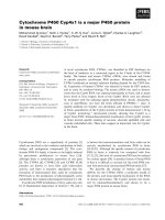
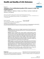
Survivin expression was observed both in the nuclei
and the cytoplasm of ovarian cancer cells, predomi-
nantly in the former (Figure 1). Nuclear and cytoplas-
mic survivin expression of any intensity was observed
in 92% and 74% of the tumours, respectively. Distribu-
tion of the nuclear and cytoplasmic survivin expression
in all patients and in the subgroups treated with either
chemotherapeutic regimen has been presented in
Table 2.
There were no associations between the cytoplasmic
and nuclear survivin expression, nor between survivin
Figure 1 Various patterns o f survivin expression in four different ovarian carcinomas (400×, hematoxylin counterstain): a) negative
survivin expression (clear cell carcinoma; FIGO IIIC), b) survivin expression absent in the nucleus but present in the cytoplasm (serous
carcinoma; FIGO IIIB), c) survivin expression present in the nucleus only (serous carcinoma; FIGO IV), d) survivin expression present in
the nucleus and cytoplasm (serous carcinoma; FIGO IV).
Felisiak-Golabek et al. Journal of Ovarian Research 2011, 4:20
/>Page 4 of 9
expression and the clinic opathological variables studied
nor TP53 accumulation.
Survivin expression in the taxane/platinum-treated group
We analysed both cytoplasmic and nucle ar survivin
expression; only t he nuclear expression related to the
clinical endpoints. This was observed in the TP53(+)
subgro up an d, to a lesser degree, in the group compris-
ing all patients, but not in the TP53(-) subgroup.
Analysis of survivin expression as a continuous variable
In the univariate analysis, high nuclear survivin expres-
sion positively influenced disease-free survival (HR 0.48,
95% CI 0.30-0.76, p = 0.002 for the TP53(+) group; HR
0.66, 95% CI 0.48-0.91, p = 0.013 for the entire group),
overall survival (HR 0.63, 95% CI 0.43-0.91, p = 0 .016
for the TP53(+) group only) and high platinum sensitiv-
ity (OR 4.25, 95% CI 1.57-11.51, p = 0.004 for the TP53
(+) group; OR 2.40, 95% CI 1.27-4.53, p = 0.007 for the
entire group). T his was confirmed by the multivariate
analyses (Table 3), i n which the ass ociations between
survivin expression and the clinical endpoints wer e
stronger and more significant in the TP53(+) group
than in the entire group. In the TP53(+) group, high
nuclear survivin expression apparently correlated with a
lesser risk of recurrence (HR 0.44, p = 0.000) and deat h
(HR 0.64, p = 0.010), and enhanced the odds of high
platinum sensitivity (OR 5.04, p = 0.010) (Table 3).
In the TP53(+) group, the clinical importance of the
survivin expression appeared stronger and more statisti-
cally significant than that of some clinicopathological
factors (Table 3).
The residual tumour size was the clinicopathological
factor most constantly associated with the clinical end-
points (Table 3). Complete remission and platinum sen-
sitivity did not associate with nuclear survivin
expression.
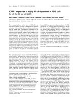
Analysis of survivin expression as a categorical variable
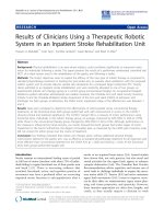
In both univariate and multivariate analyses of the med-
ian survivin score (ID ≥ 1.5 vs < 1.5), the only associa-
tion of survivin expression we observed was that with
disease-free survival (univ ariate analysis: HR 0.57, 95%
CI 0.33-0.98, p = 0.043 for the TP53(+) group [Figure
2]; multivariat e analysis: HR 0.44, 95% CI 0.25-0.79, p =
0.005 for the TP53(+) group; HR 0.63, 95% CI 0.42-0.95,
p = 0.029 for the entire group).
Survivin expression in the platinum/cyclophosphamide-
treated group
Neither nuclear nor cytoplasmic survivin expression has
been found to correlate with clinical endpoints or clini-
copathological factors in the PC/PAC patient group.
Discussion
Survivin is regarded as a potential target of molecular ther-
apy due to its strong antiapoptot ic activity. Nevertheless,
the results of numerous studies on the clinical importance
of survivin in cancer patients are inconsistent. We present
the first multivariate analysis which shows the positive
prognostic significance of survivin expression in ovarian
cancer patients. This was clearly demonstrated in patients
treated with taxane-platinum agents, but not in those trea-
ted with platinum-cyclophosphamide regimens. In addi-
tion, the highest clinical significance of survivin was
observed in patients with TP53 dysfunctional tumours.
A nu mber of authors have reported, that the expres-
sion of survivin in cancer cell nuclei was associated with
poor survival [35-37], while only a few studies have
reported a reverse correlation [38-41]. Two studies on
breast and lung cancer, have also shown the influence of
nuclear survivin expression on DFS, similar to that
obs erved in our analysis [39,41]. In case of ovarian can-
cer,theissueofsurvivin expression and tumour
response to chemotherapy has been addressed by two
groups [18,42]. One has not observed any correlation
between nuclear or cytoplasmic survivin expression and
the response to platinum/cyclophosphamide, but con-
trary to ou r results, they have not found any correlation
with the response to TP t herapy, either [18]. The other
group has reported significantly higher rates of complete
remission after tax ol-based therapy in patients with low
survivin-expressing tumours. H owever the latter group
studied cytoplasmic survivin expression only [42].
Table 2 TP53 and survivin expression in ovarian carcinomas
ALL PATIENTS TP-TREATED GROUP PC/PAC-TREATED GROUP
N = 435 N = 191 N = 244
TP53-positive carcinomas 255 (59%) 110 (58%) 145 (59%)
TP53-negative carcinomas 180 (41%) 81 (42%) 99 (41%)
Cytoplasmic survivin expression:
Low (0 + 1 scores) 363 (83%) 149 (78%) 214 (82%)
High (2 + 3 scores) 72 (17%) 42 (22%) 30 (18%)
Nuclear survivin expression:
ID score < 1.5 268 (62%) 97 (51%) 171 (70%)
ID score ≥ 1.5 167 (38%) 94 (49%) 73 (30%)
Felisiak-Golabek et al. Journal of Ovarian Research 2011, 4:20
/>Page 5 of 9
Survivinmayplayadoubleroledependingonits
cellular localisation. In the cytoplasm, it exerts an
antiapoptotic function by caspase inhibition. There is
evidence to prove that nuclear survivin is a cell-prolif-
eration promoting factor [43]. Studies conducted on
different cell lines have shown, that survivin inhibition
causes defects in cell division and suppresses prolif-
eration. Some clinicopathological studies have identi-
fied positive correlations between nuclear survivin
expression and various parameters of growth fraction
(MIB-1, PCNA and mitotic indices) in hepatocellular
carcinoma [reviewed in 4]. These findings point to
survivin expression as an unfavourable prognostic fac-
tor. However, the majority of research groups evaluat-
ing the clinical importance of survivin expression have
failed to consider the specific anti-tumour therapy
applied. It should be stressed that in this study the
positive prognostic significance of survivin expression
was found only in patients treated with taxane-plati-
num agents. This relationship may be explained by a
functional link between survivin and taxanes during
mitosis [14,15].
Table 3 Associations of nuclear survivin expression (continuous variable) with clinical endpoints in the taxane-
platinum-treated group* (multivariate Cox’s proportional hazard and logistic regression models).
All patients TP53 (+) group
N = 199 N = 110
OR/HR [95%CI] p-value OR/HR [95%CI] p-value
High platinum-sensitivity
1
Survivin expression 2.09 [1.04,4.17] 0.036 5.04 [1.47,17.18] 0.010
Residual tumour size
0 1.0 1.0
≤ 2 cm 0.17 [0.05,0.54] 0.003 -
> 2 cm 0.09 [0.03,0.31] 0.000 0.21 [0.06,0.73] 0.014
Histological Grade
Grade 2 - 1.0
Grade 3 - 0.06 [0.00,0.74] 0.028
Grade 4 - -
Disease-free survival
Survivin expression 0.67 [0.48,0.91] 0.013 0.44 [0.27,0.69] 0.000
Residual tumour size
0 1.0 1.0
≤ 2 cm 1.66 [0.99,2.78] 0.052 2.33 [1.13,5.26] 0.023
> 2 cm 1.88 [1.09,2.78] 0.022 3.09 [1.40,6.83] 0.005
Overall survival
Survivin expression - 0.64 [0.45,0.89] 0.010
Age (year)
< 53 - 1.0
≥ 53 - 1.68 [1.08,2.62] 0.020
FIGO stage
II B, IIC - -
III A, IIIB - -
III C - 1.0
IV - 2.76 [1.44,5.27] 0.002
Residual tumour size
0 1.0 1.0
≤ 2 cm 1.23 [1.22,4.07] 0.009 2.23 [1.06,4.67] 0.033
> 2 cm 3.41 [1.88,6.18] 0.000 2.99 [1.45,6.17] 0.003
Histological Grade
Grade 2 1.0 1.0
Grade 3 2.92 [1.45,5.86] 0.003 3.53 [1.45,8.61] 0.005
Grade 4 2.98 [1.44,6.15] 0.003 2.60 [1.01,6.69] 0.047
1
HPS means the complete remission with DFS longer than 24 months.
* There were no associations with survivin ID score in the TP53(-) group.
Felisiak-Golabek et al. Journal of Ovarian Research 2011, 4:20
/>Page 6 of 9
Many studies regarding cell lines have revealed the
influence of survivin expression on cancer sensitivity to
taxanes. The studies that describe and/or explain the
role of survivin in the mitotic checkpoint regulation are
of particular interest. The data obtained from HeLa cells
has shown that survivin is required for the maintenance
of the spindle assembly checkpoint arrest in the pre-
sence of taxol, and this mechanism has been shown to
be essential for taxol sensitivity [14,44]. In the presence
of taxol survivin-depleted cells were unab le to maintain
the BubR1 protein at the kinetochores (BubR1 delays
the transition to anaphase until all chromosomes are
properly aligned). Exogenous expression of the wild-type
survivin was able to restore the mitotic arrest-response
of taxol-resistant cells [15]. Nevertheless, some of the
studies analysing the biological effect of survivin sup-
pression, or its overexpression in cell lines, have shown
that survivin inhibited taxol-induced apoptosis [45,46].
Some authors have observed, that survivin overexpres-
sion (apparently the cytoplasmic survivin phosphorylated
at Thr34) significantly decreased the sensitivity of
human ovarian carcinoma cell lines to taxanes [42].
In our study patients with high survivin expression
were at a lower risk of recurrence and death, but only
in the TP53-positive group. As we have previously
shown, the TP53 s tatus may determine the clinical sig-
nificance of the expression of other proteins, particularly
of those regulated by, or interfering with TP53 in the
control of tumour cell proliferation or apoptosis
[23-25,47,48].
On the other hand, TP53 dysfunction positiv ely influ-
ences cancer sensitivity to taxane-platinum therapy
[8,22,49-51]. An increase in G2/M arrest or a loss of the
TP53-dependent post-mitotic spindle checkp oint in the
TP53 dysfunctional cells have been proposed as possible
exp lanations of this phenomenon [52,53]. Thus, in view
of the literature reports, the effects of TP53-dysfunction
and high survivin expression may possibly show syner-
gism, enhancing the response of cancer cells to taxol.
The positive prognostic importance of survivin appears
as a paradox. However, this m ay result from the survi-
vin-dependent control of the mitotic response to taxol,
rather than from antiapoptotic and proliferation-stimu-
lating survivin activity.
TP53mayplayapartinnegativesurvivinregulation.
Studies on cancer cell lines have shown that the wild-
type TP53 repressed survivin at both mRNA and protein
levels, by binding to its promoter [26,27]. Several
authors have reported an association between dysfunc-
tional TP53 status and high survivin expression. This
has been shown in ovarian (high nuclear survivin in the
TP53 mutant tumours) , pancreatic, breast and gastric
carcinomas [54-57]. We have failed to observe this cor-
relation in our large group of 435 tumours; however, we
evaluated TP53 accumulation only and it occurs with a
frequency approximately 30% lower than TP53 muta-
tions, thus the rate of TP53 dysfunctional tumours in
our group may be much higher [33].
Conclusions
In conclusion, our results show t hat the nuclear survi-
vin expression status, in combination with the TP53
status, may be of prognostic value in ovarian cancer
patients treated with taxane-platinum agents. The pre-
sent study confirms our previous observations that
analyses of carcinomas with and without TP53 accu-
mulation, may play a pivotal role in the identification
of cancer biomarkers.
List of Abbreviations used
DFS: disease-free survival; HPS: high platinum sensitivity ; ID: ID score; OS:
overall survival; PC/PAC: platinum/cyclophosphamide chemotherapy; PS:
platinum sensitivity; TP: taxane-platinum chemotherapy
Acknowledgements
We would like to thank other members of the POCSG: J. Markowska (Chair
of Gynaecologic Oncology, Medical University, Poznan, Poland), J. Debniak, J.
Emerich (Department of Gynaecologic Oncology, Medical University, Gdansk,
Poland); M. Jedryka, M. Goluda (Department of Gynaecologic Oncology,
Medical University, Wroclaw, Poland); M. Ulanska, A. Pluzanska (Department
of Gynaecologic Oncology, Medical University, Lodz, Poland); M. Klimek, K.
Urbanski (Department of Gynaecologic Oncology, Institute of Oncology,
Krakow, Poland); A. Chudecka-Glaz, I. Rzepka-Gorska (Department of
Gynaecologic Oncology, Medical University, Szczecin, Poland); I. Ziolkowska-
Seta, M. Bidzinski (Department of Gynaecologic Oncology, The Maria
Sklodowska-Curie Memorial Cancer Centre and Institute of Oncology,
Warsaw, Poland); A. Timorek, B. Spiewankiewicz (Chair and Depart ment of
Obstetrics, Gynaecology and Oncology, IInd Faculty of Medicine, Warsaw
Medical University and Brodnowski Hospital, Warsaw, Poland). We also thank
Dr Magdalena Chechlinska and Dr Malgorzata Symonides for linguistic
correction.
This study was supported by the grant no. 2 PO5A 068 27 and N N401
236134 of the Polish Ministry of Science and Higher Education.
Figure 2 Prognostic significance of nuclear survivin expression
(Kaplan-Meier curve). Patients with high nuclear survivin
expression (ID ≥ 1.5) had a significantly better disease-free survival
than patients with low nuclear survivin expression (ID < 1.5).
Felisiak-Golabek et al. Journal of Ovarian Research 2011, 4:20
/>Page 7 of 9
Author details
1
Department of Molecular Pathology, The Maria Sklodowska-Curie Memorial
Cancer Centre and Institute of Oncology, Warsaw, Poland.
2
Chair of
Gynaecologic Oncology, Medical University, Poznan, Poland.
3
Department of
Biostatistics, The Maria Sklodowska-Curie Memorial Cancer Centre and
Institute of Oncology, Warsaw, Poland.
4
Department of Anaesthesiology and
Intensive Care, The Maria Sklodowska-Curie Memorial Cancer Centre and
Institute of Oncology, Warsaw, Poland.
5
Department of Gynaecologic
Oncology, The Maria Sklodowska-Curie Memorial Cancer Centre and Institute
of Oncology, Warsaw, Poland.
6
Chair and Department of Obstetrics,
Gynaecology and Oncology, IInd Faculty of Medicine, Warsaw Medical
University and Brodnowski Hospital, Warsaw, Poland.
Authors’ contributions
AFG designed and coordinated the study, participated in
immunohistochemical analyses, drafted the manuscript. AR carried out
immunohistochemical analyses. IKRz and LSz helped to draft the manuscript.
MM carried out statistical analyses. TN, PS and BO, as well as the members
of the POCSG collected and characterized the clinical material. JK
participated in the design and coordination of the study, drafted and
critically reviewed the manuscript. All authors have read and approved the
final manuscript.
Competing interests
The authors declare that they have no competing interests.
Received: 27 June 2011 Accepted: 10 November 2011
Published: 10 November 2011
References
1. Ambrosini G, Adida C, Altieri DC: A novel anti-apoptosis gene, survivin,
expressed in cancer and lymphoma. Nat Med 1997, 3:917-921.
2. Ryan BM, O’Donovan N, Duffy MJ: Survivin: a new target for anti-cancer
therapy. Cancer Treat Rev 2009, 35:553-562.
3. Duffy MJ, O’Donovan N, Brennan DJ, Gallagher WM, Ryan BM: Survivin: a
promising tumour biomarker. Cancer Lett 2007, 249:49-60.
4. Li F, Yang J, Ramnath N, Javle MM, Tan D: Nuclear or cytoplasmic
expression of survivin: what is the significance? Int J Cancer 2005,
114:509-512.
5. McGuire WP, Hoskins WJ, Brady MF, Kucera PR, Partridge EE:
Cyclophosphamide and cisplatin compared with paclitaxel and cisplatin
in patients with stage III and stage IV ovarian cancer. N Engl J Med 1996,
334:1-6.
6. Trimble EL, Wright J, Christian MC: Treatment of platinum-resistant
ovarian cancer. Expert Opin Pharmacother 2001, 2:1299-1306.
7. Stuart GCE: First-line treatment regimens and the role of consolidation
therapy in advanced ovarian cancer. Gynecol Oncol 2003, 90:8-15.
8. Kupryjanczyk J, Kraszewska E, Ziolkowska-Seta I, Madry R, Timorek A,
Markowska J, Stelmachow J, Bidzinski M: TP53 status and taxane-platinum
versus platinum-based therapy in ovarian cancer patients: a non-
randomized retrospective study. BMC Cancer 2008, 8:1-13.
9. Fan W: Possible mechanisms of paclitaxel-induced apoptosis. Biochem
Pharmacol 1999, 57:1215-1221.
10. Kelling J, Sullivan K, Wilson L, Jordan MA: Suppression of centromere
dynamics by taxol in living osteosarcoma cells. Cancer Res 2003,
63:2794-2801.
11. Skoufias DA, Mollinari C, Lacroix FB, Margolis RL: Human survivin is a
kinetochore-associated passenger protein. J Cell Biol 2000, 151:1575-1581.
12. Bergstralh DT, Ting JP-Y: Microtubule stabilizing agents: their molecular
signaling consequences and the potential for enhancement by drug
combination. Cancer Treat Rev 2006, 32:166-179.
13. Vagnarelli P, Earnshaw WC: Chromosomal passengers: the four-
dimensional regulation of mitotic events. Chromosoma 2004, 113:211-222.
14. Carvalho A, Carmena M, Sambade C, Earnshaw WC, Wheatley SP: Survivin is
required for stable checkpoint activation in taxol-treated HeLa cells. J
Cell Sci
2003, 116:2987-2998.
15.
Zhou J, O’Brate A, Zelnak A, Giannakakou P: Survivin deregulation in beta-
tubulin mutant ovarian cancer cells underlies their compromised mitotic
response to taxol. Cancer Res 2004, 64:8708-8714.
16. Adida C, Crotty PL, McGrath J, Berrebi D, Diebold J, Altieri DC:
Developmentally regulated expression of the novel cancer anti-
apoptosis gene survivin in human and mouse differentiation. Am J
Pathol 1998, 152:43-49.
17. Fukuda S, Pelus LM: Survivin, a cancer target with an emerging role in
normal adult tissues. Mol Cancer Ther 2006, 5:1087-1098.
18. Ferrandina G, Legge F, Martinelli E, Ranelletti FO, Zannoni GF, Lauriola L,
Gessi M, Gallotta V, Scambia G: Survivin expression in ovarian cancer and
its correlation with clinico-pathological, surgical and apoptosis-related
parameters. Br J Cancer 2005, 92:271-277.
19. Athanassiadou P, Grapsa D, Athanassiades P, Gonidi M, Athanassiadou A-M,
Tsipis A, Patsouris E: The prognostic significance of COX-2 and survivin
expression in ovarian cancer. Pathol Res Pract 2008, 204:241-249.
20. Engels K, Knauer SK, Loibl S, Fetz V, Harter P, Schweitzer A, Fisseler-
Eckhoff A, Kommoss F, Hanker L, Nekljudova V, Hermanns I, Kleinert H,
Mann W, du Bois A, Stauber RH: NO signalling confers cytoprotectivity
through the survivin network in ovarian carcinomas. Cancer Res 2008,
68:5159-5166.
21. Smith-Sorensen B, Kaern J, Holm R, Dorum A, Trope C, Borresen-Dale A-L:
Therapy effect of either paclitaxel or cyclophosphamide combination
treatment in patients with epithelial ovarian cancer and relation to TP53
gene status. Br J Cancer 1998, 78:375-381.
22. Ueno Y, Enomoto T, Otsuki Y, Sugita N, Nakashima R, Yoshino K, Kuragaki C,
Ueda Y, Aki T, Ikegami H, Yamazaki M, Ito K, Nagamatsu M, Nishizaki T,
Asada M, Kameda T, Wakimoto A, Mizutani T, Yamada T, Murata Y:
Prognostic significance of p53 mutation in suboptimally resected
advanced ovarian carcinoma treated with the combination
chemotherapy of paclitaxel and carboplatin. Cancer Lett 2006,
241:289-300.
23. Kupryjanczyk J, Szymanska T, Madry R, Timorek A, Stelmachow J,
Karpinska G, Rembiszewska A, Ziolkowska I, Kraszewska E, Debniak J,
Emerich J, Ulanska M, Pluzanska A, Jedryka M, Goluda M, Chudecka-Glaz A,
Rzepka-Gorska I, Klimek M, Urbanski K, Breborowicz J, Zielinski J,
Markowska J: Evaluation of clinical significance of TP53, BCL-2, BAX and
MEK 1 expression in 229 ovarian carcinoma treated with platinum-based
regimen. Br J Cancer 2003, 88:848-854.
24. Kupryjanczyk J, Madry R, Plisiecka-Halasa J, Bar J, Kraszewska E, Ziolkowska I,
Timorek A, Stelmachow J, Emerich J, Jedryka M, Pluzanska A, Rzepka-
Gorska I, Urbanski K, Zielinski J, Markowska J: TP53 status determines
clinical significance of ERBB2 expression in ovarian cancer. Br J Cancer
2004, 91:1916-1923.
25. Ziolkowska-Seta I, Madry R, Kraszewska E, Szymanska T, Timorek A,
Rembiszewska A, Kupryjanczyk J: TP53, BCL-2 and BAX analysis in 199
ovarian cancer patients treated with taxane-platinum regimens. Gynecol
Oncol 2009, 112:179-184.
26. Hoffman WH, Biade S, Zilfou JT, Chen J, Murphy M: Transcriptional
repression of the anti-apoptotic survivin gene by wild type p53. J Biol
Chem 2002, 277
:3247-3257.
27.
Mirza A, McGuirk M, Hockenberry TN, Wu Q, Ashar H, Black S, Wen SF,
Wang L, Kirschmeier P, Bishop WR, Nielsen LL, Pickett CB, Liu S: Human
survivin is negatively regulated by wild-type p53 and participates in
p53-dependent apoptotic pathway. Oncogene 2002, 21:2613-2622.
28. Creasman WJ: Announcement, FIGO stages 1988, revisions. Gynecol Oncol
1989, 35:125-127.
29. Tavassoli FA, Devilee P: pathology and genetics of tumours of the breast and
female genital organs World Health Organisation classification of tumours
IARC Press Lyon; 2003.
30. Barber HR, Sommers SC, Snyder R, Kwon TH: Histologic and nuclear
grading and stromal reactions as indices for prognosis in ovarian cancer.
Am J Obstet Gynecol 1975, 121:795-807.
31. Miller AB, Hoogstraten B, Staquet M, Winkler A: Reporting results of cancer
treatment. Cancer 1981, 47:207-214.
32. Christian MC, Trimble EL: Salvage chemotherapy for epithelial ovarian
carcinoma. Gynecol Oncol 1994, 55:143-150.
33. Dansonka-Mieszkowska A, Ludwig AH, Kraszewska E, Kupryjanczyk J:
Geographical variations in TP53 mutational spectrum in ovarian
carcinomas. Ann Hum Genet 2006, 70:594-604.
34. Ball E, Bond J, Franc B, DeMicco C, Wynford-Thomas D: An
immunohistochemical study of p16INK4a expression in multistep
thyroid tumourigenesis. Eur J Cancer 2007, 43:194-201.
35. Grabowski P, Kuhnel T, Muhr-Wilkenshoff F, Heine B, Stein H, Hopfner M,
Germer CT, Scherubl H: Prognostic value of nuclear survivin expression in
oesophageal squamous cell carcinoma. Br J Cancer 2003, 88:115-119.
Felisiak-Golabek et al. Journal of Ovarian Research 2011, 4:20
/>Page 8 of 9
36. Lu B, Gonzalez A, Massion PP, Shyr Y, Shaktour B, Carbone DP, Hallahan DE:
Nuclear survivin as a biomarker for non-small-cell lung cancer. Br J
Cancer 2004, 91:537-540.
37. Martinez A, Bellosillo B, Bosch F, Ferrer A, Marce S, Villamor N, Ott G,
Montserrat E, Campo E, Colomer D: Nuclear survivin expression in mantle
cell lymphoma is associated with cell proliferation and survival. Am J
Pathol 2004, 164:501-510.
38. Trieb K, Lehner R, Stulnig T, Sulzbacher I, Shroyer KR: Survivin expression in
human osteosarcoma is a marker for survival. Eur J Surg Oncol 2002,
29:379-382.
39. Kennedy SM, O’Driscoll L, Purcell R, Fitz-simons N, McDermott EW, Hill AD,
O’Higgins NJ, Parkinson M, Linehan R, Clynes M: Prognostic importance of
survivin in breast cancer. Br J Cancer 2003, 88:1077-1083.
40. Knauer SK, Kramer OH, Knosel T, Engels K, Rodel F, Kovacs AF, Dietmaier W,
Klein-Hitpass L, Habtemichael N, Schweitzer A, Brieger J, Rodel C, Mann W,
Petersen I, Heinzel T, Stauber RH: Nuclear export is essential for the
tumour-promoting activity of survivin. FASEB J 2007, 21:207-216.
41. Vischioni B, van der Valk P, Span SW, Kruyt FAE, Rodriguez JA, Giaccone G:
Nuclear localisation of survivin is a positive prognostic factor for survival
in advanced non-small-cell lung cancer. Ann Oncol 2004, 15:1654-1660.
42. Zaffaroni N, Pennati M, Colella G, Perego P, Supino R, Gatti L, Pilotti S,
Zunino F, Daidone MG: Expression of the anti-apoptotic gene survivin
correlates with taxol resistance in human ovarian cancer. CMLS 2002,
59:1406-1412.
43. Fortugno P, Wall NR, Giodini A, O’Connor DS, Plescia J, Padgett KM,
Tognin S, Marchisio PC, Altieri DC: Survivin exists in immunochemically
distinct subcellular pools and is involved in spindle microtubule
function. J Cell Sci 2002, 115:575-585.
44. Sudo T, Nitta M, Saya H, Ueno NT: Dependence of paclitaxel sensitivity on
a functional spindle assembly checkpoint. Cancer Res 2004, 64:2502-2508.
45. Li F, Ambrosini G, Chu EY, Plescia J, Tognin S, Marchisio PC, Altieri DC:
Control of apoptosis and mitotic spindle checkpoint by survivin. Nature
1998, 396:580-584.
46. Giodini A, Kallio MJ, Wall NR, Gorbsky GJ, Tognin S, Marchisio PC,
Symons M, Altieri DC: Regulation of microtubule stability and mitotic
progression by survivin. Cancer Res 2002, 62:2426-2467.
47. Kupryjanczyk J: Why TP53 status does not associate with clinical
endpoints in ovarian cancer: facts and hypotheses. Gynecol Oncol 2006,
100:5-7.
48. Ludwig AH, Murawska M, Panek G, Timorek A, Kupryjanczyk J: Androgen,
progesterone, and FSH receptor polymorphisms in ovarian cancer risk
and outcome. Endocr Relat Cancer 2009, 16
:1005-1016.
49. Jones NA, Turner J, McIlwrath AJ, Brown R, Dive C: Cisplatin- and
paclitaxel-induced apoptosis of ovarian carcinoma cells and the
relationship between bax and bak up-regulation and the functional
status of p53. Mol Pharmacol 1998, 53:819-826.
50. Lavarino C, Pilotti S, Oggionni M, Gatti L, Perego P, Bresciani G, Pierotti MA,
Scambia G, Ferrandina G, Fagotti A, Mangioni C, Lucchini V, Vecchione F,
Bolis G, Scarfone G, Zunino F: p53 gene status and response to platinum/
paclitaxel-based chemotherapy in advanced ovarian carcinoma. J Clin
Oncol 2000, 18:3936-3945.
51. Gadducci A, Di Cristofano C, Zavaglia M, Giusti L, Menicagli M, Cosio S,
Naccarato AG, Genazzani AR, Bevilacqua G, Cavazzana AO: P53 gene status
in patients with advanced serous epithelial ovarian cancer in relation to
response to paclitaxel-plus platinum-based chemotherapy and long-
term clinical outcome. Anticancer Res 2006, 26:687-693.
52. Wahl AF, Donaldson KL, Fairchild C, Lee FY, Foster SA, Demers GW,
Galloway DA: Loss of normal p53 function confers sensitisation to taxol
by increasing G2/M arrest and apoptosis. Nat Med 1996, 2:72-79.
53. Cassinelli G, Supino R, Perego P, Polizzi D, Lanzi C, Pratesi G, Zunino F: A
role for loss of p53 function in sensitivity of ovarian carcinoma cells to
taxanes. Int J Canacer 2001, 92:738-747.
54. Cohen C, Lohmann ChM, Cotsonis G, Lawson D, Santoianni R: Survivin
expression in ovarian carcinoma: correlation with apoptotic markers and
prognosis. Mod Pathol 2003, 16:574-583.
55. Lu C-D, Altieri DC, Tanigawa N: Expression of a novel antiapoptosis gene,
survivin, correlated with tumour cell apoptosis and p53 accumulation in
gastric carcinomas. Cancer Res 1998, 58:1808-1812.
56. Jinfeng M, Kimura W, Sakurai F, Moriya T, Takeshita A, Hirai I:
Histopathological study of intraductal papillary mucinous tumour of the
pancreas: special reference to the roles of Survivin and p53 in
tumourigenesis of IPMT. Int J Gastrointest Cancer 2002, 32:73-81.
57. Vegran F, Boidot R, Oudin C, Defrain C, Rebucci M, Lizard-Nacol S:
Association of p53 gene alternations with the expression of
antiapoptotic survivin splice variants in breast cancer. Oncogene 2007,
26:290-297.
doi:10.1186/1757-2215-4-20
Cite this article as: Felisiak-Golabek et al.: Nuclear survivin expression is
a positive prognostic factor in taxane-platinum-treated ovarian cancer
patients. Journal of Ovarian Research 2011 4:20.
Submit your next manuscript to BioMed Central
and take full advantage of:
• Convenient online submission
• Thorough peer review
• No space constraints or color figure charges
• Immediate publication on acceptance
• Inclusion in PubMed, CAS, Scopus and Google Scholar
• Research which is freely available for redistribution
Submit your manuscript at
www.biomedcentral.com/submit
Felisiak-Golabek et al. Journal of Ovarian Research 2011, 4:20
/>Page 9 of 9