Báo cáo hóa học: " A bioactive flavonoid from Pavetta crassipes K. Schum" potx
Bạn đang xem bản rút gọn của tài liệu. Xem và tải ngay bản đầy đủ của tài liệu tại đây (261.07 KB, 5 trang )
ORIGINAL Open Access
A bioactive flavonoid from Pavetta crassipes K.
Schum
Isaac A Bello
*
, George I Ndukwe, Oladimeji T Audu and James D Habila
Abstract
Background: In our continued search for bioactive compounds from plants, conscious effort is being made to
rapidly analyze ethnobotanical plants used for treating various ailments by traditional healers before this
information is irrevocably lost as societies advance and rural commun ities become urbanized.
Results: A compound isolated from the aqueous extract of Pavetta crassipes leaves showed activity against some
pathogenic microorganisms which included Streptococcus pyogenes, Corynebacterium ulcerans, Klebsiella
pneumoniae, Neisseria gonorrhoeae, Pseudomonas aeruginosa, and Escherichia coli at a concentration < 50 mg/mL.
The compound had minimum inhibitory concentration ranging from 6.25 to 12.5 mg/mL and minimum
bactericidal concentration ranging from 12.5 to 25 mg/mL. The compound was identified using 1D and 2D NMR
experiments and comparison with literature data as quercetin-3-O-rutinoside.
Conclusions: This has supported the ethnomedicinal use of the plant, confirmed its activity, and has also provided
an easy and simple method for isolating this compound which has a lot of pharmaceutical and cosmetic
applications from a new source.
Keywords: bio-activity, rutin, Pavetta crassipes, antimicrobial, phytochemistry, structure elucidation
Background
Plants have a long his tory of use all over the world for
the treatment of different diseases and compl aints. In
certain African countries, up to 90% of the population
still relies exclusively on plants as a source of medicines
and many of these plants have been documented [1].
The available knowledge on the use of plant prepara-
tions in traditional medicine is enormous but if this i s
not rapidly researched, indications as to the usefulness
of this vegetable treasure-house will be lost with suc-
ceeding generations [1].
Africa is reputed for the extraordinary richness of its
flora, totalling several tens of thousands of species.
Environmental degradation provides a threat to biologi-
cal diversity, but the sub-Saharan region still boasts of a
wide variety of indigenous species. Based on careful
observation and a judicious choice of plants, it is possi-
ble to discover interesting new natural products [2].
Pavetta crassipes K. Schum. (Rubiaceae) is a low shrub
of the savannah. In Nigeria, the leaves of this plant are
used medicinally in the management of respiratory
infections and abdominal disorders. The leaves are also
used in Tanzania in the treatment of gonorrhoeae. In
Central Africa, the acid infusion of the leaves is taken as
a cough remedy [3]. The leaves are eaten by some native
tribes pounded up with other food, or boiled in the
slightly fermented water in which cereals have been left
to steep, and mixed with pap. The sap is a coagulant of
rubber latex [4].
Alkaloid extracts from the plants have been shown to
have significant anti-malarial activity [5]. The ethanol
extract has been shown to lower the blood pressures of
cats and rats in a dose-dependent manner [6].
In view of these medicinal uses, P. crassipes is a good
candidate for screening for bioactive compounds. It is
imperative that a study of the plant be carried out with
a view to justifying the claims by the traditional users
and possibly isolating and characterizing the compound
(s) responsible for the perceived activity. We now report
the isolation and character ization of a bioacti ve com-
pound from the leaves of P. crassipes and its antimicro-
bial properties.
* Correspondence:
Department of Chemistry, Ahmadu Bello University, Zaria, Nigeria
Bello et al. Organic and Medicinal Chemistry Letters 2011, 1:14
/>© 2011 Bello et al; licensee Springer. This is an Open Access article distributed under the terms of the Creative Commons Attribution
License ( which permits unrestricted use, distribution, and reproduction in any medium,
provided the original work is properly cited.
Results
Phytochemical screening
The phytochemical studies revealed the presence of fla-
vonoids in the leaves of the plant. Extraction of the
leaves led to the isolation of a flavonoid glycoside.
Antimicrobial screening
The results of the antimicrobial studies showed that the
compound had a remarkable activity at 50 mg/mL
against six of the ten microorganisms tested.
Spectroscopy
The compound was analyzed using
1
HNMR,
13
CNMR,
DEPT, COSY, NOESY, HMBC, and HSQC exp eriments.
Comparison of the results with literature data [7-11]
confirmed the compound as quercetin-3-O-rutinoside.
Discussion
Flavonoids are widely distributed in plants. They are
known to be responsible for t he yellow or red/blue pig-
mentations in flowers and also provide protection from
attack by microorganisms and insects. The widespread
distribution of flavonoids, their variety, and their rela-
tively low t oxicity compared to other active plant meta-
bolites (for instance alkaloids) had led to many animals,
including humans, ingesting significant quantities in their
diet without problems. Flavonoids have been referred to
as “nature’s biological response modifiers” because of the
strong experimental evidence of their inherent ability to
modify the bo dy’s reaction to allergens, viruses, and car-
cinogens. They show anti-allergic, anti-inflammatory,
anti-microbial, and anti-cancer activity [12].
Antimicrobial studies showed that the plant had zones
of inhibition ranging from 15 to 22 mm. It however
could not inhibit the growth of S.aureus,B.subtilis,S.
typhii and C. albicans. The zones of inhibition showed
that the compound had remarka ble activity when com-
pared to standard drugs [13].
MIC and MBC studies showed that the compound
inhibited the growths of Streptococcus pyogenes, Kleb-
siella pneumoniae,andNeisseria gonorrhoeae at a con-
centration of 12.5 mg/mL wit h an MBC at 25 mg/mL.
Corynebacterium ulcerans, Escherichia coli,andPseudo-
monas aeruginosa were all inhibited at a concentration
of 6.25 mg/mL with co rresponding MBC at 12.5 mg/mL
(Table 1).
The
1
H NMR spectrum summarized in Table 2 shows
the following signals in the aromatic region with pat-
terns similar to those of flavonoids [14]. Doublets at δ
6.19 (J = 1.88 Hz), 6.41 (J = 1.8 Hz), 7.53 (J =8.08Hz),
7.55 (J = 7.56 Hz) 6.85 (J = 7.84 Hz), and a singlet at
12.62 which corresponds to protons attached to the car-
bon atoms at positions C-6, C-8, C-2’,C-6’,C-5’,and
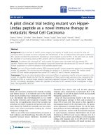
the -OH at C-5, respectively (Figure 1). The signal at δ
0.97 (J = 6.12 Hz) corresponds to the signal expected
from the methyl group of a rhamnose moiety. The sig-
nal at δ 5.32 (J = 7.44 Hz) indicates that the anomeric
glucoseprotonwasinthebetaconfiguration,whilethe
signal at δ:4.37(J = 7.6 Hz) indicates that the anomeric
rhamnose proton is in the alpha configuration [15]. The
signals between δ 3.00 and 4.00 belong to the other pro-
tons of the sugar moiety.
The
13
C NMR spectrum summarized in Table 2 indi-
cated a total of 27 carbon atoms. Fifteen of which were
methine (CH) carbon atoms, one was a methyl (CH
3
)
Table 1 Summary of MIC and MBC of the compound
(mg/mL)
Organisms MIC MBC
E. coli 6.25 12.5
P. aeruginosa 6.25 12.5
S. pyogenes 12.5 25.0
C. ulcerans 6.25 12.5
K. pneumoniae 12.5 25.0
N. gonorrhoeae 12.5 25.0
Table 2
13
C and
1
H chemical shifts assignments for the
compound
Position
13
C (400 MHz, DMSO-d
6
)
1
H (400 MHz, DMSO-d
6
)
2 156.4
3 133.2
4 177.3
5 156.6 12.62 (1H, s, 5-OH)
6 98.6 6.19 (1H, d, J = 1.88)
7 164.0
8 93.6 6.41 (1H, d, J = 1.80)
9 161.1
10 103.9
1’ 121.1
2’ 115.2 7.53 (1H, d, J = 8.08)
3’ 144.6
4’ 148.3
5’ 116.2 6.85 (1H, d, J = 7.84)
6’ 121.6 7.55 (1H, d, J = 7.56)
1
G
101.1 5.32 (1H, d, J = 7.44)
2
G
73.9 3.08 (1H, d, J = 9.28)
3
G
75.8 3.23 (1H, d, J =6)
4
G
69.9 3.26-3.36 (3H)
5
G
76.3 3.21 (1H, d, J = 5.52)
6
G
66.9 3.26-3.36 (3H)
1
R
100.7 4.37 (1H, d, J = 7.6)
2
R
70.3 3.04 (1H, d, J = 2.68)
3
R
70.5 3.69 (1H, d, J = 10.4)
4
R
71.8 3.26-3.36 (3H)
5
R
68.2 3.39 (1H, d, J = 1.76)
6
R
17.6 0.97 (3H, d, J = 6.12)
Bello et al. Organic and Medicinal Chemistry Letters 2011, 1:14
/>Page 2 of 5
carbon atom, one was a methy lene (CH
2
) carbon, and
ten were quaternary (C) carbon atoms confirmed from
the DEPT 90 and DEPT 135 experiments.
The methine (CH) signals at δ 98.6 and 93.6 belong to
the A-ring (Figure 1) at positions 6 and 8, respectively,
while the signals at 116.2, 115.2, and 121.6 belong to
the B-ring (Figure 1) at positions 2’,5’,and6’,respec-
tively, and the signals at 101.1, 73.9, 75.8, 69.9, 76.3,
100.7, 70.3, 70.5, 71.8, and 68.2 are located on the disac-
charide moiety. The methyl (CH
3
)signalatδ 17.6 was
attributed to the terminal methyl group on the rham-
nose unit at position 6. The methylene (CH
2
) signal at δ
66.9 was attributed to the CH
2
carbon at position six of
the glucose unit. The quaternary (C) carbon atoms at δ
156.4, 133.2, 177.3, 161.1, 16 4.0, 156.6, and 103.9 are on
the A-ring while the signals at δ 121.1, 144.6, and 148.3
arelocatedontheB-ring.Thesignalsatδ 101.1, 73.9,
75.8, 69.9, 76.3, 100.7, 70.3, 70.5, 71.8, 68.2, 66.9, and
17.6 are consistent with those of rutinosyl (Table 2).
These assignments were confirmed by the COSY,
NOESY, HSQC, and HMBC experiments.
Conclusions
The results from this research have supported the eth-
nomedicinal uses of this plant in the treatment of
respiratory infections, abdominal disorders, gonorrhea,
and as a cough remedy. These diseases can be caused by
the respective microorganisms tested. The compound
was purified by re-crystallization and characterized as
quercetin-3O-rutinoside. Further studies are going on to
establish other phytochemicals in the plant.
Methods
Extraction
The fresh plant (1 kg) was extracted using hot water
and filtered. A yellow solid (13.5 g) w as precipitated on
standing for a few hours. It was filtered using a Buchner
funnel and trap under vacuum and re-crystallized from
redistilled methanol to yield yellow needle-like crystals
(4.52 g).
Phytochemical screening
Phytochemical analysis was carried out on the re-crys-
tallized compound using the method set out by Brain
and Turner [16] and Trease and Evans [17].
Shinoda’s test for flavonoids
About 5 mg of the compound was dissolved in ethanol.
3 mg magnesium powder was then added followed by
few drops of conc. HCl. An orange coloration indicated
the presence of flavonoids.
Figure 1 Quercetin-3-O-rutinoside. Structure of the isolated compound.
Bello et al. Organic and Medicinal Chemistry Letters 2011, 1:14
/>Page 3 of 5
Ferric chloride test for flavonoids
About 5 mg of the compound was dissolved in ethanol
(2 mL). A few drops of 10% ferric chloride solution
were added. A green-blue coloration indicated the pre-
sence of a phenolic hydroxyl group.
Sodium hydroxide test for flavonoids
About 5 mg of the compound was dissolved in water,
warmed, and filtered; t o this solution (2 mL), 10% aqu-
eous sodium hydroxide was added. This produced a yel-
low coloration. A change in color from yellow to
colorless on addition of dilute hydrochloric acid was an
indication for the presence of flavonoids.
Antimicrobial screening
The antimicrobial activity was determined using some
pathogenic microorganisms. The microorganisms were
obtained from the Department of Medical Microbiology,
Ahmadu Bello University Teaching Hospital, Zaria,
Nigeria. All isolates were checked for purity and main-
tained in slants of blood agar.
Asolutionof0.5gofthecompoundwasmadeusing
10 mL DMSO. This solution was used to check the anti-
microbial activity of the compound. A control experi-
ment was also set up using DMSO.
Blood agar base (Oxoid, England) was prepared
according to t he manufacturer’ s instructions. This was
then sterilized at 121°C for 15 min using an autoclave
and was allowed to cool. The sterilized medium (20 mL)
was pipetted into sterilized Petri dishes, covered, and
allowed to cool and solidify.
The Petri dishes containing the medium were seeded
with the test organisms by the spread plate technique
and were left to dry for half an hour.
Filter paper disks were cut and sterilized at 160°C for
30 min. The sterilized paper disks were then dropped
into the solutions of the extracts and were dried at 45°
C.Thedrieddiskswerethenplantedonthemedium
previously seeded with the test organisms. The plates
were incubated a t 37°C for 24 h after which they were
inspected for the zones o f inhibition of growth. The
zones were measured and recorded in millimeters by
the use of a pair of dividers and a ruler.
Minimum inhibition concentration
Minimum inhibition concentration (MIC) of the com-
pound was carried out on the microorganisms that were
susceptible to it and was carried out using the broth dilu-
tion method as described by Bauer et al. [18]. Nutrient
broth (Oxoid, England) was prepared according to the
manufacturer’s instructions. 10 mL each was dispensed
into five sets of screw cap test tubes and sterilized at 121°
C for 15 min. The test tubes were allowed to cool down.
McFarland’s turbidity standard scale number 0.5 was
prepared. 10 mL normal saline was used to make a
turbid suspension of the microorganis ms. Dilution of
the microorganisms was done continuously in the nor-
mal saline until the turbidity matched that of the
McFarland’s scale by visual comparison. At this point,
the microorganisms had a density of 3 × 10
8
cfu/mL.
Serial dilution of the c ompound was made using the
nutrient broth and the following concentrations were
obtained: 50, 25, 12.5, 6.25, and 3.125 mg/mL. Having
obtained the different concentrations, 1 mL of the
microorganism in the normal saline was inoculated into
the different concentrations of the compound in the
broth and was incubated at 37°C for 24 h. The lowest
concentration that showed no turbidity (clear solution)
was recorded as the MIC.
Minimum bactericidal/fungicidal concentration
This was carrie d out to determine whether the microor-
ganisms could be completely killed or their growth
could only be inhibited.
Blood agar base (Oxoid, England) was prepared
according to the manufacturer’s instructions. The solu-
tion was sterilized at 121°C for 15 min using an auto-
clave and poured into sterilized Petri dishes. The
contents of the MIC test tubes in the serial dilution
were sub-cultured on the Petri dishes by dipping a ster-
ile wire loop into each test tube and streaked on the
surfaces of the Petri dish es. The Petri dishes were incu-
bated at 37°C for 24 h after which they were observed
for growth. The minimum bactericidal/fungicidal con-
centration (MBC/MFC) was the Petri dish with the low-
est concentration of the compound that had no growth
of the microorganisms.
Acknowledgments
We would like to appreciate the World Bank, STEP-b, IOT, Nigeria, for
sponsoring part of this project. IAB thanks Petroleum Technology
Development Fund, Nigeria for local study scholarship.
Competing interests
The authors declare that they have no competing interests.
Received: 22 June 2011 Accepted: 4 October 2011
Published: 4 October 2011
References
1. Hostettmann K, Marston A, Ndjoko K, Wolfender JL (2000) The potential of
African plants as a source of drugs. Curr Org Chem 4:973–1010.
doi:10.2174/1385272003375923.
2. Hostettmann K, Marston A (1990) Studies in natural products chemistry.
Elsevier, Amsterdam,7: p 405
3. Watt JM, Breyer-Brandwijk MG (1962) Medicinal and poisonous plants of
southern and eastern Africa. E. and S. Livingstone, Edinburgh p 901
4. Dalziel JM (1956) Useful plants of west tropical Africa. Crown Agents for
Overseas Government, London p 407
5. Sanon S, Azas N, Gasquet M, Ollivier E, Mahiou V, Barro N, Cuzin-Ouattara N,
Traore AS, Esposito F, Balansard G, Timon-David P (2003) Antiplasmodial
activity of alkaloid extracts from Pavetta crassipes (K. Schum) and
Acanthospermum hispidum DC, two plants used in traditional medicine in
Burkina Faso. Parasitol Res 90(4):314–317. doi:10.1007/s00436-003-0859-9.
Bello et al. Organic and Medicinal Chemistry Letters 2011, 1:14
/>Page 4 of 5
6. Amos S, Akah PA, Binda L, Enwerem NM, Ogundaini A, Wambebe C,
Hussaini IM, Gamaniel KS (2003) Hypotensive activity of the ethanol extract
of Pavetta crassipes leaves. Biol pharm bull 26(12):1674–1680. doi:10.1248/
bpb.26.1674.
7. Brasseur T, Angenot L (1986) Flavonol glycosides from leaves of Strychnos
variabilis. Phytochemistry 25(2):563–564. doi:10.1016/S0031-9422(00)85534-X.
8. Yasukawa K, Takido M (1987) A flavonoid glycoside from Lysimachia
mauritiana. Phytochemistry 26(4):1224–1226. doi:10.1016/S0031-9422(00)
82393-6.
9. Agrawal PK (1992) NMR spectroscopy in the structural elucidation of
oligosaccharides and glycosides. Phytochemistry 31(10):3307–3330.
doi:10.1016/0031-9422(92)83678-R.
10. Webby RF, Markham KR (1990) Flavanol 3-O-triglycosides from Actinidia
species. Phytochemistry 29(1):289–292. doi:10.1016/0031-9422(90)89052-B.
11. Wenkert E, Gottlieb HE (1977) Carbon-13 nuclear magnetic resonance
spectroscopy of flavonoid and isoflavonoid compounds. Phytochemistry
16(11):1811–1816. doi:10.1016/0031-9422(71)85095-1.
12. Cushnie TPT, Lamb AJ (2005) Antimicrobial activity of flavonoids. Int J
Antimicrob Agents 26(5):343–356. doi:10.1016/j.ijantimicag.2005.09.002.
13. Tijjani MB, Bello IA, Aliyu AB, Olurishe T, Maidawa SM, Habila JD,
Balogun EO (2009) Phytochemical and antimicrobial studies of root extract
of Cochlospermum tinctorium A. Rich. (Cochlospermaceae). Res J Med Plants
3(1):16–22. doi:10.3923/rjmp.2009.16.22.
14. Agrawal PK, Bansal MC (1989) Carbon-13 NMR of flavonoids. Elsevier, New
York,39: pp 287–293 chap 6
15. Mabry TJ, Markham KR, Thomas MB (1970) Systematic identification of
flavonoids. Springer-Verlag, New York p 268
16. Brain KR, Turner TD (1975) The practical evaluation of phytochemicals.
Wright Science Technical, Bristol pp 56–64
17. Trease GE, Evans WC (2000) Pharmacognosy. Saunders Publishers, London,
15 pp 42–44 221-229, 246-249, 304-306, 331-332, 391-393
18. Bauer AW, Kirby WMM, Sherris JC, Turk M (1966) Antibiotic susceptibility
testing by a standardized single disc method. Am J Clin Pathol 45:493–496
doi:10.1186/2191-2858-1-14
Cite this article as: Bello et al.: A bioactive flavonoid from Pavetta
crassipes K. Schum. Organic and Medicinal Chemistry Letters 2011 1:14.
Submit your manuscript to a
journal and benefi t from:
7 Convenient online submission
7 Rigorous peer review
7 Immediate publication on acceptance
7 Open access: articles freely available online
7 High visibility within the fi eld
7 Retaining the copyright to your article
Submit your next manuscript at 7 springeropen.com
Bello et al. Organic and Medicinal Chemistry Letters 2011, 1:14
/>Page 5 of 5