Báo cáo hóa học: " An alternative route for the synthesis of silicon nanowires via porous anodic alumina masks" potx
Bạn đang xem bản rút gọn của tài liệu. Xem và tải ngay bản đầy đủ của tài liệu tại đây (3.05 MB, 7 trang )
NANO EXPRESS Open Access
An alternative route for the synthesis of silicon
nanowires via porous anodic alumina masks
Francisco Márquez
1*
, Carmen Morant
2
, Vicente López
3
, Félix Zamora
3
, Teresa Campo
2
and Eduardo Elizalde
2
Abstract
Amorphous Si nanowires have been directly synthesized by a thermal processing of Si substrates. This method
involves the deposition of an anodic aluminum oxide mask on a crystalline Si (100) substrate. Fe, Au, and Pt thin
films with thicknesses of ca. 30 nm deposited on the anodic aluminum oxide-Si substrates have been used as
catalysts. During the thermal treatment of the samples, thin films of the metal catalysts are transformed in small
nanoparticles incorporated within the pore structure of the anodic aluminum oxide mask, directly in contact with
the Si substrate. These homogeneously distributed metal nanoparticles are responsible for the growth of Si
nanowires with regular diameter by a simple heating process at 800°C in an Ar-H
2
atmosphere and without an
additional Si source. The synthesized Si nanowires have been characterized by field emission scanning electron
microscopy, high-resolution transmission electron microscopy, X-ray photoelectron spectroscopy, and Raman.
Keywords: Si NWs, AAO, masks, CVD
Introduction
One-dimensional semiconductor nanostructures have
recently attracted intense research attention due to their
novel physical properties [1-5], including electrical, mag-
netic, optic al, and mechanical, and their potential for device
applications in chemical and biological sensors, optoelec-
tronic, t ransistors, etc. [6-8]. All these properties and
potential applications can be modulated by controlling the
chemical composition and the dimensionality of the nano-
wires, during the synthesis process [9]. Different methods
have been used to synthesize Si nanowires (Si NWs) such
as vapor-liquid-solid (VLS) process [10-12], laser ablation
[13], chemical vapor deposition [14,15] or even thermal
evaporation [16,17]. Electrodeposition techniques are an
interesting alternative for nanowires growth due to the low
cost and simplicity of the process [18-20]. This methodol-
ogyusesaporousstructure,whichactsasatemplate,
whose pores are electrochemically filled with the material
of interest. This technique, however, has many technical
problems to obtain nanowires with high aspect ratio.
In this study, we present an alternative procedure to
those previously reported for the synthesis of nanowires. A
porous structure (anodic aluminum oxide membrane) acts
as an efficient template during the synthesis, controlling
the dimensionality of the Si NWs. This methodology is
based on the use of a porous membrane on which the cat-
alyst i s depos ited. The use of silicon substrates as source
for the Si NWs growth has recently been reported [21].
Nevertheless, in our study, the treatment temperature is
clearly lower, the reaction time is reduced, the diameter of
the Si NWs is regular and dependent on the synthesis
parameters and the length of the nanowires is adjustable,
controlling the growth time [22]. In this procedure, the
diameter of the Si NWs can be related to the size of metal
nanoparticles, whose dimensionality is adjustable by con-
trolling the temperature, thickness of deposited material,
and pore diameter of anodic alumina membrane used in
the process [22]. In summary, it is noteworthy that the ori-
ginality of this process lies in using the same substrate
where the catalyst is deposited, as source of silicon, avoid-
ing the use of complex systems with silicon-based vap or,
together with a template that allow us to obtain silicon
nanowires with regular dimensions.
Experimental section
Preparation of the anodic aluminum oxide templates
The synthesis of highly ordered porous alumina tem-
plates has been described elsewhere [23-28]. High-purity
* Correspondence:
1
School of Science and Technology, University of Turabo, Gurabo, 00778 PR,
USA
Full list of author information is available at the end of the article
Márquez et al. Nanoscale Research Letters 2011, 6:495
/>© 2011 Márqu ez et al; lice nsee Springer. This is an Open Access article distributed under the terms of the Creative Commons
Attribution License ( which permits unrestricted use, distribution, and reproduction in
any medium, provided the original work is properly cited.
(99.999%) aluminum sheets, used as starting material,
weredegreasedbyusingamixtureofHF,HNO
3
,HCl,
and water (1:10:20:69,%v/v) and by ultrasonication in acet-
one. After that, the aluminum sheets were annealed under
nitrogen atmosphere at 400°C for 3 h to remove mechani-
cal stresses. Next, the aluminum foils were electropolished
in a perchloric acid-ethanol solution (1:4, v/v) at 2°C. The
anodization of the aluminum foils was made in two steps.
The first anodization step was carried out using a constant
voltage source (40 V) in a 0.3 M oxalic acid solution for 24
h and at a temperature around 1°C, then the oxide layer
was removed by using a mixture of chromic and phospho-
ric acids at 30°C. The second anodization step was carried
out for 3 h under identical conditions to the first anodiza-
tion step. Afterwards, a saturated HgCl
2
solution was used
to dissolve the aluminum metal. Next, the barrier layer of
the bottom part was removed and the pore diameter was
widenedbydippingthemembraneina5wt.%H
3
PO
4
solution at 35°C for 20 min. The thickness of the free-
standing porous alumina membrane was measured by
field emission scanning electron microscopy (FESEM) to
be 10 μm with a pore diameter of ca. 60 nm.
This anodic aluminum oxide (AAO) membrane was
directly supported on a silicon (100) wafer. Other more
compact Si substrates (Si (110) or Si (111)) are not able
to generate any growth. The Si used in the growth pro-
cess of nanowires is obtained from thermally ge nerated
defects on the surface of Si (100). These defects can be
observed subsequently to the synthesis of Si NWs, as
small cracks on the substrate, with loss of material. This
Si is extracted from the single crystal and used in the
growth of the Si NWs.
The adherence of the AAO template on the silicon
substrate is produced by van der Waals forces and it
can be substantially improved by wetting the AAO
membrane in propan-2-ol/ethanol (2:1, v/v)mixture.
After that, the template supported on the Si (100) was
dried at 60°C overnight.
Deposition of the catalyst on the AAO-Si sample
Different metals (30 nm) were deposited onto the AAO/
Si samples by single ion-beam sputtering of a high-pur-
ity Au (99.999%, Goodfellow), Fe (99.95%, Goodfellow),
and Pt (99.99%, Edelmetall) targets [24,29,30]. A refer-
enced continuous Au, Fe, or Pt, film was simultane ously
deposited on a Si (100) wafer to measure the thickness
of the metal layer with a Taylor-Hobson Talystep profil-
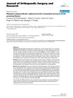
ometer. The experimental setup is shown in Figure 1.
During the metal deposition, the base vacuum was 10
-
5
Pa and the argon pressure during sputtering was 0.1
Pa. In all cases, the deposition rate (measured with a
quartz microbalance) was maintained at 2 nm min
-1
.
During the sputtering, metal atoms are deposited on the
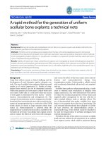
AAO surface and also inside the inner pore surface. Fig-
ure 2 shows the FESEM image of AAO masks supported
on Si (100) substrates after depositing a 30-nm-thick Fe
Figure 1 Schematic representation of the single ion-beam
sputtering system used for catalyst deposition on AAO-Si
substrates.
Figure 2 FESEM images of the AAO-Si substrates after depositing a 30-nm-thick film. A film of Fe (a), Au (b), and Pt (c) at room
temperature.
Márquez et al. Nanoscale Research Letters 2011, 6:495
/>Page 2 of 7
(a), Au (b), and Pt (c) film at room temperature. As can
be seen there, the metal deposition is homogeneously
distributed due to the constant rotation of th e sample
holder that prevents the concentration of metal atoms
in specific areas of the sample.
Thermal treatment and growth of Si NWs
The substrates were placed inside an alumina boat that
was introduced in a tube furnace with a quartz reactor
coupled, which was then heated at 800°C. The quartz
reactor is coupled to a gas mixing system with mass
flow controllers (see Figure 3).
Initially, 1,000 mL min
-1
of a mixture of hydrogen and
argon (1:7 v/v) was flowed during the heating ramp (25°
Cmin
-1
). When a temperature of 800°C was reached,
samples were maintained in these conditions for 30 min.
Finally, the flow of argon was readjusted to 1,000 mL
min
-1
and hydrogen was stopped. After that, the cooling
ramp was set at 20°C min
-1
under flowing argon during
5h.
Characterization methods
The morphology and micr ostructure of the Si NWs
grown over AAO templates were analyzed by FESEM
(Philips, FEG-XL30S, 20 kV, Philips Electronic Instru-
mentsCo.,Chicago,IL,USA)andbyhigh-resolution
transmission electron micro scopy (HRTEM, JEOL JEM-
3000F, JEOL, Tokyo, Japan). Raman spectra were also
recorded using a confocal Raman microscope (Renishaw
RM2000, Renishaw plc, Wotton-under-Edge, UK)
equipped with a laser source at 514 nm, a Leica micro-
scope, and an electrically refrigerated CCD camera. The
spectral resolution was set at 5 cm
-1
,laserpower
employed was less than 5 mW and the acquisition time
was around 2 min.
HRTEM samples were prepared by dispersing the
synthesized Si NWs in an ultras ound bath with ethanol
followed by homogenization and placing 5 μLofthis
solution onto a copper grid coated with a lacy carbon
film.
X-ray photoelectron spectroscopy (XPS) measure-
ments were performed on a PHI 3027 system, by using
the Mg Ka (1,253.6 eV) radiation of a twin anode in the
constant analyzer energy mode with a pass energy of 50
eV.
Results and discussion
Morphological characterization
During the initial stages of heat treatment, the catalyst
deposited on the AAO-Si substrate melts and is incor-
porated within the porous alumina mask, resulting in
nanoparticles with regular dimensions. These nanoparti-
cles necessarily have a size smaller than the pores of the
AAO mask and will be responsible for the constant
dimensions of the synthesized nanowires. Figure 4
shows the surface of the AAO-Si substrate, once the
Figure 3 Diagram of the CVD system and temperature ramps used in this study.
Márquez et al. Nanoscale Research Letters 2011, 6:495
/>Page 3 of 7
molten catalyst has been incorporated within the porous
structure of the membrane and before the treatment
conditions allow the nanowires growth. As can be seen
there, the catalyst can be observed as small particles
inside the porous structure of the mask.
Figure 5 shows the FESEM image of the Si NWs
obtained by using Pt as catalyst. Figure 5 shows a side
view of the nanowires grown. As can be seen there, a
high density of Si NWs emerges from the surface of the
(AAO-Si) substrate. The use of Fe or Au catalysts pro-
duced similar growths although with a lower density of
nanowires. Under these growth conditions , the only
source of silicon is the substrate Si (100). We also tested
other types of more compact silicon crystals, including
silicon Si (111) or Si (011), but in these cases, there was
no growth of nanowires. Possibly, this occurs because
during the use of more compact substrates, the tem-
perature used in treatment is not high enough to pro-
duce the evaporation of Si atoms. After the growth of
nanowires, the Si (100) single crystal shows a large num-
ber of small cracks and holes on their surface. This sili-
con which has been removed from the crystal surface
has been used in the synthesis of nanowires. Figure 6
shows a typical Si (100) surface obtained after thermal
growth of Si NWs. As can be seen there, when the
AAO mask and the Si NWs are removed from the sub-
strate, the Si surface shows the presence of defects (dark
points) with an average size and depth of around several
micrometers. The morphology and size of the synthe-
sized nanowires was also investigated by HRTEM. Fig-
ure 7 shows the HRTEM of Si NWs obtained by using
Au (Fig. 7a and 7b) and Pt (Figure 7c, d) as catalysts,
after dispersing by ultrasonic treatment of the nanowires
in ethanol. It can be seen that several nanowires, with
regular diameters are n ucleated on catalyst nanoparti-
cles. The metal nanoparticles are synthesized by using
the AAO mask supported on the Si substrate as tem-
plate. The thin metal layer deposited on the AAO-Si
substrate is melted and incorporated inside the pores in
contact with the Si surface. Since the nanoparticle size
of the patterned catalyst is uniform, the grown nano-
wires are also uniform in diameter. The averaged pore
size of the alumina mask, as determined by SEM, is
about 60 nm. The lower nanoparticle size obtained from
the alumina mask could be due to the sphericity
induced by temperature, eventually generating particles
of average size less than the predicted size. In all cases,
the Si NWs are very long (tens of micrometers) with
regular diameters of ca. 40 ± 10 nm. Inset of Figure 7a
Figure 4 FESEM image of the Pt catalyst incorporated by
thermal effect within the pore structure. Pore structure of the
AAO mask-Si before the growth of nanowires.
10
P
m
Figure 5 FESEM image of the Si NWs obtained with Pt as
catalyst.
Figure 6 SEM image of the Si (100) surface. After growth, Si NWs
and AAO template have been removed to reveal the dark points
corresponding to defects and cracks generated on the susbstrate
during the growth.
Márquez et al. Nanoscale Research Letters 2011, 6:495
/>Page 4 of 7
shows the histogram plot f or the diameter distribution
of the synthesized Si NWs.
Electron diffraction experiments on the Si NWs
observed by TEM did not result in a diffraction pattern,
evidencing the amorphous nature of this material. Upon
closer inspection of the HRTEM images of the metal
nanoparticles (inset of Figure 7b), it can be observed
that the ordered fringes are demonstrating the crystal-
line nature of the metal particles generated during the
melting process of the catalysts through the mask. On
the other hand, EDXS measurements confirmed the
composition of indiv idual Si NWs to consist of silicon
and oxygen (see the inset of Figure 7c). The oxygen sig-
nal is due to the presence of silicon oxides, possibly
located on the surface.
XPS characterization
Figure 8 shows the Si 2p and O 1s photoelectron
spectra of Si NWs obtained by using Pt as catalyst. It
is noteworthy that the XPS results obtained from
nanowires grown using other catalysts (Fe or Au)
show similar results. In order to eliminate the signal
due to the Si substrate, XPS spectra were obtained
after deposition of the Si NWs on a surface of highly
orientedpyrolyticgraphite(HOPG).TheSi2pspec-
trum (Figure 8a) shows a main peak and a shoulder at
lower binding energies. The main peak at 103.6 eV
(labeled as 3) has been attributed to Si in the oxidized
form (SiO
2
) [31]. The shoulder at lower energy has
been deconvoluted in two components at ca. 99.7 eV
(labeled as 1) and at ca. 101 eV (labeled as 2). Inter-
estingly, the peak 1 has been attributed to Si
0
[31].
The peak 2, required for the deconvolution, can be
ascribed to the presence of substoichiometric Si oxi-
des (SiO
x
) [31]. Figure 8b shows the XPS spectrum of
O 1s. As can be seen there, this band is not sym-
metric and it has been deconvolved in two compo-
nents. The main peak observed at 532.4 eV (labeled as
2) has been attributed to oxygen in SiO
2
[31]. In a
similar way as was observed with the Si 2p spectrum,
the peak at 529.9 eV (labeled as 1) ha s been assigned
to the presence of substoichiometric oxides (SiO
x
)
[31] and possibly to oxygen adsorbed on the HOPG
substrate.
SiNWs
Au
ab
cd
Pt
Pt
100 nm 100 nm
20 nm
100 nm
40 50
DIAMETER (nm)
Figure 7 HRTEM of Si NWs synthesized using Au (a, b) and Pt (c, d) as catalyst. The inset of (a) shows a histogram of the Si NWs diameter
distribution. The inset of (b) shows the Au nanoparticle. The inset of (c) corresponds to the EDX analysis of the Si NWs.
Márquez et al. Nanoscale Research Letters 2011, 6:495
/>Page 5 of 7
The results obtained by XPS and EDX indicate that
the Si NWs are constituted by Si
0
,SiO
2
,andsubstoi-
chiometric silicon oxides (SiO
x
). Moreover, studies of
electron diffraction by TEM reveal that the Si NWs are
amorphous in nature. Possibly, Si NWs are composed of
aSi
0
core surrounded by a silicon oxide shell. Different
studies on the synthesis of amorphous silica nanowires
consider that the explanation for the amorphous nano-
wires production is the growth temperature. In fact,
when temperature is not high enough, recrystallization
is not produced and, in our case, we have used a con-
stant growth temperature of 800°C.
Raman characterization
Figure 9 shows the Raman spectrum of the Si NWs
grown by using Pt as catalyst. As can be seen there, a
sharp Raman line at ca. 512 cm
-1
is obser ved. This peak
can be related to the Si-Si stretching mode. Neverthe-
less, Raman peaks at more than 510 cm
-1
(typically
around 520 cm
-1
) have been justifi ed as due to crystal-
line silicon. The above studies reveal that there was no
trace of a crystall ine phase in the synthesized Si NWs.
On the other hand, XPS analysis indicates the presence
of silicon suboxides and in this way, the Raman shift at
positions near to that corresponding to crystalline
phases can be attributed to the effect of the oxygen defi-
ciency [32].
The peak at ca. 485 cm
-1
(m) can be justified as due
to the bond Si-O of amorphous SiO
2
or also t o substoi-
chiometric oxid es. The Raman peak at ca. 584 cm
-1
(m)
has been assigned to Si-O-Si bending of silicon oxides.
The broad peak at 931 cm
-1
is due to the stretching
mode of amorphous Si-Si (vibration that is also
observe d at 512 cm
-1
). Finally, the Figure 9 shows three
peaks at ca. 678 (w), 798 (m), and 860 cm
-1
(w), that
have been associated to the stretching mode of Si-O.
Conclusions
Inthepresentwork,wehaveusedAAOmasksto
synthesize Si NWs on Si (100) substrates, by using Fe,
Au, and Pt as catalysts. In t his approach, the Si (100)
substrate acted as both silicon source and growth sub-
strate, allowing the synthesis of Si NWs with regular
dimensions.
The growth mechanism corresponds to a VLS process.
In this mechanism, the growth happens when silicon
from the Si (100) substrate diffuses into the alloy pud-
dle, favoring the melting of Si into the alloy [33].
The diameter of the nanowires ranged from ca. 30-50
nm, with an average size of ca. 40 nm and was related
to the pore size of the AAO mask. HRTEM revealed the
amorphous nature of the Si NWs, possibly due to the
Figure 8 XPS spectra of Si 2p (a) and O 1s (b), and the
corresponding deconvolution analysis.
Figure 9 Raman spectra of Si NWs.
Márquez et al. Nanoscale Research Letters 2011, 6:495
/>Page 6 of 7
low growth temperature used during the synthesis. EDX,
XPS, and Raman have shown that they are composed of
Si
0
and silicon oxides (SiO
2
-SiO
x
) possibly forming a Si
0
core surrounded by a silicon oxide shell. Nevertheless,
further research is needed to clarify this point.
Acknowledgements
The authors gratefully recognize the financial support provided by MEC
through the grants MAT2006-08158, MAT2007-66476-C02-02, MAT2010-
19804 and European Community FP6-029192. Financial supports from US
Department of Energy through the Massey Chair project at University of
Turabo and from the National Science Foundation through the contract
CHE-0959334 are also acknowledged. One of us (TC) thanks the economical
support from MICROLAN S.A. The “Servicio Interdepartamental de
Investigación (SIdI)” from Universidad Autónoma de Madrid and “Centro de
Microscopía Luis Bru” from Universidad Complutense de Madrid are
acknowledged for the use of the HRTEM and FESEM facilities.
Author details
1
School of Science and Technology, University of Turabo, Gurabo, 00778 PR,
USA
2
Departamento de Física Aplicada C-XII, Universidad Autónoma de
Madrid, Cantoblanco, 28049 Madrid, Spain
3
Departamento de Química
Inorgánica C-VIII, Universidad Autónoma de Madrid, Cantoblanco, 28049
Madrid, Spain
Authors’ contributions
FM, CM, VL, FZ, TC, and EE synthesized different samples. FM, CM, TC, and EE
characterized the synthesized samples by Raman, XPS, SEM, and TEM.
Competing interests
The authors declare that they have no competing interests.
Received: 4 April 2011 Accepted: 17 August 2011
Published: 17 August 2011
References
1. Adu KW, Gutierrez HR, Kim UJ, Sumanasekera GU, Ecklund PC: Confined
phonons in Si nanowires. Nano Lett 2005, 5:409-414.
2. Akiyama T, Nakamura K, Ito T: Structures and electronic properties of Si
nanowires grown along the [1 1 0] direction: role of surface
reconstruction. Surf Sci 2008, 602:3033-3337.
3. Clément N, Tonneau D, Dallaporta H, Bouchiat V, Fraboulet D, Mariole D,
Gautier J, Safarov V: Electronic transport properties of single-crystal
silicon nanowires fabricated using an atomic force microscope. Phys E
Low-dimens Systems and Nanostruct 2002, 13:999-1002.
4. Dalchiele EA, Martín F, Leinen D, Marotti RE, Ramos-Barrado JR: Synthesis,
structure and photoelectrochemical properties of single crystalline
silicon nanowire arrays. Thin Solid Films 2009, 518:1804-1808.
5. Guo CS, Yang XB, Zhang RQ: Remarkable effects of surface dihydride
configurations in electronic properties of < 110 > silicon nanowires.
Solid State Commun 2009, 149:1666-1669.
6. Bi X, Agarwal A, Yang KL: Oligopeptide-modified silicon nanowire arrays
as multichannel metal ion sensors. Biosens Bioelectron 2009, 24:3248-2351.
7. Bi X, Wong WL, Ji W, Agarwal A, Balasubramanian N, Yang KL:
Development of electrochemical calcium sensors by using silicon
nanowires modified with phosphotyrosine. Biosens Bioelectron 2008,
23:1442-1448.
8. Gao C, Deng SR, Wan J, Lu BR, Liu R, Huq E, Qu XP, Chen Y: 22 nm silicon
nanowire gas sensor fabricated by trilayer nanoimprint and wet etching.
Microelectron Engineer 2010, 87:927-930.
9. An X, Meng GW, Wei Q, Kong M, Zang L: SiO
2
Nanowires Growing on
Hexagonally Arranged Circular Patterns Surrounded by TiO
2
. Phys Chem
B 2006, 110:222-226.
10. David T, Buttard D, Hertog MD, Gentile P, Baron T, Ferret P, Rouvière JL:
Silicon nanowires grown in nanoporous alumina matrices on < 100 >
oriented silicon substrates investigated by electron microscopy. Superlatt
Microstruct 2008, 44:354-361.
11. Bae J, Kulkarni NN, Zhou JP, Ekerdt JG, Shih CK: VLS growth of Si
nanocones using Ga and Al catalysts. J Cryst Growth 2008, 310:4407-4411.
12. Zhang J, Xu B, Yang Y, Jiang F, Li J, Wang X, Wang S: Catalyzed-assisted
growth of well-aligned silicon oxide nanowires. J Non-Cryst Solids 2006,
352:2859-2862.
13. Fukata N, Oshima T, Okada N, Kizuka T, Tsurui T, Ito S, Murakami K: Phonon
confinement in silicon nanowires synthesized by laser ablation. Phys B:
Condensed Matter 2006, 376-377:864-867.
14. Lu M, Li MK, Kong LB, Guo XY, Li HL: Silicon quantum-wires arrays
synthesized by chemical vapor deposition and its micro-structural
properties. Chem Phys Lett 2003, 374:542-547.
15. Liu ZQ, Zhou WY, Sun LF, Tang DS, Zou XP, Li YB, Wang CY, Wang G,
Xie SS: Growth of amorphous silicon nanowires. Chem Phys Lett 2001,
341:523-528.
16. Chen J, Pan Y, Wu R: Growth mechanism of twinned SiC nanowires
synthesized by a simple thermal evaporation method. Phys E: Low-
dimensional Systems and Nanostructures 2010, 42:2335-2340.
17. Zhang RQ, Chu TS, Cheung HF, Wang N, Lee ST: Mechanism of oxide-
assisted nucleation and growth of silicon nanostructures. Mater Sci
Engineer C 2001, 16:31-35.
18. Kim K, Kim M, Cho SM: Pulsed electrodeposition of palladium nanowire
arrays using AAO template. Mater Chem Phys 2006, 96:278-282.
19. Peppler K, Janek J: Template assisted solid state electrochemical growth
of silver micro and nanowires. Electrochim Acta 2007, 53:319-323.
20. Xu CL, Li H, Zhao GY, Li HL: Electrodeposition and magnetic properties of
Ni nanowire arrays on anodic aluminum oxide/Ti/Si substrate. Appl Surf
Sci 2006, 253:1399-1403.
21. Park HK, Yang B, Kim SW, Kim GH, Young DH, Kim S H, Maeng SL:
Formation of silicon oxide nanowires directly from Au/Si and Pd-Au/Si
substrates. Phys E: Low-dimensional Systems and Nanostructures 2007,
37:158-162.
22. Márquez F, Morant C, Elizalde E, Zamora F, López V: Synthesis of silicon
nanowires. Spanish Patent; 2010, P201030501.
23. Yanagishita T, Nishio K, Masuda H: Fabrication of metal nanohole arrays
with high aspect ratios using two-step replication of anodic porous
alumina. Adv Mater 2005, 17:2241-2243.
24. Márquez F, Morant F, Pirota KR, Borrás A, Sanz JM, Elizalde E: Fabrication of
ordered crystalline zirconium nanoporous membranes by an one-step
procedure. Nano Today 2009, 4:21-26.
25. Navas D, Hernández-Vélez M, Asenjo A, Jaafar M, Baldonedo JL, Vázquez M:
Preparation and magnetic characterization of Ni membranes with
controlled highly ordered nanohole arrays. IEEE Trans Magn 2006,
42:3057-3059.
26. Masuda H, Fukuda K: Ordered metal nanohole arrays made by a two-step
replication of honeycomb structures of anodic alumina. Science 1995,
268:1466-1468.
27. Li A, Müller F, Birner A, Nielsch K, Gösele U: Fabrication and micro-
structuring of hexagonally ordered two-dimensional nanopore arrays in
anodic alumina. Adv Mater 1999, 11:483-487.
28. Lei Y, Chim WK, Zhang Z, Zhou T, Zhang L, Meng G, Phillipp F: Ordered
nanoporous nickel films and their magnetic properties. Chem Phys Lett
2003, 380:313-318.
29. Morant C, Márquez F, Campo T, Sanz JM, Elizalde E: Niobium and hafnium
grown on porous membranes. Thin Solid Films 2010, 518:6799-6803.
30. Márquez F, Morant C, Campo T, Sanz JM, Elizalde E: Ordered Metal
Nanotube Arrays Fabricated by PVD. J Nanosci Nanotechnol 2010,
10:1115-1119.
31. Moulder JF, Stickle NF, Sobol PE, Bomben KD: Handbook of X-ray
Photoelectron Spectroscopy.Edited by: Chastain J, King RC. Eden Prairie:
Physical Electronics; 1995:.
32. Nishikawa H, Shiryawa T, Nakamura R, Ohki Y, Nagaswa K, Hama Y:
Photoluminescence from defect centers in high-purity silica glasses
observed under 7.9-eV excitation. Physical Review B 1992, 45:586-591.
33. Paulose M, Varghese OK, Grimes CA: Synthesis of gold-silica composite
nanowires through solid-liquid-solid phase growth. J Nanosci
Nanotechnol 2003, 3:341-346.
doi:10.1186/1556-276X-6-495
Cite this article as: Márquez et al.: An alternative route for the synthesis
of silicon nanowires via porous anodic alumina masks. Nanoscale
Research Letters 2011 6:495.
Márquez et al. Nanoscale Research Letters 2011, 6:495
/>Page 7 of 7