Báo cáo hóa học: " Innovative gait robot for the repetitive practice of floor walking and stair climbing up and down in stroke patients" pdf
Bạn đang xem bản rút gọn của tài liệu. Xem và tải ngay bản đầy đủ của tài liệu tại đây (2.94 MB, 10 trang )
JNER
JOURNAL OF NEUROENGINEERING
AND REHABILITATION
Hesse et al. Journal of NeuroEngineering and Rehabilitation 2010, 7:30
/>Open Access
RESEARCH
© 2010 Hesse et al; licensee BioMed Central Ltd. This is an Open Access article distributed under the terms of the Creative Commons
Attribution License ( which permits unrestricted use, distribution, and reproduction in
any medium, provided the original work is properly cited.
Research
Innovative gait robot for the repetitive practice of
floor walking and stair climbing up and down in
stroke patients
Stefan Hesse*
1
, Andreas Waldner
2,3
and Christopher Tomelleri
2
Abstract
Background: Stair climbing up and down is an essential part of everyday's mobility. To enable wheelchair-dependent
patients the repetitive practice of this task, a novel gait robot, G-EO-Systems (EO, Lat: I walk), based on the end-effector
principle, has been designed. The trajectories of the foot plates are freely programmable enabling not only the practice
of simulated floor walking but also stair climbing up and down. The article intended to compare lower limb muscle
activation patterns of hemiparetic subjects during real floor walking and stairs climbing up, and during the
corresponding simulated conditions on the machine, and secondly to demonstrate gait improvement on single case
after training on the machine.
Methods: The muscle activation pattern of seven lower limb muscles of six hemiparetic patients during free and
simulated walking on the floor and stair climbing was measured via dynamic electromyography. A non-ambulatory,
sub-acute stroke patient additionally trained on the G-EO-Systems every workday for five weeks.
Results: The muscle activation patterns were comparable during the real and simulated conditions, both on the floor
and during stair climbing up. Minor differences, concerning the real and simulated floor walking conditions, were a
delayed (prolonged) onset (duration) of the thigh muscle activation on the machine across all subjects. Concerning
stair climbing conditions, the shank muscle activation was more phasic and timely correct in selected patients on the
device. The severely affected subject regained walking and stair climbing ability.
Conclusions: The G-EO-Systems is an interesting new option in gait rehabilitation after stroke. The lower limb muscle
activation patterns were comparable, a training thus feasible, and the positive case report warrants further clinical
studies.
Background
The annual stroke incidence is approximately 180 per
100.000 inhabitants in the industrialized world [1]. Three
months after a stroke, a third of the surviving patients are
still wheelchair-dependent, and the gait velocity and
endurance are significantly reduced in approximately 80%
of the ambulatory patients [2]. Accordingly, the restora-
tion and improvement of walking functions is a primary
concern with respect to the aspired social and vocational
reintegration.
To achieve this goal, a task specific repetitive training
seems most promising [3]. The conventional physiother-
apy instead focuses on strengthening and practicing sin-
gle movements or various neurofacilitation techniques,
but these methods do not stress gait practice.
One treatment approach to increase steps number dur-
ing training sessions is the treadmill training with partial
body weight support. [4,5]. However the assignment of
human resources for manual assistance in this method is
considerable; up to three therapists have to place the
paretic limb during the swing phase and to shift the
patient's weight onto the stance limb.
Consequently, gait machines followed, either applying
an exoskeleton [6-9] (e.g. Lokomat, LOPES, ALEX,
AutoAmbulator) or an end-effector principle [10-12] (e.g.
Gait Trainer GT I, HapticWalker, LokoHelp). The exo-
skeleton is equipped with programmable drives or pas-
* Correspondence:
1
Medical Park Humboldtmühle Berlin, Department Neurological Rehabilitation
Charité - University Medicine, 13507 Berlin, Germany
Full list of author information is available at the end of the article
Hesse et al. Journal of NeuroEngineering and Rehabilitation 2010, 7:30
/>Page 2 of 10
sive elements which flex the knees and hips during the
swing phase, whereas with the other principle the feet are
placed on foot plates, whose trajectories simulate the
stance and swing phases. Clinical trials in stroke patients
revealed non-equivocal results for the Lokomat [13] and
a consistently superior effect for the GT I [14] with
respect to the restoration of gait. A head-to-head com-
parison of the clinical effectiveness between existing
machines is missing. An accelerometry-based biome-
chanical comparison between the Lokomat and the GT I
showed comparable mechanical constrains that may alter
leg accelerations and decelerations during stance and
swing phases [15].
The currently commercially available gait machines
(Lokomat, AutoAmbulator, LokoHelp and GT I) are lim-
ited to the repetitive exercise of walking on the floor. Stair
climbing up and down, however, is an essential part of
everyday's mobility, and recent reports indicated that
only 5 to 25% of stroke patients can master one floor at
their discharge home from early rehabilitation. [16]. To
improve the outcome, the patients should frequently
practise stair climbing up and down in line with the task-
specific repetitive approach. However the inherent physi-
cal effort of the therapists limit the intended intensity, a
further burden is the risk of falls on the stairs.
The Haptic Walker [17], an end-effector based robot
with fully programmable trajectories, was the first device
to additionally enable harness-secured patients the repet-
itive practice of stair climbing up and down without over-
stressing therapists. The dimensions and the required
high voltage, resulting from the goal to achieve a maxi-
mum acceleration of 3,5 g during the push-off and a max-
imum speed of 5 km/h as during natural gait of healthy
subjects [18], limited its clinical utility.
Accordingly, the present work introduces a newly
developed gait robot (G-EO-Systems; EO, latin: I walk)
for the treatment of stroke patients (Figure 1). Its specifi-
cations included smaller dimensions and an energy sup-
ply of 230 V. The main aim of the present study was to
compare limb muscle activation patterns of hemiparetic
subjects during real and simulated floor walking, and
during real and simulated stair climbing up by means of
dynamic electromyography. Comparable muscle activa-
tion patterns between the real and simulated conditions,
and the lack of obviously deviant patterns induced by the
gait robot should help to dissipate any fears of the induc-
tion of a pathological gait on the machine. The second
aim was to demonstrate gait improvement on a single
case after a five weeks training with the new machine.
Methods
Patients
Six subacute stroke patients participated. They all had
suffered a supratentorial ischemia resulting in a right
(left) hemiparesis in three cases each, the stroke interval
reached from 6 to 14 weeks. Their age was less than 75
years, all of them could walk independently at least a dis-
tance of 20 m at a velocity of more than 0.25 m/s, and
could climb 10 stairs in an alternate fashion, the use of
technical aids or hand rails was allowed. The lower limb
spasticity was mild to moderate, the modified Ashworth
score (0-5) to assess hip, knee and ankle tonus, did not
exceed a value of 2 for any of the joints. All patients
understood the purpose and content of the protocol,
approved by the local ethical committee, none of them
had any other orthopaedic or neurological disease
impairing gait, nor an apparent heart failure.
The device
The device (Figure 2) followed the end effector principle.
The harness secured patient stood on two foot plates,
whose trajectories were completely programmable. The
two foot plates were connected each by a pivoting arm to
two moving sledges. The foot's forward motion was given
by the movement of the principal sledge, which was con-
nected to the transmission belt of the linear guide (Figure
3). The transmission belt was driven by a 1.500 W servo-
motor fixed to the back end of the linear guide. The for-
ward and backward excursion of the principal sledge
ensured the control of the step length. The mechanic
design for the control of the step height was realized
implementing the scissor principle. The second sledge on
the linear guide moved relatively to the principal sledge.
A rod ensured the connection of the relative sledge to the
pivoting arm. Nearing the relative sledge to the principal
sledge closed the scissor, providing the pivoting arm to
get lifted, and vice versa. The servomotor responsible for
the relative motion was fixed under the relative sledge
and connected to the principal sledge by a screw axle. A
third completely programmable 400 W drive was fixed on
the arm and transferred the rotation through a transmis-
sion belt to an external axle, which was aligned to the
ankle, controlling the plantar- and dorsiflexion during the
steps.
The motion control of each servomotor was provided
by an industrial personal computer, which was coupled to
the eight motion controllers responsible for each pro-
grammable degree of freedom on the machine, three for
each leg and two for the CoM control. All the electronics
and the electric parts were placed in the control box in
the back end of the device.
The maximal step length corresponded to 55 cm, the
maximal achievable angles were ± 90°. The step height in
the workspace was 40 cm, allowing the patient to climb a
standardized step of 18 cm. The maximal possible gait
velocity was 0,6 m/s, corresponding to an acceleration
peak of 10 m/s
2
.
Hesse et al. Journal of NeuroEngineering and Rehabilitation 2010, 7:30
/>Page 3 of 10
The feet were placed in two snowshoe bindings on a
steel plate, which was fixed to the basis plate by magnets.
The plate loosened in all three directions of the footplate,
if a limit momentum of 4 Nm was exceeded. Hand rails at
both sides were settable vertically and laterally. The
patient's body weight support system was fixed by an alu-
minium chassis. It consisted of an electrical patient lift
system, intended for helping the patients to stand up
from the wheelchair, and a drive activating a three roll
mechanism. The patent lifter's belt passed through the
three rolls mechanism and was attached to patient's har-
ness. The belt got shortened by the mechanism's motion,
ensuring the vertical motion of patient's centre of mass
(CoM). The two ends of a rope were fixed to patient's har-
ness at hip height to control the lateral motion of patient's
CoM. Another drive moved the rope.
A ramp allowed wheelchair access into the device from
behind, to be followed by getting in the snowshoes, fas-
tening them, securing the patient lifter's belt to the har-
ness, standing up with the assistance of the therapist, and
a last check before starting therapy.
The trajectories of the foot plates during the floor walk-
ing condition were taken from healthy subjects' data in
the literature, the same applied to the vertical and hori-
zontal movements of the CoM [19,20]. To simulate the
stair walking condition up and down, the stair climbing of
healthy subjects was assessed with the help of an active
marker system based on ultrasound (Zebris). The rele-
vant markers for the determination of the gait trajectories
were placed at the following positions: toe, metatarsale V,
heel and hip. The trajectory raw data, recorded by the
motion capture system, got processed by a filtering block.
First processing was a average filtering considering the
actual sample and the two subsequent samples. After the
average filter, data were filtered by a second order Butter-
worth low pass filter. The last smoothing procedure was
given by a regularization of the jerk [21], the third deriva-
tive of the position information.
The data relative to the marker placed on the metatar-
sale V provided the information for the up- and down
movement and the forward-backward movement of the
feet. The data of the metatarsale V got transformed into
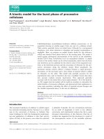
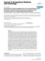
Figure 1 Wheelchair-bound left hemiparetic patient practising stair climbing. On the gait robot, the therapist assists the extension of the affect-
ed knee (left), the same patient practising stair climbing with the help of two therapists (right).
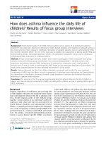
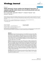
Figure 2 The G-EO-Systems Robot. A three-dimensional view of the
new gait robot with freely programmable foot plates, the patient lifter,
body weight support system, handrails and the ramp.
Hesse et al. Journal of NeuroEngineering and Rehabilitation 2010, 7:30
/>Page 4 of 10
hip relative coordinates by subtracting the coordinates of
the hip (marker placed on the trochanter major) to the
coordinates of the metatarsale V. By this transformation,
the hip was considered as fixed in the forward-backward
direction as there is no progression on the floor during
the simulated gait on the robot.
The data relative to the marker placed on the heel pro-
vided the information necessary to find the inclination of
the foot plates. The position coordinates of the metatar-
sale V were subtracted to the coordinates of the heel. The
arcustangens of this arithmetical operation provided the
inclination of the foot plates.
The movement of the CoM during stair climbing was
that measured with the ultrasound system, the marker
was attached to the hip, and confronted with the available
data in the literature [22,23].
The graphic user interface (GUI) showed the actual tra-
jectories for any of the conditions on-line, so that the
therapist was able to control and to correct it. Changes
could be made for step length, step height, the toe off and
the initial contact inclination angles of the feet. For a per-
fect match of the listed trajectory settings, the patient was
fixed in the snowshoe bindings in such a way that the
marked position of the metatarsale V in the binding cor-
responded to the patient's metatarsale V. The therapist
could further adapt the excursions of the CoM in the ver-
tical and horizontal directions and the relative position of
the suspension point with respect to the foot plates. The
PC memorized the treatment conditions of each individ-
ual patient. The dimensions of the CE-certified machine
(medical device directive 93/42/EEC) were 2.800 mm ×
1.200 mm × 2.300 mm, the net weight was 850 kg, the
power supply was 230 V.
Intervention
The gait was subsequently analysed during the following
four conditions: 1. hemiparetic walking on the floor at
self selected speed, 2. simulated walking on the machine
at comparable speed, cadence, and stride length in a
highly symmetric fashion, 3. stair climbing up for one
flight at self-selected pace in alternate fashion, and 4. sim-
ulated stair climbing at comparable step rise and cadence.
The 10 m test helped to derive the mean basic cycle
parameters to be set on the machine for each individual
subject (condition #2). The time needed for a 10 m walk-
ing distance at self selected speed was taken with the help
of a stop watch, and the number of steps counted. The
speed (cadence) was calculated in m/sec (steps/min), the
stride length (m) according to the formula: speed divided
by twice the cadence. On the machine, the patients not
only walked at comparable basic cycle parameters as on
the floor, additionally in a highly physiological fashion as
the limb-dependent cycle parameters, the gait symmetry,
step height, the initial contact and toe off angles were set
according to values of healthy subjects of comparable
height. The vertical (lateral) movement of the hip was set
to 2 cm (2,5 cm) in all subjects.
To set the stair climbing condition parameters (#4), the
patients climbed one flight in an alternate fashion, stair
by stair. The time needed was taken, to calculate the
speed (stairs/min), the normed step rise was 18 cm. The
initial contact and toe off angles corresponded to the
floor walking trajectory. The vertical (lateral) displace-
ment of the hip was set to 5 cm (2,5 cm). There was no
displacement of the trajectory in the forward or back-
ward direction.
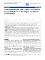
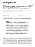
Figure 3 Three-dimensional sketch of the motion mechanism of the gait robot. The components are numbered as follows: 1. principal drive, 2.
relative drive, 3. drive for the foot angle, 4. principal sledge, 5. relative sledge, 6. linear guide, 7. screw axle.
Hesse et al. Journal of NeuroEngineering and Rehabilitation 2010, 7:30
/>Page 5 of 10
Assessment procedures
Before the actual assessment, the patients practised the
conditions several times to get acquainted to, and to
derive the parameters for the conditions #2 and #4. If
necessary a metronome helped with pacing. Next the
machine parameters were set, the patients instrumented,
and the four conditions were assessed in a random order
within one session. For each condition, the assessment
time was 30 seconds, or at least 10 strides.
The gait analysis system (Infotronic) consisted of over-
shoe slippers of various sizes with 8 force sensors inte-
grated to assess the limb-dependent cycle parameters.
Data were collected at 1000 Hz, amplified and memo-
rized by a portable data logger worn by the patient.
The electromyographic activity of seven lower limb
muscles of the affected side (Mm. tibialis anterior, gas-
trocnemius, vastus medialis, vastus lateralis, rectus femo-
ris, biceps femoris, and gluteus medius) were detected by
pairs of self-adhesive surface electrodes (diameter 8 mm)
following a standardized protocol: the electrodes were
attached 2 cm apart on the muscle bellies after a conven-
tional skin preparation (shaving, cleansing, and abrasion
of keratinized epidermis). The impedance was checked
and kept below 5 kΩ. Signals (sampling rate 1.000 Hz)
were pre-amplified with standard preamplifiers of the
Infotronic system attached to the limb and memorized by
the portable data logger.
Data analysis
All gathered signals (i.e, foot contacts, electromyographic
measurements) were transmitted after the end of each
trial to a personal computer and further processed by
Infotronic and Matlab software [24]. Cycle parameters
were averaged over at least 10 strides.
The electromyographic data were digitally filtered with
a first order lowpass filter (cutoff at 500 Hz) and the sin-
gle steps were determined by the trigger signal provided
by the overshoes. The single steps were time normalized
to the mean cycle duration set to 100%, and up-sampled
using a cubic spline [25,26]. The muscle activation pat-
terns for each stride were then smoothed using a 150-
point root-mean-square (RMS) algorithm. After calculat-
ing the RMS of the activation patterns, the mean EMG
pattern for the gait cycle was generated by averaging all
the individual stride cycles taken by the subject during
the 30-seconds data collection sequence. No signal pro-
cessing occurred for the amplitudes of the measured
muscles. In a first step, two experienced raters visually
checked the muscle activity patterns of each individual
subject for any obvious differences across the four condi-
tions. In a second step, the mean onset and offset points
of the activation patterns were determined by means of
thresholding the EMG envelope. The onset and offset
points were subsequently rounded, to lower the accuracy
of the measured data down to 5% of the gaitcycle. The
mean error of this loss of accuracy was determined to be
1,25% of the whole gait cycle, calculated as the half of the
maximum error possible of the rounding process. Statis-
tics (Wilcoxon test, p < 0.05) compared the on- and offset
points and duration of activity between conditions #1 and
#2, respectively between conditions #3 and #4. Patients
with a tonic or absent muscle activity pattern were
excluded.
Results
Results of the EMG analysis
For the floor walking condition, the pattern of the thigh
muscles (Mm. vastus medialis, lateralis, glutues medius)
was comparable during the real and simulated conditions
across all subjects. Minimal deviations were a delayed
onset and a prolongation of the activation of the Mm.
vastus medialis, lateralis during the simulated walking (p
< 0.05). Instead of the vastus medialis muscle, two sub-
jects more activated the vastus lateralis on the machine
(Figure 4). For the shank muscles, deviations became
apparent for two subjects. The tibialis anterior muscle
remained rather silent during the real and the simulated
floor walking, whereas the activity of the gastrocnemius
muscle showed a tonic activation pattern during the real
and a phasic, but less intense, activation pattern during
the simulated walking on the floor (Figure 5). For the
remaining four subjects no clear differences became
apparent. Table 1 resumes the relevant data of the activa-
tion patterns of the shank and thigh muscles for both
floor walking conditions.
For the stair climbing condition, the activation pattern
and the amplitudes of the thigh muscles were comparable
during both conditions. For the shank muscles, distinct
differences became apparent in three out of the six
patients, in the sense that the tibialis anterior muscle was
activated in a timely correct fashion (p < 0.05). At the
same time, the activation pattern of the gastrocnemius
muscle became more phasic. In the other three patients,
the activity of both muscles was rather low and tonic and
did not differ across conditions. Table 2 shows the rele-
vant data of the activation patterns of the shank and thigh
muscles during real and simulated stair climbing.
Clinical case report
The patient was a 72 year old male. A supratentorial isch-
emic stroke in the territory of the right middle cerebral
artery had resulted in a severe hemiparesis left with a
neglect syndrome. Five weeks after stroke onset, he was
able to sit at the edge of the bed (feet placed on the floor
and holding on with the non-affected upper limb), but
could not transfer, stand or walk independently. He
required the help of at least one person for these tasks.
His Functional Ambulation Category (FAC, 0-5, 0 = not
Hesse et al. Journal of NeuroEngineering and Rehabilitation 2010, 7:30
/>Page 6 of 10
able to walk at all, 5 = independent including stair climb-
ing) score was 1, his Rivermead Mobility Index was 3. The
lower limb muscle strength was 22, assessed with the help
of the Motricity Index (MI, 0-100). His upper limb was
plegic, and he was partially dependent in the basic activi-
ties of living with a Barthel Index (0-100) of 25.
His comprehensive rehabilitation programme, which
had started two weeks earlier, included regular physio-
and occupational therapy. Tasks repetitively practiced
were the transfer into the wheelchair, sitting balance on
the bench, standing with physical help or in a standing
frame, and making the first steps alongside the bench
with the help of two therapists.
To start the locomotor therapy, the team then decided
to additionally use the G-EO-Systems every workday for a
total of 25 sessions à 25 - 30 min. During the first five ses-
sions, the harness-secured patient practiced simulated
floor walking at a velocity of 0,25 m/sec, the step length
(cadence) was 37,5 cm (40 steps/minute). The relative
body weight support was 30%. One therapist helped with
donning and doffing, which each took three to five min-
utes, and with manually assisting the knee extension of
the paretic side throughout the session. Including breaks
due to exhaustion (the maximum heart rate not to exceed
was set at 120 beats/min), the net training time was
approximately 15 min totalling 600 steps per session in
the first week. During the next three weeks, the BWS was
gradually reduced to 10% BWS, and the speed increased
to 0,33 m/sec during the floor walk condition, manual
assistance of knee extension was no longer required. Fur-
thermore, the subject also practiced stair climbing up for
a net of five to eight minutes during each session. The
cadence started at 35 steps/min, the step rise (step run)
was 18 (30) cm. Over the three weeks the cadence was
gradually increased to a value of 48 steps/min, the other
parameters remained unchanged. Initially the training of
stair climbing had to be stopped every two to five minutes
as the heart rate exceeded the level of 120 beats/min, in
the last week he sustained eight minutes stair climbing
training without a break. Physical assistance with knee
extension of the paretic side was required throughout the
training of stair climbing.
Subjectively, he rated the locomotor training positive
but demanding, particularly during the simulated stair
climbing. He noticed a constant improvement in his
mobility and corporal fitness, a view shared by his thera-
pists.
At the end of the five week period he was able to trans-
fer, and to stand up independently. While standing he had
to hold on with the non-paretic hand, and, with the help
of a quadricane, he could walk a distance of 20 m without
physical support, corresponding to a FAC value of 4. For
climbing one flight he needed the physical assistance of
one therapist. The lower limb muscle strength was 59
Figure 4 EMG of the thigh muscles of the affected side in a hemiparetic subject. The left column shows the activation pattern of the muscle
during the real condition, the right column refers to the simulated condition. Note a delayed onset and prolonged activity of the thigh muscles on
the machine. The blue lines show the EMG activation pattern of the thigh muscles, the green lines represent the standard deviation of the EMG en-
velope.
Hesse et al. Journal of NeuroEngineering and Rehabilitation 2010, 7:30
/>Page 7 of 10
(initial value 22), assessed again with the help of the
Motricity Index (0-100), his Rivermead Mobility Index
(0-15) increased to 7 (initial value 3) and the Barthel
Index (0-100) up to 65 (initial value 25). Table 3 shows the
clinical parameters of the present clinical case report.
Discussion
The article presents a newly developed gait robot to
intensify the gait rehabilitation including stair climbing of
stroke patients. The dynamic EMG of selected lower limb
muscles of six ambulatory hemiparetic subjects con-
firmed rather comparable muscle activation patterns dur-
ing the real and simulated walking on the floor, and a
more timely correct pattern of the shank muscles during
the simulated stair climbing on the machine as compared
to the real walking condition.
The G-EO-Systems followed the intention of the Hap-
ticWalker, but specifications included smaller dimensions
Figure 5 EMG of the shank muscles of the affected side in a hemiparetic subject. The left column shows the activation pattern of the muscle
during the real condition, the right column refers to the simulated condition. Note the timely correct activation of the Mm. tibialis anterior and the
more phasic pattern of the Mm. gastrocnemius on the machine. The blue lines show the EMG activation pattern of the shank muscles, the green lines
represent the standard deviation of the EMG envelope.
Table 1: Muscle activation data of six hemiplegic patients for the real and the simulated floor walking condition.
real walking on the floor simulated walking
on the floor
tibialis
anterior
gastrocnemius vastus
medialis
vastus
lateralis
tibialis
anterior
gastrocnemius vastus
medialis
vastus
lateralis
1 silent silent 90-10 90-10 silent silent 90-40 0-25
2 tonic tonic 80-40 80-40 tonic tonic 90-50 80-40
3 silent silent 90-30 90-30 silent silent 90-35 90-40
4 tonic tonic 85-40 85-40 40-10 20-55 silent 90-50
5 silent tonic 85-35 90-30 silent tonic 80-50 80-50
6 55-20 90-50 80-50 0-70 90-20 15-45 30-60 30-60
The left side of the table shows the data of the shank and thigh muscles relative to the real floor walking condition, the left side of the table shows
the data of the shank and thigh muscles relative to the simulated floor walking condition. Note a slight delay in the onset of the mean activation
times for the simulated condition in respect to the real condition.
Hesse et al. Journal of NeuroEngineering and Rehabilitation 2010, 7:30
/>Page 8 of 10
and an energy supply of 230 V to ensure a maximum clin-
ical applicability. The device met the specifications, how-
ever at the price of a reduced maximum acceleration of 1
g during push-off and a reduced maximum speed of 2,3
km/h when compared to the HapticWalker. Those limita-
tions were tolerated, as the primary purpose of the G-EO-
Systems was the treatment of gait-impaired stroke
patients.
Compared to the technical features of the Lokomat, the
currently most applied gait machine, force feedback and a
virtual reality application have not yet been realized. Fur-
ther, an exoskeleton-based system supports the knee dur-
ing the stance phase, whereas an end-effector based
machine may require manual assistance or a functional
electrical stimulation of the quadriceps, depending on the
grade of paresis, to stabilize the knee during the stance
phase. This may be of particular importance in severely
affected spinal cord injury patients.
Hidler et al. [24] had studied the activation pattern of
healthy subjects on the treadmill and on the Lokomat.
The authors observed significant differences in the spatial
and temporal muscle activation patterns across various
portions of the gait cycle between treadmill and robot-
assisted walking, a finding partly confirmed for the
LOPES [27], another exoskeleton-based system. Activity
in the quardiceps and hamstrings was significantly higher
during the swing phase of Lokomat walking than tread-
mill walking, while activity in the ankle flexor and exten-
sor muscles was reduced throughout most of the gait
cycle in the Lokomat.
The present study assessed the dynamic EMG of hemi-
paretic subjects which renders any comparison with
those of healthy subjects difficult given the fact the mus-
cle activation pattern of hemiparetic subjects can vary
considerably [28]. The authors therefore concentrated on
the intra-individual comparison across real and simulated
walking conditions, a statistical comparison was only
meaningful in the case of a uniform change induced by
the machine.
Unanimously, the onset (duration) of the activation of
the quadriceps muscles was delayed (prolonged)
throughout the whole stance phase on the G-EO-Systems
during the floor walking condition. A less hard impact
during the initial contact requiring less stabilization force
during the initial contact, and the lack of a metatarsal
joint impeding the tibia advancement requiring a pro-
longed activation may have been the principal cause. For
the shank muscles, the activation patterns were rather
comparable during both floor walking conditions, the
individual patterns varied considerably in line with the
types described by Knuttson and Richards [29], either an
extremely low muscle activity, or in the sense of a patho-
logical coactivation during part of the gait cycle, thus dis-
rupting the normal sequential shift of activity in
antagonistic muscles. During the real stair climbing, the
individually disturbed activation pattern of the shank
muscles remained, whereas it became more physiological
during the simulated stair climbing on the machine in
three patients. The coactivation was replaced by an alter-
nating and timely correct pattern. A potential explanation
is the more physiological movement on the machine, as
the programmed trajectories rendered the gait perfectly
symmetric thus prolonging the stance phase of the
affected leg on the machine as compared to the real con-
ditions. At the same time, the patients were instructed
and assisted by the therapist to flex the hip and knee in
the pre-swing and to propel the body upward by a strong
activation of the shank muscles. Further, the harness pro-
vided safety.
Contrary to the findings with exoskeleton-based sys-
tems in healthy subjects [24], the thigh muscles of the
hemiparetic subjects did not become active during the
Table 2: Muscle activation data of six hemiplegic patients for the real and the simulated stair climbing condition.
real stair
climbing
simulated
stair climbing
tibialis
anterior
gastrocnemius vastus
medialis
vastus
lateralis
tibialis
anterior
gastrocnemius vastus
medialis
vastus
lateralis
1 silent silent tonic Tonic silent silent tonic tonic
2 tonic tonic 90-55 85-55 tonic tonic 85-50 85-45
3 silent silent silent 90-40 silent silent 90-40 85-45
4 tonic tonic 90-40 90-50 40-60 5-45 80-50 80-50
5 silent silent 80-40 80-30 55-95 10-40 85-40 85-40
6 tonic tonic 90-10 Tonic 50-20 20-55 80-20 90-20
The left side of the table shows the data of the shank and thigh muscles relative to the real stair climbing, the left side of the table shows the data
of the shank and thigh muscles relative to the simulated stair climbing. Note a more phasic activation of the shank muscles and the correct
activation of the thigh muscles, for what concerns activation time and duration.
Hesse et al. Journal of NeuroEngineering and Rehabilitation 2010, 7:30
/>Page 9 of 10
swing phase on the G-EO-Systems, confirming the results
of the HapticWalker [30]. Of course, any comparison
between healthy and hemiparetic subjects is question-
able, but one may speculate that the hemiparetic subjects
had not to counterbalance any additional inertia or move-
ment restriction imposed by an exoskeleton.
The single case report of a non ambulatory patient does
not allow any conclusions on the effectiveness of the gait
robot, which was applied additionally to his conventional
programme. The patient regarded the training positive,
the body weight support (net treatment time) was con-
stantly reduced (extended), and his gait functions
improved. Relevant side effects, such as acute arthritis of
the lower limb joints or of cardiovascular origin, did not
occur.
Conclusions
The new gait robot enables stroke patients the repetitive
practice of not only walking on the floor but also stairs
climbing up and down. Major deviations of the lower
limb muscle activation patterns of six ambulatory
patients did not occur while practising on the machine,
rather selected patients exhibited a less coactivated pat-
tern of the shank muscles during the simulated stair
climbing up. Any fears of the practice of a less physiologi-
cal muscle activation pattern on the machine in stroke
patients are thus not warranted. The positive single case
report does not allow any conclusions on the effective-
ness of the device; it will help to design future clinical
studies, urgently needed.
Consent
Written informed consent was obtained from the patient
for publication of this case report and accompanying
images. A copy of the written consent is available for
review by the Editor-in-Chief of this journal.
Competing interests
Reha-Technologies GmbH, Bozen, Italy holds the international patent on the
device presented. The author SH is a shareholder of the company.
Authors' contributions
SH conceived the device and drafted the article. AW supported the draft of the
article and the analysis of the gathered data. CT worked on the data acquisi-
tion, the subsequent signal processing, and helped in the draft of the present
manuscript. All authors read and approved the final manuscript.
Acknowledgements
The project was both supported by a BMBF grant (BioFuture) of the German
government, and by a grant of the Autonome Provinz Bozen-Südtirol, Italy.
Thanks to Anita Bardeleben, Cordula Werner, Mirjam Schimanke and Georg
Nitzsche for their skilful support during the assessment of the dynamic EMG.
A special thank to therapists of Villa Melitta and Medical Park for their support
during the development of the G-EO-Systems.
Author Details
1
Medical Park Humboldtmühle Berlin, Department Neurological Rehabilitation
Charité - University Medicine, 13507 Berlin, Germany,
2
Privatklinik Villa Melitta,
Neurological Rehabilitation, 39100 Bozen, Italy and
3
Research Department for
Neurorehabilitation South Tyrol, 39100 Bozen, Italy
References
1. Kolominsky-Rabas PL, Heuschmann PU: Incidence, etiology and long-
term prognosis of stroke. Fortschr Neurol Psychiatr 2002, 70:657-62.
2. Jorgensen HS, Nakayama H, Raaschou HO, Olsen TS: Recovery of walking
function in stroke patients: the Copenhagen stroke study. Arch Phys
Med Rehabil 1995, 76:27-32.
3. Carr J, Shepherd R: Stroke Rehabilitation: Guidelines for exercises and
training. London: Butterworth Heinemann; 2003.
4. Barbeau H, Visintin M: Optimal outcomes obtained with body-weight
support combined with treadmill training in stroke subjects. Arch Phys
Med Rehabil 2003, 84(10):1458-65.
5. Dobkin BH, Apple D, Barbeau H, Basso M, Behrman A, Deforge D, Ditunno
J, Dudley G, Elashoff R, Fugate L, Harkema S, Saulino M, Scott M: Methods
for a randomized trial of weight-supported treadmill training versus
conventional training for walking during inpatient rehabilitation after
incomplete traumatic spinal cord injury. Neurorehabil Neural Repair
2003, 17(3):153-67.
6. Colombo G, Joerg M, Schreier R, Dietz V: Treadmill training of paraplegic
patients using a robotic orthosis. J Rehabil Res Dev 2000, 37(6):693-700.
7. Veneman JF, Kruidhof R, Hekman EE, Ekkelenkamp R, Van Asseldonk EH,
van der Kooij H: Design and evaluation of the LOPES exoskeleton robot
for interactive gait rehabilitation. IEEE Trans Neural Syst Rehabil Eng 2007,
15(3):379-86.
8. Mankala K, Banala S, Agrawal S: Novel swing-assist un-motorized
exoskeletons for gait training. J Neuroeng Rehabil 2009, 6:24.
Received: 28 September 2009 Accepted: 28 June 2010
Published: 28 June 2010
This article is available from: 2010 Hesse et al; licensee BioMed Central Ltd. This is an Open Access article distributed under the terms of the Creative Commons Attribution License ( .0), which permits unrestricted use, distribution, and reproduction in any medium, provided the original work is properly cited.Journa l of Neuro Engineeri ng and Reh abilitat ion 2010, 7:30
Table 3: Clinical Data of a single case patient before and
after the therapy on the G-EO- Systems.
Age 72
Sex male
Diagnosis supratentorial,
ischemic stroke
Hemiparesis left
initial value final value
FAC 1 4
RMI 3 7
MI 22 59
BI 25 65
The assessed parameters at the beginning and at the end of the
treatment period were the Functional Ambulation Category (FAC, 0-
5), the Rivermead Mobility Index (RMI, 0-15), the Motricity Index (MI,
0-100) and the Barthel Index (BI, 0-100). Note an improvement of all
the clinical parameters after five weeks of treatment on the gait
robot.
Hesse et al. Journal of NeuroEngineering and Rehabilitation 2010, 7:30
/>Page 10 of 10
9. Mantone J: Getting a leg up? Rehab patients get an assist from devices
such as HealthSouth's AutoAmbulator, but the robots' clinical benefits
are still in doubt. Mod Healthc 2006, 36(7):58-60.
10. Hesse S, Uhlenbrock D: A mechanized gait trainer for restoration of gait.
J Rehab Res Dev 2000, 37(6):701-8.
11. Schmidt H, Werner C, Bernhardt R, Hesse S, Krüger J: Gait rehabilitation
machines based on programmable footplates. J Neuroeng Rehabil 2007,
4:2.
12. Freivogel S, Mehrholz J, Husak-Sotomayor T, Schmalohr D: Gait training
with the newly developed 'LokoHelp'-system is feasible for non-
ambulatory patients after stroke, spinal cord and brain injury. A
feasibility study. Brain Inj 2008, 22(7-8):625-32.
13. Husemann B, Müller F, Krewer C, Heller S, Koenig E: Effects of locomotion
training with assistance of a robot-driven gait orthosis in hemiparetic
patients after stroke. A randomized controlled pilot study. Stroke 2007,
38(2):349-54.
14. Pohl M, Werner C, Holzgraefe M, Kroczek G, Mehrholz J, Wingendorf I,
Hoölig G, Koch R, Hesse S: Repetitive locomotor training and
physiotherapy improve walking and basic activities of daily living in
subacute, nonambulatory stroke patients: a single-blind, randomised
multi-centre trial (DEutsche GAngtrainerStudie, DEGAS). Clinical
Rehabilitation 2007, 21(1):17-27.
15. Regnaux JP, Saremi K, Marehbian J, Bussel B, Dobkin BH: An
accelerometry-based comparison of 2 robotic assistive devices for
treadmill training of gait. Neurorehabil Neural Repair 2008, 22(4):348-54.
16. Paolucci S, Braagoni M, Coiro P, De Angelis D, Fusco FR, Morelli D,
Venturiero V, Pratesi L: Quantification of the probability of reaching
mobility indipendend at discharge from the rehabilitation hospital in
non walking early ischemic stroke patient: a multivariate study.
Cerebrovasc Dis 2008, 26(1):16-22.
17. Schmidt H, Sorowka D, Hesse S, Bernhardt R: Development of a robotic
walking simulator for gait rehabilitation. Biomed Tech (Berl) 2003,
48(10):281-6.
18. Behrman AL, Harkema SJ: Locomotor training after human spinal cord
injury: A series of case studies. Physical Therapy 2000, 80(7):688-700.
19. Winter DA: Foot trajectory in human gait: a precise and multifactorial
motor control task. Phys Ther 1992, 72(1):45-53.
20. Winter DA: Biomechanics and control of human movement. Second
edition. Wiley Inter Science; 1990:212-32.
21. Hogan N: The organizing principle for a class of voluntary movements.
Journal of Neuroscience 1984, 11(4):2745-54.
22. Zachazewski JE, Riley PO, Krebs DE: Biomechanical analysis of body mass
transfer during stair ascent and descent of healthy subjects. J Rehabil
Res Dev 1993, 30(4):412-22.
23. McFadyen BJ, Winter DA: An integrated biomechanical analysis of
normal stair ascent and descent. J Biomech 1988, 21(9):733-44.
24. Hidler JM, Wall AE: Alterations in muscle activation patterns during
robotic-assisted walking. Clin Biomech 2005, 20:184-93.
25. Giakas G, Baltzopoulos V: A comparison of automatic filtering
techniques applied to biomechanical walking data. J Biomech 1997,
30:847-50.
26. Simons W, Yang K: Differentiation of human motion data using
combined spline and least squares concept. Journal of Biomechanical
Engineering 1991, 113:348-51.
27. van Asseldonk EH, Veneman JF, Ekkelenkamp R, Buurke JH, van der Helm
FC, van der Kooij H: The Effects on Kinematics and Muscle Activity of
Walking in a Robotic Gait Trainer During Zero-Force Control. IEEE Trans
Neural Syst Rehabil Eng 2008, 16(4):360-70.
28. Den Otter AG, Guerts AC, Mulder T, Duysens J: Abnormalities in the
temporal patterning of lower extremity muscle activity in hemiparetic
gait. Gait Posture 2007, 25(3):342-52.
29. Knuttson E, Richards C: Different types of disturbed motor control in
gait of hemiparetic patients. Brain 1979, 102:405-30.
30. Hussein S, Schmidt H, Volkmar M, Werner C, Helmich I, Piorko F, Krüger J,
Hesse S: Muscle coordination in healthy subjects during floor walking
and stair climbing in robot assisted gait training. Conf Proc IEEE Eng Med
Biol Soc 2008:1961-4.
doi: 10.1186/1743-0003-7-30
Cite this article as: Hesse et al., Innovative gait robot for the repetitive prac-
tice of floor walking and stair climbing up and down in stroke patients Jour-
nal of NeuroEngineering and Rehabilitation 2010, 7:30