Biomedical Engineering Trends in Materials Science Part 8 pdf
Bạn đang xem bản rút gọn của tài liệu. Xem và tải ngay bản đầy đủ của tài liệu tại đây (10.15 MB, 30 trang )
Biomedical Engineering, Trends in Materials Science
202
Xiong, L. & Yang, L. (2003). Quantitative analysis of osteoblast behaviour on
microgroovedhydroxyapatite and titanium substrata. J. Biomed. Mater. Res., 66A,
677-687
Yap, A.S. & Kovacs, E.M. (2003). Direct cadherin – activated cell signaling: a view from the
plasma membrane. J. Cell Biol., 160, 11-16
Yamada, K. (1991). Adhesive recognition sequences. J. Biol. Chem., 266, 12809-12812
Yang, Y.C. & Change, E. (2001). Influence of residual stress on bonding strength and fracture
of plasma–sprayed hydroxyapatite coatings on Ti-6AL-4V substrate. Biomaterials,
22, 1827-1836
Yang, J.; Lian, J.; Dong, Q. & Guo, Z. (2004). Nano structured films formed on the AlSi 329
stainless steel by Nd:YAG pulsed laser irradiation. Appl. Surf. Sci., 229, 2-8
Ziats, N.P.; Miller, K.M. & Anderson, J.M. (1988). In vitro and in vivo interactions of cells
with biomaterials. Biomaterials, (Ibid), 9, 5-13
Zhu, X. & Assoian, R. K. (1995). Integrin-dependent activation of MAP kinase: a link to
shape-dependent cell proliferation. Mol. Biol. Cell, 6, 273-282
9
Novel Titanium Manganese Alloys and
Their Macroporous Foams for
Biomedical Applications
Prepared by Field Assisted Sintering
Faming Zhang and Eberhard Burkel
Physics of New Materials, University of Rostock
August Bebel Str.55, 18055 Rostock
Germany
1. Introduction
In this chapter, a novel titanium (Ti) alloy and foam suitable for biomedical applications will
be introduced. As we know, Ti and its alloys are widely used as biomaterials especially for
orthopedic implants in load bearing sites as dental and orthopedic implants and heart
valves, due to their high mechanical properties, corrosion resistance and biocompatibility
(Geetha et al., 2009). Pure Ti was once used as biomaterial, but its disadvantage as implant
materials is low strength and insufficient hardness. Therefore, the Ti6Al4V alloy is
preferentially in clinical use because of its favourable mechanical properties. However, some
studies showed that the vanadium (V) and aluminium (Al) release in Ti6Al4V alloy could
induce Alzheimer’s disease, allergic reaction and neurological disorders (Mark & Waqar,
2007). Therefore, the exploration of high strength new Ti alloys without Al and V for
medical implants has gained great attention in the past years and it is still ongoing. Al and V
free alloys containing non-toxic elements such as iron (Fe),
niobium (Nb), zirconium (Zr),
tantalum (Ta), molybdenum (Mo), nickel (Ni), gold (Au), or silicon (Si), etc. were
investigated (Zhang, Weidmann et al, 2010). As long-term load-bearing implants in clinic,
the incorporation of porous structures into the Ti and its alloys could lead to a reliable
anchoring of host tissue into the porous structure, and allow mechanical interlocking
between bone and implant (Li et al, 2005). The porous structure is preferable for Ti and its
alloys used as bone implants. Many techniques have been applied to produce Ti foams in
recent years. Nevertheless, there are still problems to be solved in the field of Ti foams for
biomedical applications (Zhang, Otterstein et al., 2010): the difficulty to create controlled
porosity and pore sizes, the insufficient knowledge of porous structure-property
relationships, the requirements of new sintering techniques with rapid energy transfer and
less energy consumption and so on.
The Ti alloys and foams are difficult to be produced from the liquid state due to high
melting point, high reactive activity at high temperature above 1000 ºC and contamination
susceptibility. The production of Ti alloys and foams via a powder metallurgy (PM) route is
attractive due to the ability to produce net-shaped components. Because of their stable
Biomedical Engineering, Trends in Materials Science
204
surface oxide film (TiO
2
), the Ti alloys are difficult to be sintered by traditional PM sintering
techniques. Thus, the spark plasma sintering (SPS), a pulsed electric current field assisted
sintering technique has been introduced to prepare the Ti alloys. Spark plasma sintering,
commonly also defined as field assisted sintering (FAST) or pulsed electric current sintering
(PECS) is a novel pressure assisted pulsed electric current sintering process utilizing ON-
OFF DC pulse energizing. Due to the repeated application of an ON-OFF DC pulse voltage
and current between powder materials, the spark discharge point and the Joule heating
point (local high temperature-state) are transferred and dispersed to the overall specimen
(Munir & Anselmi-Tamburini, 2006). The SPS process is based on the electrical spark
discharge phenomenon: a high energetic, low voltage spark pulse current momentarily
generates spark plasma at high localized temperatures, from several to ten thousand
degrees between the particles resulting in optimum thermal and electrolytic diffusion.
During SPS treatment, powders contained in a die can be processed for diverse novel bulk
material applications, for example nanostructured materials (Gao et al., 1999), functional
gradated materials (Lou et al., 2003), hard alloys (Zhang et al., 2004), biomaterials (Gu et al.,
2004), porous ceramics (Jayaseelan et al., 2002) and diamonds (Zhang et al., 2005) etc. The
research group of the author (E.B) has applied the SPS technique also for the synthesis of
new materials such as nanostructured magnets, quasicrystals, nanoceramics and Ti alloys
(Nicula, Cojocaru et al., 2007; Nicula, Turquier, et al., 2007; Nicula, Lüthen et al., 2007).
The preparation of dense Ti alloys by using the SPS was reported extensively, but still fewer
studies were on porous Ti foams (Zhang, Otterstein et al, 2010). The SPS studies on porous
Ti alloys were mainly using low temperature and low pressure to decrease the relative
density of samples. The samples exhibited pore sizes of some tens of micrometers and a
porosity in the range of 20-45%. As bone foams, high porosity (>50%) and macropore size
(>200 μm) are essential requirements for the bone growth and the osteoconduction.
We aim at
1. the exploration of new elements within Ti alloys for biomedical applications,
2. the development of new methods to prepare Ti foams for biomedical applications,
3. the deep understanding of the relationships between the microstructure and properties
of the new Ti alloy and foams.
Manganese (Mn) is one of the essential trace elements in human body. In recent decades
research has discovered the special role manganese plays as a co-factor in the formation of
bone cartilage and bone collagen, as well as in bone mineralization (Brown, 2006). The Mn is
also beneficial to the normal skeletal growth and development. It is important for enzymes
in the body like the superoxide dismutase and, therefore, involved in the elimination of
radicals (Zhang, Weidmann et al., 2010).
Titanium-manganese (TiMn) alloys have been
extensively used in aerospace and hydrogen storage, but not yet in biomedicine. The results
in our group showed that the Mn incorporation into the Ti-Al-V alloy could enhance the cell
adhesion properties (Nicula, Lüthen et al., 2007). In this chapter, the Mn element was
incorporated into the Ti system and TiMn alloys with different Mn amounts were prepared
by SPS technique. The preparation process, microstructures, mechanical properties,
cytotoxicity and cell proliferation properties of the TiMn alloys were investigated for
exploration of their biomedical applications. Macroporous Ti foams with controlled
architectures were also prepared using the SPS technique and subsequently modified with
TiO
2
nanostructures. The relationship between the properties and the porous architectures
was analyzed and discussed.
Novel Titanium Manganese Alloys and
Their Macroporous Foams for Biomedical Applications Prepared by Field Assisted Sintering
205
2. Major raw materials and methods
• The precursor Ti and Mn powders with purities above 99.0% were obtained from Alfa
Aesar, Germany. The space holder materials for preparation of Ti foams with 99.0%
purity were also obtained form Alfa Aesar and sieved in the range of 100 to 1000 μm.
• The mechanical alloying of the alloy powders is completed using a high energy
planetary ball milling machine (Retsch PM400, Germany). The SPS experiments were
performed using a Model HPD-25/1 FCT spark plasma sintering system (FCT systeme
GmbH, Rauenstein, Germany).
• The analysis of the phase transformation of the alloys was conducted with a differential
scanning calorimetry (DSC, DSC 404 C Pegasus®, Germany). The microstructure
analysis was performed using X-ray diffraction (XRD, Bruker D8, Germany) and
Scanning electron microscopy (SEM, Zeiss Supra 25, Germany). The Ti foam
architecture was examined by using X-ray microcomputed tomography (Micro-CT, GE,
USA).
• The hardness and the elastic modulus of the dense alloys were measured by Universal
CETR Nano+Micro tester with a model UNMT-1 multi-specimen test system. The
mechanical behaviour of the Ti foams was investigated by uniaxial compression
experiments at room temperature. The plateau stress and further elastic modulus
measurements were carried out on a universal testing machine Zwick Roell Z050.
• The human osteoblastic cells MG-63 (osteosarcoma cell line, ATCC, LGC Promochem)
were used to investigate the in vitro biocompatibility of the TiMn alloys. The
cytotoxicity of the alloys were measured by the methyltetrazolium salt (3-(4,5-
dimethylthiazol-2-yl)-5 -(3-carboxy -methoxyphenyl)) (MTS) method. The flow
cytometry for determining the cells proliferation property on the alloys was also
performed.
• The surface modification of the Ti foams was conducted by soaking in a strong alkali
solution and heat treatment. The in vitro bioactivity of the modified foams was tested
using a simulated body fluid solution in a shaking bath kept at 37.0 °C.
3. Titanium Manganese alloys
3.1 Phase diagram of the TiMn alloys
The binary phase diagram of TiMn alloys is shown in Fig. 1. It shows the conditions at
which thermodynamically distinct phases can occur in equilibrium. The TiMn alloy
powders were designed by varying the amount of Mn in the Ti with 2, 5, 8 and 12 (wt.%)
compositions on the base of phase diagram. In Fig. 1, the locations of the phases of the Ti-2,
5, 8, 12 wt.% Mn alloys discussed in this work are indicated as straight lines in the phase
diagram. The phase compositions of the TiMn alloys with Mn below 12 wt.% are all Ti2Mn2
phase.
3.2 Preparation of the TiMn alloys
The TiMn alloy powders with 2, 5, 8 and 12 wt.% Mn compositions were mixed and
mechanical alloyed for various hours in a high energy ball milling machine. Fig. 2 shows the
XRD patterns of the pure Ti and Mn powders and of the TiMn alloy powders after 60 hours
mechanical alloying. The pure Ti and Mn peaks completely disappeared and TiMn phases
Biomedical Engineering, Trends in Materials Science
206
were formed after 60 hours of mechanical alloying. The pure Ti powders show the α-Ti
phase (PDF# 65-3362) with hexagonal structure and the pure Mn powders the α-Mn phase
(PDF# 32-0637) with cubic structure. The synthesized TiMn powders contain the α-TiMn
phase (PDF# 07-0132) with tetragonal structure. There are no obvious changes in the phase
compositions with increasing Mn amount up to 12 wt% in Ti, which corresponds to the
binary phase diagram of the TiMn alloy (Fig. 1). The powders are analyzed by SEM
revealing agglomerates with mean particle sizes of 4-5 µm in diameter with a narrow size
distribution. The EDX spectra indicate that the Ti, Mn peaks belong to the TiMn powder.
The C and O peaks are resulting from adsorption of air, and the small Fe peak is due to the
contamination from the steel balls and vials during the mechanical alloying.
12 8 5 2
Fig. 1. Binary phase diagram of TiMn alloy showing the phases of the Ti-2, 5, 8, 12 wt.% Mn
alloys.
The phase transformation behaviors of the TiMn alloy powders were analyzed by using
differential scanning calorimetry. Fig. 3 shows the transformation temperatures of the TiMn
alloys in comparison with the pure Ti. In the case of pure Ti, the transformation temperature
from α to β phase occurs at about 840 ºC. The transformation temperature in Ti2Mn is at
about 735 ºC while that of the Ti5Mn alloys is at about 700 ºC. The transformation
temperatures are at about 665 ºC and at about 660
ºC in Ti8Mn and Ti12Mn alloys,
respectively. With increasing amount of Mn, the transformation temperature decreased to a
lower temperature value. The addition of Mn in Ti has depressed the transformation
temperature from the α to the β phase. The elements V, Mo, Nb, Fe, Cr, etc are all β
stabilizers and an addition of these elements depresses the β transition temperature. The
results in Fig. 3 show that the Mn has decreased the transformation temperature from the α
to the β phase. The influence of manganese on the α to β transition temperature is
significant. It is confirmed that the Mn is a β stablizing addition element for Ti metals.
Novel Titanium Manganese Alloys and
Their Macroporous Foams for Biomedical Applications Prepared by Field Assisted Sintering
207
30 40 50 60 70
α-Mn( 332)
α-Mn( 330)
α-Ti (101)
2 theta (Deg.)
Ti
Internsity
α-Ti(100)
α-Ti(002)
a-Ti(102)
α-Ti(110)
α-Ti(103)
α-TiMn(320)
α-TiMn(400)
α-TiMn(112)
α−TiMn(630)
Mn
Ti2Mn
Ti5Mn
Ti8Mn
Ti12Mn
Fig. 2. XRD patterns of the Ti, Mn powders, and TiMn alloy powders prepared by
mechanical alloying showing the formation of α-TiMn phases.
100 200 300 400 500 600 700 800 900 1000 1100
-180
-160
-140
-120
-100
-80
-60
-40
-20
0
20
Ti5Mn
DSC (mW/mg)
Temperature (
o
C)
α to β transformation temperature
Ti12Mn
Ti8Mn
Ti
Ti2Mn
Endo
Fig. 3. Phase transformation behaviours of the TiMn alloys measured by DSC showing the
decrease of the α to β transformation temperatures with increasing Mn amount.
After characterization, the alloy powders were subjected to the SPS furnace. The relative
densities of the spark plasma sintered pure Ti at different temperatures and the TiMn alloys
with various Mn amounts were also investigated. With a temperature increase from 550°C
Biomedical Engineering, Trends in Materials Science
208
to 800ºC, the relative density of the Ti metal increased from 68% to 99%. The relative density
of the TiMn alloys increased with higher Mn amount. The Ti8Mn alloys showed 99%
relative density after sintering at 700ºC for 5 min. The SPS method reduces the sintering
temperature of Ti and TiMn alloys. The Mn addition increased the relative density of Ti
metal during the SPS treatment. Finally, high density Ti metal was prepared by using the
SPS application at 750ºC for 5 min and high density TiMn alloys were sintered at 700ºC with
a holding time of 5 min. By using the traditional sintering techniques, high temperatures of
1100-1300ºC would be required to get pure or alloyed high density Ti.
The SPS has
decreased the sintering temperature of Ti and TiMn alloys. The Mn has increased the
relative density of Ti alloy, which is due to the lower β transformation temperatures in the
TiMn alloys. The low sintering temperature is ascribed to the ionization of particles by local
sparks during SPS. Pulsed current generated plasmas are expected to lead surface activation
of the powder particles, melting the titanium oxide films and forming neck junctions among
powder particles at a lower temperature (Zhang, Weidmann, 2009).
3.3 Microstructures of the TiMn alloys
Fig. 4 (a) shows X-ray diffraction (XRD) patterns of the spark plasma sintered Ti, Mn and
TiMn samples. The pure Ti and Mn still retain the α-Ti and the α-Mn phases because of the
lower sintering temperature of 700ºC during the SPS treatment. However, most of the TiMn
alloys show the β-TiMn phase (PDF# 11-0514) with cubic structure. There is still a small
amount of the α-TiMn phase in the alloy; therefore, the TiMn sample is an α+β phase alloy.
The synthesized alloy has α+β microstructures which are similar to those of an Ti6Al4V
alloy.
The SEM micrograph of the fracture surface of the spark plasma sintered Ti8Mn sample is
shown in Fig.4 (b). There are very few micropores in the fracture surface of the TiMn alloys.
The grain size of the Ti8Mn alloys is about 500 nm indicating an ultrafine microstructure
and the fracture mode of the alloy is primary intergranular cracking. During the SPS, a
simultaneous pressure impact causes a plastic flow of the powders, which enables the
creation of the dense Ti alloys with ultrafine microstructures at high heating rates, lower
temperature and short holding time.
3.4 Properties of the TiMn alloys
The mechanical properties of the TiMn alloys are shown in Fig. 5. The microindentation
hardness results show that the hardness value tended to rise with increasing Mn contents
(Fig. 5a). The hardness values of all TiMn alloys are significantly higher than that of pure Ti.
The pure Ti shows a hardness of 1.60 GPa ± 0.20 GPa; Ti2Mn 2.40 GPa ± 0.25 GPa; Ti5Mn
3.65 GPa ± 0.29 GPa; Ti8Mn 4.98 GPa ± 0.32 GPa and Ti12Mn 5.28 GPa ± 0.37 GPa. The
detected hardness value (5.28 GPa ± 0.37 GPa) of the Ti12Mn alloy is comparable to that of
the pure Mn (5.44 GPa ± 0,34 GPa). From statistical analysis, the hardness values of the
TiMn alloys are significantly higher than that of pure Ti.
The elastic modulus results are
shown in Fig. 5(b). The pure Ti is 105.3 GPa ± 6.0 GPa, Ti2Mn 83.3 GPa ± 3.0 GPa, Ti5Mn
95.0 GPa ± 5.0 GPa, Ti8Mn 106 GPa ± 4.1 GPa, and Ti12Mn 122 GPa ± 6.2 GPa, Mn
68.72 GPa ± 4.3 GPa. The ductility results of the TiMn alloys are shown in Fig. 5 (c). The pure
Ti exhibits 25.0% ± 2.0% ductility, Ti2Mn 21.3% ± 2.4%, Ti5Mn 18.2% ± 2.2%, Ti8Mn
15.0% ± 1.3% and Ti12Mn 11.7% ± 1.9%. The ductility decreased with increasing Mn
amounts in the TiMn alloy. For comparison, the mechanical properties of the Ti6Al4V
Novel Titanium Manganese Alloys and
Their Macroporous Foams for Biomedical Applications Prepared by Field Assisted Sintering
209
alloy were also measured with the same methods. This shows a hardness of 4.3 GPa ± 0.3
GPa, an elastic modulus of 122 GPa ± 4.0 GPa, and a ductility of 14.0 GPa ± 1.5 GPa which
are almost identical with reported literature values (Barbieri et al., 2007). The Ti2Mn,
Ti5Mn and Ti8Mn alloys possess lower elastic modulus and higher ductility than the
Ti6Al4V alloy.
30 40 50 60 70
α-TiMn (222)
α-TiMn (400)
β-TiMn (600)
2 Theta (Deg.)
α-TiMn (320)
β-TiMn (330)
Intensity
α-Ti (100)
α-Ti (002)
α-Ti (101) α-Ti (102)
α-Mn (330)
α-Mn (332)
α-Mn (510)
SPSed Ti
SPSed Ti5Mn
SPSed Ti8Mn
SPSed Ti2Mn
SPSed Ti12Mn
SPSed Mn
(a)
(b)
Fig. 4. XRD patterns of the spark plasma sintered Ti, Mn and TiMn alloys showing the TiMn
alloys are α+β phase alloy (a) and SEM micrograph of the fracture surface of a Ti8Mn alloy.
Biomedical Engineering, Trends in Materials Science
210
0
20
40
60
80
100
120
140
Young's modulus (GPa)
Ti
Ti2Mn
Ti5Mn
Ti8Mn
Ti12Mn
Mn
Ti6Al4V
0
1
2
3
4
5
6
7
Hardness (GPa)
Ti
Ti2Mn
Ti5Mn
Ti8Mn
Ti12Mn
Mn
Ti6Al4V
0
5
10
15
20
25
30
Ductility (%)
Ti
Ti2Mn
Ti5Mn
Ti8Mn
Ti12Mn
Ti6Al4V
(a)
(b)
(c)
Fig. 5. Hardness (a) and Elastic modulus (b) of the Ti, Mn, TiMn and Ti6Al4V alloys
obtained by microindentation tests, as well as ductility values at room temperature (c) of the
TiMn alloys
Novel Titanium Manganese Alloys and
Their Macroporous Foams for Biomedical Applications Prepared by Field Assisted Sintering
211
The TiMn alloys provide higher hardness and elastic modulus than those of the pure Ti. The
Ti5Mn and Ti8Mn alloys show comparable hardness but lower elastic modulus compared to
the Ti6Al4V alloy. The increment of the hardness and elastic modulus of the TiMn alloys is
ascribed to the formation α+β TiMn phases which are intermetallic phases with excellent
mechanical properties. The Ti6Al4V alloy was chosen for orthopedic implant for several
reasons. Excellent ductility is one of the most important reasons for its wide use in
biomedical industry. The ductility of Ti6Al4V alloy is measured to be 14% at room
temperature. The ductility of the TiMn alloy decreased from 21.3% (Ti2Mn) to 11.7%
(Ti12Mn) with increase of Mn amount. However, the Ti2Mn, Ti5Mn, Ti8Mn alloys have
higher ductility than that of the Ti6Al4V. Compared with the Ti6Al4V, the Ti2Mn alloy
presents lower hardness (2.4 GPa) with better elastic modulus (83.3 GPa) and ductility
(21.3%). The Ti5Mn alloy exhibits comparable hardness (3.65 GPa) and better elastic
modulus (95.0 GPa) and ductility (18.2%) and the Ti8Mn alloy shows better hardness (4.98
GPa) and elastic modulus (106 GPa) with a comparable ductility (15.0%). In the light of their
mechanical properties, the Ti2Mn, Ti5Mn and Ti8Mn alloys are suitable as biomedical
implants.
Fig. 6 represents the cytotoxicity and cell proliferation results of the TiMn alloys. The tissue
culture polystyrene (TCPS) was used as a control material. The MG-63 osteoblast cell
viability (%) of the pure Ti and TiMn alloys by MTS assay is shown in Fig. 6(a). The
cytotoxicity increases with increasing amount of the Mn contents in the Ti alloy. Cell's
viability on pure Mn and Ti12Mn was about 50 % and 72 %, respectively (p<0.05). However,
cells on the Ti5Mn and Ti8Mn alloys were also influenced concerning viability without
statistical difference (p>0.05), but it reached comparative high values (89 %, 86 %,
respectively) comparable with that of pure Ti (93 %). The proliferation of MG-63 osteoblasts
on the TiMn alloys using flow cytometric cell proliferation analysis is demonstrated in Fig. 6
(b). The percentage of cells on the pure Ti and TiMn alloys decreases in contrast to the TCPS
control (53.67 %). The number of proliferating cells on TiMn alloys (Ti2Mn 41.17%, Ti5Mn
40.50 %, Ti8Mn 41.57% and Ti12Mn 39.99 %) is reduced compared with that of pure Ti
(48.93 %), however, with p>0.05 not significantly and all acceptable for biomedical
applications. However, the percentage of proliferating cells grown on pure Mn is
significantly reduced to 35.87 % (p<0.05). The student t-test, an established statistical
method, shows that the proliferation of MG-63 osteoblast cells on TiMn alloys is not
remarkably inhibited. Only Mn is significantly decreased (p<0.05). The decrease in pure Mn
is about 27% from the Ti value. It is indicated that only a very high amount of Mn inhibits
cell proliferation. Combining the cytotoxicity and cell proliferation results, leads to the
assumption that the amount of Mn below 8 wt.% has a negligible effect on the cytotoxicity
and cell proliferation of all tested Ti alloys.
Some commercial Ti alloys also contain Mn as an alloying component. The Mn has been
doped in magnesium alloy with 1.2 wt. % and it was found that the Mn has no toxicity and
can improve the corrosion resistance and mechanical properties of Mg (Xu et al., 2007). The
Mn was doped to tri-calcium phosphate bioceramics and showed good cell compatibility
(Sima et al., 2007). Recently, a Fe-35Mn alloy was prepared and showed higher strength and
ductility, degradable properties. These observations make it suitable for biodegradable stent
applications (Hermawan et al., 2007). The values concerning cytotoxicity and cell
proliferation of the TiMn alloys demonstrate a dependency on the Mn concentration. A
lower Mn content (<8 wt.%) in Ti has a low effect on the cytotoxicity and cell proliferation
properties (p>0.05). In general, the Ti2Mn, Ti5Mn and Ti8Mn were comparable in viability
Biomedical Engineering, Trends in Materials Science
212
and cell proliferation properties with pure Ti. The Ti6Al4V alloy was firstly used in
aerospace industry, and then applied in biomedical field as bone and dental implants. Until
now, the Ti8Mn alloy as one of the typical α+β Ti alloys has been extensively used in
aerospace industry because of its excellent mechanical properties. Our research here
suggests that the application of the Ti8Mn alloy could be extended to biomedical field. As
well as the Ti2Mn and Ti5Mn alloys, they exhibit higher ductility and lower elastic modulus
than those of Ti6Al4V. The lower values of the elastic modulus of metals for joint prosthesis
could decrease the stress-shielding effect in bone-implant coupling. The Ti2Mn, Ti5Mn and
Ti8Mn alloys all exhibit acceptable cytotoxicity and cell proliferation of the human
osteoblasts. Consequently, all the Ti2Mn, Ti5Mn and Ti8Mn alloys have a potential for the
use in the biomedical field as new bone substitutes and dental implants.
0
10
20
30
40
50
60
70
80
90
100
110
120
*
*
Ti2Mn
Ti5Mn
Mn
Ti12Mn
Ti8Mn
Ti
TCPS
Cell viability (%)
0
10
20
30
40
50
60
70
80
*
Ti8Mn
% of Cells
Ti
TCPS
Ti2Mn
Ti5Mn
Ti12Mn
Mn
(a)
(b)
Fig. 6. Cytotoxicity (a) and cell proliferation (b) of MG-63 osteoblasts on the Ti and TiMn
alloys showing the comparable cell viability of Ti2Mn, Ti5Mn and Ti8Mn alloys with that of
pure Ti, and the proliferation of osteoblasts was not inhibited on TiMn alloys but only
significantly on pure Mn. Mean ±SD, n=5, Student t-test * p<0.05.
Novel Titanium Manganese Alloys and
Their Macroporous Foams for Biomedical Applications Prepared by Field Assisted Sintering
213
4. Macroporous Titanium foams
4.1 Preparation and microstructures of the Titanium foams
High density pure Ti, and TiMn alloys were prepared by using the SPS in the above section.
In this section, the preparation of Ti and TiMn foams by using the SPS will be introduced.
Firstly, the pure Ti foams were prepared by the free pressureless SPS method developed by
Zhang et al. (Zhang et al., 2008). The Ti powders were mixed with 15 wt. % of NH
4
HCO
3
and 2 wt. % of TiH
2
powder as pore forming agents. Then the powder mixture was sintered
at 1000°C by the SPS under a pressureless condition. Using 3D reconstruction by topographical
methods is the most realistic way to get information on the internal structure of the foams in
a non-destructive way. Fig. 7 shows the 3D reconstructions of the obtained Ti foams. The 3D
cropped isometric view of cross sections in this Ti foam shows the non-uniform pore
distribution and poor interconnectivity (Fig. 7a). The Micro-CT 2D top view and side views
show that the macropore shapes are in irregular cross sections and randomly distributed
(Fig. 7b-d). The 3D cropped internal surface exhibits pore size of 410 ± 90 μm. The XRD
results indicate that these Ti foams by free pressureless SPS method are in β-Ti phase
(Ibrahim, Zhang et al, 2011).
1000 um
1000 um
1000 um
(a) (b)
(c) (d)
Fig. 7. Micro-CT 3D reconstructions of the Ti foam with NH
4
HCO
3
and TiH
2
as pore forming
agents produced at 1000°C by free pressureless SPS method with an isometric view (a), 2D
top view (b), left side view (c), and right side view (d). Scale spacing, 1000 μm.
Biomedical Engineering, Trends in Materials Science
214
Alternatively, Ti foams with NaCl as spacer material were prepared at 700 ºC by SPS under
50 MPa. Fig. 8 shows the 3D μ-CT reconstructions of the obtained Ti foams. This spark
plasma sintered titanium foams shows 55% porosity and 250 μm pore size. The 3D cropped
isometric view of cross sections in the Ti foam shows the uniform pore distribution and
interconnected 3D porous structures with a high porosity (Fig. 8 a). The Micro-CT 2D top
view and side views show that the macropore shapes are in square cross sections, uniform
distribution of pore sizes with high interconnectivity (Fig. 8b-d). The 3D surface, the cell
wall thickness and the connectivity were examined by the Micro-CT in a non-destructive
way. The 3D cropped internal surfaces exhibit highly porous structures and interconnectivity
with pore sizes of 243±50 μm and a cell wall average thickness of 20.4 μm. The XRD results
indicate that these Ti foams are in α-Ti phase.
500 um
500 um
(a) (b)
(c) (d)
Fig. 8. Micro-CT 3D reconstructions of the Ti foam with NaCl as spacer material prepared by
SPS at 700 ºC under 50 MPa with an isometric view (a), 2D top view (b), left side view (c),
and right side view (d). Scale spacing, 500 μm.
The results in Fig.7 and Fig. 8 indicate that the Ti foams have been prepared successfully by
using the SPS technique. The foams prepared by the SPS and NaCl dissolution method show
better interconnectivities than those prepared by the free pressureless SPS method. High
Novel Titanium Manganese Alloys and
Their Macroporous Foams for Biomedical Applications Prepared by Field Assisted Sintering
215
interconnectivity of the foams will support the osteconduction of bone tissue. Therefore, Ti
foams with different pore sizes and porosities were prepared by the SPS and NaCl
dissolution method. The influence of the weight ratio and particle size of NaCl on the
porosity and pore size of Ti foams with corresponding SPS parameters is shown in Table 1.
The pore sizes of the sintered foams were measured from the SEM images. This shows a
mean pore size of about 125 μm in the foams with the NaCl spacing material being in the
range of 88-149 μm, a mean pore size of 250 μm with NaCl of the sizes 224-297 μm, a pore
size of 400 μm with NaCl of 388-500 μm sizes and a pore size of 800 μm with NaCl of 784-
1000 μm sizes. After the porosity characterization by the Archimedes method, it was noticed
that more NaCl particles were needed to obtain the same porosity in the large pore sized
foams. To achieve a porosity of 55% in the 125 μm foams, the weight ratio of Ti:NaCl is
about 1:1.28. However, the weight ratio of Ti:NaCl is about 1:1.75 in the 800 μm foams for
the same porosity. This might be due to the decreased specific surface area in the large sized
NaCl particles as spacer materials.
Ti powder NaCl powder
Weight ratio
(Ti: NaCl)
Porosity Pore size
SPS parameters
(Temperature,
dwell time)
10-30 μm
88-149 μm
(170-100 mesh)
1:1.28 ~55% ~125 μm 700 ºC, 8 min
1:0.72 ~30% ~250 μm 700 ºC, 8 min
1:0.93 ~45% ~250 μm 700 ºC, 8 min
550 ºC, 8 min
600 ºC, 8 min
650 ºC, 8 min
700 ºC, 8 min
730 ºC, 8 min
750 ºC, 5 min
1:1.32 ~55% ~250 μm
800 ºC, 3 min
10-30 μm
149-297 μm
(100-50 mesh)
1:1.64 ~70% ~250 μm 700 ºC, 8 min
10-30 μm
354-500 μm
(45-35 mesh)
1:1.46 ~55% ~400 μm 700 ºC, 8 min
10-30 μm
707-1000 μm
(25-18 mesh)
1:1.75 ~55% ~800 μm 700 ºC, 8 min
Table 1. The influence of the weight ratio and particle size of NaCl on the porosity and pore
size of the Ti foams with corresponding SPS parameters.
The SEM micrographs of the Ti foams with the same porosity of 55% but different pore sizes
of 125 μm, 250 μm, 400 μm, and 800 μm are shown Fig. 9. All the foams from 125 to 800 μm
exhibit highly interconnected porous structures and uniform pore distributions. It is found
Biomedical Engineering, Trends in Materials Science
216
that the pores have irregular quadratic cross sections. They are similar to those of the initial
NaCl particles of cuboid shapes.
Fig. 10 shows the SEM micrographs of the porous Ti foams with the same pore size of 250
μm with different porosities of 30 %, 45%, 55% and 70%. The thickness of the pore walls in
the 30% porosity foams is about 100 μm, decreasing to 50 μm in 45% porosity foams, and to
20 μm in 55%, finally ending at 10 μm in 70% porosity foams. The interconnectivity was also
enhanced with the increase in porosity. The 30% and 45% porosity foams show poor
interconnectivity because of the lower porosity. But the 55% and 70% higher porosity
samples show good interconnectivity. The macropores are in square cross sections in all the
Ti foams with different porosities.
(a)
(b)
(c)
(d)
Fig. 9. SEM micrographs of the Ti foams with the same porosity of 55% but different pore
sizes of 125 (a), 250 (b), 400 (c) and 800 μm (d). Scale bars, 300 μm.
Novel Titanium Manganese Alloys and
Their Macroporous Foams for Biomedical Applications Prepared by Field Assisted Sintering
217
(a) (b)
(c)
(d)
Fig. 10. SEM micrographs of the Ti foams with the same pore size of 250 μm but different
porosities of 30 % (a), 45% (b), 55% (c) and 70% (d). Scale bars, 300 μm.
4.3 Mechanical properties of the Ti foams
Fig. 11 shows the effect of pore size and porosity on the plateau stress and Young’s modulus
of the porous Ti foams. The measured plateau stress and Young’s modulus of the Ti foams
were compared with values calculated following Gibson-Ashby model. According to the
Gibson-Ashby model, the relationship between the relative plateau stress and relative
density is given by (Wen et al., 2002):
()
32
00
C
σσ ρρ
= (1)
where σ is the plateau stress of the foams, σ
0
is the yield stress of the dense material; C is a
constant 0.3 from the data of cellular metals and polymers (Gibson & Ashby, 1997), ρ is the
Biomedical Engineering, Trends in Materials Science
218
density of the foams, ρ
0
is the density of the dense material. The density of the pure Ti is 4.5
g/ cm
3
with yield stress of 692 MPa (Long & Rack, 1998). The density of the Ti foam with
55% porosity and 250 μm pore size is 1.69 g/cm
3
. Substituting these values in Equation 1,
the theoretical value was calculated to be 47.78 MPa, which is comparable to the measured
plateau stress 45.1±3.0 MPa.
100 200 300 400 500 600 700 800 900
20
25
30
35
40
45
50
55
100 200 300 400 500 600 700 800 900
20
25
30
35
40
45
50
55
(g/cm
3
)
Density
1.25
1.461.69
Pore size (um)
Experimental
1.81
Plateau stress (MPa)
Calculated
100 200 300 400 500 600 700 800 900
6
8
10
12
14
16
18
20
22
100 200 300 400 500 600 700 800 900
6
8
10
12
14
16
18
20
22
(g/cm
3
)
Density
1.25
1.461.69
Pore size (um)
Experimental
1.81
Young's modulus (GPa)
Calculated
30 40 50 60 70
20
30
40
50
60
70
80
90
100
110
30 40 50 60 70
20
30
40
50
60
70
80
90
100
110
Porosity (%)
Experimental
2.54
2.19
1.69
1.22 g/cm
3
Density
Plateau stress (MPa)
Calculated
30 40 50 60 70
5
10
15
20
25
30
35
40
30 40 50 60 70
5
10
15
20
25
30
35
40
Porosity (%)
Experimental
2.54
2.19
1.69
1.22 g/cm
3
Density
Young's modulus (GPa)
Calculated
(c) (d)
(a) (b)
Fig. 11. The effects of pore sizes (a, b) and porosities (c, d) on the plateau stress and Young’s
modulus of the Ti foams.
According to the Gibson-Ashby model, the relationship between the relative Young’s
modules and relative density is given by (Wen et al., 2002):
()
2
00
EE A
ρρ
=
(2)
where E is the Young’s modulus of the foams, E
0
is the Young’s modulus of the dense
materials, A is a constant of 1 including data of metals, rigid polymers, elastomers and
glasses. The Young’s modulus of the pure Ti is 105 GPa according to the Equation 2 (Long &
Rack, 1998). The measured Young’s modulus of the above Ti foams with 55% porosity is
13.46 GPa ± 1.4 GPa. Substituting the values into Equation 2, gives the theoretical value of
14.81 GPa which is also comparable to the measured one. All the Ti foams prepared by the
SPS were measured and calculated.
Novel Titanium Manganese Alloys and
Their Macroporous Foams for Biomedical Applications Prepared by Field Assisted Sintering
219
As seen from the Fig. 11, it can be deduced that all the experimental data agree with the
Gibson-Ashby model (1) and (2) in the present study. The relationship between the pore
sizes and the mechanical properties of the Ti foams is shown in Fig. 11 (a, b). The plateau
stress decreased from 49.7 MPa ±3.8 MPa to 27.2 MPa ± 3.0 MPa with the pore size increase
(Fig. 11a). The Young’s modulus reduced from 18.3 GPa ±2.0 GPa to 8.9 GPa ±1.5 GPa with
the pore size increase (Fig. 11b). It coarsely obeys a linear decay with the pore size increase.
The effect of the porosity on the mechanical properties of the Ti foams is shown in Fig. 11 (c,
d). The plateau stress decreased from 94.2 MPa ± 5.9 MPa to 28.8MPa ± 3.3 MPa, and the
Young’s modulus decreased from 36.1 GPa ± 3.5 GPa to 6.2 GPa ± 1.8 GPa with porosity
increase. It generally obeys the rule of exponential decline with the porosity increase.
The plateau stress and Young’s modulus coarsely obey linear declines with the pore size
increase and exponential decay with the porosity increase. Liu found that the plateau stress
of the porous hydroxylapatite ceramics decreases linearly with increasing macropore size
for a given total porosity (Liu, 1997). In this study, we found the plateau stress and Young’s
modulus coarsely obey linear declines with the pore size increase (Fig. 11 a, b). Rice RW has
proposed a function for the relationship of the porosity and the strength of porous solids
(Rice, 1993),
(
)
0
exp cp
σσ
=−
(3)
where σ
0
is zero-porosity strength, σ is the strength at pore volume fraction p, and the
constant c is related directly to the pore characteristics such as pore shape and size. In this
study, we used the same Ti powder and NaCl spacer material; therefore, σ
0
and c can be
considered as constant. According to the above function, the strength (σ) should decrease
exponentially as the pore volume fraction (p) increases. Our results in Fig. 11 (c, d) are well
in accordance with the above function.
4.4 Surface modification
Using the SPS and NaCl dissolution method, Ti8Mn foams were also prepared at SPS
temperature of 700 ºC under 50 MPa. Fig.12 (a) shows the SEM micrograph of the Ti8Mn
foams. It shows pore sizes of about 300 μm with interconnected pore distributions. The
porosity of the Ti8Mn foam is 65% determined by the Archimedes principal method. These
Ti8Mn foams process a compressive plateau stress of 68.5 MPa ± 13 MPa and an elastic
modulus of 32.3 GPa ± 1.8 GPa. Additionally, the obtained TiMn foams were surface
modified by immersing in a NaOH solution at 60 ºC for several hours. The foams were
washed and heat treated at 600 ºC (Takemoto, 2006). The SEM micrographs of a Ti8Mn foam
sample with a surface modified by TiO
2
nanostructures are shown in Fig. 12 (b-d). The
micrographs at different magnifications show that oriented nanowire structures cover all the
surfaces of the pore walls in the TiMn foams. The X-ray diffraction results confirmed the
surface TiO
2
nanostructures as anatase/rutile phases.
Finally, the in vitro bioactivity of surface modified Ti8Mn foams was tested by suspending
in polystyrene bottles containing simulated body fluid solution at 37.0°C in a shaking bath.
At certain times, the samples were taken out, rinsed with deionized water, and dried in an
oven. The results show that these anatase/rutile phases of the TiO
2
nanostructures on the
Ti8Mn foams have very high in vitro bioactivity. They formed apatite (hydroxyapatite) on
the pore walls of the Ti8Mn foams after only 3 days soaking in the simulated body fluid
(Fig.13). The EDX analysis indicates the precipitation of bone-like biomimetic apatite. The
Biomedical Engineering, Trends in Materials Science
220
deposition of the apatite on the pore walls is a biomineralization process where the TiO
2
nanostructures provide proper nucleation sites. This high in vitro bioactivity of the TiO
2
modified Ti8Mn foams indicates a high bone-bonding ability of these foams in vivo.
(a)
(b)
(c)
(d)
Fig. 12. SEM micrograph of the Ti8Mn foams prepared by the SPS and NaCl dissolution
method (a), and morphologies of the surface modified TiO
2
nanostructures on the porous
wall of the foams at different magnifications (b-d).
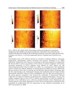
Fig. 13. SEM micrograph (a) and EDX (b) of the TiO
2
modified Ti8Mn foam immersed in
simulated body fluid for 3 days showing the apatite formation.
Novel Titanium Manganese Alloys and
Their Macroporous Foams for Biomedical Applications Prepared by Field Assisted Sintering
221
Implants sometimes were used to substitute bone defects in tumour or spine surgery.
Porous Ti and its alloy foam with their osteoconductive properties are an ideal alternative
bone graft. The porous structure with pore sizes of 200-500 μm of the Ti foams may be able
to permit bone cell penetration and tissue integration. The plateau stress of the human
vertebral bone (load-bearing site) ranges from 24 to 43 MPa, and femoral cancellous bone
(load-bearing site) is in the range of 48-80 MPa (Zhang et al, 2007). The average Young’s
modulus of compact bone of human ranges 7-30 GPa (Zhang et al, 2007). The plateau stress
of the presented Ti and Ti8Mn foams in the range of 27-94 MPa is comparable to that of the
cancellous bone which is sufficient for biomedical applications. For biomedical applications,
the main problem of Ti and Ti alloys in clinical view is their high Young’s modulus. Stress
shielding is known to lead to bone resumption and eventual loosening of the implant. The
dense Ti generally showed much higher Young’s modulus (70-120 GPa) than that of human
bone. Thus, the porous structures were incorporated in the Ti and Ti alloys. In this study,
the porous Ti and Ti8Mn foams show lower Young’s modulus values (6.2-36.1 GPa) than
that of dense ones which are comparable to those of natural compact bone (7-30 GPa). The
macroporous Ti and Ti8Mn foams with plateau stress 27.2-94.2 MPa and Young’s modulus
6.2-36.1 GPa have a potential to be used as bone implants. The low Young’s modulus of
titanium foams is desirable to reduce the amount of stress shielding of the bone into which
the foam is implanted. Thus, the good biocompatibility, the interconnected porous structure
achieved by the SPS and the NaCl dissolution method and the observed mechanical
properties comparable to those of human bones make pure Ti and Ti8Mn foams to ideal
bone implant materials.
5. Conclusions and outlook
The α+β type TiMn alloys with high relative density and ultrafine microstructures were
prepared by using mechanical alloying for 60 hours and spark plasma sintering at 700
o
C for
5 min. The Mn reduced the α to β transformation temperature of Ti and was confirmed as a
β stabilizer element. The hardness increased significantly ranging from 2.4 GPa (Ti2Mn) to
5.28 GPa (Ti12Mn), the elastic modulus ranging from 83.3 GPa (Ti2Mn) to 122 GPa (Ti12Mn)
and the ductility decreased ranging from 21.3% (Ti2Mn) to 11.7% (Ti12Mn) with increasing
manganese content in the Ti. Concentrations of Mn below 8 wt.% in titanium reveal
negligible effects on the metabolic activity and the cell proliferation of human osteoblasts.
Therefore, the Mn could be used in lower concentrations as an alloying element for
biomedical titanium. The Ti2Mn, Ti5Mn and Ti8Mn alloys all have a potential for use as
new bone substitutes and dental implants.
Macroporous Ti foams were successfully fabricated by the free pressureless SPS technique.
Micro-CT results showed the non-uniform pore distribution and poor interconnectivity in
these foams. Alternatively, macroporous pure Ti foams with porosities of 30-70% and pore
sizes of 125-800 μm were prepared by using SPS and NaCl dissolution method. The Ti foams
prepared by SPS at 700
o
C for 8 min under 50 MPa showed pure α-Ti phase structure. The Ti
foams consist of interconnected macropores with square cross sections. The plateau stress
and Young’s modulus agree with the Gibson-Ashby models, and coarsely obey linear
declines with the pore size increase and exponential decays with the increase of porosity.
Ti8Mn foams were also prepared in α+β phases with a porosity of 65% and pore sizes of 300
μm by using the SPS and NaCl dissolution method. TiO
2
nanostructures in anatase/rutile
phases were modified on the pore walls of the Ti8Mn foam uniformly by NaOH solution
Biomedical Engineering, Trends in Materials Science
222
soaking and heat treatment. This surface modified TiMn foam exhibited high in vitro
bioactivity with a fast apatite-forming ability in the simulated body fluid. The Ti and Ti8Mn
foams processed by SPS and NaCl dissolution method showed mechanical properties within
those of human bone range making these materials to be ideal bone implant foams.
As load bearing and long term hard tissue repair materials, Ti and its alloys are the most
outstanding metallic materials nowadays. The modification (processing and/or surface) of
the clinic used Ti alloys, and the exploration of new Ti alloy systems for biomedical
applications are still the tasks for the future. For the Ti foams, the development of
processing techniques to create controlled porosity, pore sizes and interconnectivity is still
required. The relationship between the relative density and mechanical properties of the Ti
foams can be predicted well with the Gibson-Ashby model. However, the relationships
between the porosity-functional properties (thermal, flow, transport, absorption and so on)
are not well modelled yet. The effects of pore architecture, pore size, pore interconnectivity,
inter-connective pore size on the mechanical and functional properties of Ti foams are still
not clear, and need more investigations. Energy saving is one of the hot issues in 21
st
century. There are high requirements of new sintering techniques with rapid energy transfer
and less energy consumption to produce dense Ti alloys and foams. The SPS is considered
as a novel field sintering technique for fast preparation of diverse bulk materials with a near
net shape. The future highlights will be the preparation of nanostructured Ti alloys and the
processing of Ti foams with complex shapes by using the SPS technique. The application of
the SPS in preparation of the biomedical Ti alloys and foams has perspective future.
6. Acknowledgements
Funding for this research was supported by the DFG-Deutschen Forschungsgemeinschaft
(German Research Foundation) with grant No. GRK1505/1 (Welisa). The authors
acknowledge the group of PD Dr. Barbara Nebe in Department of Cell Biology of Rostock
University for the help in the cell experiments, and the group of PD Dr. Ulrich Beck in
Department of Electrical Engineering and Informatics of Rostock University for the help in
the SEM experiments.
7. References
Barbieri, F.C.; Otani, C.; Lepienski, C.M.; Urruchi, W.I.; Maciel, H.S.; Petraconia, G. (2007).
Nanoindentation study of Ti6Al4V alloy nitrided by low intensity plasma jet
process. Vacuum, 67: 457–461.
Brown, S.E. (2006). Bone Nutrition, In: Scientific Evidence for Musculoskeletal, Bariatric, and
Sports Nutrition, Editor: Ingrid Kohlstadt, CRC Press, ISBN: 978-0849337246, US.
Gao, L.; Wang, H.Z.; Hong, J.S.; Miyamoto, H.; Miyamoto, K.; Nishikawa, Y.; Torre,
S.D.D.L.(1999). Mechanical properties and microstructure of nano-SiC-Al2O3
composites densified by spark plasma sintering. Journal of the European Ceramic
Society.19(5): 609-613.
Geetha, M.; Singh, A.K.; Asokamani, R.; and Gogia, A.K. (2009). Ti based biomaterials, the
ultimate choice for orthopaedic implants – A review. Progress in Materials Science,
54: 397-425.
Gibson L. J. & Ashby M.F. (1997). Cellular solids: structure and properties. Cambridge
University Press, ISBN: 0-521-49560-1.UK.
Novel Titanium Manganese Alloys and
Their Macroporous Foams for Biomedical Applications Prepared by Field Assisted Sintering
223
Gu, Y.W.; Khor, K.A.; Cheang, P. (2004). Bone-like apatite layer formation on hydroxyapatite
prepared by spark plasma sintering (SPS). Biomaterials. (25)18: 4127-34.
Hermawan, H.; Dube, D.; Mantovani, D. (2007). Development of degradable Fe-35Mn alloy
for biomedical applications. Advanced Materials Research.15-17: 107-122.
Ibrahim, A; Zhang, F; Otterstein, E.; and Burkel, E. (2011). Processing of porous Ti and
Ti5Mn foams by spark plasma sintering. Materials & Design. 32(1):146-153.
Jayaseelan, D. D.; Kondo, N.; Brito, M.E.; Ohji, T. (2002). High-strength porous alumina
ceramics by the pulse electric current sintering technique. Journal of the American
Ceramic Society. 85(1): 267-269.
Li, J.P.; Li, S.H.; Van Blitterswijk, C.A.; de Groot, K. (2005). A novel porous Ti6Al4V:
characterization and cell attachment. J Biomed Mater Res A, 73: 223-233.
Liu, D.M. (1997). Influence of porosity and pore size on the compressive strength of porous
hydroxyapatite ceramic. Cera. Inter. 23: 135-139.
Long, M.; Rack, H. J. (1998). Titanium alloys in total joint replacement—a materials science
perspective. Biomaterials. 19, 1621-1639.
Lou, Y.; Pan, W.; Li, S.; Wang, R.; Li, J. (2003). A novel functionally graded materials in the
Ti-Si-C system. Materials Science and Engineering A. 345( 1-2):99-105.
Mark, J.J.; & Waqar, A. (2007). Surface Engineered Surgical Tools and Medical Devices, Springer,
ISBN: 978-0-387-27028-9, US.
Munir, Z. A.; Anselmi-Tamburini, U. (2006). The effect of electric field and pressure on the
synthesis and consolidation of materials: A review of the spark plasma sintering
method. J Materials Science. 41:763–777.
Nicula, R.; Cojocaru, V.D.; Stir, M.; Hennicke, J.; Burkel, E (2007). High-energy ball-milling
synthesis and densification of Fe–Co alloy nanopowders by field-activated
sintering (FAST). Journal of Alloys and Compounds, 434-435: 362-366.
Nicula, R.; Lüthen, R.; Stir, M.; Nebe, J.B.; Burkel, E. (2007). Spark plasma sintering synthesis
of porous nanocrystalline titanium alloys for biomedical applications. Biomolecular
Engineering. 24: 564-567.
Nicula, R.; Turquier, F.; Stir, M.; Kodash, V.Y.; Groza, J.R.; Burkel E. (2007). Quasicrystal
phase formation in Al–Cu–Fe nanopowders during field-activated sintering
(FAST). Journal of Alloys and Compounds. 434-435: 319-323.
Rice, R.W. (1993). Comparison of physical property-porosity behaviour with minimum solid
area models. J. Mater. Sci. 8: 2187-2190.
Sima, F.; Socol, G.; Axente, E.; Mihailescu, I.N.; Zdrentu, L.; Petrescu, S.M.; Mayer, I. (2007).
Biocompatible and bioactive coatings of Mn2+-doped β-tricalcium phosphate
synthesized by pulsed laser deposition. Applied Surface Science. 254: 1155-1159.
Takemoto, M.; Fujibayashi, S.; Neo, M.; Suzuki, J.; Matsushita, T.; Kokubo, T.; Nakamura, T.
(2006). Osteoinductive porous titanium implants: effect of sodium removal by
dilute HCl treatment. Biomaterials. 27:2682-2691.
Wen, C. E.; Yamada, Y.; Shimojima, K.; Chino, Y.; Asahina, T.; Mabuchi, M. (2002).
Processing and mechanical properties of autogenous titanium implant materials. J.
Mater. Sci.: Mater. Med. 2002,13, 397-401.
Xu, L. P.; Yu, G.; Zhang, E.; Pan, F.; Yang, K. (2007). In vivo corrosion behavior of Mg-Mn-
Zn alloy for bone implant application.
J Biomed Mater Res A. 83: 703–711.
Biomedical Engineering, Trends in Materials Science
224
Zhang, F.; Shen, J.; Sun, J. (2004). The Effect of Phosphorus additions on Densification, Grain
growth and Properties of nanocrystalline WC/Co composites. Journal of Alloys and
Compounds. 385(1-2): 96-103.
Zhang, F.; Shen, J.;Sun,J.; Zhu, Y.Q.;Wang, G.; and McCartney, G. (2005) Conversion of
Carbon Nanotubes to Diamond by a spark plasma sintering. Carbon. 43 (6): 1254-
1258.
Zhang, F.; Chang, J.; Lu, J.; Lin, K.; and Ning, C. (2007). Bioinspired structure of bioceramics
for bone regeneration in load-bearing sites. Acta Biomaterialia, 3(6): 896-904.
Zhang, F.; Lin, K.; Chang, J.; Lu, J.; and Ning, C. (2008). Spark plasma sintering of
macroporous calcium phosphate scaffolds from nanocrystalline powders. Journal of
the European Ceramic Society. 28 (3): 539-545.
Zhang, F.; Weidmann, A.; Nebe, B. J. ; Burkel, E. (2009) . Preparation of TiMn alloy by
mechanical alloying and spark plasma sintering for biomedical applications. Journal
of Physics: Con. Series. 144: 012007.
Zhang, F.; Weidmann, A.; Nebe, J.B.; Beck, U.; Burkel., E. (2010). Preparation,
microstructures, mechanical properties, and cytocompatibility of TiMn alloys for
biomedical applications. J Biomed Mater Res Part B,Appl Biomater, 94: 406–413.
Zhang, F.; Otterstein E.; Burkel E. (2010). Spark plasma sintering, microstructures and
mechanical properties of macroporous titanium foams. Advanced Engineering
Materials. 12(9): 863-872.
0
Development and Application of Low-Modulus
Biomedical Titanium Alloy Ti2448
Rui Yang, Yulin Hao and Shujun Li
Shenyang National Laboratory for Materials Science,
Institute of Metal Research, Chinese Academy of Sciences
P. R. China
1. Introduction
Economic development leads to improved living standard, but is also attended by the
following consequences: increased number of senile people who, due to degenerative diseases
such as arthritis, may need medical assistance in maintaining their convenience of mobility,
increased volume of transportation in terms of the number of cars and associated traffic
accidents, and increased amount of leisure time channeled to sports that have a higher
than average risk of injuries. All these require orthopaedic surgeries and cause increased
consumption of biomedical materials.
Load bearing orthopedic implants must satisfy the following requirements (Wang, 1996; Long
& Rack, 1999): First of all they are ideally without cytotoxicity, and this places stringent
restriction to the choice of alloying elements. Secondly, their long service life coupled with
the variety of human activity demands excellence in mechanical properties, primarily high
strength and high fatigue resistance, but low elastic modulus. This is a big challenge because
for crystalline materials their strength and elastic modulus tend to increase or decrease
simultaneously. Thirdly, wear resistance is important because wear causes not only implant
loosening but also harmful reactions if the wear debris is deposited in the tissue. Finally,
biochemical compatibility requires the implanted materials to possess superior corrosion
resistance in body environment and be bioactive. The first two aspects clearly fall into the
domain of alloy design; the last two, though closely related to alloy type and composition, are
normally the subjects of surface modification.
Judging from the above requirements titanium alloys stand out as the best class of implant
materials due to a combination of acceptable biocompatibility and good properties such as
high strength, low density, relatively low elastic modulus and excellent corrosion resistance.
While Ti–6Al–4V is used earliest in biomedical engineering and is still a benchmark among
biomedical alloys (Froimson et al., 2007), it was not purpose-designed. In terms of cytotoxicity,
vanadium is toxic both in the elemental state and in the form of oxide (Wapner, 1991;
Eisenbarth et al., 2004), and there exits some correlation between V and Al ions released
from the alloy and long-term health problems such as Alzheimer disease, neuropathy and
ostemomalacia (Nag et al., 2005). These facts highlight the importance of careful choice of
alloying additions when designing new alloys specifically for biomedical use.
The main mechanical effect of an implant on the bone relates to stress shielding, i.e., reduction
in bone stress in vivo following the introduction of the implant. The stress needed by cells
around the implant is thus shielded and the cells do not survive. The change in stress
10