Báo cáo hóa học: " Preparation and properties of copper-oil-based nanofluids" ppt
Bạn đang xem bản rút gọn của tài liệu. Xem và tải ngay bản đầy đủ của tài liệu tại đây (646.65 KB, 7 trang )
NANO EXPRESS Open Access
Preparation and properties of copper-oil-based
nanofluids
Dan Li
1*
, Wenjie Xie
2
and Wenjun Fang
3*
Abstract
In this study, the lipophilic Cu nanoparticles were synthesized by surface modification method to improve their
dispersion stability in hydrophobic organic media. The oil-based nanofluids were prepared with the lipophilic Cu
nanoparticles. The transport properties, viscosity, and thermal conductivity of the nanofluids have been measured.
The viscosities and thermal conductivities of the nanofluids with the surface-modified nanoparticles have higher
values than the base fluids do. The composition has more significant effects on the thermal conductivity than on
the viscosity. It is valuable to prepare an appropriate oil-based nanofluid for enhancing the heat-transfer capacity of
a hydrophobic system. The effects of adding Cu nanoparticles on the thermal oxidation stability of the fluids were
investigated by measuring the hydroperoxide concentration in the Cu/kerosene nanofluids. The hydroperoxide
concentrations are observed to be clearly lower in the Cu nanofluids than in their base fluids. Appropriate amounts
of metal nanoparticles added in a hydrocarbon fuel can enhance the thermal oxidation stability.
Introduction
Nanofluid is a novel heat-transfer fluid prepared by dis-
persing nanometer-sized solid particles in traditional
heat-transfer fluid to increase thermal conductivity and
heat-transfer performance. Nanofluid was coined by
Choi and colle agues [1-3] in 1995 at Argonne National
Laboratory of the USA. Nanofluids wit h water, ethylene
glycol, or oil as the base fluid were o f great significance
primarily because of their enhanced thermal properties.
There are compelling needs in many industr ial fields to
develop oil-based heat transfer fluids with significantly
higher thermal conductivity for energy-efficient heat
exchangers. Many efforts have been focused on the oil-
based nanofluids. Transformer oil, mineral oil, silicon
oil, hydrocarbon fuels, and some organic solutions are
used as the base fluids for studying nanoflu ids. The dis-
persion and thermal conductivities of the oil-based
nanofluids containing Cu, CuO, Ag, or Al
2
O
3
particles
have been recently reported [4-6].
When nanoparticles are introduced into oil, the parti-
cles are usually sedimented within several minutes
because of the poor compatibility between the
nanoparticles and the base oil. The agglomerated parti-
cles are g radually settled over time, which leads to the
poor stability and low heat-transfer capability of the sus-
pensions. Thus, an appropriate lipophilic modification
process is needed for the formation of a stable oil-based
nanofluid. Surface modification on metallic particles
with hydrophobic ligands and addition of dispersant can
be employed to improve the compatibility between the
nanoparticles a nd the oil-based fluid. T he o rganic
ligands with long hydrocarbon chains coordinated to the
nanoparticles prevent the particles from clustering, and
the surface-modified nanoparticles possess good disper-
sion behavior in oils [4,7-9].
Kerosene, a typical hydrocarbon fuel, circulated in air-
craft for cooling can serve as the primary thermal sink
by dissipating waste heat from aircraft subsystems. How-
ever, it has relatively low thermal conductivity. As is
well known, a kerosene-based nanofluid can improve
the heat transfer property and cooling capacity. In t his
study, we attempted to synthesize lipophilic Cu nano-
particles and to prepare oil-based nanofluids. The
hydrophobic layers formed on the surface of copper
nanoparticles can protect the particles against oxidation
and improve dispersion stability of oil-based nanofluids
[10-12], which are important for exploiting the potential
benefits and applications of the enhanced thermal prop-
erties of the nanofluids. In the meanwhile, the effects of
* Correspondence: ;
1
Department of Chemistry and Chemical Engineering, Weifang University,
Weifang 261061, China
3
Department of Chemistry, Zhejiang University, Hangzhou 310027, China
Full list of author information is available at the end of the article
Li et al. Nanoscale Research Letters 2011, 6:373
/>© 2011 Li et al; licensee Springer. This is an Open Access article distributed under the terms of the Creative Com mons Attribu tion
License ( which permits unrestricted use, distribution, and reproduction in any medium,
provided the original work is properly cited.
the lipophilic Cu nanoparticles on the viscosity, thermal
conductivity, and thermal oxidation stability of the
nanofluids are also investigated.
Experimental
Materials and preparation of ligand
All the materials and solvents used in this study, P
2
S
5
,
cetyl alcohol, anhydrous ammonia, benzene, cupric acet-
ate, ethanol, sodium hypophosphite, hydrazine hydrate
solution (85%), toluene, decahydronaphthalene, and
dichloromethane were analytic grade agents.
The Cu nanoparticles were prepared and modified by
O, O-di-n-cetyldithiophosphoric acid. The O, O-di-n-
cetyldithiophosphate [13] was synthesized by heating
P
2
S
5
(0.02 mol) and cetyl alcohol (0.07 mol) at 80°C for
3 h. The suspension was cooled to room temperature
followed by the addition of 50 mL dichloromethane.
The mixture was filtered, followed by evaporation of the
solvent. Anhydrous ammonia was subsequently bubbled
through the solution under stirring. The ligand, a mmo-
nium (O, O)-dialkyldithiophosphate, was then precipi-
tated and recrysta llized in benzene, washed wit h
absolute ethyl ether, and dried in vacuum.
Preparation and characterization of Cu nanoparticles
Cupric acetate (0.002 mol) was dissolved in 20 mL deio-
nized water used as the precursor of Cu nanoparticles.
A mixture of the ligand (O, O-di-n-cetyldithiopho-
sphate) and sodium hypophosphite (NaH
2
PO
2
, 0.0015
mol) in 100 mL solvent of ethanol/water was stirred
uniformly at 60°C. The solution of cupric acetate was
introduced dropwise into the mixture, and the reaction
system turned from colorless solution to yellow suspen-
sion. Then, the hydrazine solution (10 mL) was added
to the mixture, and a dark colloid was observed. The
mixture was stirred at 60°C for 0.5 h and then cooled to
room temperature. The precipitate was separated by
centrifugation and was washed subsequently with water
and ethanol. After separation, the nanoparticles were
dried in a vacuum oven at 45°C for 2 h.
The surface-modified Cu nanoparticles with various
molar ratios of P to Cu (1:2, 1:5, and 1:10) were pre-
pared by fixing the concentrations of copper salt and
reductant, and varying the concentration of O, O-di-n-
cetyldithiophosphate. Because the ligands act as particle
protectors through coordinating the S-containing end
groups on the copper particle surfaces and the hydro-
phobic carbon tails are pointed outward from the parti-
cles, the resulting copper nanoparticles with the
modification layers should be hydrophobic and be dis-
persed in nonpolar solvents.
The phase properties of the surface-modified Cu
nanoparticles were characterized by X-ray powder dif-
fraction (XRD) using a Thermo X-ray diffractometer
(Bruker, Germany) with monochromatized Cu Ka
radiation (l = 1.5405 Å). The differential sc anning
calorimeter (DSC/TG, NETZSCH STA 409 PC/PG)
was used to analyze the thermal decomposition process
of the particles with a heating rate of 10 K/min in N
2
withaflowrateof20mL/min. Transmission electron
microscopy (TEM) images were taken with JEM-
200CX (JEOL, Japan) instrument using an operating
voltage of 160 kV. Scanning electron microscopy
(SEM) images were taken with field-emission scanning
electronmicroscope,andtheenergydispersiveX-ray
analysis (EDX) was carried out on the SEM equipped
with energy-dispersive spectrometer (FEI SIRION-100,
GENENIS-4000, Netherlands). A Nexus 470 Fourier
transform infrared spectrometer (NICOLET, USA) was
employed to observe the changes of organic functional
groups.
Preparation of nanofluids
Three types of nanofluids were prepared by dispersing
different mass fractions of the surface-modified Cu
nanoparticles in kerosene, toluene, and decahydro-
naphthalene as the base liquids without a dispersant.
The samples w ere homogenized for about 5 min using
an ultrasonic disrupter to ensure proper dispersion of
the nanoparticles. The color of the suspension was
observed to be puce.
Measurements on viscosity and thermal conductivity
A capillary visc ometer was utilized to det ermine the
viscosities of the Cu nanofluids. The viscometer was
filled with 15 mL nanofluid and was submerged into a
thermostat ic bath with a resolution of 0.01 K. The verti-
cal angle of the viscometer was accurately controlled
with a special tripod. The flow time was measured with
a stopwatch to an accuracy of 0.01 s. The viscometer
was calibrated with twice-distilled water. Each viscosity
value of the nanofluid was reported by averaging over
three consecutive runs. The flow time was reproducible
to be ± 0.2 s and the uncertainty of viscosity was within
± 0.002 mPa s. The densities of all the nanofluids were
measured by a 10-mL capillary-type pycnometer, which
was calibrated with deionized double-distilled water.
The dynamic viscosity, h, was calculated according to
the equation:
η =
νρ
(1)
where r and ν arethedensityandkinematicviscosity
of the nanofluid, respectively, at the same temperature.
Measurements of the thermal conductivities of Cu
nanofluids were performed by means of a computer-
controlled transient calorimeter [14]. The schematic dia-
gram of the apparatus has been described previously in
detail [15]. The nanofluid samples were added into the
Li et al. Nanoscale Research Letters 2011, 6:373
/>Page 2 of 7
thermal conductivity cell, and a series of voltage differ-
ences (ΔV) of the unbalanced bridge were recorded with
the time at each temperature. These data were utilized
to calculate the slope of the voltage against time (dV/dt)
of the unbalanced bridge. The thermal conductivities of
the base fluids and nanofluids were calculated from the
established equation between l and dV/dt,andthe
enhanced ratios of thermal conductivity were then
obtained. All the measurements were performed at
atmospheric pressure.
Thermal-oxidation tests
The Cu/ker osene-based nanofluids (0.1% Cu nanoparti-
cles) were thermally oxidized in an isothermal appara-
tus. Each test tube containing 100-mL sample of Cu
nanofluid was placed in the heated test well. The inves-
tigated samples were subjected to thermal oxidation at
120 or 140°C. The temperature remained steady within
± 1°C. The flow meters were employed to regulate the
oxygen flow with the rate of 30 mL/min into each sam-
ple by means of a gas dispersion tube. A small number
of aliquots (<0.5 mL) of the samples were removed from
the test tubes at fixed time intervals for the hydroperox-
ide measurements. The hydroperoxides formed in the
samples during the thermal oxidization process were
determined through measuring the absorption spectra of
the iodine-starch solutions using ultraviolet-visible spec-
trometry [16,17].
Results and discussion
Characterization of surface-modified Cu nanoparticles
Depending on the concentration of the ligand (O, O-di-
n-cetyldithiophosphate), different generated products of
surface-modified Cu nanoparticles have been obtained.
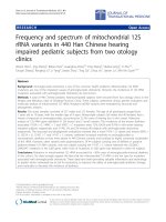
The XRD patterns of several samples are shown in
Figure 1. Figure 1a gives the powder XRD pattern of the
O, O-di-n-cetyldithiophosphate.Figure1b,c,dgives
those of the products with molar ratios of P to Cu of
1:2, 1:5, and 1:10, respectively. The XRD pattern with P:
Cu of 1:2 (Figure 1b) or 1:10 (Figure 1d) only exhibits
the peaks of ligand or Cu, respectively. The XRD pattern
shown in Figure 1c gives three characteristic peaks
which can be indexed as face-centered cubic (fcc) struc-
ture Cu (111), (200), and (220). No visible XRD peaks
arisi ng from the impurity phase such as CuO and Cu
2
O
are found. It is difficult for the formati on of the core of
Cu in the reaction solution when the ratio of li gand is
too high. H owever, the ligand is not sufficient to modify
the Cu particles produced in the reduct ion process,
when the ratio of the ligand is too low. Therefore, the
resultant prod uct with P:Cu molar ratio of 1:5 is appro-
priate for preparing nanofluids. The characterizations
and studies discussed in this section are focused on this
composition.
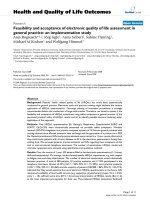
Infrared spectra of O, O-di-n-cetyldithiophosphate and
surface-modified Cu nanop articles are shown in Figure
2. As shown in Figure 2a, the absorptions at 2918 and
2850 cm
-1
are assigned to the stretching vibrations of
CH
2
groups, and the band at 1470 cm
-1
corresponds to
the deformation vibration of CH
2
groups. The absorp-
tion at 720 cm
-1
is due to the rocking vibration of the
long chain alkanes [(CH
2
)
n
, n >4].Theabsorptions
from 930 to 1050 cm
-1
are attributed to the stretching
20 40 60 80
2
T
q
a
d
(220)
(200)
(110)
c
b
Figure 1 XRD patterns of several samples : (a) O, O-di -n-
cetyldithiophosphate and surface-modified Cu products with molar
ratios of P to Cu of (b) 1:2; (c) 1:5; and (d) 1:10.
4000 3500 3000 2500 2000 1500 1000 50
0
b
3187
2849
2917
667
721
964
1421
1467
818
1470
wavelen
g
th/nm
721
991
a
Figure 2 Infrared spectra of (a) O, O-di-n-cetyldithio phosphate,
and (b) surface-modified Cu nanoparticles.
Li et al. Nanoscale Research Letters 2011, 6:373
/>Page 3 of 7
vibration of O-CH
2
. The absorptions at 687 and 670
cm
-1
are attributed to the stretching vibrations of P = S
group, whi le the absorption at 582 cm
-1
is attributed to
the stretching vibrations of P-S group. The absorption
at 1400 cm
-1
is assigned to the stretching vibrations of
NH
4
+
. As shown in Figure 2b, the bands of C-H and O-
CH
2
are also observed in the surface-modified Cu nano-
particles, while the absorption peaks of P = S and P-S
shifts, and the bands of N-H mostly disappear.
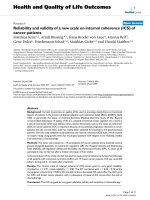
Figure 3 shows the TG and DTA curves of O, O-di-n-
cetyldithiophosphate and its surface-modified Cu nano-
particles, respectively. It is seen from the TG curve that
O, O-di-n-cetyldithiophosphate and Cu nanoparticles
begintoloseweightat110and210°C,respectively.An
obvious mass loss ranging from 210 to 350°C is
observed for the Cu nanoparticles, and the total mass
loss is about 40%. From the TG analyses, it can be con-
cluded that the modification agent is coated on Cu
nanocores through strong interaction, but not a mixture
or simple absorption between Cu nanoparticles and
modification agent. If the products comprise the mix-
ture of Cu nanoparticles and modification agent, then
the modification layers can be rinsed off in the synthesis
proceeding, and very l arge amount of mass loss in the
TG curve should not occur.
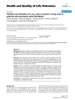
Figure 4 shows an SEM image (Figure 4a), an EDX
spectrum (Figure 4b), a TEM image, and HTEM image
of the surface-modified Cu nanoparticles. Nanopar ticles
with diameter in the range of 40-60 nm can be seen
from the SEM image. The EDX analysis indicates that
the Cu mass fraction in the prepared nanoparticles is
60-62%.Thisisconsistentwith the TG analysis. Figure
4c depicts a TEM image and the corresponding selected
area electron diffraction (SAED) pattern. The micro-
graph reveals that the surface-modified Cu nanoparticles
consist of spherical particles. The diffraction pattern
furth er proves an fcc structure. The lattice fringes of Cu
nanoparticles observed by close inspection with HRTEM
are shown in Figure 4d.
The Cu nanoparticles are surface-modified by the
organic ligands containing hydrocarbon tail. The coating
layers should not easily separate from the surface of the
Cu nanoparticles when the Cu nanoparticles are dis-
persed in the oil-based fluids. The lipophilic surface-
modified Cu nanoparticles should be dispersed in hydro-
phobic solvents, such as toluene, chloroform, and liquid
paraffin. It shoul d not be dispersed in water and should
not stay at the aqueous-organic interface. Therefore , the
dispersion capability of Cu nanoparticles in hydrophobic
solvents is improved by the surface modification, which
enables the surface-modified Cu nanoparticles to be
used as additives in oils.
Viscosities and thermal conductivities of nanofluids
The effects of both temperature and mass fraction of the
nanoparticles on the viscosities of the nanofluids were
investigated. Figure 5 shows the results of viscosity mea-
surements for different fluid-based nanofluids at the
temperature range from 20 to 60°C. The viscosity of a
nanofluid decreases with increasing temperature, in a
manner similar to that of a pure base liquid. It increases
somewhat with increasing concentration of the nanopar-
ticles. The addition of nanoparticles with 1% of mass
fraction leads to no more than 5% increase of the visc-
osity. Therefore, the formatio n of nanofluids has no sig-
nificant effect upon the viscous resistance.
Thermal conductivities of the nanofluids for different
fluid-based nanofluids as a function of mass fraction of
nanoparticles at 25°C are represented in Figure 6a. It
can be seen that the thermal conductivity of Cu nano-
fluid increases with increasing mass fraction of nanopar-
ticles for different fluid-based nanofluids. The
relationship between the thermal conductivity enhance-
ment and the mass fraction is nonlinear. The
100 200 300 400 500
50
60
70
80
90
100
Temperature/ qC
TG/%
b
0
2
4
6
8
10
12
14
16
18
20
DTA
/(
mW
/
mg
)
100 200 300 400 500
0
20
40
60
80
100
TG/%
Temperature/ qC
a
-4
-2
0
2
4
6
8
10
12
14
DTA/(mW/mg)
Figure 3 TG/DTA curves: (a) O, O-di-n-cetyldithiophosphate, and (b) surface-modified Cu nanoparticles.
Li et al. Nanoscale Research Letters 2011, 6:373
/>Page 4 of 7
temperature effects on the enhancement of effective
thermal conductivity are investigated by measuring the
thermal conductivities of Cu/kerosene-based nanofluids
at different temperatures,asshowninFigure6b.It
demonstrates that the thermal co nductivities of the oil-
based nanofluids increase clearly with the fluid tempera-
ture. The thermal conductivity of kerosene-ba sed nano-
fluid increases by about 10, 13, and 14.6 % with 1.0%
(mass fraction) Cu nanoparticles at 25, 40, and 50°C,
respectively. As the heat transfer in solid-liquid suspen-
sion occurs at the particle-fluid interface [18], an
increase of the interfacial area can lead to efficient heat-
transfer properties. Because the modified layers cap the
copper cores and the metal surfaces do not directly con-
tact with the base fluid, the surface-modified Cu nano-
particles are less effective than the uncoated Cu
particles as far as the thermal-conductivity enhancement
is concerned.
Hydroperoxides in the Cu/kerosene-based nanofluids
The hydroperoxides are the intermediates in the autoxi-
dation reactions of hydrocarbon fuels. The
hydroperoxide concentration is important for character-
izing the thermal oxidation of a kerosene. Figure 7 gives
the hydroperoxide concentration as a function of time
in Cu/kerosene-based nanofluids and in kerosene with-
out Cu nanoparticles thermal-oxidized at 120 a nd 140°
C. As shown in Figure 7, the change of hydroperoxide
concentration in the nanofluid oxidized at 120°C is
nearly the same as that of the blank kerosene. At 140°C,
the hydroperoxide concentrations in the nanofluid mea-
sured within 3 h are very low. It is clear that the hydro-
peroxide concentrations in the nanofluids are much
lowe r than those in the blank kerosene during the ther-
mal oxidation process. The Cu nanoparticles can signifi-
cantly reduce the forma tion of the hydropero xides in
the kerosene. During the thermal oxidation at 140°C,
the Cu nanoparticles deposit and react with oxygen.
Therefore, the black CuO were found in the bottom o f
reactor. It indicated that the Cu nanoparticles were oxi-
dized before the kerosene was oxidized. At lower tem-
peratures, the coating layers on the surfaces of the
nanoparticle s prevent the Cu cores from oxidation. At
higher temperatures, however, the coatings open or
a
b
c
d
Figure 4 ( a) SEM image; (b) EDX spectrum of the surface-modified Cu n anoparticles; (c) TEM images SAED pattern; and (d) HTEM
image.
Li et al. Nanoscale Research Letters 2011, 6:373
/>Page 5 of 7
release from the surfaces, giving the opportunity for
oxygen molecules to gain access to the Cu cores. The
Cu nanoparticles then react with the oxygen before the
kerosen e is oxidized [19]. As a result, the hydroperoxi de
concentrations are observed to be re latively low in the
Cu nanofluids. Appropriate amounts of metal nanoparti-
cles added into a hydrocarbon fuel can enhance its ther-
mal oxidation stability.
Conclusions
The Cu oil-based nanofluids have been prepared by dis-
persing Cu nanoparticles modified with O, O-di-n-cetyl-
dithiophosphate in kerosene, toluene, or
decahydronaphthalene. The modified ligand is e ffective
in improving the lipophilic property of Cu nanoparticles.
The modified layers can be effectively coated on the sur-
faces of the Cu nanoparticles even when they are
20 30 40 50 60
0.7
0.8
0.9
1.0
1.1
1.2
1.3
1.4
1.5
1
.
6
K
/ (mm
2
s
-1
)
Temperature/qC
0
0.25%
0.5%
1 %
a
20 30 40 50 60
0.35
0.40
0.45
0.50
0.55
0.60
0.65
K
/ (mm
2
s
-1
)
Temperature/qC
0
0.25%
0.5%
1%
b
20 30 40 50 60
1.0
1.2
1.4
1.6
1.8
2.0
2.2
2.4
2.6
2.8
3.0
K
/ (mm
2
s
-1
)
Tem
p
erature/qC
0
0.25%
0.5%
1 %
c
Figure 5 Viscosities of Cu nanofluids: (a) Cu/kerosene; (b) Cu/toluene; and (c) Cu/decahydronaphthalene.
0.000 0.005 0.010 0.015
0.115
0.120
0.125
0.130
0.135
0.140
0.145
0.150
0.155
Thermal conductivity/(W m
-1
K
-1
)
Particle mass fraction
toluene
kerosene
decahydronaphthalene
a
0.0000 0.0025 0.0050 0.0075 0.0100
0.105
0.110
0.115
0.120
0.125
0.130
0.135
0.140
0.145
0.150
Thermal conductivity/(W m
-1
K
-1
)
Particle mass fraction
25 qC
40 qC
50 qC
b
Figure 6 Thermal conductivity of nanofluids: (a) Variation of thermal conductivity of nanofluids at 25°C with mass fraction of nanoparticles;
(b) variation of thermal conductivity with temperature for Cu/kerosene-based nanofluids.
Li et al. Nanoscale Research Letters 2011, 6:373
/>Page 6 of 7
dispersed in the oil-based fluids. The thermal conductiv-
ity of nanofluids increases with the mass fraction of
nanoparticles to some extent. The hydroperoxide con-
centrations are observed to be low er in the Cu nano-
fluids than in their base fluids. Appropriate amounts of
metal nanoparticles added into a hydrocarbon fuel can
enhance its thermal oxidation stability.
Abbreviations
EDX: energy dispersive X-ray analysis; SAED: selected area electron
diffraction; SEM: scanning electron microscopy; TEM: transmission electron
microscopy; XRD: X-ray powder diffraction.
Author details
1
Department of Chemistry and Chemical Engineering, Weifang University,
Weifang 261061, China
2
Qianjiang College, Hangzhou Normal University,
Hangzhou 310027, China
3
Department of Chemistry, Zhejiang University,
Hangzhou 310027, China
Authors’ contributions
DL: conceived of the study, carried out the experimental analyses,
performed the XRD analyses, TEM characterizations and drafted the
manuscript, WX: conceived the study, and participated in its design and
coordination, WF: conceived the study, and participated in its design and
coordination. All authors read and approved the final manuscript.
Competing interests
The authors declare that they have no competing interests.
Received: 29 January 2011 Accepted: 5 May 2011 Published: 5 May 2011
References
1. Eastman JA, Choi SUS, Li S, Thompson LJ, Lee S: Enhanced thermal
conductivity through the development of nanofluids: nanophase and
nanocomposite materials II. Pennsylvania: Mater Res Soc 1997, 457:3-11.
2. Lee S, Choi SUS, Li S, Eastman JA: Measuring thermal conductivity of
fluids containing oxide nanoparticles. ASME J Heat Transfer 1999,
121:280-289.
3. Xuan Y, Li Q: Heat transfer enhancement of nanofluids. Int J Heat Fluid
Flow 2000, 21:58-64.
4. Choi C, Yoo HS, Oh JM: Preparation and heat transfer properties of
nanoparticle-in-transformer oil dispersions as advanced energy-efficient
coolants. Curr Appl Phys 2008, 8:710-712.
5. Murshed SMS, Leong KC, Yang C: Investigations of thermal conductivity
and viscosity of nanofluids. Int J Therm Sci 2008, 47:560-568.
6. Zhu H, Zhang C, Tang Y, Wang J: Novel synthesis and thermal
conductivity of CuO nanofluid. J Phys Chem C 2007, 111:1646.
7. Stouwdam JW, Veggel FCJM: Improvement in the luminescence
properties and processability of LaF
3
/Ln and LaPO
4
/Ln nanoparticles by
surface modification. Langmuir 2004, 20:11763-11771.
8. Sun L, Zhang Z, Wu Z, Dang H: Synthesis and characterization of DDP
coated Ag nanoparticles. Mater Sci Eng A 2004, 379:378-383.
9. Zhou J, Wu Z, Zhang Z, Liu W, Xue Q: Tribological behavior and
lubricating mechanism of Cu nanoparticles in oil. Tribol Lett 2000,
8:213-219.
10. Kanninen P, Johans C, Merta J, Kontturi K: Influence of ligand structure on
the stability and oxidation of copper nanoparticles. J Colloid Interface Sci
2008, 318:88-95.
11. Song X, Sun S, Zhang W, Yin Z: A method for the synthesis of spherical
copper nanoparticles in the organic phase. J Colloid Interface Sci 2004,
273:463-469.
12. Khanna PK, Kale TS, Shaikh M, Rao NK, Satyanarayana CVV: Synthesis of
oleic acid capped copper nano-particles via reduction of copper salt by
SFS. Mater Chem Phys 2008, 110:21-25.
13. Gianini M, Caseri WR, Gramlich V, Suter UW: Synthesis and characterization
of liquid platinum compounds. Inorg Chim Acta 2000, 299:199-208.
14. Wang C, Yang M: A new calorimeter for measuring rapidly the thermal
conductivity of liquids. Thermochim Acta 1995, 255:365-370.
15. Li D, Fang W, Xie W, Xing Y, Guo Y, Lin R: Preparation of well-dispersed
silver nanoparticles for oil-based nanofluids. Ind Eng Chem Res 2010,
49:1697-1702.
16. Cimato A, Facorro G, Aguirre F, Hager A, De Paoli T, Ihlo J: A
spectrophotometric method for the determination of hydroperoxides in
liposomes. Spectrochim Acta A 1998, 54:2001-2008.
17. Li D, Fang W, Xing Y, Guo Y, Lin R: Spectroscopic studies on thermal-
oxidation stability of hydrocarbon fuels. Fuel 2008, 87:3286-3291.
18. Karthikeyan NR, Philip J, Raj B: Effect of clustering on the thermal
conductivity of nanofluids. Mater Chem Phys 2008, 109:50-55.
19. Yetter RA, Risha GA, Son SF: Metal particle combustion and
nanotechnology. Proc Combust Inst 2009, 32:1819-1838.
doi:10.1186/1556-276X-6-373
Cite this article as: Li et al.: Preparation and properties of copper-oil-
based nanofluids. Nanoscale Research Letters 2011 6:373.
Submit your manuscript to a
journal and benefi t from:
7 Convenient online submission
7 Rigorous peer review
7 Immediate publication on acceptance
7 Open access: articles freely available online
7 High visibility within the fi eld
7 Retaining the copyright to your article
Submit your next manuscript at 7 springeropen.com
0246810
0.00
0.02
0.04
0.06
0.08
0.10
0.12
0.14
Hydroperoxide concentration/(mmol/g)
Tim
e/
h
blank 140 qC
+ 0.1% 140 qC
blank 120 qC
+0.1% 120 qC
Figure 7 The change of hydroperoxide concentration in the
nanofluid oxidized at 120°C and 140°C.
Li et al. Nanoscale Research Letters 2011, 6:373
/>Page 7 of 7