Báo cáo hóa học: " A comparative study of non-covalent encapsulation methods for organic dyes into silica nanoparticles" pptx
Bạn đang xem bản rút gọn của tài liệu. Xem và tải ngay bản đầy đủ của tài liệu tại đây (679.34 KB, 12 trang )
NANO EXPRESS Open Access
A comparative study of non-covalent
encapsulation methods for organic dyes into
silica nanoparticles
Aurélien Auger
*
, Jorice Samuel, Olivier Poncelet and Olivier Raccurt
Abstract
Numerous luminophores may be encapsulated into silica nanoparticles (< 100 nm) using the reverse
microemulsion process. Nevertheless, the behaviour and effect of such luminescent molecules appear to have
been much less studied and may possibly prevent the encapsulation process from occurring. Such nanospheres
represent attractive nanoplatforms for the development of biotargeted biocompatible luminescent tracers. Physical
and chemical properties of the encapsulated molecules may be affected by the nanomatrix. This study examines
the synthesis of different types of dispersed silica nanoparticles, the ability of the selected luminophores towards
incorporation into the silica matrix of those nanoobjects as well as the photophysical properties of the produced
dye-doped silica nanoparticles. The nanoparticles present mean diameters between 40 and 60 nm as shown by
TEM analysis. Mainly, the photophysical characteristics of the dyes are retained upon their encapsulation into the
silica matrix, leading to fluorescent silica nanoparticles. This feature article surveys recent research progress on the
fabrication strategies of these dye-doped silica nanoparticles.
Introduction
The development and need for silica-based fluorescent
nanoparticles as markers in biological applications such
as sensing and imaging have spread significantly since
the 1990s [1-3]. Fluorescent labelling of biomolecules
has been established as an essential tool in many biolo-
gical investigations. Recently, significant advances have
led to a large variety of labelling reagents based on inor-
ganic (quantum dots [4], lanthanide-doped oxides [5,6],
metallic gold [7,8]) or organic nanomaterials (latex,
polystyrene and polymethylmethacrylate) [9]. Indeed,
small luminescent molecules like organic dyes displaying
high quantum yield can be encapsulated into oxide
nanoparticles, specifically into silica, by sol-gel. These
new fluorescent probes can be developed for the field of
biological assays and have reached great expectations
[10,11]. The wide range and variety of fluorophores
available nowadays facilitate the targeting of suitable
applications for the newly prepared nanoparticle
materials.
Organics dyes have been known for some time now to
be used in biology for fluorescent labelling. Although
those dyes possess a certai n number of drawbacks
including a short Stokes shift, poor photochemical stabi-
lity, sensibility to the buffer composition (quenching or
decomposition due to the pH), susceptibility to photo-
bleaching and decomposition under repeated excitation,
they remain used extensively and considerably as a
result of t heir low cost, commercial availability and ease
of use. Furthermore, modern research has developed
organic dyes which exhibit better chemical and optical
properties. Examples involve fluorescein [12,13], rhoda-
mine [14,15], cyanine [13,16], alexa dyes [13,17], oxa-
zines [18,19], porphyri ns [20] and phthalocyanines [21],
just to name a few. Even if fluorescence detection exhi-
bits a sh arp sensitivity, most of the organic fluorophores
used as luminescent biomarkers present drawbacks.
Therefore, hydrophobicity (causin g a poor sol ubility into
biological buffers) (collisional), quenching in aqueous
media and irreversible photodegradation under intense
excitation light [11,22], requires encapsulation so that to
produce monodisperse and more robust emitters from
organic dye molecules and amorphous silica. Further-
more, a supplementary advantage to encapsulation of
* Correspondence:
CEA Grenoble, Department of Nano Materials, NanoChemistry and
NanoSafety Laboratory (DRT/LITEN/DTNM/LCSN), 17 rue des Martyrs, 38054
Grenoble Cedex 9, France
Auger et al. Nanoscale Research Letters 2011, 6:328
/>© 2011 Auger et al; licensee Springer. This is an Open Access article distributed under the terms of the Cre at ive Commons At tributi on
License (ht tp://creativecommons.org/l icenses/by/2.0), which permits unrestricted use, distribution, and reproduction in any medium,
provided the original work is properly cited.
organic dyes into silica beads is to enhance the detection
limit by encapsulating a larger number of fluorophores
molecules by synthesised probes. The technique of
encapsulation of fluorophores into silica beads prevents
from interaction of fluorophores with the buffer. Finally,
silica functionalisation is a well-known and a well-devel-
oped chemistry, and the incorporation of dyes into silica
nanoparticles offer a great potential for customising the
surface independently to the dye structure.
Traditionally, there are two chemical approaches for
incorporating organic dyes into silica nanoparticles. The
first approach consists of using covalent bonding of the
dye with the silicated matrix [23-25]. On the c ontrary,
the second approach has been described as using non-
covalent or non-bonding process (i.e. by electrostatic
interactions), by entrapping the dye into the siloxane
matrix [26]. Relatively few examples (involving rhoda-
mine and ruthenium complexes) have been reported in
the literature, and covalent binding of the dye to the
silica network is usually the preferred method. Sol-gel
synthesis of silica b eads can also be undertaken by two
types of sol-gel methods: the Stöber [27] and the micro-
emulsion methods [28]. It is obvious that the best
method for incorporation of a dye into silica beads is by
the covalent bonding approach but it requires the dye to
possess sufficient chemical groups towards functionalisa-
tion and chemical reaction between the dye and the sili-
cated precursors. This concept might sometimes
enhance considerably the difficulty of the dye prepara-
tion. Consequently, the non-covalent approach repre-
sents a promising way and more attention should be
paid to its investigation since it exhibits a low-cost
method, and that this pro cess does not emphasise the
limitation of the chosen dye.
According to the Stöber method, the incorporation
yield of the dye into the silica beads under non-cova-
lent bonding is poor and dependant of the absorption
force between the dye itself and the silica precursor
[15]. However, t he microemulsion process avoids that
drawback, controlling the quantity of incorporated dye
into silica beads by utilising a water soluble dye. For
reminding, the first method has been developed in the
late 1960s by Stöber et al. [27]. The mild synthetic
protocol consists of the hydrolysis and condensation
of silica alkoxide precursors (such as tetraethoxysilane,
TEOS) in ethanol solution in the presence of aqueous
ammonium hydroxide mixture as a catalyst to gener-
ate electrostatically stabilised, spherical and monodis-
perse particles. Indeed, homogeneous nucleation forms
silica particles of tens to hundreds of nanometres in
size [28,29]. Even if this method is rather simple and
that it can involve t he incorporation of both organic
and inorganic markers [19], the fact is that the particle
size may not be uniform and besides different
modifications of the particle surface are not easily
achieved and might require covalent binding to
achieve proper encapsulation. The second approach
for the synthesis of uniform organic dye-doped silica
nanoparticles of different sizes can be achieved by a
reverse microemulsion method [30-33]. Reverse
microemulsion techniques rely on the stabilisation of
water nanodroplets (by surfactant molecules) formed
in an oil solution (water in oil (W/O) emulsion) which
act as nanoreactors, where silane derivatives hydrolysis
and formation of nanoparticles take place, entrapping
dye molecules [11,26]. Furthermore, the nanoreactor
environment within the reverse micelle has been
yielded highly monodisperse nanoparticles and an
increase in the incorporation of nonpolar molecules
has been observed [34] because the particle’ sdimen-
sion was limited by the volume of the micelle. The
microemulsion method produced hydrophilic and
fairly uniform-sized nanoparticles and allows easy
modulation of the nanoparticle surfaces for various
applications. Moreover, it has been determined that
the size of the nanoparticles is controlled by para-
meters such as the hydrolysis reagent, the nature of
surfactant, the reaction time and the oil/water ratio,
just to name a few [28].
Dye encapsulation c an be achieved either by covalent
bond of the dye with silica precursors before the hydro-
lysis or by first solubilising the dye in the core (small
reactors) of the microemulsion and then carrying out
the polymerisation. As a matter of fact, the covalently
dye-doped silica nanoparticles have launched a promis-
ing field towards the development and investigation of
luminescent biomarkers. Manystudiesonthistopic
were reported [11,28,30-32], principally since 1992, van
Blaaderen and co-wor kers [23,24] described for the first
time covalently incorporating organic fluorophores into
the silica matrix by coupling them to reactive organosili-
cates. This approach affords ver satility with regard to
the placement of the dye molecules within the silica
nanoparticle. The non-covalent approach has re cently
been subjected to investigation by Tan and co-workers,
who reported that fluorophores (e.g. rhodami ne 6G) can
be captured at high co ncentrations in silica nanoparticle
cores produced by means of a reverse microemulsion
process [34-36]. The water-soluble fluorophores are
confined in the polar core of the inverse micelles in
which h ydrolysis as well as nanoparticle formation take
place, leading to the dye incorporation into the s ol-gel
matrix of the nanoparticles [37].
Encapsulation of hydrophobic molecules by reverse
microemulsion has also been investigated [15]. Further
to their study, Deng et al. [38] described the use of a
silica precursor, hexadecyltrimethoxysilane (HDTMOS),
mixed with a hydrophobic fluorophores, methylene blue
Auger et al. Nanoscale Research Letters 2011, 6:328
/>Page 2 of 12
(MB).Thismixture,onceaddedtoTEOS,allowedthe
hydrophobic dye to be dragged in the silica nanoparti-
cles during the synthetic process. The ratio of
HDTMOS/MD and the synthetic procedure have been
optimised to measure the incorporation rate of the dye
by means of fluoresce nce spectroscopy. However, the
lack of covalent connection between the fluorophores
and the silica core imply that the dye molecules can
leak out of the nanoparticles over time, inducing reduc-
tion of brightness of the material, amplification of back-
ground signal and exposition of the fluorophores to
their environment.
Different requirements should characterise those
nanoparticles to achieve the desired properties. There-
fore, photostability, brightness as well as monodisper-
sivity of the synthesised nanoparticles should be
targeted and focussed on. To the best of our knowl-
edge, most of the reports concentrated on the incor-
poration of dyes or fluorophores through covalent
bonds into colloidal silica spheres [39-43], which can
greatly decrease the leakage from the silica matrix.
Nevertheless very few studies have been carried out
that focus on the nature of the fluorophores used for
encapsulation and their eff ects either on the efficiency
of the loading or the leaching of the dye-doped nano-
particle s in a systemat ic manner. A m ajor understand-
ing of these phenomenons will provide the elemental
basis for the effective application of these silica nano-
particles in the topics of bioanalysis and bioseparation.
In this study, we report the effect of the nature of the
fluorophores molecules on the particle size, polydisper-
sity, loading and fluorescence spectra of dye-doped
silica nanoparticles produced by the reverse microe-
mulsion sol-gel synthesis.
Materials and met hods
Materials
Triton
®
X100 (TX-100), 1-hexanol anhydrous (≥99% ),
cyclohexane reagent plus
®
(≥99%), aqueous ammonia
(NH
4
OH) solution (25%), tetramethylortho silicate
(TMOS, 98%), tetrae thylorthosilicate (TEOS, 98%), etha-
nol, Cardiogreen (ICG), Fluorescein, Rhodamine B, Pro-
pyl Astra Blue Io dide (PABI), 4,4’ ,4” ,4’’’-(Porphine-
5,10,15,20-tetrayl)tetrakis(benzoic acid) (PPC), IR 806,
Nile Blue A perchlorate (NBA), 1,1’,3,3,3’,3’-Hexamethy-
lindotricarbocyanine iodide (HITC), all purchased from
Aldrich, were used without further purification. Water
was purified with a Mi lli-Q system (Millipore, Bedford,
MA, USA) including a SynergyPak
®
unit. T he exclusive
Jetpore
®
, ultrapure grade mixed-bed ion-exchange resin,
was also used in this unit. Water achieved resistivity
above 18.0 MΩ · cm at 25°C. A C 3.12 centr ifuge
(Jouan, France) and a SONOR EX DIGITEC sonification
water-bath (Roth, France) were used.
Synthesis
General method of dye encapsulation
Silica nanoparticles were synthesised using a reverse
microemulsion method, as des cribed by Bagwe et al. [28]
in the lit erature. Consequently, a quater nary microemul-
sion consisted of mixing Triton X-100 (4.2 ml), 1-hexa-
nol (4.1 ml) and cyclohexane (18.76 ml) under a vigorous
stirring at room temperature, followed by additions of a
concentrated aqueous solution of the selected dye in
water (200 μL at 0.1 M), water (1.00 mL), aqueous
ammonia NH
4
OH (250 μL at 25%) and TEOS (250 μL)
or TMOS (250 μL) in that order. The mixture was
allowed to stir for 24 h at room temperature and a subse-
quent addition of ethanol (100 mL) disrupted the inverse
micelles. Particles were recovered by centrifugation (6000
× g for 15 min) and washed thoroughly three time with
ethanol and once with water. Ultrasonification was used
to disperse nanopart icles aggregated into the washing
solvent and to inc rease the desorption rate of surfactant
from the surface of the synthesised nanoparticles.
Capping of silica nanoparticles
Capping was achieved by add ing TMOS (25 μL) to the
reverse micellar system prior to disruption with ethanol.
After stirring for 24 h at room temperature, the colloidal
solution was subjected to a thermal treatment (30 min
at 70°C), before separating and washing the so-formed
capped silica nanoparticles with ethanol and water as in
the procedure described above.
Characterisation: transmission electron microscopy (TEM)
The morphology and sizes of dye-doped silica nanopar-
ticles were obtained utilising a transmission electron
microscope (JEOL 2000 FX). The sample for TEM was
prepared by plunging a 200 mesh carbon-coated copper
grid, 30-50 nm thickness (Euromedex, France) in the
desired nanoparticle-con taining aqueous solution just
after dispersion by ultrasonification. Furt her to the ev a-
poration of the water, the particles were observed at a n
operating voltage of 200 kV. Once the samples were
imaged, TEM micro graphs of dye-doped silica nanopar-
ticles were c onverted to digitised images using imaging
software (IMIX, PGT). Furthermore, elemental analysis
of the samples could be performed by energy dispersion
RX spectroscopy (EDS).
Particle sizing
The hydrodynamic diameter and dispersivity of the silica
nanoparticles were determined by dynamic l ight scatter-
ing (DLS) Technique u sing a Zetasizer Nano ZS from
Malvern Instruments. The light scattering measurements
were performed using a 633-nm red laser in a back-scat-
tering geometry (θ = 180°). The particle s ize was ana-
lysed using a dilute suspension of particles in deionized
(or ultrapure) water.
Auger et al. Nanoscale Research Letters 2011, 6:328
/>Page 3 of 12
Fluorescence measurements
All fluorescence measurements were performed at room
temperature on a steady-state FS920 spectrofluorimeter
(Edinburgh Instruments, UK,, Edinburgh, ) with a high
spectral resolution (signal to noise ratio > 6000:1), using
water as t he solvent, and either a 1-cm cell or a 1-mm
quartz cell, the latter oriented at -45° to the direction of
the excitation light beam. The spect rofluori meter covers
the wavelength range from 200 to 1670 nm using two
detectors: a photomultiplier R928 for UV-Vis scans (up
to 870 nm) and a solid InGas TE G8605-2 3 detector for
IR scans. The excitation sourc e is a continuous Xenon
Arc lamp (450 W) coupled to two Czerny-Turner
DMX300X 1800 tr/mn monochromators, one for UV
excitation (focal length 300 nm) and one for visible
wavelength (focal length 500 nm). Fluorescence intensity
values were integrated over the wavelength region speci-
fied. Data were recorded in a comparative manner, caus-
ing the same aperture of slits.
Transmission measurements we re also rec orded on a
steady-state FS920 spectrofluorimeter (Edinburgh Instru-
ments, UK) equipped with a Si solid detector and covering
the wavelength range from 200 to 900 nm. For each sam-
ple, the reference spectrum of transmission was measured
with the pure solvent (deionised water), and was sub-
tracted from each sample transmission spectrum. Mea-
surements were realised using 1 cm × 1 cm quartz cells.
Results and discussion
Preparation of dye-doped nanoparticle dispersions
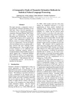
We have synthesised luminescent probes based on silica
nanoparticles embedded with different hydrophilic a nd
organic dyes ( Figure 1). The criteria and the paramet ers
required to prope rly encapsulating those fluorophores
within the silica shell have been investigated and seem
to differ from one fluorophor e to another. The success
of relatively good encapsulation tends to be related to
the structure of the selected dye. The first series of silica
nanoparticles, 1a-h, was prepared using the recently
developed W/O microemulsion method proposed by
Bagwe et al. [28] This regular synthesis involved the use
of Triton X100, n- hexanol, cyclohexane and water to
prepare the microemulsion. The desired dye (Rhoda-
mine B, Fluorescein, PABI, PPC, IR 806, NBA, HITC,
ICG see Figure 1 for full names and structures) was dis-
solved in the aqueous phase at a concentration of 0.1 M
in 200 μl,andinjectedintheW/Omicroemulsionsys-
tem. The second step involves the hydrolysis of TEOS
initiated by the addition of aqueous ammonia to the
reaction mixture that results in the formation of mono-
disperse spherical particles of amorphous silica.
The second series of silica nanopa rticles, 2a-f, was
prepared in an identical way but using another silica
precursor tetramethoxysilane (TMOS) for further
capping of the produced nanoparticles, this second silica
precursor was added after 24 h of reaction into the
microemulsion to create a denser silica shell. A thermal
treatment was effected at the end of the process so that
to densify the silica network. This protocol was devel-
oped to investigate if the capping followed by a thermal
treatment would r e-enforce the encapsulation process
and t herefore behave more efficiently towards the
encapsulation phenomenon. Indeed, it is known that the
use of TMOS instead of TEOS produces a denser silica
network, emphasising the encapsulation of the selected
fluorophores. The use of TMOS is expecting to consoli-
date the silica shell of the produced materials by gener-
ating a denser silica network within the nanomaterials
as suggested for the capping of the series 2. In addition,
it is known from the literature that using standard con-
ditions, the rate of hydrolysis of TEOS to a gel i s about
10 days, w hereas those of TMOS and tetra-n-butoxysi-
lane (TBOS) are 2 and 25 days, respectively [44-47].
The third series of silica nanoparticles, 3a-f, was pro-
duced by mixing porous silica nanoparticles, which pores
were functionalised with 3-(mercaptopropyl)triethoxysi-
lane [48], with the proper aqueous solution of the
required fluorophore. The thiol functionalities are design
to bind and therefore trap t he fluorophores within the
pores of the silica nanoparticl es. Finally, the fourth series
of silica nanoparticles, 4a-f, was prepared exactly as the
first series, 1, except that the silicon derivative used for
hydrolysis was TMOS [49]. Further to washings f our
silica nanoparticles series (1-4) were isolated which phy-
sical properties were further investigated.
Characterisation of nanoparticles
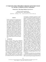
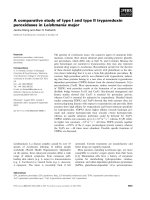
Figure 2 shows TEM images of three different series (1,
2 and 4) of silica nanoparticles prepared in this study.
No example illustrates the series 3. Indeed, due to the
porosity of the material o btained at the extremely low
pressure required for TEM analysis, the sample was
(collapsed) crushed on itself and the pictures observed
were not characteristic of the material. Cryo-TEM ana-
lysis of th e material i s under investi gation in our labora-
tories and will be reported in a different manuscript.
Overall, the resulting luminescent probes are spherical
in shape, and average diameters of 44 ± 3, 47 ± 4 and
41 ± 4 nm have been observed for samp les of each ser-
ies ca. 1b, 2b and 4a, respectively. The images also
showed that the particles were monodispersed. Further
TEM images of samples 1g 48 ± 4 nm and 1 h 46 ± 3
nm are also available in Figure 2 so that to emphasise
the size homogeneity obtained for different samples of
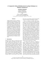
the series 1. Dynamic laser light scattering me asure-
ments show that the hydrodynamic diameters (the
apparent diameter of the hydrated/solvated particles) of
each particle of each series (1-4) are slightly larger than
Auger et al. Nanoscale Research Letters 2011, 6:328
/>Page 4 of 12
the dry particle diameters observed from the TEM. The
hydrodynamic diameters of the lumin escent nanoparti-
clesmaybeconsiderablylargerthantheir‘ dry’ dia-
meters due to the existence of a water layer surrounding
the hydrophilic silica network. Therefore the following
diameters of 58, 50, 51 and 44 nm were recorded for
samples of each series, ca. 1c, 2d, 3e and 4c, respec-
tively, as ill ustrated in Figure 3. Overall t he TEM and
DLS analyses have confirmed similar sizes, morphologies
and dispersivity of the silica nanoparticles prepared
using the different protocols.
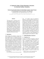
Spectroscopic properties of aqueous photoresponsive
nanoparticle dispersions
The principal tools used in this study to characterise the
dye’s encapsulation into silica matrix are the absorption
Propyl Asrtra Blue Iodide (PABI)
4,4’,4’’,4’’’-(porphine-5,10,15,20-
tetrayl)tetrakis(benzoic acid) (PPC)
IR 806 Nile Blue A perchlorate
1,1',3,3,3',3'-Hexamethylindotricarbocyanine iodide
(HITC) Cardiogreen
Rh
oda
min
e
B Fl
uo
r
esce
in
N
N
N
N
N
N
N
N
Cu
R
R
R
R=
SN
H
O
O
N
SO
O
O
CH
3
O
N
NH
N
HN
R
R
R
R
O
OH
R=
N N
SO
3
SO
3
Na
Cl
O
N
H
2
N N
ClO
4
N N
I
N N
SO
3
SO
3
Na
ON N
OH
O
Cl
O
O
O
H
O
O
H
Figure 1 Names and structures of the different dyes and fluorophores used during the study.
Auger et al. Nanoscale Research Letters 2011, 6:328
/>Page 5 of 12
and fluorescence spectroscopies. The photochromic
properties displayed by the nanoparticles a re indicative
of the successful incorporation of dyes into the nanopar-
ticles. Indeed, to detect the correct encapsulation of the
desired dye within the silica network of the nanoparti-
cles, the fluorescence and/or the absorption of the aqu-
eous solution of the prepared nanoparticles was
measured. S uch measurements informed us of the suc-
cessful encapsulation. The following dyes have been sub-
jected to encapsulation by four different methods
described in the paragraph above.
Fluorescence and absorption measurements of every
sample were recorded, and when a specific sample of
nanoparticles exhibited such prop erties, it was immed i-
ately compared to the fluorescence or the absorption of
the free-d ye dissolved in water. Refer ences spectra of the
different dyes in water had to be recorded so that to be
able to compare the fluorescence recorded of the different
fluorophores alone and also the fluorescence recorded of
the fluorophores once encapsulated into the silica matrix.
The content or concentration of fluorescent dye in
silica nanoparticles tends to influence the fluorescence
intensity of nanoparticles dispersions. The quantity of
encapsulated dye is not relevant to ou r study. Therefore,
since the study m ainly focuses on the incorporation and
not the quantity of dyes into the silica matrix, all
absorption and fluorescent spectra were normalised
arbitrarily. Furthermore, self-quenching of fluorescence
has been determined for each fluorophores used to
establish the appropriate amount of chromophore to
incorporate into the nanoparticles to e nsure high fluor-
escence intensity and at the same time to avoid fluores-
cence self-quenching. The dyes selected for the study
were: PABI, PPC, IR 806, NBA, HITC and ICG (Figure
1). We also reproduced the encapsulation of fluorescein
and rhodamine with the standard microemulsion sol-gel
process as the successful encapsulation of those two
dyes has been investigated and optimised in our labora-
tories [50]. It is important to mention that all dyes and
fluorophores selected for t his study are commercially
available and their hydrophilic structural character con-
fer them good to excellent water solubility. High con-
centration such as 0.1 M in water was therefore
employed for the synthetic processes 1-4.
B C
D
A
E
F
Figure 2 TEM images of silica nanoparticles with different average sizes. (A) 1b (44 ± 3 nm), (B) 2b (46 ± 3 nm), (C) 1 h (46 ± 3 nm), (D)
2b (47 ± 4 nm), (E) 4b (40 ± 3 nm) and (F) 4a (41 ± 4 nm). Scale bar: 100 nm.
0
5
10
15
20
25
30
1 10 100 1000 10000
1c
2d
3e
4c
Number (%)
Size diameter
(
nm
)
Figure 3 Dynamic light scattering measurements of
synthesized dye-doped silica nanoparticles of each series (1c
58 nm, 2d 50 nm, 3e 51 nm and 4c 44 nm).
Auger et al. Nanoscale Research Letters 2011, 6:328
/>Page 6 of 12
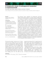
Fluorescein and rhodamine
Furthermore, fluorescein and rhodamine B were suc-
cessfully encapsulated by the method 1. The fluores-
cence data, excitation and emission wavelengths,
observed for sample 1 h were identical to those
recorded for a solutio n of fr ee fluorescein in wa ter as
illustrated in Figure 4. Indeed, the fluorescence maxi-
mum, at 513 nm upon an excitation at 488 nm for sam-
ple 1 h, indicated that the fluorescein had been
encapsulated into the silica nanoparticles. A freshly pre-
pared solution of fluorescein into water also exhibited
maxima excitation and emission wavelengths at 486 and
513 nm, respectively. The same phenomena were
observed for the sample 1g consisting of rhodamine B
encapsulated into silica nanoparticles. Both, the aqueous
solutions of free rhodamine B and of sample 1g dis-
played maxima excitation and emission wavelengths at
555 and 577 nm, respectively. A slight shift and different
shapes in the excitation band of the fluorescein was
observed which is attributed to the incorporation of the
fluorescent dye and its interaction with the silica net-
work. Those results indicated that the silica encapsula-
tion by microemulsion was suitable for encapsulation of
hydrophilic chromophores and was consistent with the
literature [15,51-54]. It was therefore decided after those
fluorescence measuremen ts (Figure 4) that no better
encapsulation could be achieved by other processes and
no further investigation of those two dyes were tested. It
was also important to notice that non-covalent encapsu-
lation of those dyes has been reported earlier on in the
literature [52].
3.3.2. PABI
The transmission spectra of pure PABI dye and PABI
nanoparticles were measur ed in aqueous so lution
(Figure 5). Since the PABI dye is not fluorescent, the
encapsulation phenomenon could be checked by trans-
mission measurements. The pure dye solution showed
three t ypical absorption peaks characteristic of t he aro-
matic macrocyclic π-electron of phthalocyanine dyes.
Absorption maxima were recorded at 342 nm (B-band),
612 nm (vibrationa l band) and 668 nm (Q-band). The
transmission spectra for the pure PABI and the samples
1a, 2a, 3a and 4a d isplayed almost the same profile in
aqueous solution, though there was only a very slight
red-shift (1-2 nm) for their absor bance maxima when
compared to each PABI nanoparticles prepared respec-
tively. Those results indicate that the four methods of
encapsulation used were successful. The PABI dye
400 450 500 550 600 650 700
Fluorescein exc
Fluorescein em
1h exc
1h em
Intenisity
(
a.u.
)
Wavelength (nm)
Fl
uoresce
i
n
450 500 550 600 650 700 750
Rhodamine B exc
Rhodamine B em
1g exc
1g em
Intenisity (a.u.)
Wavelen
g
th
(
nm
)
Rhodamine B
Figure 4 Excitation and emission spectra of aqueous solutions of (left) fluorescein and silica nanoparticles doped with fluorescein 1 h,
and (right) rhodamine B and silica nanoparticles doped with rhodamine B 1g.
300 400 500 600 700 800
PABI
PABI
1a
2a
3a
4a
Intenisty
(
a.u.
)
Wavelen
g
th
(
nm
)
Figure 5 Transmittance spectra of aqueous solutions of PABI
and silica nanoparticles doped with PABI (1a, 2a, 3a and 4a).
Auger et al. Nanoscale Research Letters 2011, 6:328
/>Page 7 of 12
seemed to be proper towa rds encapsulation conditions.
Once embedded into the silica nanoparticles (samples
1a, 2a, 3a and 4a), the flat and rigid aromatic core of the
phthalocyanine derivative can no longer escape, and
remain well trapped within the silica network. Further-
more, phthalocyanine dyes are well-known to aggregate
and generate π-stacking, and such phenomenon could
emphasise the stability of those dyes towards encapsula-
tion. The ordering of the π-stacking o f the PABI mole-
cules can favour their insertion into the silica network.
Also, the π-stacking could be generated into the micelle,
enhancing the rigidity of the organically bulk structure
and therefore favouring the encapsulation process. Addi-
tionally, the interactions between the nitrogen atoms of
the four imino bridges of the phthalocyanine aromatic
core of th e PABI, and the hanging hydroxyl of the silica
core-shell facilitate further the encapsulat ion. The inter-
actions of the dye to encapsulate with the silica network
of the nanopa rticles added to the rigidity of its aromatic
core confer excellent conditions towards encapsulation.
Prior to the results obtained with PABI, such conditions
have been reported for the encapsulation of fluorescein
1 h and rhodamine B 1g. Similarly, those molecules pos-
sess reasonably flat and rig id aromatic co res, in part due
to the conjugated system, emphasising the aromaticity
and the stability of those dyes, and also due to the spiro
cent re contained in the structure of the fluorescein, and
the lack of freedom towards the vertical bond in the
molecule of rhodamine B, between the oxo-anthracenyl
analogue core and the vertical ortho-carboxyphenyl sub-
stituent. The latest could introduce atropisomerism,
exhibiting blocked isomers leading to rigid structures
lacking of three-dimensional freedom, and therefore
facilitating the encapsulation process.
3.3.3. PPC porphyrin
Further incorporation of flat and rigid aromatic core
organic dye has been investigated. The PPC porphyrin
was chosen due to the structural similarity to the planar
PABI molecule. But, exhibiting fluorescence, the PPC
porphyrin was chosen to study the impa ct of encapsula-
tion towards the fluorescent properties of this family o f
compounds. Indeed the PABI and the PPC molecules
possess an a romatic core consisting of 18- π electrons,
which emphasise the stability and the electrochromic
properti es of this family of intensely coloured dyes. The
PPC dye exhibits fluorescence whereas the PABI detec-
tion was limited to absorption measurements. Excitation
and emission spectra of a pure aqueous solution of PPC
are illustrated in Figure 6.
The excitation spectrum displays a s plitted maximum
peak at 407 and 421 nm due to symmetry of the PPC
molecule. Then the emission peaks were recorded at
647 and 706 nm. The fluorescence measurements of the
silica-based samples 1b, 2b, 3b and 4b showed identical
excitation and emission spectra than those exhibited by
the free-PPC in water. As can be seen in Figure 6, the
silica-based encapsulations showed a well-resolved coa-
lesced peak for the excitation maxima at 415 nm. This
phenomenon is typical of a loss of symmetry and of an
ordered state of the organic molecules. This phenom-
enon could also be attributed to embedding stress
which would result from the interaction of the organic
dye with the silica matrix. This effect was o bserved for
each process (1-4). The different encapsulation pro-
cesses studied (1-4) did not alter whatsoever the emis-
sion spectra. As for the PABI experiments (1a, 2a, 3a
and 4a), the successful encapsulation of PPC by mean of
the four processes described earlier is a consequence of
the flatness and rigidity of the aromatic macrocyclic
core of the PPC porphyrin, as well as the possible inter-
action of the four nitrogens, of the residual pyrroles
included in the porphyrin ar omatic core, with the hang-
ing hydroxyl substituents of the silica matrix.
IR 806
IR 806 is a water-soluble near-infrared cyanine dye.
Usually these dyes are known to have narrow and
intense absorption bands in the near-IR spectral region,
and to possess good photostability. A solution of free IR
806 dye was used for fluorescence measurements in
water.
The results are presented in Figure 7, and show three
excitation peaks upon emission at 806 nm. The main
excitation peak was observed as a sharp peak at 824 nm.
Esp ecially notewo rthy was the observation of signifi cant
overlapping secondary peaks at 702 and 746 nm,
400 500 600 700 80
0
PPC exc
PPC em
1b exc
1b em
2b exc
2b em
3b exc
3b em
4b exc
4b em
Intenisty
(
a.u.
)
Wavelen
g
th
(
nm
)
PP
C
Figure 6 Excitation and emission spectra of aqueous solutions
of PPC and silica nanoparticles doped with PPC (1b, 2b, 3b
and 4b).
Auger et al. Nanoscale Research Letters 2011, 6:328
/>Page 8 of 12
equivalent in intensity. The emission peak was recorded
at 837 nm. A comparison of the excitation and emission
spectra measured for silica-based samples 1c, 2c, 3c and
4c gave various results. Fluorescence was measured but
not recorded for samples 1c and 2c indicating t he non-
encapsulation of the IR 806 dye under those conditions.
Most probably, the encapsulation’ sfailuresimplythat
the kinetic rate of hydrolysis of the TEOS prevent from
ideal encapsulation conditions. Slow hydrolysis to pro-
duce the silica network can e mphasise the exclusion of
the molecule as well as an enhancement of the porosity
of the silica network of the nanoparticles [55]. Hence,
two straightforward explanations come to mind, either
the dye is excluded during the growth o f the silica
matrix of the nanoparticle, or it is first encapsulated
then released during the different w ashing steps due to
the porosity of the silica network. Opposite results were
observed for experiments 3c and 4c which encapsula-
tions were successful. Fluorescent spectra of sample 3 c
are illustrated in Figure 5. The single excitation peak
and emission peak were recorded at 827 and 839 nm,
respectively. The slight bathochromic shift observed (2-3
nm) suggests an effect/influence of the confined IR 806
dye into the silica nanoparticles. Fluorescent spectra of
sample 4c are also shown in Figure 7. Importan t hypso-
chromic shifts are observed as well as disappearance of
the main sharp excitation peak occurring at 824 nm.
The single excitation peak was recorded at 660 nm and
the corresponding emission peak was observed at
743 nm. The encapsulation of IR 806 in the silica net-
work of the nanoparticles tends to totally quench the
low energy transition, therefore exhibiting only the
secondary or high energy transition. Measurements of
fluorescence of an aqueous solution of IR 806 did not
exhibit luminescence at 743 nm upon e xcitation at 660
nm. The induced shift effect was observed and resulted
from the confinement of the fluorescent dye w ithin the
silica particle, when prepared with TMOS. Subsequently,
it is reasonable to assume that the interactions of the
hydroxyl groups of the silica network with the IR 806
fluorescent dye tend to block preferably the radiative
transitions at 806 nm than those at 743 nm. Further-
more, the successful encapsulation can result in the use
of TMOS instead of TEOS which possess a faster rate
of hydrolysis and build a denser silica network embed-
ding more efficiently the IR 806 dye as explained in the
paragraph above.
NBA
The synthesis of nanosensors based on silica nanoparti-
clesembeddedwitharigidfluorophorescalledNBA
was undertaken. NBA is commonly used a s a fluores-
cent laser dye. An aqueous solution of free-NBA exhib-
ited an excitation peak at 634 nm and an em ission peak
at 677 nm as illustrated in Figure 8.
Further attempts towards encapsulation of NBA using
the four different methods detailed earlier on proved to
be successful. Indeed reasonably similar maxima excita-
tion and emission wavelengths were recorded in close
range to those observed for the free-NBA. Subsequently,
samples 1d, 2d, 3d and 4d gave excitation peaks at 641,
633, 633 and 637 nm, respectively, whereas the corre-
sponding emission peaks w ere showed at 674, 674, 675
and 675 nm, respectively. The encapsulation tends to
560 600 640 680 720 760 800 840
IR 806 exc
IR 806 em
3c exc
3c em
4c exc
4c em
Intenisty
(
a.u.
)
Wavelength (nm)
IR
806
Figure 7 Excitation and emission spectra of aqueous solutions
of IR 806 and silica nanoparticles doped with IR 806 (3c, 4c).
450 500 550 600 650 700 750 800 850
NBA exc
NBA em
1d exc
1d em
2d exc
2d em
3d exc
3d em
4d exc
4d em
Intenisty (a.u.)
Wavelength (nm)
NBA
Figure 8 Excitation and emission spectra of aqueous solutions
of NBA and silica nanoparticles doped with NBA (1d, 2d, 3d
and 4d).
Auger et al. Nanoscale Research Letters 2011, 6:328
/>Page 9 of 12
influence mostly the excitation peaks (Δl
ex
=8nm)
than the emission peaks (Δ l
em
= 3 nm). The water-solu-
ble molecule of NBA dye was encapsulated successfully
due in part to its rigid aromatic core. As for the PABI
and PPC molecules, the rigidity of the aromatic core
added to the presence of heteroatoms in the molecule
of NBA tends to enhance the embedding process.
HITC and+ ICG
Finally, two cyanine-based near-infrared absorbing dyes
(HITC and ICG) were subjected to the four methods of
encapsulations involved in this study. Those dyes are
commercially available due to their photographic sensi-
tivity and infrared lasers absorption, essential properties
to the printing industry. It is also important to notice
that, currently, the organic dye ICG is the only near-
infrared fluorophores approved by FDA for use in vivo
in humans [56]. Aqueous solutions of free-HITC and
free-ICG displayed sharp excitation peaks a t 734 and
776 nm, respective ly, as well as sharp emission peaks at
790 and 806 nm as indicated in Figure 9.
Under encapsulation conditions of methods 1-4, HITC
embedding occurred for samples 3e and 4e. Fluores-
cence was measured for 3e (l
ex
= 738 nm, l
em
= 758
nm) and 4e (l
ex
= 741 nm, l
em
= 759 nm), whereas no
fluorescence could be recorded neither for samples 1e
nor 2e. Furthermore, in the case of ICG, while sampl e
3f displayed a well-resolved fluorescence with an excita-
tion peak at 780 nm an d an emissi on peak at 820 nm,
samples 1f, 2f and 4f did not exhibit any fluorescence.
The poor chemical and photostability of cyanine-based
dyes especially in aqueous environments u nder basic
conditions, as well as their strong tendency to form
aggregates might decrease their ability towards the
encapsulation process (1-4). Also cyanine-based dyes
must be monomolecular and possess planar rigid geo-
metries to be efficient at absorbing and emitting light.
Therefore, the poor rigidit y of both cyanine-based mole-
cules, HITC and ICG, indicates that it is a relevant cri-
terion to take into account when proceeding to
encapsulation of those dyes into silica nanoparticles.
Samples 3e and 3f illustrated the successful encapsula-
tion of HITC and ICG. This is in part due to the poros-
ity of the silica nanoparticles, and also accentuated by
thefactthatthoseporesarefunctionalised with thiols
(SH) that can bind and entrap organic dyes via hydrogen
bondings and electrostatic forces.
Conclusions
To conclude, these experiments have allowed us to
establish and optimise criteria and principles towards
efficient encapsulation of dyes by reverse microemulsion
process involving non-covalen t embeddement. Table 1
summarises the successful encapsulations as well as the
techniques of characterisation used. The study of their
luminescent properties or the ir quenching was also
described.
i. Hydrophilic Vs hydrophobic ch aracter the single
use of TEOS allowed us to encapsulate hydrophilic
molecules essentially. In order to embed molecules
rather hydrophobic than hydrophilic into silica
nanoparticles, t he use of an ad ditional silica precur-
sor was considered to induce interactions of the
silica with the selected dye via hydrogen bondings.
500 550 600 650 700 750 800 850
HITC exc
HITC em
1e exc
1e em
3e exc
3e em
4e exc
4e em
Intenisty
(
a.u.
)
Wavelength (nm)
HITC
600 650 700 750 800 850
I
CG
ICG exc
ICG em
3f exc
3f em
Intensity (a.u.)
Wavelength (nm)
Figure 9 Excitation and emission spectra of aqueous solutions of (left) HITC and silica nanoparticles doped with HITC 3e and 4e, and
(right) ICG and silica nanoparticles doped with ICG 3f.
Auger et al. Nanoscale Research Letters 2011, 6:328
/>Page 10 of 12
Also, the choice of such silica precursors can be dri-
ven by their faster rates of hydrolysis to avoid steric
exclusion.
ii. Molecular rigidity (isomerism) the rigidity of the
dye has a propensity to favour the encapsulation
process. The porosity and the density of the silica
network of the so-formed nanoparticles are inv olved
in an efficient e ncapsulation. Indeed, a denser silica
matrix can generate rigid assembli ng mole cules
within the network, and therefore depending on the
rate of hydrolysis of the chosen silica precursors.
iii. Fluorescence display the embeddement of a fluor-
escent dye does not prevent the display of fluores-
cence. Upon encapsulation, the dye-doped silica
nanoparticles exhibit relatively similar excitation and
emission spectra than the free dye. Interaction of the
silica network and the chosen fluorescent dye should
not cause quenching of its emission or influe nce the
probability of some luminescent transitions between
excited and ground states of the selected organic dye
(as for IR 806). Blocking or shifting of the major
radiative tr ansit ion may be due to t he interaction of
the dye with the silica network or the locking of the
isomerism. In couple of experiments, shifted peaks
were observed, and those phenomena were attribu-
ted to inter action and/or affinity of the silica for the
selected organic dye. Further interaction could give
rise to total quenching of the fluorescent dye. Repla-
cing or substituting the free hydroxyl groups of the
silica by a different silica precursor during the synth-
esis has a tendency to modulate those interactions
and to, some time s, recover an emphasi se d emission
signal from the selected fluorescent dye.
Abbreviations
DLS: dynamic light scattering; HDTMOS: hexadecyltrimethoxysilane; MB:
methylene blue; NBA: Nile Blue A perchlorate; PABI: propyl astra blue iodide;
TEOS: tetraethoxysilane; TMOS: tetramethoxysilane; TBOS: tetra-n-
butoxysilane; TEM: transmission electron microscopy; W/O: water in oil.
Acknowledgements
The authors thank Nathalie Scheer-Pelissier for providing the transmission
electronic microscopy characterisations.
Authors’ contributions
The work presented here was carried out in collaboration between all
authors. AA, OR and OP defined the research theme. AA and OR designed
methods and experiments and also coordinated the present study. AA
carried out the laboratory experiments, interpreted the results and wrote the
paper. OR performed the luminescence measurements and analyzed the
data. OP co-designed experiments and discussed analyses. JS: performed the
syntheses of silica nanoparticles with rhodamine B and fluorescein dyes and
the luminescence measurement of these samples. All authors have
contributed to, seen, read and approved the manuscript.
Competing interests
The authors declare that they have no competing interests.
Received: 13 September 2010 Accepted: 13 April 2011
Published: 13 April 2011
References
1. Bruchez M, Moronne M, Gin P, Weiss S, Alivisatos AP: Semiconductor
nanocrystals as fluorescent biological labels. Science 1998, 281:2013.
2. Chan WCW, Nie S: Quantum dot bioconjugates for ultrasensitive
nonisotopic detection. Science 1998, 281:2016.
3. De M, Ghosh PS, Rotello VM: Applications of Nanoparticles in Biology. Adv
Mater 2008, 20:1.
4. Pinaud F, Michalet X, Bentolila LA, Tsay JM, Doose S, Li JJ, Iyer G, Weiss S:
Advances in fluorescence imaging with quantum dot bio-probes.
Biomaterials 2006, 27:1679.
5. Wang F, Chatterjee DK, Li Z, Zhang Y, Fan X, Wang M: Synthesis of
polyethylenimine/NaYF4 nanoparticles with upconversion fluorescence.
Nanotechnology 2006, 17:5786.
6. Ye Z, Tan M, Yuan J: Novel fluorescent europium chelate-doped silica
nanoparticles: preparation, characterization and time-resolved
fluorometric application. J Mater Chem 2004, 14 :851.
7. Daniel M, Astruc D: Gold nanoparticles: Assembly, supramolecular
chemistry, quantum-size-related properties, and applications toward
biology, catalysis, and nanotechnology. Chem Rev 2004, 104:293.
8. West JL, Halas NJ: Engineered nanomaterials for biophotonics
applications: Improving sensing, imaging, and therapeutics. Annu Rev
Biomed Eng 2003, 5:285.
9. Pham HH, Gourevich J, Oh JK, Jonkman JEN, Kumacheva A: A multidye
nanostructured material for optical data storage and security data
encryption. Adv Mater 2004, 16:516.
10. Sharma P, Brown S, Walter G, Santra S, Moudgil B: Nanoparticles for
bioimaging. Adv Colloid Interface Sci 2006, 123-126:471.
11. Yan J, Estévez MC, Smith JE, Wang K, He X, Wang L, Tan W: Dye-doped
nanoparticles for bioanalysis. Nanotoday 2007, 2:44.
12. Wang F, Tan WB, Zhang Y, Fan X, Wang M: Luminescent nanomaterials for
biological labelling. Nanotechnology 2006, 17:R1.
13. Ow H, Larson DR, Srivastava M, Baird BA, Webb WW, Wiesner U: Bright and
stable core-shell fluorescent silica nanoparticles. Nano Lett 2005, 5:113.
14. Larson DR, Ow H, Vishwasrao HD, Heikal AA, Wiesner U, Webb WW: Silica
nanoparticle architecture determines radiative properties of
encapsulated fluorophores. Chem Mater 2008, 20:2677.
15. Ethiraj AS, Hebalkar N, Kharrazi S, Urban J, Sainkar SR, Kulkarni SK:
Photoluminescent core-shell particles of organic dye in silica.
J Luminescence 2005, 114:15.
16.
Bringley JF, Penner TL, Wang R, Harder JF, Harrison WJ, Buonemani L: Silica
nanoparticles encapsulating near-infrared emissive cyanine dyes.
J Colloid Interface Sci 2008, 320:132.
Table 1 Reminder of successful and unsuccessful
encapsulations of the different dyes (a-h) and the uses of
the different methods of encapsulation (i.e. series 1-4)
investigated during the study
Dyes Series
1234
a PABI ✓✓✓✓
a
b PPC ✓
a
✓
a
✓✓
a
c IR 806 X
b
X ✓✓
b
d NBA ✓✓
b
✓✓
e HITC X X ✓
b
✓
f ICG X X ✓ X
g RHOD B ✓
a
N/A N/A N/A
h FLUO ✓
a
N/A N/A N/A
All successfully encapsulated samples have been further characterised by
fluorescent measurements.
a
Characterised by TEM analysis.
b
Characterised by DLS measurements.
Auger et al. Nanoscale Research Letters 2011, 6:328
/>Page 11 of 12
17. Aslan K, Wu M, Lakowicz JR, Geddes CD: Fluorescent core-shell Ag@SiO2
nanocomposites for metal-enhanced fluorescence and single
nanoparticle sensing platforms. J Am Chem Soc 2007, 129:1524.
18. Addison CJ, Brolo AG: Nanoparticle-containing structures as a substrate
for surface-enhanced Raman scattering. Langmuir 2006, 22:8696.
19. Shibata S, Taniguchi T, Yano T, Yamane M: Formation of water-soluble
dye-doped silica particles. J Sol-Gel Sci Technol 1997, 10:263.
20. Imahori H, Mitamura K, Shibano Y, Umeyama T, Matano Y, Isoda S, Araki Y,
Ito O: A photoelectrochemical device with a nanostructured SnO2
electrode modified with composite clusters of porphyrin-modified silica
nanoparticle and fullerene. J Phys Chem B 2006, 110:11399.
21. Chen X, Zou J, Zhao T, Li Z: Preparation and fluoroimmunoassay
application of new red-region fluorescent silica nanoparticles.
J Fluorescence 2007, 17:235.
22. Lakowicz JR: Principles of Fluorescent Spectroscopy. New York: Kluwer;, 2
1999.
23. Verhaeg NAM, van Blaaderen A: Dispersions of rhodamine-labeled silica
spheres - synthesis, characterization, and fluorescence confocal scanning
laser microscopy. Langmuir 1994, 10:1427.
24. van Blaaderen A, Vrij A: Synthesis and characterization of colloidal
dispersions of fluorescent, monodisperse silica spheres. Langmuir 1992,
8:2921.
25. Lu Y, Yin Y, Mayers BT, Xia Y: Modifying the surface properties of
superparamagnetic iron oxide nanoparticles through a sol-gel approach.
Nano Lett 2002, 2:183.
26. Yamauchi H, Ishikawa T, Kondo S: Surface characterization of ultramicro
spherical-particles of silica prepared by w/o microemulsion method.
Colloids & Surfaces 1989, 37:71.
27. Stöber W, Fink A, Bohn E: Controlled growth of monodisperse silica
spheres in micron size range. J Colloid Interface Sci 1968, 26:62.
28. Bagwe RP, Yang C, Hilliard LR, Tan W: Optimization of dye-doped silica
nanoparticles prepared using a reverse microemulsion method.
Langmuir 2004, 20:8336.
29. Nyffenegger R, Quellet C, Ricka J: Synthesis of fluorescent, monodisperse,
colloidal silica particles. J Colloid Interface Sci 1993, 159:150.
30. Chang S, Liu L, Asher SA: Creation of templated complex topological
morphologies in colloidal silica. J Am Chem Soc 1994, 116:6745.
31. Burns A, Ow H, Wiesner U: Fluorescent core-shell silica nanoparticles:
towards “Lab on a Particle” architectures for nanobiotechnology.
Chem
Soc Rev 2006, 35:1028.
32. Zhao X, Bagwe RP, Tan W: Development of organic-dye-doped silica
nanoparticles in a reverse microemulsion. Adv Mater 2004, 16:173.
33. Arriagada FJ, Osseo-Asare K: Synthesis of nanosize silica in a nonionic
water-in-oil microemulsion: Effects of the water/surfactant molar ratio
and ammonia concentration. J Colloid Interface Sci 1999, 211:210.
34. Wang L, Wang K, Santra S, Zhao X, Hilliard L, Smith J, Wu Y, Tan W:
Watching silica nanoparticles glow in the biological world. Anal Chem
2006, 78:646.
35. Yang HH, Qu HY, Lin P, Li SH, Ding MT, Xu JG: Nanometer fluorescent
hybrid silica particle as ultrasensitive and photostable biological labels.
Analyst 2003, 128:462.
36. Hai XD, Tan MQ, Wang GL, Ye ZQ, Yuan JL, Matsumoto K: Preparation and
a time-resolved fluoroimmunoassay application of new europium
fluorescent nanoparticles. Anal Sci 2004, 20:245.
37. Santra S, Yang HS, Dutta D, Stanley JT, Holloway PH, Tan WH, Moudgil BM,
Mericle RA: TAT conjugated, FITC doped silica nanoparticles for
bioimaging applications. Chem Commun 2004, 2810.
38. Deng T, Li J, Jiang J, Shen G, Yu R: Preparation of near-IR fluorescent
nanoparticles for fluorescence-anisotropy-based immunoagglutination
assay in whole blood. Adv Funct Mater 2006, 16:2147.
39. van Blaaderen A, Vrij A: Synthesis and characterization of monodisperse
colloidal organo-silica spheres. J Colloid Interface Sci 1993, 156:1.
40. Santra S, Wang KM, Tapec R, Tan WH: Development of novel dye-doped
silica nanoparticles for biomarker application. J Biomed Opt 2001, 6:160.
41. Santra S, Zhang P, Wang KM, Tapec R, Tan WH: Conjugation of
biomolecules with luminophore-doped silica nanoparticles for
photostable biomarkers. Anal Chem 2001, 73:4988.
42. Bagwe RP, Zhao XJ, Tan WH: Bioconjugated luminescent nanoparticles for
biological applications. J Dispersion Sci Technol 2003, 24:543.
43. Lian W, Litherland SA, Badrane H, Tan WH, Wu DH, Baker HV, Gulig PA,
Lim DV, Jin SG: Ultrasensitive detection of biomolecules with fluorescent
dye-doped nanoparticles. Anal Biochem 2004, 334:135.
44. Senarath-Yapa MD, Phimphivong S, Coym JW, Wirth MJ, Aspinwall CA,
Saavedra SS: Preparation and characterization of poly(lipid)-coated,
fluorophore-doped silica nanoparticles for biolabeling and cellular
imaging. Langmuir 2007, 23:12624.
45. Husing N, Schubert U, Mezei R, Fratzl P, Riegel B, Kiefer W, Kohler D,
Mader W: Formation and structure of gel networks from Si(OEt)(4)/(MeO)
(3)Si(CH2)(3)NR ‘(2) mixtures (NR ‘(2) = NH2 or NHCH2CH2NH2). Chem
Mater 1999, 11:451.
46. Cattey H, Audebert P, Sanchez C, Hapiot P: Electrochemical investigations
on liquid-state polymerizing systems: Case of sol-gel polymerization of
transition metal alkoxides. J Phys Chem B 1998, 102:1193.
47. Audebert P, Divisia-Blohorn B, Cohen-Addad JP: Electroactive probe
diffusion through DMF/polyacrylonitrile gels: free volume behaviour.
Polymer Bull 1997, 39:225.
48. Auger A, Poncelet O: Process for preparing porous silica particles, the
silica particles obtained and their use, EP2228127. 2010.
49. Arkles B: Kirk-Othmer Encyclopedia of Chemical Technology. New York:
Wiley;, 4 1997:22:69-81.
50. Samuel J, Raccurt O, Poncelet O, Auger A, Ling WL, Cherns P, Grünwald D,
Tillement O: Surface characterizations of fluorescent-functionalized silica
nanoparticles: from the macroscale to the nanoscale. J Nanopart Res
2010, 12:2255-2265.
51. Ha S, Camalier CE, Beck GR, Lee J: New method to prepare very stable
and biocompatible fluorescent silica nanoparticles. Chem Commun 2009,
2881.
52. Samuel J, Raccurt O, Poncelet O, Tillement F, Tardif F: Dispersion and
incorporation of optical nanotracers. Technical Proceedings of 2008 NSTI
Nanotechnology Conference and Trade Show 2008, 1:230.
53. Karolin J, Geddes CD, Wynne K, Birch DJS: Nanoparticle metrology in sol-
gels using multiphoton excited fluorescence. Meas Sci Technol 2002,
13:21.
54. Samuel J, Tallec G, Cherns P, Ling WL, Raccurt O, Poncelet O, Imbert D,
Mazzanti M: Lanthanide-chelate silica nanospheres as robust multicolor
Vis-NIR tags. Chem Commu 2010, 46(15):2647.
55. Finnie KS, Bartlett JR, Barbé CJA, Kong L: Formation of silica nanoparticles
in microemulsions. Langmuir 2007, 23:3017.
56. Gao Y, Cui Y, Levenson RM, Chung LWK, Nie S: In vivo cancer targeting
and imaging with semiconductor quantum dots. Nat Biotechnol 2004,
22:969.
doi:10.1186/1556-276X-6-328
Cite this article as: Auger et al.: A comparative study of non-covalent
encapsulation methods for organic dyes into silica nanoparticles.
Nanoscale Research Letters 2011 6:328.
Submit your manuscript to a
journal and benefi t from:
7 Convenient online submission
7 Rigorous peer review
7 Immediate publication on acceptance
7 Open access: articles freely available online
7 High visibility within the fi eld
7 Retaining the copyright to your article
Submit your next manuscript at 7 springeropen.com
Auger et al. Nanoscale Research Letters 2011, 6:328
/>Page 12 of 12