Báo cáo hóa học: " Quasi-radial growth of metal tube on si nanowires template" pptx
Bạn đang xem bản rút gọn của tài liệu. Xem và tải ngay bản đầy đủ của tài liệu tại đây (2.68 MB, 8 trang )
NANO EXPRESS Open Access
Quasi-radial growth of metal tube on si
nanowires template
Zhipeng Huang
1*
, Lifeng Liu
2
, Nadine Geyer
2
Abstract
It is reported in this article that Si nanowires can be employed as a positive template for the controllable
electrochemical deposition of noble metal tube. The deposited tube exhibits good crystallinity. Scanning electron
microscope and transmission electron microscope characterizations are conducted to reveal the growth process of
metal tube, showing that the metal tube grows quasi-radially on the wall of Si nanowire. The quasi-r adial growth
of metal enables the fabrication of thickness-defined metal tube via changing deposition time. Inner-diameter-
defined metal tube is achieved by choosing Si nanowires with desired diameter as a template. Metal tubes with
inner diameters ranging from 1 μm to sub-50 nm are fabricated.
Introduction
Owing to a considerably enhanced surface-to-volume
ratio compared to bulk, one-dimensional metallic tubular
structure has shown promising application potential in
the fields of energy storage and conversion [1,2], catalysis
[3-5], and magnetism [6,7], and therefore has gained
increasing attention. Similar to the case of other nanos-
tructures, controllable fabrication is essential for the
device application of tubular structure. Various
approaches (e.g., electrochemical deposition [ 8-10], elec-
troless deposition [11,12]), etc., have been developed to
fabricate metal tubes. Meanwhile, temp lates with specific
aspect ratio and packing manner are used to define the
geometries of nanotubes. Nowadays, two insulating
masks, namely, porous anodic aluminum oxide (AAO)
and ion-track-etched polymer membrane, are widely
used for the fabrication of nanotubes. However, chemical
modificat ion (introducing molecular anchor) of pore wall
[9,13,14] or metal pre-deposition (as seed layer) on pore
wall [12,15] is necessary before the fabrication of metal
tube, which wi ll inevitably introduce impurity to the
deposited structures [12]. On the other hand, during
electrochemical deposition, metal grows along axial
direction in the isolating template [8], which makes it dif-
ficult for controlling independently the thickness and
length of tubular structure . From these points of view,
conducting or semi-conducting t emplate is more favo r-
able for the fabrication of metal tube, because the modifi-
cation of template surface is unnecessary and the growth
is hopefully radial. Macroporous silicon (Si) [16-18] and
InP [19] have been used as templates for the fabricati on
of metal tube. However, the feature size in macroporous
Si is usually larger than several hundreds of nanometer
due to a well-known 2W
sc
rule [20], where W
sc
is the
thickness of space charge layer in Si subst rat e at Si/solu-
tion interface. Moreover, only the tube of less noble
metal has been demonstrated on the macroporous Si
template, whereas the electrochemical deposition of
noble metal leads to wire or pillar, because noble metal
grows axially from the bottom of pores in the macropor-
ous Si template [16,17].
Si nanowire would be an alternative candidate as a
positive template for the deposition of metal tube, due to
its intrinsic semi-c onducting property and wide diameter
range. Especially, template-based metal-assisted chemical
etching [21-25] enables precise control over the diameter,
length, orientation relative to substrate, packing manner,
and cross-sectional shape of Si nanowires. In this article,
it is reported that highly ordered array of Si nanowires
fabricated by template-based metal-assisted chemical
etching can be used as a positive template f or the con-
trollable electrochemical deposition of noble metal (Au)
tube. It is i ndicated by scanning electron microscope
(SEM) and transmission electron microscope (TEM) that
metal grows quasi-radially on the sidewall of Si nanowire.
Therefore, the length and thickness of metal tube can be
* Correspondence:
1
Functional Molecular Materials Centre, Scientific Research Academy, Jiangsu
University, Zhenjiang 212013, P. R. China.
Full list of author information is available at the end of the article
Huang et al. Nanoscale Research Letters 2011, 6 :165
/>© 2011 Huang et al; licensee Springer. This is an Open Access article distributed unde r the terms of the Creative Commons Attribution
License ( which permits unrestricted use, distribution, and reproduction in any medium,
provided the original work is properly cited.
independently controlled. On the other hand, metal tubes
with the inner diameter ranging from 1 μm to sub-50 nm
are obtained by choosing Si nanowires with desired dia-
meters as a template.
Experimental
Si nanowire templates were fabricated by template-based
metal-assisted chemical etching [21,23,24] of Si sub-
strates (r:1-10Ωcm, n-type substrates for samples are
shown in Figures 1, 2, 3, 4, 5, 6, 7a and 7b, and p-type
substrates for samples are shown in Figure 7c). Except
the one used in Figure 7, the Si nanowire t emplates
used in this article were fabricated by the metal-assisted
chemical etching combined with nanosphere lithogra-
phy. In brief, polystyrene (PS) spheres were assembled
into monolayer hexagonal array onto a Si substrate.
Then the diameter of PS spheres was reduced by reac-
tive ion etching. Afterwa rd, a silver (Ag) mesh with
ordered pores was obtained by depositing Ag onto the
Si substrate with arrays of diam eter-reduced PS spheres
[21]. Subsequently, the Si substrates loaded with Ag
mesh were etched in an etchant composed of HF, H
2
O
2
,
and de-ionized water for a certain time. Afterward, the
Ag mesh was removed by a 3-min concentrated HNO
3
treatment, and t he Si substrate with Si nanowires was
rinsed with copious amount of de-ionized water. For the
Si nanowires templates used in Figure 7, AAO mem-
brane was used as template instead of PS spher e for the
deposition of Ag mesh, as reported by Huang et al. [23].
The diameter of Si nanowires was defined by the dia-
meter of the pre-defined mask, and the length of Si
nanowires was determined by the etching time.
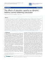
Metal was galvanostatically deposited onto Si nanowires
in a two-e lectrode setup (Fig ure 1). A home-b uilt T eflon
electrochemical cell was used to ensure that only the sur-
face with Si nanowires was exposed to a plating solution.
During plating, Si nanowires on a Si substrate acted as a
working electrode, and a platinum wire worked as a coun-
ter electrode. For the deposition of gold (Au) tube, com-
mercial plating solution (25 mM, Goldplattierbad GP 204,
from Heimerle+Meule GmbH, Germany) was used. A
Keithley 2400 power supply was used as a current source,
and the current density during the deposition was adjusted
to 1 mA/cm
2
. The plating experiments were carried out in
ambient condition at room temperature. No special atten-
tion had to be paid to the contact between backside of Si
substrate and Cu electrode. No discernable difference was
found between samples plated with and without GaIn
eutectic (as an ohmic c ontact) between Si substrate and
Cu plate.
After plating, surf ace morphologies an d element analy-
sis of the Si nanowires with metal tube were character-
ized by a SEM (JSM 7001F, JEOL) equipped with energy
dispersive X-ray analysis system (EDXA, Inca Energy-
350, Oxford Instruments, UK). To reveal the thicknesses
of tubular structures, TEM (JEM 2100, JEOL) characteri-
zation was carried out. For the TEM characterization, the
Si substrates with metal tubes were subjected to a con-
centrated NaOH solution (4.5 M, 50°C, 3 h) to release
metal tubes from Si nanowires. Afterward, the metal
tubes were extracted via centrifugation, and were rinsed
with ethano l until the pH value of solution equaled 7.
Finally, the metal tubes/ethanol s olution was dropped
onto TEM grids.
Results and discussion
In a typical electrochemical deposition experiment, Au
was deposited onto Si nanowires with average diameter
of ca. 550 nm. During the deposition, a small number of
bubbles were observed on the Si nanowire substrate in
the electrochemical deposition of Au, which might be
due to hydrogen evolution from the Si template. After
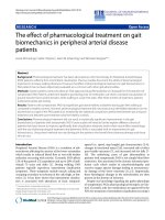
electrochemical deposition, Au was found to be homoge-
neously deposited onto the template in a large area, exhi-
biting bright contrast in SEM images (Figure 2a). The
deposited Au film covers fully the side wall of Si nano-
wires, resulting in Au tube (Figure 2b,c). Interestingly, it
is revealed that the Au i s deposited not only onto the
sidewall of Si nanowire, but also to the plateau between
Si nanowires (Figure 2c), implying that the electrochemi-
cal deposition uniformly occurred on the entire Si surface
irrespective of the surface morphology. It was conf irmed
by EDXA (Figure 2d) that the deposited film is Au. Au
tube deposited on Si nanowire exhibits good crystallinity,
as e videnced by the h igh-resolution TEM (HR-TEM)
image (Figure 2e) of an Au tube released from Si nano-
wire template and the corresponding selected area elec-
tron diffraction (SAED) pattern (inset of Figure 2e).
Neither surface modification nor remova l of surface
Si oxide, which formed because of slow oxidation of as-
prepared Si nanowires in the air, was necessary before the
electrochemical deposition of Au tubes shown in Figure 2.
Control experiments were performed, in which surface
Figure 1 Schematic illustration showing the e xperimental
setup of electrochemical depositing metal onto Si nanowires.
Huang et al. Nanoscale Research Letters 2011, 6 :165
/>Page 2 of 8
oxide was removed by HF-treatment (3.4 wt.%, 5 min)
before th e electrochemical de position. The morphologies
of Au tubes on Si nanowire templates with or without HF
treatment did not exhibit discernable difference. The pre-
sence or the ab sence of surface oxide fi lm is very impor-
tant in electrochemical deposition. Oxide film of the
non-HF treatment templates might have somehow been
removed in electrochemical bath. However, it is hard to
give solid evidence of oxide removal, because the detail
information of commercial available Au plating solution is
unknown, and the surface oxide will form again in several
minutesintheairevenifitwasremovedbytheplating
Figure 2 (a-c) The bird’ s-eye view of SEM images of Au tube deposited on an ordered array of Si nanowires.Therectanglein
(a) encloses a region which is magnified into (b), and the rectangle in (b) encloses a region which is magnified into (c). (d) EDX spectrum of
an Au tube/Si nanowires sample. (e) HR-TEM image of an Au tube released from Si nanowire, and (inset of e) the [110] zone axis SAED pattern
of the Au tube. The white lines indicate projection of atoms on (111) plane along [110] direction. (f) Applied potentials versus deposition times
for the deposition in the dark (black line) and under room light illumination (gray line), respectively.
Huang et al. Nanoscale Research Letters 2011, 6 :165
/>Page 3 of 8
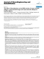
Figure 3 The bird’s-eye view of SEM images of the samples subjected to electrodepositions under the current density of (a) 2 mA/cm
2
for 40 min and (b) 1 mA/cm
2
for 80 min, respectively, and (c) the sample immersed in the plating solution without applied potential.
The diameters, the lengths, and the inter-wire distances between nanowires of samples used in (a) and (b) were identical.
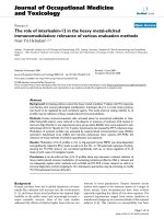
Figure 4 SEM images of Si nanowires deposited with Au for 5 min. (a) Low magnification image showing the morphologies of the who le
wires. (b-d) High magnification SEM images showing in detail the morphologies of the top, middle, and root part of a single nanowire,
respectively. The rectangles in (a) enclose the regions which are magnified into (b-d).
Huang et al. Nanoscale Research Letters 2011, 6 :165
/>Page 4 of 8
solution during the deposition, introducing difficulty to
any ex situ TEM characterization.
The depositions were performed in the dark, and under
the front-side room light illumination. No discernable
morphological difference was found in the resulting Au
tubes on corresponding Si templates. The applied poten-
tials during the depositions were recorded, and shown in
Figure 2f. The potential necessary for the experiment in
the dark is higher than that under illumination. The light
irradiating the Si substrate induced photo-generated elec-
tron-hole pairs in the template, and the photo-excited
electrons could arrive at the Si/solution interface and
reduce Au ions bec ause of the applied external potential.
Accordingly, only a less applied potential is needed to
drive the same amount of electrons to the Si/solution
interface in the case of deposition under illumination
than in that of deposition in the dark.
The depositions were performed under different cur-
rent densities. Figure 3a,b shows clearly that the thick-
ness of the deposited Au under 2 mA/cm
2
was larger
than that under 1 mA/cm
2
, even if the deposition tim e
under 1 mA/cm
2
(80 min) was two times of that under
2mA/cm
2
(40 min). The clearance between Si n anowires
has been totally filled by the deposited Au in the sample
shown in Figure 3a, whereas the gap between Si nano-
wires appears in the sample shown in Figure 3b. If the Si
nanowire template was immersed into the plating solu-
tion while no potential was applied, then neither the Au
particle nor the tube was found on the wall of Si template
(Figure 3c). Therefore, the results sho wn in Figure 3
proved definitely that the deposition of Au in this experi-
ment was because of electrochemical process, but not of
electroless plating.
For the electrochemical deposition of metal onto
macroporous Si, there are three typical deposition
modes, which represent the deposition proceeding from
pore bottom to pore opening [16,26,27], the deposition
proceeding from the opening of pores [27], as well as
the deposition occurring homogeneously on the entire
surfaceofporewall[16,17].Thehomogeneousdeposi-
tion occurs only for the deposition of less noble metal,
whereas no radial growth on sidewall has been found
for the noble metals so far. Therefore, macroporous Si
has not yet been employed as a template for the electro-
chemical deposition of noble metal tube.
Noble metal tube is achieved with the use of Si nano-
wires as a template in t his experiment. To explore the
growth process of Au tube on Si nanowires template, the
morphology of Au-deposited Si nanowires at the initial
stage of deposition was investigated. For a deposition time
of 5 min, the top (Figure 4b) and the middle (Figure 4c)
parts of a Si nanowire are fully covered by Au layer, while
the bottom part of a Si nanowires and the plateau between
nanowires are loaded with isolated Au particles (Figure
4d). Especially, the density of Au particle on the plateau
between Si nanowires is apparen tly lower than that on
the bottom part of a Si nanowire. To further investigate
the growth process of Au tube, the thicknesses of an Au
Figure 5 The thicknesses along a typical Au nanotube. (a) The relationship between the thicknesses of an Au tube and the distances of the
measured points from the root of the Au tube. (b) Low TEM image of the measured Au tube. The thickness values are measured from higher
magnification TEM images.
Huang et al. Nanoscale Research Letters 2011, 6 :165
/>Page 5 of 8
tube at different sites apart from the root of an Au tube
were measured, as shown in Figure 5a. It is shown that the
top and middle parts possess almost the same thickness,
while the root part of the Au tube is thinner t han the
remaining part of the tube. The morphologies of different
parts of Au-deposited structures with short (Figure 4) and
long (Figure 5) deposition times suggest that the growth of
Au proceeds quasi-radially on the Si nanowires.
The mechanism of quasi-radial growth remains
unclear so far. The difference between morphologies of
Au on the top/middle parts (continuous film) and that
of root part (isolated particles) of a Si nanowire might
be in duced by a mass transfer effect. Sinc e the electro-
chemical depos ition could take place everywhere on the
exposed Si surface, the me tal ions at the deposition
front are consumed quickly once the electrochemical
Figure 6 Typical TEM images of Au tubes deposited with (a) 20 min, (b) 40 min, and (c) 60 min. (d) Relationship between tube thickness and
deposition time.
Figure 7 SEM images of Au tubes deposited on SiNWs with differ ent diameters (a) 1 μm, (b) 450 nm, and (c) 45 nm.Insetsin(a) and
(b) show respective close cross-sectional views revealing the Au tube on Si nanowires. Arrow 1 in (c) indicates a broken tube structure. Arrow 2
in (c) indicates a Si nanowire template.
Huang et al. Nanoscale Research Letters 2011, 6 :165
/>Page 6 of 8
deposition starts. The subsequent supply of metal ions
from bulk solution will be preferentially transported to
the top/middle parts of the Si nanowires. In this case,
the metal ions that can finally reach the root part will
be much less because of the consumption of the top/
middle part during the depo sition, thus resulting in a
thick top/middle part and a thin root part of the Au
tubes.
The quasi-radial growth of Au on Si nanowires
implies that the thickness of Au tube increases linearly
with the deposition time, while the length of Au tube
remains constant. The assumption has been confirmed
by a series of control experiments (Figure 6). As shown
by the TEM images of Au tube during different deposi-
tion times (Figure 6a-c), the thickness of wall in an Au
tube does increase approximately linearly with the
deposition time (Figure 6d). The results presented here
suggest that the wall thickness of metal tube can be
controlled by changing the deposition time, whereas the
length of metal tube can be independently controlled via
choosing Si nanowires template with a desired length.
By further increasing the deposition time, the gap
between Si nanowires is filled with the deposited Au.
Consequently, the deposited Au evolves from tubular
structure to a thick film with straight channels.
As mentioned above, by template-based metal-assisted
chemical etching, the diamete r of Si nanowires can be
precisely controlled, and Si nanowires with diameters
ranging from sub-10 nm to one micron have been
achieved [21,23]. Accordingly, the inner diameter of an
Au nanotube fabricated with Si nanowires as a positive
template can be tuned in a wide range. Figure 7 shows a
series of Au nanotubes with different inner diameters.
Tubular structure wit h inner diameter as small as
45 nm was fabricated with Si nanowires from the AAO
mask method (Figure 7c). The Si nanowires bend and
stick together before the electrochemical deposition, and
therefore bundles of Au tube are found (Figure 7c). The
bending of nanowires and the formation of bundle are
common phenomena for 1D nanostructure fabricated
via solution-based method, due to surface tension force
exerted on the nanowires during the drying of the sam-
ple [21,28]. The bending and bundling could be avoided
or relieved by a supercritical drying process [24], thus
potentially allowing the formation of isolated metal
nanotube arrays with small tube diameters.
Conclusions
In conclusion, Si nanowires have been employed as
a template for the fabrication o f noble metal tube by
the electrochemical method. The growth of metal
on Si nanowires proceeds quasi-radially, as suggested by
SEM and TEM characterizations. This growth behavior
enables precise control over the thickness of the
deposited metal tube. Metal tubes with inner diameters
ranging from 1 μm down to 45 nm are obtained by elec-
trochemical deposition on the Si nanowires with pre-
ferred diameter.
Abbreviations
AAO: anodic aluminum oxide; EDXA: energy dispersive X-ray analysis; HR-
TEM: high-resolution TEM; PS: polystyrene; SAED: selected area electron
diffraction; SEM: scanning electron microscope; TEM: transmission electron
microscope.
Acknowledgements
This study was supported by the research foundation of Jiangsu University,
P. R. China (Grant 09JDG043), and the National Natural Science Foundation
of China (Grant 61006049).
Author details
1
Functional Molecular Materials Centre, Scientific Research Academy, Jiangsu
University, Zhenjiang 212013, P. R. China.
2
Max Planck Institute of
Microstructure Physics, Weinberg 2, D-06120 Halle/Saale, Germany.
Authors’ contributions
ZH carried out the etching experiments for Si nanowire templates and the
electrodepositons, the SEM and TEM characterizations, as well as drafted the
manuscript. LL participated in the electrodeposition and SEM
characterization. NG carried out the RIE experiments during the fabrication
of Si nanowires. All authors read and approved the final manuscript.
Competing interests
The authors declare that they have no competing interests.
Received: 6 May 2010 Accepted: 23 February 2011
Published: 23 February 2011
References
1. Che GL, Lakshmi BB, Fisher ER, Martin CR: Carbon nanotubule membranes
for electrochemical energy storage and production. Nature 1998,
393:346.
2. Steigerwalt ES, Deluga GA, Lukehart CM: Pt-Ru/carbon fiber
nanocomposites: Synthesis, characterization, and performance as anode
catalysts of direct methanol fuel cells. A search for exceptional
performance. J Phys Chem B 2002, 106:760.
3. Sanchez-Castillo MA, Couto C, Kim WB, Dumesic JA: Gold-nanotube
membranes for the oxidation of CO at gas-water interfaces. Angew Chem
Int Ed 2004, 43:1140.
4. An W, Pei Y, Zeng XC: CO oxidation catalyzed by single-walled helical
gold nanotube. Nano Lett 2008, 8:195.
5. Zhang XY, Dong DH, Li D, Williams T, Wang HT, Webley PA: Direct
electrodeposition of Pt nanotube arrays and their enhanced
electrocatalytic activities. Electrochem Commun 2009, 11:190.
6. Chae WS, Hwang IW, Jung JS, Kim YR: Optical and magnetic properties
induced by structural confinement of ternary chalcogenide in AlMCM-41
nanotube. Chem Phys Lett 2001, 341:279.
7. Singh AK, Briere TM, Kumar V, Kawazoe Y: Magnetism in transition-metal-
doped silicon nanotubes. Phys Rev Lett 2003, 91:146802.
8. Brumlik CJ, Martin CR: Template Synthesis of Metal Microtubules. JAm
Chem Soc 1991, 113:3174.
9. Bao JC, Tie CY, Xu Z, Zhou QF, Shen D, Ma Q: Template synthesis of an
array of nickel nanotubules and its magnetic behavior. Adv Mater 2001,
13:1631.
10. Liu LF, Zhou WY, Xie SS, Song L, Luo SD, Liu DF, Shen J, Zhang ZX,
Xiang YJ, Ma WJ, Ren Y, Wang CY, Wang G: Highly efficient direct
electrodeposition of Co-Cu alloy nanotubes in an anodic alumina
template. J Phys Chem C 2008, 112:2256.
11. Wirtz M, Martin CR: Template-fabricated gold nanowires and nanotubes.
Adv Mater 2003, 15:455.
12. Rohan JF, Casey DP, Ahern BM, Rhen FMF, Roy S, Fleming D, Lawrence SE:
Coaxial metal and magnetic alloy nanotubes in polycarbonate templates
by electroless deposition. Electrochem Commun 2008, 10:1419.
Huang et al. Nanoscale Research Letters 2011, 6 :165
/>Page 7 of 8
13. Peng TY, Yang HP, Dai K, Pu XL, Hirao K: Fabrication and characterization
of CdS nanotube arrays in porous anodic aluminum oxide templates.
Chem Phys Lett 2003, 379:432.
14. Li N, Li XT, Yin XJ, Wang W, Qiu SL: Electroless deposition of open-end Cu
nanotube arrays. Solid State Commun 2004, 132:841.
15. Lee W, Scholz R, Niesch K, Gosele U: A template-based electrochemical
method for the synthesis of multisegmented metallic nanotubes. Angew
Chem Int Ed 2005, 44:6050.
16. Ogata YH, Kobayashi K, Motoyama M: Electrochemical metal deposition
on silicon. Curr Opin Solid State Mater Sci 2006, 10:163.
17. Kobayashi K, Harraz FA, Izuo S, Sakka T, Ogata YH: Microrod and microtube
formation by electrodeposition of metal into ordered macropores
prepared in p-type silicon. J Electrochem Soc 2006, 153:C218.
18. Fukami K, Sakka T, Ogata YH, Yamauchi T, Tsubokawa N: Multistep filling of
porous silicon with conductive polymer by electropolymerization. Phys
Status Solidi A 2009, 206:1259.
19. Tiginyanu I, Monaico E, Monaico E: Ordered arrays of metal nanotubes in
semiconductor envelope. Electrochem Commun 2008, 10:731.
20. Lehmann V, Ronnebeck S: The Physics of Macropore Formation in Low-
Doped p-Type Silicon. J Electrochem Soc 1999, 146:2968.
21. Huang ZP, Fang H, Zhu J: Fabrication of silicon nanowire arrays with
controlled diameter, length, and density. Adv Mater 2007, 19:744.
22. Peng KQ, Zhang ML, Lu AJ, Wong NB, Zhang RQ, Lee ST: Ordered silicon
nanowire arrays via nanosphere lithography and metal-induced etching.
Appl Phys Lett 2007, 90:163123.
23. Huang ZP, Zhang XX, Reiche M, Liu LF, Lee W, Shimizu T, Senz S, Gösele U:
Extended arrays of vertically aligned sub-10 nm diameter [100] Si
nanowires by metal-assisted chemical etching. Nano Lett 2008, 8:3046.
24. Chang SW, Chuang VP, Boles ST, Ross CA, Thompson CV: Densely Packed
Arrays of Ultra-High-Aspect-Ratio Silicon Nanowires Fabricated using
Block-Copolymer Lithography and Metal-Assisted Etching. Adv Funct
Mater 2009, 19:2495.
25. de Boor J, Geyer N, Wittemann JV, Gösele U, Schmidt V: Sub-100 nm
silicon nanowires by laser interference lithography and metal-assisted
etching. Nanotechnology 2010, 21:095302.
26. Fang C, Foca E, Xu SF, Carstensen J, Foll H: Deep silicon macropores filled
with copper by electrodeposition. J Electrochem Soc 2007, 154:D45.
27. Fukami K, Kobayashi K, Matsumoto T, Kawamura YL, Sakka T, Ogata YH:
Electrodeposition of noble metals into ordered macropores in p-type
silicon. J Electrochem Soc 2008, 155:D443.
28. Ahn M, Heilmann RK, Schattenburg ML: Fabrication of ultrahigh aspect
ratio freestanding gratings on silicon-on-insulator wafers. J Vac Sci
Technol B 2007, 25:2593.
doi:10.1186/1556-276X-6-165
Cite this article as: Huang et al.: Quasi-radial growth of metal tube on si
nanowires template. Nanoscale Research Letters 2011 6:165.
Submit your manuscript to a
journal and benefi t from:
7 Convenient online submission
7 Rigorous peer review
7 Immediate publication on acceptance
7 Open access: articles freely available online
7 High visibility within the fi eld
7 Retaining the copyright to your article
Submit your next manuscript at 7 springeropen.com
Huang et al. Nanoscale Research Letters 2011, 6 :165
/>Page 8 of 8