New Developments in Biomedical Engineering 2011 Part 10 docx
Bạn đang xem bản rút gọn của tài liệu. Xem và tải ngay bản đầy đủ của tài liệu tại đây (4 MB, 40 trang )
NewDevelopmentsinBiomedicalEngineering352
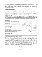
Fig. 5. Filtering principles of light propagating inside a biological tissue. Superficial and
deep regions are marked as 1 and 2, respectively.
Registration of the co- and cross-linear polarizer output channels allows the determination
of the degree of polarization (DOP), which is defined as:
II
II
I I
DOP
I I
(7)
where <
I
>
and <I
> are the mean intensity of the co- and cross-polarized speckle patterns.
Subtracting the cross-polarized pattern from the co-polarized pattern suppresses the volume
scattering.
Spectral filtering (Demos et al., 2000) is based on the spectral dependence of skin attenuation
coefficients (Salomatina et al., 2006). Shorter wavelengths are attenuated more heavily in a
scattering medium and yield a higher output of scattered light than longer wavelengths.
Therefore region 1 for the blue light is expected to be shallower than the red light, and, we
should thus use the blue laser for skin roughness measurements (Tchvialeva et al., 2008).
In another study (Tchvialeva et al., 2009), we adopted the above filtering techniques for
speckle roughness estimation of the skin. However, our experiment showed that the filtered
signals still contained sufficient volume-scattered signals and overestimated the skin
roughness. Therefore, we formulate a mathematical correction to further adjust the speckle
contrasts to their surface reflection values.
3.2.3 Speckle contrast correction
The idea of speckle contrast correction for eliminating the remaining volume scattering was
inspired by the experimental evidence arising from the co-polarized contrast vs. DOP as
shown in Figure 6 (Tchvialeva, et al., 2009). There is a strong correlation between the co-
polarized contrast and DOP (r = 0.777, p < 0.0001).
0
0.2
0.4
0.6
0.8
0 0.2 0.4 0.6 0.8
DOP
Speckle Contract
Fig. 6. The linear fit of the experimental points for co-polarized contrast vs. DOP.
We assume (at least as a first approximation) that this linear relation is valid for the entire
range of DOP from 0 to 1. We also know that weakly scattered light has almost the same
state of polarization as incident light (Sankaran et al., 1999; Tchvialeva, et al., 2008). If the
incident light is linearly polarized (DOP = 1), light scattered by the surface should also have
DOP
surf
= 1. Based on this assumption, we can compute speckle contrast for surface scattered
light by linearly extrapolating the data for DOP = 1. The corrected contrast is then applied to
the calibration curve for the blue laser (Figure 4) and is mapped to the corrected roughness
value.
3.2.4 Comparing in-vivo data for different body sites
To compare skin roughness obtained by our prototype with other
in-vivo data, we
conducted an experiment with 34 healthy volunteers. Figure 7 shows preliminary data for
speckle roughness and standard deviation for various body sites. We also looked up the
published
in-vivo roughness values for the same body site and plot these values against our
roughness measurements. Measured speckle roughness are consistent with published
values. Currently, we are in the process of designing a study to compare the speckle
roughness with replica roughness.
SkinRoughnessAssessment 353
Fig. 5. Filtering principles of light propagating inside a biological tissue. Superficial and
deep regions are marked as 1 and 2, respectively.
Registration of the co- and cross-linear polarizer output channels allows the determination
of the degree of polarization (DOP), which is defined as:
II
II
I I
DOP
I I
(7)
where <
I
>
and <I
> are the mean intensity of the co- and cross-polarized speckle patterns.
Subtracting the cross-polarized pattern from the co-polarized pattern suppresses the volume
scattering.
Spectral filtering (Demos et al., 2000) is based on the spectral dependence of skin attenuation
coefficients (Salomatina et al., 2006). Shorter wavelengths are attenuated more heavily in a
scattering medium and yield a higher output of scattered light than longer wavelengths.
Therefore region 1 for the blue light is expected to be shallower than the red light, and, we
should thus use the blue laser for skin roughness measurements (Tchvialeva et al., 2008).
In another study (Tchvialeva et al., 2009), we adopted the above filtering techniques for
speckle roughness estimation of the skin. However, our experiment showed that the filtered
signals still contained sufficient volume-scattered signals and overestimated the skin
roughness. Therefore, we formulate a mathematical correction to further adjust the speckle
contrasts to their surface reflection values.
3.2.3 Speckle contrast correction
The idea of speckle contrast correction for eliminating the remaining volume scattering was
inspired by the experimental evidence arising from the co-polarized contrast vs. DOP as
shown in Figure 6 (Tchvialeva, et al., 2009). There is a strong correlation between the co-
polarized contrast and DOP (r = 0.777, p < 0.0001).
0
0.2
0.4
0.6
0.8
0 0.2 0.4 0.6 0.8
DOP
Speckle Contract
Fig. 6. The linear fit of the experimental points for co-polarized contrast vs. DOP.
We assume (at least as a first approximation) that this linear relation is valid for the entire
range of DOP from 0 to 1. We also know that weakly scattered light has almost the same
state of polarization as incident light (Sankaran et al., 1999; Tchvialeva, et al., 2008). If the
incident light is linearly polarized (DOP = 1), light scattered by the surface should also have
DOP
surf
= 1. Based on this assumption, we can compute speckle contrast for surface scattered
light by linearly extrapolating the data for DOP = 1. The corrected contrast is then applied to
the calibration curve for the blue laser (Figure 4) and is mapped to the corrected roughness
value.
3.2.4 Comparing in-vivo data for different body sites
To compare skin roughness obtained by our prototype with other
in-vivo data, we
conducted an experiment with 34 healthy volunteers. Figure 7 shows preliminary data for
speckle roughness and standard deviation for various body sites. We also looked up the
published
in-vivo roughness values for the same body site and plot these values against our
roughness measurements. Measured speckle roughness are consistent with published
values. Currently, we are in the process of designing a study to compare the speckle
roughness with replica roughness.
NewDevelopmentsinBiomedicalEngineering354
Fig. 7. In-vivo skin rms roughness obtained by our speckle device and by published values
of fringe projection systems. The number of samples measured by the speckle prototype is
denoted within the parentheses after the body sites.
4. Conclusion
Skin roughness is important for many medical applications. Replica-based techniques have
been the
de facto method until the recent development of fringe projection, an area-
topography technique, because short data acquisition time is most crucial for
in-vivo skin
application. Similarly, laser speckle contrast, an area-integrating approach, also shows
potential due to its acquisition speed, simplicity, low cost, and high accuracy. The original
theory developed by Parry was for opaque surfaces and for light source with a Gaussian
spectral profile. We extended the theory to polychromatic light sources and applied the
method to a semi-transparent object, skin. Using a blue diode laser, with three filtering
mechanisms and a mathematical correction, we were able to build a prototype which can
measure rms roughness
R
q
up to 100 μm. We have conducted a preliminary pilot study with
a group of volunteers. The results were in good agreement with the most popular fringe
project methods. Currently, we are designing new experiments to further test the device.
5. References
Articus, K.; Brown, C. A. & Wilhelm, K. P. (2001). Scale-sensitive fractal analysis using the
patchwork method for the assessment of skin roughness
, Skin Res Technol, Vol. 7, No. 3,
pp. 164-167
Bielfeldt, S.; Buttgereit, P.; Brandt, M.; Springmann, G. & Wilhelm, K. P. (2008).
Non-invasive
evaluation techniques to quantify the efficacy of cosmetic anti-cellulite products
, Skin Res
Technol,
Vol. 14, No. 3, pp. 336-346
Bourgeois, J. F.; Gourgou, S.; Kramar, A.; Lagarde, J. M.; Gall, Y. & Guillot, B. (2003).
Radiation-induced skin fibrosis after treatment of breast cancer: profilometric analysis, Skin
Res Technol,
Vol. 9, No. 1, pp. 39-42
Briers, J. (1993). Surface roughness evaluation. In:
Speckle Metrology, Sirohi, R. S. (Eds), by
CRC Press
Callaghan, T. M. & Wilhelm, K. P. (2008).
A review of ageing and an examination of clinical
methods in the assessment of ageing skin. Part 2: Clinical perspectives and clinical methods
in the evaluation of ageing skin
, Int J Cosmet Sci, Vol. 30, No. 5, pp. 323-332
Cheng, C.; Liu, C.; Zhang, N.; Jia, T.; Li, R. & Xu, Z. (2002).
Absolute measurement of roughness
and lateral-correlation length of random surfaces by use of the simplified model of image-
speckle contrast
, Applied Optics, Vol. 41, No. 20, pp. 4148-4156
Connemann, B.; Busche, H.; Kreusch, J.; Teichert, H M. & Wolff, H. (1995).
Quantitative
surface topography as a tool in the differential diagnosis between melanoma and naevus
,
Skin Res Technol, Vol. 1, pp. 180-186
Connemann, B.; Busche, H.; Kreusch, J. & Wolff, H. H. (1996).
Sources of unwanted variabilitv
in measurement and description of skin surface topography
, Skin Res Technol, Vol. 2, pp.
40-48
De Paepe, K.; Lagarde, J. M.; Gall, Y.; Roseeuw, D. & Rogiers, V. (2000).
Microrelief of the skin
using a light transmission method
, Arch Dermatol Res, Vol. 292, No. 10, pp. 500-510
Death, D. L.; Eberhardt, J. E. & Rogers, C. A. (2000).
Transparency effects on powder speckle
decorrelation
, Optics Express, Vol. 6, No. 11, pp. 202-212
del Carmen Lopez Pacheco, M.; da Cunha Martins-Costa, M. F.; Zapata, A. J.; Cherit, J. D. &
Gallegos, E. R. (2005).
Implementation and analysis of relief patterns of the surface of
benign and malignant lesions of the skin by microtopography
, Phys Med Biol, Vol. 50, No.
23, pp. 5535-5543
Demos, S. G.; Radousky, H. B. & Alfano, R. R. (2000).
Deep subsurface imaging in tissues using
spectral and polarization filtering
, Optics Express, Vol. 7, No. 1, pp. 23-28
Egawa, M.; Oguri, M.; Kuwahara, T. & Takahashi, M. (2002).
Effect of exposure of human skin
to a dry environment
, Skin Res Technol, Vol. 8, No. 4, pp. 212-218
Fischer, T. W.; Wigger-Alberti, W. & Elsner, P. (1999).
Direct and non-direct measurement
techniques for analysis of skin surface topography
, Skin Pharmacol Appl Skin Physiol, Vol.
12, No. 1-2, pp. 1-11
Fricke-Begemann, T. & Hinsch, K. (2004).
Measurement of random processes at rough surfaces
with digital speckle correlation
, J Opt Soc Am A Opt Image Sci Vis, Vol. 21, No. 2, pp.
252-262
Friedman, P. M.; Skover, G. R.; Payonk, G. & Geronemus, R. G. (2002a).
Quantitative
evaluation of nonablative laser technology
, Semin Cutan Med Surg, Vol. 21, No. 4, pp.
266-273
Friedman, P. M.; Skover, G. R.; Payonk, G.; Kauvar, A. N. & Geronemus, R. G. (2002b).
3D
in-vivo optical skin imaging for topographical quantitative assessment of non-ablative laser
technology
, Dermatol Surg, Vol. 28, No. 3, pp. 199-204
Fujii, H. & Asakura, T. (1977).
Roughness measurements of metal surfaces using laser speckle,
JOSA, Vol. 67, No. 9, pp. 1171-1176
SkinRoughnessAssessment 355
Fig. 7. In-vivo skin rms roughness obtained by our speckle device and by published values
of fringe projection systems. The number of samples measured by the speckle prototype is
denoted within the parentheses after the body sites.
4. Conclusion
Skin roughness is important for many medical applications. Replica-based techniques have
been the
de facto method until the recent development of fringe projection, an area-
topography technique, because short data acquisition time is most crucial for
in-vivo skin
application. Similarly, laser speckle contrast, an area-integrating approach, also shows
potential due to its acquisition speed, simplicity, low cost, and high accuracy. The original
theory developed by Parry was for opaque surfaces and for light source with a Gaussian
spectral profile. We extended the theory to polychromatic light sources and applied the
method to a semi-transparent object, skin. Using a blue diode laser, with three filtering
mechanisms and a mathematical correction, we were able to build a prototype which can
measure rms roughness
R
q
up to 100 μm. We have conducted a preliminary pilot study with
a group of volunteers. The results were in good agreement with the most popular fringe
project methods. Currently, we are designing new experiments to further test the device.
5. References
Articus, K.; Brown, C. A. & Wilhelm, K. P. (2001). Scale-sensitive fractal analysis using the
patchwork method for the assessment of skin roughness
, Skin Res Technol, Vol. 7, No. 3,
pp. 164-167
Bielfeldt, S.; Buttgereit, P.; Brandt, M.; Springmann, G. & Wilhelm, K. P. (2008).
Non-invasive
evaluation techniques to quantify the efficacy of cosmetic anti-cellulite products
, Skin Res
Technol,
Vol. 14, No. 3, pp. 336-346
Bourgeois, J. F.; Gourgou, S.; Kramar, A.; Lagarde, J. M.; Gall, Y. & Guillot, B. (2003).
Radiation-induced skin fibrosis after treatment of breast cancer: profilometric analysis, Skin
Res Technol,
Vol. 9, No. 1, pp. 39-42
Briers, J. (1993). Surface roughness evaluation. In:
Speckle Metrology, Sirohi, R. S. (Eds), by
CRC Press
Callaghan, T. M. & Wilhelm, K. P. (2008).
A review of ageing and an examination of clinical
methods in the assessment of ageing skin. Part 2: Clinical perspectives and clinical methods
in the evaluation of ageing skin
, Int J Cosmet Sci, Vol. 30, No. 5, pp. 323-332
Cheng, C.; Liu, C.; Zhang, N.; Jia, T.; Li, R. & Xu, Z. (2002).
Absolute measurement of roughness
and lateral-correlation length of random surfaces by use of the simplified model of image-
speckle contrast
, Applied Optics, Vol. 41, No. 20, pp. 4148-4156
Connemann, B.; Busche, H.; Kreusch, J.; Teichert, H M. & Wolff, H. (1995).
Quantitative
surface topography as a tool in the differential diagnosis between melanoma and naevus
,
Skin Res Technol, Vol. 1, pp. 180-186
Connemann, B.; Busche, H.; Kreusch, J. & Wolff, H. H. (1996).
Sources of unwanted variabilitv
in measurement and description of skin surface topography
, Skin Res Technol, Vol. 2, pp.
40-48
De Paepe, K.; Lagarde, J. M.; Gall, Y.; Roseeuw, D. & Rogiers, V. (2000).
Microrelief of the skin
using a light transmission method
, Arch Dermatol Res, Vol. 292, No. 10, pp. 500-510
Death, D. L.; Eberhardt, J. E. & Rogers, C. A. (2000).
Transparency effects on powder speckle
decorrelation
, Optics Express, Vol. 6, No. 11, pp. 202-212
del Carmen Lopez Pacheco, M.; da Cunha Martins-Costa, M. F.; Zapata, A. J.; Cherit, J. D. &
Gallegos, E. R. (2005).
Implementation and analysis of relief patterns of the surface of
benign and malignant lesions of the skin by microtopography
, Phys Med Biol, Vol. 50, No.
23, pp. 5535-5543
Demos, S. G.; Radousky, H. B. & Alfano, R. R. (2000).
Deep subsurface imaging in tissues using
spectral and polarization filtering
, Optics Express, Vol. 7, No. 1, pp. 23-28
Egawa, M.; Oguri, M.; Kuwahara, T. & Takahashi, M. (2002).
Effect of exposure of human skin
to a dry environment
, Skin Res Technol, Vol. 8, No. 4, pp. 212-218
Fischer, T. W.; Wigger-Alberti, W. & Elsner, P. (1999).
Direct and non-direct measurement
techniques for analysis of skin surface topography
, Skin Pharmacol Appl Skin Physiol, Vol.
12, No. 1-2, pp. 1-11
Fricke-Begemann, T. & Hinsch, K. (2004).
Measurement of random processes at rough surfaces
with digital speckle correlation
, J Opt Soc Am A Opt Image Sci Vis, Vol. 21, No. 2, pp.
252-262
Friedman, P. M.; Skover, G. R.; Payonk, G. & Geronemus, R. G. (2002a).
Quantitative
evaluation of nonablative laser technology
, Semin Cutan Med Surg, Vol. 21, No. 4, pp.
266-273
Friedman, P. M.; Skover, G. R.; Payonk, G.; Kauvar, A. N. & Geronemus, R. G. (2002b).
3D
in-vivo optical skin imaging for topographical quantitative assessment of non-ablative laser
technology
, Dermatol Surg, Vol. 28, No. 3, pp. 199-204
Fujii, H. & Asakura, T. (1977).
Roughness measurements of metal surfaces using laser speckle,
JOSA, Vol. 67, No. 9, pp. 1171-1176
NewDevelopmentsinBiomedicalEngineering356
Fujimura, T.; Haketa, K.; Hotta, M. & Kitahara, T. (2007).
Global and systematic demonstration
for the practical usage of a direct in vivo measurement system to evaluate wrinkles
, Int J
Cosmet Sci,
Vol. 29, No. 6, pp. 423-436
Gautier, S.; Xhauflaire-Uhoda, E.; Gonry, P. & Pierard, G. E. (2008).
Chitin-glucan, a natural
cell scaffold for skin moisturization and rejuvenation
, Int J Cosmet Sci, Vol. 30, No. 6, pp.
459-469
Goodman, J. W. (2006).
Speckle Phenomena in Optics: Theory and Application, Roberts and
Company Publishers
Handels, H.; RoS, T.; Kreusch, J.; Wolff, H. H. & Poppl, S. J. (1999).
Computer-supported
diagnosis of melanoma in profilometry
, Meth Inform Med, Vol. 38, pp. 43-49
Hashimoto, K. (1974).
New methods for surface ultrastructure: Comparative studies of scanning
electron microscopy, transmission electron microscopy and replica method
, Int J Dermatol,
Vol. 13, No. 6, pp. 357-381
Hocken, R. J.; Chakraborty, N. & Brown, C. (2005).
Optical metrology of surface, CIRP Annals -
Manufacturing Technology,
Vol. 54, No. 2, pp. 169-183
Hof, C. & Hopermann, H. (2000).
Comparison of replica- and in vivo-measurement of the
microtopography of human skin
, SOFW Journal, Vol. 126, pp. 40-46
Humbert, P. G.; Haftek, M.; Creidi, P.; Lapiere, C.; Nusgens, B.; Richard, A.; Schmitt, D.;
Rougier, A. & Zahouani, H. (2003).
Topical ascorbic acid on photoaged skin. Clinical,
topographical and ultrastructural evaluation: double-blind study vs. placebo
, Exp
Dermatol,
Vol. 12, No. 3, pp. 237-244
Hun, C.; Bruynooghea, M.; Caussignacb, J M. & Meyrueisa, P. (2006). Study of the
exploitation of speckle techniques for pavement surface,
Proc of SPIE 6341, pp.
63412A,
International Organization for Standardization Committee (2007).
GPS-Surface texture:areal-
Part 6: classification of methods for measuring surface structure, Draft 25178-6
Jacobi, U.; Chen, M.; Frankowski, G.; Sinkgraven, R.; Hund, M.; Rzany, B.; Sterry, W. &
Lademann, J. (2004).
In vivo determination of skin surface topography using an optical
3D device
, Skin Res Technol, Vol. 10, No. 4, pp. 207-214
Jaspers, S.; Hopermann, H.; Sauermann, G.; Hoppe, U.; Lunderstadt, R. & Ennen, J. (1999).
Rapid in vivo measurement of the topography of human skin by active image triangulation
using a digital micromirror device mirror device
, Skin Res Technol, Vol. 5, pp. 195-207
Kampf, G. & Ennen, J. (2006).
Regular use of a hand cream can attenuate skin dryness and
roughness caused by frequent hand washing
, BMC Dermatol, Vol. 6, pp. 1
Kawada, A.; Konishi, N.; Oiso, N.; Kawara, S. & Date, A. (2008).
Evaluation of anti-wrinkle
effects of a novel cosmetic containing niacinamide
, J Dermatol, Vol. 35, No. 10, pp. 637-
642
Kim, E.; Nam, G. W.; Kim, S.; Lee, H.; Moon, S. & Chang, I. (2007).
Influence of polyol and oil
concentration in cosmetic products on skin moisturization and skin surface roughness
, Skin
Res Technol,
Vol. 13, No. 4, pp. 417-424
Korting, H.; Megele, M.; Mehringer, L.; Vieluf, D.; Zienicke, H.; Hamm, G. & Braun-Falco, O.
(1991).
Influence of skin cleansing preparation acidity on skin surface properties,
International Journal of Cosmetic Science, Vol. 13, pp. 91-102
Lagarde, J. M.; Rouvrais, C. & Black, D. (2005).
Topography and anisotropy of the skin surface
with ageing
, Skin Res Technol, Vol. 11, No. 2, pp. 110-119
Lagarde, J. M.; Rouvrais, C.; Black, D.; Diridollou, S. & Gall, Y. (2001).
Skin topography
measurement by interference fringe projection: a technical validation
, Skin Res Technol,
Vol. 7, No. 2, pp. 112-121
Lee, H. K.; Seo, Y. K.; Baek, J. H. & Koh, J. S. (2008).
Comparison between ultrasonography
(Dermascan C version 3) and transparency profilometry (Skin Visiometer SV600)
, Skin
Res Technol,
Vol. 14, pp. 8-12
Lehmann, P. (1999).
Surface-roughness measurement based on the intensity correlation function of
scattered light under speckle-pattern illumination
, Applied Optics, Vol. 38, No. 7, pp.
1144-1152
Lehmann, P. (2002).
Aspect ratio of elongated polychromatic far-field speckles of continuous and
discrete spectral distribution with respect to surface roughness characterization
, Applied
Optics,
Vol. 41, No. 10, pp. 2008-2014
Leonard, L. C. (1998).
Roughness measurement of metallic surfaces based on the laser speckle
contrast method
, Optics and Lasers in Engineering, Vol. 30, No. 5, pp. 433-440
Leveque, J. L. (1999).
EEMCO guidance for the assessment of skin topography. The European
Expert Group on Efficacy Measurement of Cosmetics and other Topical Products
, J Eur
Acad Dermatol Venereol,
Vol. 12, No. 2, pp. 103-114
Leveque, J. L. & Querleux, B. (2003).
SkinChip, a new tool for investigating the skin surface in
vivo
, Skin Res Technol, Vol. 9, No. 4, pp. 343-347
Levy, J. L.; Servant, J. J. & Jouve, E. (2004).
Botulinum toxin A: a 9-month clinical and 3D in vivo
profilometric crow's feet wrinkle formation study
, J Cosmet Laser Ther, Vol. 6, No. 1, pp.
16-20
Li, L.; Mac-Mary, S.; Marsaut, D.; Sainthillier, J. M.; Nouveau, S.; Gharbi, T.; de Lacharriere,
O. & Humbert, P. (2006a).
Age-related changes in skin topography and microcirculation,
Arch Dermatol Res, Vol. 297, No. 9, pp. 412-416
Li, Z.; Li, H. & Qiu, Y. (2006b).
Fractal analysis of laser speckle for measuring roughness, SPIE,
Vol. 6027, pp. 60271S
Lu, R S.; Tian, G Y.; Gledhill, D. & Ward, S. (2006).
Grinding surface roughness measurement
based on the co-occurrence matrix of speckle pattern texture
, Applied Optics, Vol. 45, No.
35, pp. 8839–8847
Lukaszewski, K.; Rozniakowski, K. & Wojtatowicz, T. W. (1993).
Laser examination of cast
surface roughness
, Optical Engineering, Vol. 40, No. 9, pp. 1993-1997
Markhvida, I.; Tchvialeva, L.; Lee, T. K. & Zeng, H. (2007).
The influence of geometry on
polychromatic speckle contrast
, Journal of the Optical Society of America A, Vol. 24, No. 1,
pp. 93-97
Mazzarello, V.; Soggiu, D.; Masia, D. R.; Ena, P. & Rubino, C. (2006).
Melanoma versus
dysplastic naevi: microtopographic skin study with noninvasive method
, J Plast Reconstr
Aesthet Surg,
Vol. 59, No. 7, pp. 700-705
Ning, Y. N.; Grattan, K. T. V.; Palmer, A. W. & Meggitt, B. T. (1992).
Coherence length
modulation of a multimode laser diode in a dual Michelson interferometer configuration
,
Applied Optics, Vol. 31, No. 9, pp. 1322–1327
Parry, G. (1984). Speckle patterns in partially coherent light. In:
Laser Speckle and Related
Phenomena, Dainty, J. C.
(Eds), pp. 77-122, Springer-Verlag, Berlin; New York
Peters, J. & Schoene, A. (1998).
Nondestructive evaluation of surface roughness by speckle
correlation techniques
, SPIE, Vol. 3399, pp. 45-56
SkinRoughnessAssessment 357
Fujimura, T.; Haketa, K.; Hotta, M. & Kitahara, T. (2007).
Global and systematic demonstration
for the practical usage of a direct in vivo measurement system to evaluate wrinkles
, Int J
Cosmet Sci,
Vol. 29, No. 6, pp. 423-436
Gautier, S.; Xhauflaire-Uhoda, E.; Gonry, P. & Pierard, G. E. (2008).
Chitin-glucan, a natural
cell scaffold for skin moisturization and rejuvenation
, Int J Cosmet Sci, Vol. 30, No. 6, pp.
459-469
Goodman, J. W. (2006).
Speckle Phenomena in Optics: Theory and Application, Roberts and
Company Publishers
Handels, H.; RoS, T.; Kreusch, J.; Wolff, H. H. & Poppl, S. J. (1999).
Computer-supported
diagnosis of melanoma in profilometry
, Meth Inform Med, Vol. 38, pp. 43-49
Hashimoto, K. (1974).
New methods for surface ultrastructure: Comparative studies of scanning
electron microscopy, transmission electron microscopy and replica method
, Int J Dermatol,
Vol. 13, No. 6, pp. 357-381
Hocken, R. J.; Chakraborty, N. & Brown, C. (2005).
Optical metrology of surface, CIRP Annals -
Manufacturing Technology,
Vol. 54, No. 2, pp. 169-183
Hof, C. & Hopermann, H. (2000).
Comparison of replica- and in vivo-measurement of the
microtopography of human skin
, SOFW Journal, Vol. 126, pp. 40-46
Humbert, P. G.; Haftek, M.; Creidi, P.; Lapiere, C.; Nusgens, B.; Richard, A.; Schmitt, D.;
Rougier, A. & Zahouani, H. (2003).
Topical ascorbic acid on photoaged skin. Clinical,
topographical and ultrastructural evaluation: double-blind study vs. placebo
, Exp
Dermatol,
Vol. 12, No. 3, pp. 237-244
Hun, C.; Bruynooghea, M.; Caussignacb, J M. & Meyrueisa, P. (2006). Study of the
exploitation of speckle techniques for pavement surface,
Proc of SPIE 6341, pp.
63412A,
International Organization for Standardization Committee (2007).
GPS-Surface texture:areal-
Part 6: classification of methods for measuring surface structure, Draft 25178-6
Jacobi, U.; Chen, M.; Frankowski, G.; Sinkgraven, R.; Hund, M.; Rzany, B.; Sterry, W. &
Lademann, J. (2004).
In vivo determination of skin surface topography using an optical
3D device
, Skin Res Technol, Vol. 10, No. 4, pp. 207-214
Jaspers, S.; Hopermann, H.; Sauermann, G.; Hoppe, U.; Lunderstadt, R. & Ennen, J. (1999).
Rapid in vivo measurement of the topography of human skin by active image triangulation
using a digital micromirror device mirror device
, Skin Res Technol, Vol. 5, pp. 195-207
Kampf, G. & Ennen, J. (2006).
Regular use of a hand cream can attenuate skin dryness and
roughness caused by frequent hand washing
, BMC Dermatol, Vol. 6, pp. 1
Kawada, A.; Konishi, N.; Oiso, N.; Kawara, S. & Date, A. (2008).
Evaluation of anti-wrinkle
effects of a novel cosmetic containing niacinamide
, J Dermatol, Vol. 35, No. 10, pp. 637-
642
Kim, E.; Nam, G. W.; Kim, S.; Lee, H.; Moon, S. & Chang, I. (2007).
Influence of polyol and oil
concentration in cosmetic products on skin moisturization and skin surface roughness
, Skin
Res Technol,
Vol. 13, No. 4, pp. 417-424
Korting, H.; Megele, M.; Mehringer, L.; Vieluf, D.; Zienicke, H.; Hamm, G. & Braun-Falco, O.
(1991).
Influence of skin cleansing preparation acidity on skin surface properties,
International Journal of Cosmetic Science, Vol. 13, pp. 91-102
Lagarde, J. M.; Rouvrais, C. & Black, D. (2005).
Topography and anisotropy of the skin surface
with ageing
, Skin Res Technol, Vol. 11, No. 2, pp. 110-119
Lagarde, J. M.; Rouvrais, C.; Black, D.; Diridollou, S. & Gall, Y. (2001).
Skin topography
measurement by interference fringe projection: a technical validation
, Skin Res Technol,
Vol. 7, No. 2, pp. 112-121
Lee, H. K.; Seo, Y. K.; Baek, J. H. & Koh, J. S. (2008).
Comparison between ultrasonography
(Dermascan C version 3) and transparency profilometry (Skin Visiometer SV600)
, Skin
Res Technol,
Vol. 14, pp. 8-12
Lehmann, P. (1999).
Surface-roughness measurement based on the intensity correlation function of
scattered light under speckle-pattern illumination
, Applied Optics, Vol. 38, No. 7, pp.
1144-1152
Lehmann, P. (2002).
Aspect ratio of elongated polychromatic far-field speckles of continuous and
discrete spectral distribution with respect to surface roughness characterization
, Applied
Optics,
Vol. 41, No. 10, pp. 2008-2014
Leonard, L. C. (1998).
Roughness measurement of metallic surfaces based on the laser speckle
contrast method
, Optics and Lasers in Engineering, Vol. 30, No. 5, pp. 433-440
Leveque, J. L. (1999).
EEMCO guidance for the assessment of skin topography. The European
Expert Group on Efficacy Measurement of Cosmetics and other Topical Products
, J Eur
Acad Dermatol Venereol,
Vol. 12, No. 2, pp. 103-114
Leveque, J. L. & Querleux, B. (2003).
SkinChip, a new tool for investigating the skin surface in
vivo
, Skin Res Technol, Vol. 9, No. 4, pp. 343-347
Levy, J. L.; Servant, J. J. & Jouve, E. (2004).
Botulinum toxin A: a 9-month clinical and 3D in vivo
profilometric crow's feet wrinkle formation study
, J Cosmet Laser Ther, Vol. 6, No. 1, pp.
16-20
Li, L.; Mac-Mary, S.; Marsaut, D.; Sainthillier, J. M.; Nouveau, S.; Gharbi, T.; de Lacharriere,
O. & Humbert, P. (2006a).
Age-related changes in skin topography and microcirculation,
Arch Dermatol Res, Vol. 297, No. 9, pp. 412-416
Li, Z.; Li, H. & Qiu, Y. (2006b).
Fractal analysis of laser speckle for measuring roughness, SPIE,
Vol. 6027, pp. 60271S
Lu, R S.; Tian, G Y.; Gledhill, D. & Ward, S. (2006).
Grinding surface roughness measurement
based on the co-occurrence matrix of speckle pattern texture
, Applied Optics, Vol. 45, No.
35, pp. 8839–8847
Lukaszewski, K.; Rozniakowski, K. & Wojtatowicz, T. W. (1993).
Laser examination of cast
surface roughness
, Optical Engineering, Vol. 40, No. 9, pp. 1993-1997
Markhvida, I.; Tchvialeva, L.; Lee, T. K. & Zeng, H. (2007).
The influence of geometry on
polychromatic speckle contrast
, Journal of the Optical Society of America A, Vol. 24, No. 1,
pp. 93-97
Mazzarello, V.; Soggiu, D.; Masia, D. R.; Ena, P. & Rubino, C. (2006).
Melanoma versus
dysplastic naevi: microtopographic skin study with noninvasive method
, J Plast Reconstr
Aesthet Surg,
Vol. 59, No. 7, pp. 700-705
Ning, Y. N.; Grattan, K. T. V.; Palmer, A. W. & Meggitt, B. T. (1992).
Coherence length
modulation of a multimode laser diode in a dual Michelson interferometer configuration
,
Applied Optics, Vol. 31, No. 9, pp. 1322–1327
Parry, G. (1984). Speckle patterns in partially coherent light. In:
Laser Speckle and Related
Phenomena, Dainty, J. C.
(Eds), pp. 77-122, Springer-Verlag, Berlin; New York
Peters, J. & Schoene, A. (1998).
Nondestructive evaluation of surface roughness by speckle
correlation techniques
, SPIE, Vol. 3399, pp. 45-56
NewDevelopmentsinBiomedicalEngineering358
Phillips, K.; Xu, M.; Gayen, S. & Alfano, R. (2005).
Time-resolved ring structure of circularly
polarized beams backscattered from forward scattering media
, Optics Express, Vol. 13, No.
20, pp. 7954-7969
Rapini, R. (2003). Clinical and Pathologic Differential Diagnosis. In:
Dermatology, Bolognia, J.
L., Jorizzo, J. L. and Rapini, R. P.
(Eds), Mosby, London
Rohr, M. & Schrader, K. (1998).
Fast Optical in vivo Topometry of Human Skin (FOITS) -
Comparative Investigations with Laser Profilometry
, SOFW Journal, Vol. 124, pp. 52-59
Rosén, B G.; Blunt, L. & Thomas, T. R. (2005).
On in-vivo skin topography metrology and
replication techniques
, Phys.: Conf. Ser., Vol. 13, pp. 325-329
Salomatina, E.; Jiang, B.; Novak, J. & Yaroslavsky, A. N. (2006).
Optical properties of normal
and cancerous human skin in the visible and near-infrared spectral range
, J Biomed Opt,
Vol. 11, No. 6, pp. 064026
Sankaran, V.; Everett, M. J.; Maitland, D. J. & Walsh, J. T., Jr. (1999).
Comparison of polarized-
light propagation in biological tissue and phantoms
, Opt Lett, Vol. 24, No. 15, pp. 1044-
1046
Segger, D. & Schonlau, F. (2004).
Supplementation with Evelle improves skin smoothness and
elasticity in a double-blind, placebo-controlled study with 62 women
, J Dermatolog Treat,
Vol. 15, No. 4, pp. 222-226
Setaro, M. & Sparavigna, A. (2001).
Irregularity skin index (ISI): a tool to evaluate skin surface
texture
, Skin Res Technol, Vol. 7, No. 3, pp. 159-163
Sprague, R. A. (1972).
Surface Roughness Measurement Using White Light Speckle, Applied
Optics,
Vol. 11, No. 12, pp. 2811-2816
Stockford, I. M.; Morgan, S. P.; Chang, P. C. & Walker, J. G. (2002).
Analysis of the spatial
distribution of polarized light backscattered from layered scattering media
, J Biomed Opt,
Vol. 7, No. 3, pp. 313-320
Tchvialeva, L.; Zeng, H.; Lui, H.; McLean, D. I. & Lee, T. K. (2008). Comparing in vivo Skin
surface roughness measurement using laser speckle imaging with red and blue
wavelengths,
The 3rd world congress of noninvasive skin imaging, pp. Seoul, Korea,
May 7-10, 2008
Tchvialeva, L.; Zeng, H.; Markhvida, I.; Dhadwal, G.; McLean, L.; McLean, D. I. & Lui, H.
(2009). Optical discrimination of surface reflection from volume backscattering in
speckle contrast for skin roughness measurements,
Proc of SPIE BiOS 7161 pp.
71610I-716106, San Jose, Jan. 24-29, 2009
Contact
Tim K. Lee, PhD
BC Cancer Research Centre
Cancer Control Research Program
675 West 10th Avenue
Vancouver, BC
Canada V5Z 1L3
Tel: 604-675-8053
Fax: 604-675-8180
Email:
Off-axisNeuromuscularTrainingforKneeLigamentInjuryPreventionandRehabilitation 359
Off-axis Neuromuscular TrainingforKneeLigament Injury Prevention
andRehabilitation
YupengRen,Hyung-SoonPark,Yi-NingWu,FrançoisGeigerandLi-QunZhang
X
Off-axis Neuromuscular Training for Knee
Ligament Injury Prevention and Rehabilitation
Yupeng Ren, Hyung-Soon Park, Yi-Ning Wu,
François Geiger
, and Li-Qun Zhang
Rehabilitation Institute of Chicago and Northwestern University
Chicago, USA
1. Introduction
Musculoskeletal injuries of the lower limbs are associated with the strenuous sports and
recreational activities. The knee was the most often injured body area, with the anterior
cruciate ligament (ACL), the most frequently injured body part overall (Lauder et al., Am J
Prev. Med., 18: 118-128, 2000). Approximately 80,000 to 250,000 ACL tears occur annually in
the U.S. with an estimated cost for the injuries of almost one billion dollars per year (Griffin
et al. Am J Sports Med. 34, 1512-32). The highest incidence is in individuals 15 to 25 years
old who participate in pivoting sports (Bahr et al., 2005; Griffin et al., 2000; Olsen et al., 2006;
Olsen et al., 2004). Considering that the lower limbs are free to move in the sagittal plane
(e.g., knee flexion/extension, ankle dorsi-/plantar flexion), musculoskeletal injuries
generally do not occur in sagittal plane movements. On the other hand, joint motion about
the minor axes (e.g., knee valgus/varus (synonymous with abduction/adduction), tibial
rotation, ankle inversion/eversion and internal/external rotation) is much more limited and
musculoskeletal injuries are usually associated with excessive loading/movement about the
minor axes (or called off-axes)
(Olsen et al., 2006; Yu et al., 2007; Olsen et al., 2004; Boden et
al., 2000; Markolf et al., 1995; McNair et al., 1990). The ACL is most commonly injured in
pivoting and valgus activities that are inherent to sports and high demanding activities, for
example. It is therefore critical to improve neuromuscular control of off-axis motions (e.g.,
tibial rotation / valgus at the knee) in order to reduce/prevent musculoskeletal injuries.
However, there are no convenient and effective devices or training strategies which train
off-axis knee neuromuscular control in patients with knee injuries and healthy subjects
during combined major-axis and off-axis functional exercises. Existing rehabilitation/
prevention protocols and practical exercise/training equipment (e.g., elliptical machines,
stair climbers, steppers, recumbent bikes, leg press machines) are mostly focused on sagittal
plane movement (Brewster et al., 1983, Vegso et al., 1985, Decarlo et al., 1992, Howell et al.,
1996, Shelbourne et al., 1995). Training on isolated off-axis motions such as
rotating/abducting the leg alone in a static seated/standing position is unlikely to be
practical and effective. Furthermore, many studies have shown that neuromuscular control
is one of the key factors in stabilizing the knee joint and avoiding potentially injurious
motions. Practically neuromuscular control is modifiable through proper training
19
NewDevelopmentsinBiomedicalEngineering360
(Myklebust et al., 2003; Olsen et al., 2005; Hewtt et al., 1999; Garaffa et al., 1996). It is
therefore very important to improve neuromuscular control about the off-axes in order to
reduce knee injuries and improve recovery post injury/surgical reconstruction.
The proposed training program that addresses the specific issue of off-axis movement
control during sagittal plane stepping/running functional movements will be helpful in
preventing musculoskeletal injuries of the lower limbs during strenuous and training and in
real sports activities. Considering that ACL injuries generally do not occur in sagittal plane
movement (McLean et al., 2004; Zhang and Wang 2001; Park et al. 2008), it is important to
improve neuromuscular control in off-axis motions of tibial rotation and abduction. A
pivoting elliptical exercise machine is developed to carry out the training which generates
perturbations to the feet/legs in tibial rotations during sagittal plane elliptical movement.
Training based on the pivoting elliptical machine addresses the specific issue of movement
control in pivoting and potentially better prepare athletes for pivoting sports and helps
facilitate neuromuscular control and proprioception in tibial rotation during dynamic lower
extremity movements. Training outcome can also be evaluated in multiple measures using
the pivoting elliptical machine.
2. Significance for Knee Ligament Injury Prevention/Rehabilitation
An off-axis training and evaluation mechanism could be designed to help subjects improve
neuromuscular control about the off-axes external/internal tibial rotation, valgus/varus,
inversion/eversion, and sliding in mediolateral, anteroposterior directions, and their
combined motions (change the “modifiable” factors and reduce the risk of ACL and other
lower limb injuries). Practically, an isolated tibial pivoting or frontal plane valgus/varus
exercise against resistance in a seated posture, for example, is not closely related to
functional weight-bearing activities and may not provide effective training. Therefore, off-
axis training is combined with sagittal plane movements to make the training more practical
and potentially more effective. In practical implementations, the off-axis pivoting training
mechanism can be combined with various sagittal plane exercise/training machines
including the elliptical machines, stair climbers, stair steppers, and exercise bicycles.
This unique neuromuscular exercise system on tibial rotation has significant potential for
knee injury prevention and rehabilitation.
1) Unlike previous injury rehabilitation/prevention programs, the training components
of this program specifically target major underlying mechanisms of knee injuries associated
with off-axis loadings.
2) Combining tibial rotation training with sagittal plane elliptical movements makes the
training protocol practical and functional, which is important in injury
rehabilitation/prevention training.
3) Considering that tibial rotation is naturally coupled to abduction in many functional
activities including ACL injury scenarios, training in tibial rotation will likely help control
knee abduction as well. Practically, it is much easier to rotate the foot and adjust tibial
rotation than to adduct the knee.
4) Training-induced neuromuscular changes in tibial rotation properties will be quantified
by strength, laxity, stiffness, proprioception, reaction time, and instability (back-and-forth
variations in footplate rotation) in tibial rotation. The quantitative measures will help us
evaluate the new rehabilitation/training methods and determine proper training dosage
and optimal outcome (reduced recovery time post injury/surgery, alleviation of pain, etc.)
5) Success of this training program will facilitate identification of certain neuromuscular risk
factors or screening of “at-risk” individuals (e.g. individuals with greater tibial rotational
instability and higher susceptibility of ACL injuries); so early interventions can be
implemented on a subject-specific basis.
6) The training can be similarly applied to patients post-surgery/post-injury rehabilitation
and to healthy subjects for injury prevention.
7) Although this article focuses on training of the knee, the training involves ankle and
hip as well. Practically, in most injury scenarios, the entire lower limb (and trunk) in
involved with the feet on the ground, so the proposed exercise will likely help ankle/hip
training/rehabilitation as well.
3. Pivoting Elliptical System Design
Various neuromuscular training programs have been used to prevent non-contact ACL
injury in female athletes (Caraffa et al., 1996; Griffin et al., 2006; Heidt et al., 2000; Hewett et
al., 2006; Mandelbaum et al., 2005; Pfeiffer et al., 2006). The results of these programs were
mixed; with some showing significant reduction of injury rate and some indicating no
statistical difference in the injury rate between trained and control groups. Thus it is quite
necessary to design a new system or method with functional control and online assessments.
More exercise information will be detected and controlled with this designing system, which
will be developed with controllable strengthening and flexibility exercises, plyometrics,
agility, proprioception, and balance trainings.
3.1 Pivoting Elliptical Machine Design with Motor Driven
A special pivoting elliptical machine is designed to help subjects improve neuromuscular
control in tibial rotation (and thus reduce the risk of ACL injuries in pivoting sports).
Practically, isolated pivoting exercise is not closely related to functional activities and may
not be effective in the training. Therefore, in this method, pivoting training is combined with
sagittal plane stepping movements to make the pivot training practical and functional.
The traditional footplates of an elliptical machine are replaced with a pair of custom
pivoting assemblies (Figure.1). The subject stands on each of the pivoting assemblies
through a rotating disk, which is free to rotate about the tibial rotation axis. The subject’s
shoes are mounted to the rotating disks through a toe strap and medial and lateral shoe
blockers, which makes the shoe rotate together with the rotating disk while allowing the
subject to get off the machine easily and safely. Each rotating disk is controlled by a small
motor through a cable-driven mechanism. An encoder and a torque sensor mounted on the
servomotor measure the pivoting angle and torque, respectively. A linear potentiometer is
used to measure the linear movement of the sliding wheel on the ramp and thus determine
the stride cycle of the elliptical movement. Practically, the pivoting elliptical machine
involves the ankle and hip as well as the knee. Considering that the entire lower extremities
and trunk are involved in an injury scenario in pivoting movements, it is appropriate to
train the whole lower limb together instead of only training the knee. Therefore, the
proposed training will be useful for the purpose of rehabilitation after ACL reconstruction
with the multiple joints of the lower limbs involved. Mechanical and electrical stops plus
Off-axisNeuromuscularTrainingforKneeLigamentInjuryPreventionandRehabilitation 361
(Myklebust et al., 2003; Olsen et al., 2005; Hewtt et al., 1999; Garaffa et al., 1996). It is
therefore very important to improve neuromuscular control about the off-axes in order to
reduce knee injuries and improve recovery post injury/surgical reconstruction.
The proposed training program that addresses the specific issue of off-axis movement
control during sagittal plane stepping/running functional movements will be helpful in
preventing musculoskeletal injuries of the lower limbs during strenuous and training and in
real sports activities. Considering that ACL injuries generally do not occur in sagittal plane
movement (McLean et al., 2004; Zhang and Wang 2001; Park et al. 2008), it is important to
improve neuromuscular control in off-axis motions of tibial rotation and abduction. A
pivoting elliptical exercise machine is developed to carry out the training which generates
perturbations to the feet/legs in tibial rotations during sagittal plane elliptical movement.
Training based on the pivoting elliptical machine addresses the specific issue of movement
control in pivoting and potentially better prepare athletes for pivoting sports and helps
facilitate neuromuscular control and proprioception in tibial rotation during dynamic lower
extremity movements. Training outcome can also be evaluated in multiple measures using
the pivoting elliptical machine.
2. Significance for Knee Ligament Injury Prevention/Rehabilitation
An off-axis training and evaluation mechanism could be designed to help subjects improve
neuromuscular control about the off-axes external/internal tibial rotation, valgus/varus,
inversion/eversion, and sliding in mediolateral, anteroposterior directions, and their
combined motions (change the “modifiable” factors and reduce the risk of ACL and other
lower limb injuries). Practically, an isolated tibial pivoting or frontal plane valgus/varus
exercise against resistance in a seated posture, for example, is not closely related to
functional weight-bearing activities and may not provide effective training. Therefore, off-
axis training is combined with sagittal plane movements to make the training more practical
and potentially more effective. In practical implementations, the off-axis pivoting training
mechanism can be combined with various sagittal plane exercise/training machines
including the elliptical machines, stair climbers, stair steppers, and exercise bicycles.
This unique neuromuscular exercise system on tibial rotation has significant potential for
knee injury prevention and rehabilitation.
1) Unlike previous injury rehabilitation/prevention programs, the training components
of this program specifically target major underlying mechanisms of knee injuries associated
with off-axis loadings.
2) Combining tibial rotation training with sagittal plane elliptical movements makes the
training protocol practical and functional, which is important in injury
rehabilitation/prevention training.
3) Considering that tibial rotation is naturally coupled to abduction in many functional
activities including ACL injury scenarios, training in tibial rotation will likely help control
knee abduction as well. Practically, it is much easier to rotate the foot and adjust tibial
rotation than to adduct the knee.
4) Training-induced neuromuscular changes in tibial rotation properties will be quantified
by strength, laxity, stiffness, proprioception, reaction time, and instability (back-and-forth
variations in footplate rotation) in tibial rotation. The quantitative measures will help us
evaluate the new rehabilitation/training methods and determine proper training dosage
and optimal outcome (reduced recovery time post injury/surgery, alleviation of pain, etc.)
5) Success of this training program will facilitate identification of certain neuromuscular risk
factors or screening of “at-risk” individuals (e.g. individuals with greater tibial rotational
instability and higher susceptibility of ACL injuries); so early interventions can be
implemented on a subject-specific basis.
6) The training can be similarly applied to patients post-surgery/post-injury rehabilitation
and to healthy subjects for injury prevention.
7) Although this article focuses on training of the knee, the training involves ankle and
hip as well. Practically, in most injury scenarios, the entire lower limb (and trunk) in
involved with the feet on the ground, so the proposed exercise will likely help ankle/hip
training/rehabilitation as well.
3. Pivoting Elliptical System Design
Various neuromuscular training programs have been used to prevent non-contact ACL
injury in female athletes (Caraffa et al., 1996; Griffin et al., 2006; Heidt et al., 2000; Hewett et
al., 2006; Mandelbaum et al., 2005; Pfeiffer et al., 2006). The results of these programs were
mixed; with some showing significant reduction of injury rate and some indicating no
statistical difference in the injury rate between trained and control groups. Thus it is quite
necessary to design a new system or method with functional control and online assessments.
More exercise information will be detected and controlled with this designing system, which
will be developed with controllable strengthening and flexibility exercises, plyometrics,
agility, proprioception, and balance trainings.
3.1 Pivoting Elliptical Machine Design with Motor Driven
A special pivoting elliptical machine is designed to help subjects improve neuromuscular
control in tibial rotation (and thus reduce the risk of ACL injuries in pivoting sports).
Practically, isolated pivoting exercise is not closely related to functional activities and may
not be effective in the training. Therefore, in this method, pivoting training is combined with
sagittal plane stepping movements to make the pivot training practical and functional.
The traditional footplates of an elliptical machine are replaced with a pair of custom
pivoting assemblies (Figure.1). The subject stands on each of the pivoting assemblies
through a rotating disk, which is free to rotate about the tibial rotation axis. The subject’s
shoes are mounted to the rotating disks through a toe strap and medial and lateral shoe
blockers, which makes the shoe rotate together with the rotating disk while allowing the
subject to get off the machine easily and safely. Each rotating disk is controlled by a small
motor through a cable-driven mechanism. An encoder and a torque sensor mounted on the
servomotor measure the pivoting angle and torque, respectively. A linear potentiometer is
used to measure the linear movement of the sliding wheel on the ramp and thus determine
the stride cycle of the elliptical movement. Practically, the pivoting elliptical machine
involves the ankle and hip as well as the knee. Considering that the entire lower extremities
and trunk are involved in an injury scenario in pivoting movements, it is appropriate to
train the whole lower limb together instead of only training the knee. Therefore, the
proposed training will be useful for the purpose of rehabilitation after ACL reconstruction
with the multiple joints of the lower limbs involved. Mechanical and electrical stops plus
NewDevelopmentsinBiomedicalEngineering362
enable switch will be used to insure safe pivoting. Selection of a small but appropriately
sized motor with 5~10 Nm torque will make it safe for the off-axis loading to the knee joint
and the whole lower limb.
Fig. 1. A pivoting elliptical machine with controlled tibial rotation (pivoting) during sagittal
stepping movement. The footplate rotation is controlled by two servomotors and various
perturbations can be applied flexibly
3.2 Design Pivoting Training Strategies
The amplitude of perturbation applied to the footplate rotation during the elliptical
movement starts from moderate level and increase to a higher level of perturbations, within
the subject’s comfort limit. The subjects are encouraged to exercise at the level of strong
tibial rotation. The perturbations can be adjusted within pre-specified ranges to insure safe
and proper training. If needed, a shoulder-chest harness can be used to insure subject’s
safety.
Fig. 2. the main principle of the training challenge levels
Figure 2 shows the main principle of the training challenge levels involved in the off-axis
training. The flowchart will help the subject/operator decide and adjust the
training/challenge levels. The subject can also reach their effective level by adjsuting the
challenge level.
Fig. 3. Elliptical Running Cycling exercise modes with different control commands
Sinusoidal, square and noise signals will be considered to generate perturbation torque
commands, which control the pivoting movements, as shown in Figure 3. The subject is
asked to resist the pivoting perturbations and keep the foot at the neutral target position in
the VR environment during the elliptical stepping/running movement.
The duration, interval, frequency and amplitude of each control signal are adjusted by the
microcontroller. As the exercise feedback, the instability of the lower limb perturbation will
be displayed on the screen. In addition, the specific perturbation timing during the
stepping/running movement will be controlled according to the different percentage of the
stepping/running cycling (e.g. A%, B%), as shown in Figure 3. The different torque
comands will provide different intensities and levels of the lower limb exercise.
According to the the training challenge levels, two training modes have been developed.
The operation parameters for the trainers and therapists would be optimized and siplimfied,
so that it would be easy for the users to understand and adjust to the proper training levels.
We put those optimized parameters on the control panel as the default parameters and also
create a “easy-paraterm” with 10 steps for quick use.
Training Mode 1: The footplate is perturbed back and forth by tibial rotation (pivoting)
torque during the sagittal plane stepping/running movement. The subject is asked to resist
the foot/tibial rotation torque and keep the foot pointing forward and lower limb aligned
properly while doing the sagittal movements. Perturbations are applied to both footplates
simultaneously during the pivoting elliptical training. The perturbations will be random in
timing or have high frequency so the subject can not predict and reaction to the individual
perturbation pulses. The tibial rotation/mediolateral perturbation torque/position
amplitude, direction, frequency, and waveform can be adjusted conveniently. The
perturbations will be applied throughout the exercise but can also be turned on only for
selected time if needed.
Training Mode 2: The footplate is made free to rotate (through back-drivability control
which minimizes the back-driving torque at the rotating disks or by simply releasing the
cable driving the rotating disk) and the subject needs to maintain stability and keep the foot
straight during the elliptical stepping exercise. Both of the modes are used to improve
neuromuscular control in tibial rotation (Fig. 4).
To make the training effective and keep subjects safe during the pivoting exercise, specific
control strategies will be evaluated and implemented. Pivoting angle, resistant torque,
Off-axisNeuromuscularTrainingforKneeLigamentInjuryPreventionandRehabilitation 363
enable switch will be used to insure safe pivoting. Selection of a small but appropriately
sized motor with 5~10 Nm torque will make it safe for the off-axis loading to the knee joint
and the whole lower limb.
Fig. 1. A pivoting elliptical machine with controlled tibial rotation (pivoting) during sagittal
stepping movement. The footplate rotation is controlled by two servomotors and various
perturbations can be applied flexibly
3.2 Design Pivoting Training Strategies
The amplitude of perturbation applied to the footplate rotation during the elliptical
movement starts from moderate level and increase to a higher level of perturbations, within
the subject’s comfort limit. The subjects are encouraged to exercise at the level of strong
tibial rotation. The perturbations can be adjusted within pre-specified ranges to insure safe
and proper training. If needed, a shoulder-chest harness can be used to insure subject’s
safety.
Fig. 2. the main principle of the training challenge levels
Figure 2 shows the main principle of the training challenge levels involved in the off-axis
training. The flowchart will help the subject/operator decide and adjust the
training/challenge levels. The subject can also reach their effective level by adjsuting the
challenge level.
Fig. 3. Elliptical Running Cycling exercise modes with different control commands
Sinusoidal, square and noise signals will be considered to generate perturbation torque
commands, which control the pivoting movements, as shown in Figure 3. The subject is
asked to resist the pivoting perturbations and keep the foot at the neutral target position in
the VR environment during the elliptical stepping/running movement.
The duration, interval, frequency and amplitude of each control signal are adjusted by the
microcontroller. As the exercise feedback, the instability of the lower limb perturbation will
be displayed on the screen. In addition, the specific perturbation timing during the
stepping/running movement will be controlled according to the different percentage of the
stepping/running cycling (e.g. A%, B%), as shown in Figure 3. The different torque
comands will provide different intensities and levels of the lower limb exercise.
According to the the training challenge levels, two training modes have been developed.
The operation parameters for the trainers and therapists would be optimized and siplimfied,
so that it would be easy for the users to understand and adjust to the proper training levels.
We put those optimized parameters on the control panel as the default parameters and also
create a “easy-paraterm” with 10 steps for quick use.
Training Mode 1:
The footplate is perturbed back and forth by tibial rotation (pivoting)
torque during the sagittal plane stepping/running movement. The subject is asked to resist
the foot/tibial rotation torque and keep the foot pointing forward and lower limb aligned
properly while doing the sagittal movements. Perturbations are applied to both footplates
simultaneously during the pivoting elliptical training. The perturbations will be random in
timing or have high frequency so the subject can not predict and reaction to the individual
perturbation pulses. The tibial rotation/mediolateral perturbation torque/position
amplitude, direction, frequency, and waveform can be adjusted conveniently. The
perturbations will be applied throughout the exercise but can also be turned on only for
selected time if needed.
Training Mode 2:
The footplate is made free to rotate (through back-drivability control
which minimizes the back-driving torque at the rotating disks or by simply releasing the
cable driving the rotating disk) and the subject needs to maintain stability and keep the foot
straight during the elliptical stepping exercise. Both of the modes are used to improve
neuromuscular control in tibial rotation (Fig. 4).
To make the training effective and keep subjects safe during the pivoting exercise, specific
control strategies will be evaluated and implemented. Pivoting angle, resistant torque,
NewDevelopmentsinBiomedicalEngineering364
reaction time and standard deviation of the rotating angle, those above recording
information will be monitored to insure proper and safe training. The system will return to
the initial posture if one of those variables is out of range or reaches the limit.
(a) Training Mode (b) Evaluation Mode
Fig. 4. The pivoting elliptical machine with controlled tibial rotation during sagittal plane
elliptical running movement. The footplate rotation is controlled by a servomotor and
various perturbations are applied. The EMG measurement is measured for the evaluation.
3.3 Using Virtual Reality Feedback to Guide Trainers in Pivoting Motion
Real-time feedback of the footplate position is used to update a virtual reality display of the
feet, which is used to help the subject achieve proper foot positioning (Fig. 5). A web camera
is used to capture the lower limb posture, which is played in real-time to provide qualitative
feedback to the subject to help keep the lower limbs aligned properly. The measured
footplate rotation is closely related to the pivoting movements. The pivoting training using
the pivoting device may involve ankle and hip as well as the knee. However, considering
the trunk and entire lower extremities are involved in an injury scenario in pivoting sports,
it is more appropriate to train the whole lower limb together instead of training the knee in
isolation. Therefore, the pivot training is useful for the purpose of lower limb injury
prevention and/or rehabilitation with the multiple joints involved.
Fig. 5. Real-time feedback of the footplate position is used to update a virtual reality display
of the feet, which is used to help the subject achieve proper foot positioning
A variety of functional training modes have been programmed to provide the subjects with
a virtual reality feedback for lower limb exercise. The perturbation timing of pivoting
movements will be adjusted in real-time to simulate specific exercise modes at the proper
cycle points (e.g. A%, B%), as shown in Figure 3. According to the VR feedback on the
screen, the subjects need to give the correct movement response to maintain the foot
pointing forward and aligned with the target position for neuromuscular control training of
the lower limbs (Fig. 5). The VR system shows both the desired and actual lower limb
posture/foot positions according to signals measured in real time, the subject needs to
correct their running or walking posture to track the target (Fig. 5)
4. Evaluation Method Design and Experimental Results
4.1 Evaluation Method for the neuromuscular and biomechanical properties of the low
limb with the pivoting train
The neuromuscular and biomechanical properties could be evaluated as follows:
The subject will stand on the machine with the shoes held to the pivoting disks. The
evaluations can be done at various lower limb postures. Two postures are selected. First, the
subject stands on one leg with the knee at full extension and the contralateral knee flexed at
about 45º. Measurements will be done at both legs, one side after the other. The flexed knee
posture is helpful in separating the tibial rotation from femoral rotation, while the extended
side provides measurements of the whole lower limb. The second posture will be the
reverse of the first one. The testing sequence will be randomized to minimize learning effect.
Several measures of neuromuscular control in tibial rotation could be taken at each of the
postures as follows:
1. Stiffness: At a selected posture during the elliptical running movement, the
servomotor will apply a perturbation with controlled velocity and angle to the
footplate, and the resulting pivoting rotation and torque will be measured. Pivoting
stiffness will be determined from the slope of the torque-angle relationship at the
common positions and at controlled torque levels (Chung et al., 2004; Zhang and Wang
2001; Park et al. 2008).
2. Energy loss: For joint viscoelasticity, energy loss will be measured as the area enclosed
by the hysteresis loop (Chung et al., 2004).
3. Proprioception: The footplate will be rotated by the servomotor at a standardized slow
velocity and the subject will be asked to press a handheld switch as soon as she feels
the movement. The perturbations will be applied randomly to the left or right leg and
internal or external rotation. The subject will be asked to tell the side and direction of
the slow movement at the time she presses the switch. The subject will be blind-folded
to eliminate visual cues.
4. Reaction time to sudden twisting perturbation in tibial rotation: Starting with a
relaxed condition, the subject’s leg will be rotated at a controlled velocity and at a
random time. The subject will be asked to react and resist the tibial rotation as soon as
he feels the movement. Several trials will be conducted, including both left and right
legs and both internal and external rotation directions.
5. Stability (or instability) in tibial rotation will be determined as the variation of foot
rotation (in degrees) during the elliptical running movement.
Muscle strength will be measured while using the pivoting elliptical machine. With the
pivoting disk locked at a position of neutral foot rotation, the subject will perform maximal
voluntary contraction (MVC) in tibial external rotation and then in tibial internal rotation.
The MVC measurements will be repeated twice for each direction.
Off-axisNeuromuscularTrainingforKneeLigamentInjuryPreventionandRehabilitation 365
reaction time and standard deviation of the rotating angle, those above recording
information will be monitored to insure proper and safe training. The system will return to
the initial posture if one of those variables is out of range or reaches the limit.
(a) Training Mode (b) Evaluation Mode
Fig. 4. The pivoting elliptical machine with controlled tibial rotation during sagittal plane
elliptical running movement. The footplate rotation is controlled by a servomotor and
various perturbations are applied. The EMG measurement is measured for the evaluation.
3.3 Using Virtual Reality Feedback to Guide Trainers in Pivoting Motion
Real-time feedback of the footplate position is used to update a virtual reality display of the
feet, which is used to help the subject achieve proper foot positioning (Fig. 5). A web camera
is used to capture the lower limb posture, which is played in real-time to provide qualitative
feedback to the subject to help keep the lower limbs aligned properly. The measured
footplate rotation is closely related to the pivoting movements. The pivoting training using
the pivoting device may involve ankle and hip as well as the knee. However, considering
the trunk and entire lower extremities are involved in an injury scenario in pivoting sports,
it is more appropriate to train the whole lower limb together instead of training the knee in
isolation. Therefore, the pivot training is useful for the purpose of lower limb injury
prevention and/or rehabilitation with the multiple joints involved.
Fig. 5. Real-time feedback of the footplate position is used to update a virtual reality display
of the feet, which is used to help the subject achieve proper foot positioning
A variety of functional training modes have been programmed to provide the subjects with
a virtual reality feedback for lower limb exercise. The perturbation timing of pivoting
movements will be adjusted in real-time to simulate specific exercise modes at the proper
cycle points (e.g. A%, B%), as shown in Figure 3. According to the VR feedback on the
screen, the subjects need to give the correct movement response to maintain the foot
pointing forward and aligned with the target position for neuromuscular control training of
the lower limbs (Fig. 5). The VR system shows both the desired and actual lower limb
posture/foot positions according to signals measured in real time, the subject needs to
correct their running or walking posture to track the target (Fig. 5)
4. Evaluation Method Design and Experimental Results
4.1 Evaluation Method for the neuromuscular and biomechanical properties of the low
limb with the pivoting train
The neuromuscular and biomechanical properties could be evaluated as follows:
The subject will stand on the machine with the shoes held to the pivoting disks. The
evaluations can be done at various lower limb postures. Two postures are selected. First, the
subject stands on one leg with the knee at full extension and the contralateral knee flexed at
about 45º. Measurements will be done at both legs, one side after the other. The flexed knee
posture is helpful in separating the tibial rotation from femoral rotation, while the extended
side provides measurements of the whole lower limb. The second posture will be the
reverse of the first one. The testing sequence will be randomized to minimize learning effect.
Several measures of neuromuscular control in tibial rotation could be taken at each of the
postures as follows:
1. Stiffness:
At a selected posture during the elliptical running movement, the
servomotor will apply a perturbation with controlled velocity and angle to the
footplate, and the resulting pivoting rotation and torque will be measured. Pivoting
stiffness will be determined from the slope of the torque-angle relationship at the
common positions and at controlled torque levels (Chung et al., 2004; Zhang and Wang
2001; Park et al. 2008).
2. Energy loss:
For joint viscoelasticity, energy loss will be measured as the area enclosed
by the hysteresis loop (Chung et al., 2004).
3. Proprioception:
The footplate will be rotated by the servomotor at a standardized slow
velocity and the subject will be asked to press a handheld switch as soon as she feels
the movement. The perturbations will be applied randomly to the left or right leg and
internal or external rotation. The subject will be asked to tell the side and direction of
the slow movement at the time she presses the switch. The subject will be blind-folded
to eliminate visual cues.
4. Reaction time
to sudden twisting perturbation in tibial rotation: Starting with a
relaxed condition, the subject’s leg will be rotated at a controlled velocity and at a
random time. The subject will be asked to react and resist the tibial rotation as soon as
he feels the movement. Several trials will be conducted, including both left and right
legs and both internal and external rotation directions.
5. Stability (or instability)
in tibial rotation will be determined as the variation of foot
rotation (in degrees) during the elliptical running movement.
Muscle strength will be measured while using the pivoting elliptical machine. With the
pivoting disk locked at a position of neutral foot rotation, the subject will perform maximal
voluntary contraction (MVC) in tibial external rotation and then in tibial internal rotation.
The MVC measurements will be repeated twice for each direction.
NewDevelopmentsinBiomedicalEngineering366
4.2 Experimental Results: 1. Muscle activities
The subjects performed the pivoting elliptical movement naturally with rotational
perturbations at both feet. The perturbations resulted in stronger muscle activities in the
targeted lower limb muscles. Compared with the trial of the footplate-locked exercise (e.g.
like an original elliptical exerciser), the hamstrings and gastrocnemius which have
considerable tibial rotation action showed considerably increased actions during forward
stepping movement with the sequence of torque perturbation pulses (Fig. 6). for example,
comparing Fig. 6b. LG/MG EMG plots with Fig. 6a.
(a) (b)
Fig. 6. A subject performed the pivoting elliptical exercise using the pivoting elliptical
machine. (a) The footplates were locked in the elliptical movement. (b) The footplates were
perturbed by a series of torque pulses which rotate the footplates back and forth. The subject
was asked to perform the elliptical movement while maintaining the foot pointing forward.
From top to bottom, the plots show the footplate external rotation torque (tibial internal
rotator muscle generated torque was positive), sliding wheel position (a measurement of
elliptical cycle), footplate rotation angle (external rotation is positive), and EMG signals from
the rectus femoris (RF), vastus lateralis (VL), semitendinosus (ST), biceps femoris (BF),
medial gastrocnemius (MG), and lateral gastrocnemius (LG).
4.3 Experimental Results: Stability in tibial rotation
Three female and 3 male subjects were tested to improve their neuromuscular control in
tibial rotation (pivoting). Subjects quickly learned to perform the elliptical movement with
rotational perturbations at both feet naturally. The pilot training strategies showed several
training-induced sensory-motor performance improvements. Over five 30-minute training
sessions, the subjects showed obvious improvement in controlling tibial rotation, as shown
in the reduced rotation instability (variation in rotation) (Fig. 7).
Fig. 7. Stability in tibial rotation with the footplate free to rotate during the pivoting elliptical
exercise before and after 5 sessions of training using the pivoting elliptical machine. The
data are from the same female subject. Notice the considerable reduction in rotation angle
variation and thus improvement in rotation stability.
The pivoting disks were made free to rotate and the subject was asked to keep the feet stable
and pointing forward during the elliptical movements. Standard deviation of the rotating
angle during the pivoting elliptical exercise was used to measure the rotating instability,
which was reduced markedly after the training (Fig. 7), and the instability reduction was
obvious for both left and right legs (Fig. 8).
Forward Exrcise
with Footplate Freely Rotating
0.0
1.0
2.0
3.0
4.0
5.0
6.0
7.0
8.0
9.0
Left Side Right Side
Instable Angle [deg]
Before
After
Fig. 8. Rotation instability of a female subject before and after 5 sessions of training during
forward elliptical exercise with foot free to rotate. Similar results were observed in
backward pivoting elliptical movements.
Off-axisNeuromuscularTrainingforKneeLigamentInjuryPreventionandRehabilitation 367
4.2 Experimental Results: 1. Muscle activities
The subjects performed the pivoting elliptical movement naturally with rotational
perturbations at both feet. The perturbations resulted in stronger muscle activities in the
targeted lower limb muscles. Compared with the trial of the footplate-locked exercise (e.g.
like an original elliptical exerciser), the hamstrings and gastrocnemius which have
considerable tibial rotation action showed considerably increased actions during forward
stepping movement with the sequence of torque perturbation pulses (Fig. 6). for example,
comparing Fig. 6b. LG/MG EMG plots with Fig. 6a.
(a) (b)
Fig. 6. A subject performed the pivoting elliptical exercise using the pivoting elliptical
machine. (a) The footplates were locked in the elliptical movement. (b) The footplates were
perturbed by a series of torque pulses which rotate the footplates back and forth. The subject
was asked to perform the elliptical movement while maintaining the foot pointing forward.
From top to bottom, the plots show the footplate external rotation torque (tibial internal
rotator muscle generated torque was positive), sliding wheel position (a measurement of
elliptical cycle), footplate rotation angle (external rotation is positive), and EMG signals from
the rectus femoris (RF), vastus lateralis (VL), semitendinosus (ST), biceps femoris (BF),
medial gastrocnemius (MG), and lateral gastrocnemius (LG).
4.3 Experimental Results: Stability in tibial rotation
Three female and 3 male subjects were tested to improve their neuromuscular control in
tibial rotation (pivoting). Subjects quickly learned to perform the elliptical movement with
rotational perturbations at both feet naturally. The pilot training strategies showed several
training-induced sensory-motor performance improvements. Over five 30-minute training
sessions, the subjects showed obvious improvement in controlling tibial rotation, as shown
in the reduced rotation instability (variation in rotation) (Fig. 7).
Fig. 7. Stability in tibial rotation with the footplate free to rotate during the pivoting elliptical
exercise before and after 5 sessions of training using the pivoting elliptical machine. The
data are from the same female subject. Notice the considerable reduction in rotation angle
variation and thus improvement in rotation stability.
The pivoting disks were made free to rotate and the subject was asked to keep the feet stable
and pointing forward during the elliptical movements. Standard deviation of the rotating
angle during the pivoting elliptical exercise was used to measure the rotating instability,
which was reduced markedly after the training (Fig. 7), and the instability reduction was
obvious for both left and right legs (Fig. 8).
Forward Exrcise
with Footplate Freely Rotating
0.0
1.0
2.0
3.0
4.0
5.0
6.0
7.0
8.0
9.0
Left Side Right Side
Instable Angle [deg]
Before
After
Fig. 8. Rotation instability of a female subject before and after 5 sessions of training during
forward elliptical exercise with foot free to rotate. Similar results were observed in
backward pivoting elliptical movements.
NewDevelopmentsinBiomedicalEngineering368
Exercise with
Perturbing Right Side
0
2
4
6
8
10
Before
(Female)
After
(Female)
Control
(Male)
Instable Angle [deg]
Fig. 9. Rotation instability of multiple subjects before and after 5 sessions of training during
forward pivoting elliptical exercise with footplate perturbed in rotation by the servomotor.
Relevant improvement for rotation stability of the lower limb was observed when measured
under external perturbation of the footplate by the motor, as shown in Fig.9, which also
showed higher rotation instability of females as compared with males. The increased
stability following the training may be related to improvement in tibial rotation muscle
strength, which was increased after the training of multiple sessions.
4.4 Experimental Results: Proprioception and Reaction time in sensing tibia/footplate
rotation
The subjects stood on the left leg (100% body load) on the pivoting elliptical machine with
the right knee flexed and unloaded (0% body load). From left to right, the 4 groups of bars
correspond to the reaction time for external rotating (ER) the loaded left leg, the reaction
time for internal rotating (IR) the loaded left leg; the reaction time for external rotating the
unloaded right leg; and the reaction time for internal rotating the unloaded right leg.
Proprioception in sensing tibia/footplate rotation also showed improvement with the
training, as shown in Fig. 10. In addition, reaction time tends to be shorter for the loaded leg
as compared to the unloaded one and tendency of training-induced improvement was
observed (Fig. 11). Statistical analysis was not performed due to the small sample size in the
pilot study.
Before vs. After (female), vs. Male
0
0.5
1
1.5
2
2.5
3
Left-ER Right-ER
Proprioception (deg)
before
after
male
Fig. 10. Proprioception in sensing tibia/foot rotation before and after 5 sessions of training,
and the males (before training only)
0
50
100
150
200
250
300
350
400
ER(loaded) IR(loaded) ER(unloaded) IR(unloaded)
Reaction Time (msec)
female(before)
female(after)
male
Fig. 11. Reaction time of the subjects (mean±SD) to sudden external rotation (ER) and
internal rotation (IR) perturbations before and after training.
5. Discussion
A number of treatment strategies are available for ACL injuries (Caraffa et al., 1996; Griffin
et al., 2006; Heidt et al., 2000; Hewett et al., 2006; Hewett et al., 1999; Mandelbaum et al.,
2005; Myklebust et al., 2003; Petersen et al., 2005; Pfeiffer et al., 2006; Soderman et al., 2000).
It appears that the successful programs had one or several of the following training
components: traditional strengthening and flexibility exercises, plyometrics, agility,
proprioception, and balance trainings. Some programs also included sports-specific
technique training.
Improper neuromuscular control and proprioception are associated with ACL injuries, and
therefore relevant training was conducted for ACL injury prevention and rehabilitation
(Griffin et al., 2006; Caraffa et al., 1996). Griffin and co-workers reviewed some of the
applied prevention approaches (the 2005 Hunt Valley Meeting). The general outcome is that
neuromuscular training reduces the risk of ACL injuries significantly, if plyometrics,
balance, and technique training were included.
In the current exercise machine market, the elliptical machine, stepper, and bicycle do not
provide any controllable pivoting functions, therefore they are not suitable for off-axis
neuromuscular training for ACL injury rehabilitation/prevention. The current clinical and
research market needs a system which can not only implement the existing treatments and
prevention strategies but also perform off-axis rotation training for the knee injury
prevention and rehabilitation. Our controllable training system with quantitative outcome
evaluation will offer various training modes including traditional strengthening and
flexibility exercises, plyometrics, agility, proprioception, balance trainings and sports-
specific technique training. Additionally the success of this project will offer the researchers
a new tool to conduct further quantitative study in the field.
Tibial rotation training using the pivoting elliptical machine may involve ankle and hip as
well as the knee. However, considering the trunk and entire lower extremities are involved
in an injury scenario in pivoting sports, it is more appropriate to train the whole lower limb
together instead of training the knee in isolation. Therefore, the pivot training is useful for
the purpose of ACL injury prevention with the multiple joints involved.
Off-axisNeuromuscularTrainingforKneeLigamentInjuryPreventionandRehabilitation 369
Exercise with
Perturbing Right Side
0
2
4
6
8
10
Before
(Female)
After
(Female)
Control
(Male)
Instable Angle [deg]
Fig. 9. Rotation instability of multiple subjects before and after 5 sessions of training during
forward pivoting elliptical exercise with footplate perturbed in rotation by the servomotor.
Relevant improvement for rotation stability of the lower limb was observed when measured
under external perturbation of the footplate by the motor, as shown in Fig.9, which also
showed higher rotation instability of females as compared with males. The increased
stability following the training may be related to improvement in tibial rotation muscle
strength, which was increased after the training of multiple sessions.
4.4 Experimental Results: Proprioception and Reaction time in sensing tibia/footplate
rotation
The subjects stood on the left leg (100% body load) on the pivoting elliptical machine with
the right knee flexed and unloaded (0% body load). From left to right, the 4 groups of bars
correspond to the reaction time for external rotating (ER) the loaded left leg, the reaction
time for internal rotating (IR) the loaded left leg; the reaction time for external rotating the
unloaded right leg; and the reaction time for internal rotating the unloaded right leg.
Proprioception in sensing tibia/footplate rotation also showed improvement with the
training, as shown in Fig. 10. In addition, reaction time tends to be shorter for the loaded leg
as compared to the unloaded one and tendency of training-induced improvement was
observed (Fig. 11). Statistical analysis was not performed due to the small sample size in the
pilot study.
Before vs. After (female), vs. Male
0
0.5
1
1.5
2
2.5
3
Left-ER Right-ER
Proprioception (deg)
before
after
male
Fig. 10. Proprioception in sensing tibia/foot rotation before and after 5 sessions of training,
and the males (before training only)
0
50
100
150
200
250
300
350
400
ER(loaded) IR(loaded) ER(unloaded) IR(unloaded)
Reaction Time (msec)
female(before)
female(after)
male
Fig. 11. Reaction time of the subjects (mean±SD) to sudden external rotation (ER) and
internal rotation (IR) perturbations before and after training.
5. Discussion
A number of treatment strategies are available for ACL injuries (Caraffa et al., 1996; Griffin
et al., 2006; Heidt et al., 2000; Hewett et al., 2006; Hewett et al., 1999; Mandelbaum et al.,
2005; Myklebust et al., 2003; Petersen et al., 2005; Pfeiffer et al., 2006; Soderman et al., 2000).
It appears that the successful programs had one or several of the following training
components: traditional strengthening and flexibility exercises, plyometrics, agility,
proprioception, and balance trainings. Some programs also included sports-specific
technique training.
Improper neuromuscular control and proprioception are associated with ACL injuries, and
therefore relevant training was conducted for ACL injury prevention and rehabilitation
(Griffin et al., 2006; Caraffa et al., 1996). Griffin and co-workers reviewed some of the
applied prevention approaches (the 2005 Hunt Valley Meeting). The general outcome is that
neuromuscular training reduces the risk of ACL injuries significantly, if plyometrics,
balance, and technique training were included.
In the current exercise machine market, the elliptical machine, stepper, and bicycle do not
provide any controllable pivoting functions, therefore they are not suitable for off-axis
neuromuscular training for ACL injury rehabilitation/prevention. The current clinical and
research market needs a system which can not only implement the existing treatments and
prevention strategies but also perform off-axis rotation training for the knee injury
prevention and rehabilitation. Our controllable training system with quantitative outcome
evaluation will offer various training modes including traditional strengthening and
flexibility exercises, plyometrics, agility, proprioception, balance trainings and sports-
specific technique training. Additionally the success of this project will offer the researchers
a new tool to conduct further quantitative study in the field.
Tibial rotation training using the pivoting elliptical machine may involve ankle and hip as
well as the knee. However, considering the trunk and entire lower extremities are involved
in an injury scenario in pivoting sports, it is more appropriate to train the whole lower limb
together instead of training the knee in isolation. Therefore, the pivot training is useful for
the purpose of ACL injury prevention with the multiple joints involved.
NewDevelopmentsinBiomedicalEngineering370
6. References
Bahr, R. and T. Krosshaug, (2005). "Understanding injury mechanisms: a key component of
preventing injuries in sport. " Br J Sports Med, 39(6): p. 324-9.
Boden, B. P., Dean, G. S., Feagin, J. A., Jr., and Garrett, W. E., Jr., 2000. Mechanisms of
anterior cruciate ligament injury. Orthopedics. 23, 573-578.
Brewster, C., D. Moynes, and F. Jobe. (1983). Rehabilitation for anterior cruciate
reconstruction. J. Orthop. Sports Phys. Ther. 5:121-126.
Caraffa, A., Cerulli, G., Projetti, M., Aisa, G., and Rizzo, A., (1996) "Prevention of anterior
cruciate ligament injuries in soccer. A prospective controlled study of
proprioceptive training. " Knee Surg Sports Traumatol Arthrosc. 4, 19-21.
Chung, S. G., van Rey, E. M., Bai, Z., Roth, E. J., and Zhang, L Q., 2004. Biomechanical
changes in ankles with spasticity/contracture in stroke patients. Archives of Physical
medicine and Rehabilitation. 85, 1638-1646.
Decarlo, M., K. Shelbourne, J. McCarroll, and A. Retig. (1992). Traditional versus accelerated
rehabilitation following ACL reconstruction: a one-year follow-up. J. Orthop. Sports
Phys. Ther. 15:309-316.
Griffin, L. Y., Albohm, M. J., Arendt, E. A., Bahr, R., (2006). "Understanding and Preventing
Noncontact Anterior Cruciate Ligament Injuries: A Review of the Hunt Valley II
Meeting, January 2005." Am J Sports Med. 34, 1512-1532.
Griffin, L. Y., Agel, J., Albohm, M. J., Arendt, E. A., Dick, R. W., (2000). Noncontact anterior
cruciate ligament injuries: Risk factors and prevention strategies. Journal of the
American Academy of Orthopaedic Surgeons. vol.8, 141-150.
Heidt, R. S., Jr., Sweeterman, L. M., Carlonas, R. L., Traub, J. A., and Tekulve, F. X., (2000).
"Avoidance of Soccer Injuries with Preseason Conditioning." Am J Sports Med. 28,
659-662.
Hewett, T. E., Lindenfeld, T. N., Riccobene, J. V., and Noyes, F. R., (1999). The Effect of
Neuromuscular Training on the Incidence of Knee Injury in Female Athletes: A
Prospective Study. Am J Sports Med. 27, 699-706.
Hewett, T. E., Ford, K. R., and Myer, G. D., (2006). "Anterior Cruciate Ligament Injuries in
Female Athletes: Part 2, A Meta-analysis of Neuromuscular Interventions Aimed at
Injury Prevention." Am J Sports Med. 34, 490-498.
Howell, S. and M. Taylor, (1992). Brace-free rehabilitation, with early return to activity, for
knees reconstructed with a double-looped semitendinosus and gracilis graft. J. Bone
Joint Surg. 78A:814-823, 1996.
Mandelbaum, B. R., Silvers, H. J., Watanabe, D. S., Knarr, J. F., Thomas, S. D., Griffin, L. Y.,
Kirkendall, D. T., and Garrett, W., Jr., (2005). "Effectiveness of a Neuromuscular
and Proprioceptive Training Program in Preventing Anterior Cruciate Ligament
Injuries in Female Athletes: 2-Year Follow-up. " Am J Sports Med. 33, 1003-1010.
Markolf, K.L., et al., (2005). Combined knee loading states that generate high anterior
cruciate ligament forces. J Orthop Res, 13(6): p. 930-5.
McLean, S. G., Huang, X., Su, A., and van den Bogert, A.J., (2004) "Sagittal plane
biomechanics cannot injure the ACL during sidestep cutting." Clinical Biomechanics.
19, 828-838.
McNair, P. J., Marshall, R. N., and Matheson, J. A., 1990. Important features associated with
acute anterior cruciate ligament injury. New Zealand Medical Journal. 14, 537-539.
Myklebust, G., Engebretsen, L., Braekken, I. H., Skjolberg, A., Olsen, O. E., and Bahr, R.,
(2003). Prevention of anterior cruciate ligament injuries in female team handball
players: a prospective intervention study over three seasons. Clin J Sport Med. 13,
71-8.
Olsen, O.E., et al., (2004). Injury mechanisms for anterior cruciate ligament injuries in team
handball: a systematic video analysis. Am J Sports Med, 32(4): p. 1002-12.
Olsen, O.E., et al., (2005). Exercises to prevent lower limb injuries in youth sports: cluster
randomised controlled trial. BMJ, 330(7489): p. 449.
Olsen, O.E., et al., (2006). "Injury pattern in youth team handball: a comparison of two
prospective registration methods". Scand J Med Sci Sports, 2006. 16(6): p. 426-32.
Park, H S., Wilson, N.A., Zhang, L Q., 2008. Gender Differences in Passive Knee
Biomechanical Properties in Tibial Rotation. Journal of Orthopaedic Research 26, 937-
944
Petersen, W., Braun, C., Bock, W., Schmidt, K., Weimann, A., Drescher, W., Eiling, E.,
Stange, R., Fuchs, T., Hedderich, J., and Zantop, T., (2005). A controlled prospective
case control study of a prevention training program in female team handball
players: the German experience. Arch Orthop Trauma Surg. 125, 614-621.
Pfeiffer, R. P., Shea, K. G., Roberts, D., Grandstrand, S., and Bond, L., (2006) "Lack of Effect
of a Knee Ligament Injury Prevention Program on the Incidence of Noncontact
Anterior Cruciate Ligament Injury. " J Bone Joint Surg Am. 88, 1769-1774.
Shelbourne, K. M. Klootwyk, J. Wilckens, and M. Decarlo. (1995). Ligament stability two to
six years after anterior cruciate ligament reconstruction with autogenous patellar
tendon graft and participation in accelerated rehabilitation program. Am. J. Sports
Med. 23:575-579.
Soderman, K., Werner, S., Pietila, T., Engstrom, B., and Alfredson, H., (2000). Balance board
training: prevention of traumatic injuries of the lower extremities in female soccer
players? A prospective randomized intervention study. Knee Surg Sports Traumatol
Arthrosc. 8, 356-63.
T. D. Lauder, S. P. Baker, G. S. Smith, and A. E. Lincoln, (2000). "Sports and physical training
injury hospitalizations in the army," American Journal of Preventive Medicine, vol. 18,
pp. 118–128.
Vegso, J., S. Genuario, and J. Torg. Maintenance of hamstring strength following knee
surgery. Med. Sci. Sports Exerc. 17:376-379, 1985.
Yu, B. and W.E. Garrett, Mechanisms of non-contact ACL injuries. Br J Sports Med, 2007. 41
Suppl 1: p. i47-51.
Zhang, L Q., and Wang, G., (2001). "Dynamic and Static Control of the Human Knee Joint in
Abduction-Adduction. " J. Biomech. 34, 1107-1115
Off-axisNeuromuscularTrainingforKneeLigamentInjuryPreventionandRehabilitation 371
6. References
Bahr, R. and T. Krosshaug, (2005). "Understanding injury mechanisms: a key component of
preventing injuries in sport. " Br J Sports Med, 39(6): p. 324-9.
Boden, B. P., Dean, G. S., Feagin, J. A., Jr., and Garrett, W. E., Jr., 2000. Mechanisms of
anterior cruciate ligament injury. Orthopedics. 23, 573-578.
Brewster, C., D. Moynes, and F. Jobe. (1983). Rehabilitation for anterior cruciate
reconstruction. J. Orthop. Sports Phys. Ther. 5:121-126.
Caraffa, A., Cerulli, G., Projetti, M., Aisa, G., and Rizzo, A., (1996) "Prevention of anterior
cruciate ligament injuries in soccer. A prospective controlled study of
proprioceptive training. " Knee Surg Sports Traumatol Arthrosc. 4, 19-21.
Chung, S. G., van Rey, E. M., Bai, Z., Roth, E. J., and Zhang, L Q., 2004. Biomechanical
changes in ankles with spasticity/contracture in stroke patients. Archives of Physical
medicine and Rehabilitation. 85, 1638-1646.
Decarlo, M., K. Shelbourne, J. McCarroll, and A. Retig. (1992). Traditional versus accelerated
rehabilitation following ACL reconstruction: a one-year follow-up. J. Orthop. Sports
Phys. Ther. 15:309-316.
Griffin, L. Y., Albohm, M. J., Arendt, E. A., Bahr, R., (2006). "Understanding and Preventing
Noncontact Anterior Cruciate Ligament Injuries: A Review of the Hunt Valley II
Meeting, January 2005." Am J Sports Med. 34, 1512-1532.
Griffin, L. Y., Agel, J., Albohm, M. J., Arendt, E. A., Dick, R. W., (2000). Noncontact anterior
cruciate ligament injuries: Risk factors and prevention strategies. Journal of the
American Academy of Orthopaedic Surgeons. vol.8, 141-150.
Heidt, R. S., Jr., Sweeterman, L. M., Carlonas, R. L., Traub, J. A., and Tekulve, F. X., (2000).
"Avoidance of Soccer Injuries with Preseason Conditioning." Am J Sports Med. 28,
659-662.
Hewett, T. E., Lindenfeld, T. N., Riccobene, J. V., and Noyes, F. R., (1999). The Effect of
Neuromuscular Training on the Incidence of Knee Injury in Female Athletes: A
Prospective Study. Am J Sports Med. 27, 699-706.
Hewett, T. E., Ford, K. R., and Myer, G. D., (2006). "Anterior Cruciate Ligament Injuries in
Female Athletes: Part 2, A Meta-analysis of Neuromuscular Interventions Aimed at
Injury Prevention." Am J Sports Med. 34, 490-498.
Howell, S. and M. Taylor, (1992). Brace-free rehabilitation, with early return to activity, for
knees reconstructed with a double-looped semitendinosus and gracilis graft. J. Bone
Joint Surg. 78A:814-823, 1996.
Mandelbaum, B. R., Silvers, H. J., Watanabe, D. S., Knarr, J. F., Thomas, S. D., Griffin, L. Y.,
Kirkendall, D. T., and Garrett, W., Jr., (2005). "Effectiveness of a Neuromuscular
and Proprioceptive Training Program in Preventing Anterior Cruciate Ligament
Injuries in Female Athletes: 2-Year Follow-up. " Am J Sports Med. 33, 1003-1010.
Markolf, K.L., et al., (2005). Combined knee loading states that generate high anterior
cruciate ligament forces. J Orthop Res, 13(6): p. 930-5.
McLean, S. G., Huang, X., Su, A., and van den Bogert, A.J., (2004) "Sagittal plane
biomechanics cannot injure the ACL during sidestep cutting." Clinical Biomechanics.
19, 828-838.
McNair, P. J., Marshall, R. N., and Matheson, J. A., 1990. Important features associated with
acute anterior cruciate ligament injury. New Zealand Medical Journal. 14, 537-539.
Myklebust, G., Engebretsen, L., Braekken, I. H., Skjolberg, A., Olsen, O. E., and Bahr, R.,
(2003). Prevention of anterior cruciate ligament injuries in female team handball
players: a prospective intervention study over three seasons. Clin J Sport Med. 13,
71-8.
Olsen, O.E., et al., (2004). Injury mechanisms for anterior cruciate ligament injuries in team
handball: a systematic video analysis. Am J Sports Med, 32(4): p. 1002-12.
Olsen, O.E., et al., (2005). Exercises to prevent lower limb injuries in youth sports: cluster
randomised controlled trial. BMJ, 330(7489): p. 449.
Olsen, O.E., et al., (2006). "Injury pattern in youth team handball: a comparison of two
prospective registration methods". Scand J Med Sci Sports, 2006. 16(6): p. 426-32.
Park, H S., Wilson, N.A., Zhang, L Q., 2008. Gender Differences in Passive Knee
Biomechanical Properties in Tibial Rotation. Journal of Orthopaedic Research 26, 937-
944
Petersen, W., Braun, C., Bock, W., Schmidt, K., Weimann, A., Drescher, W., Eiling, E.,
Stange, R., Fuchs, T., Hedderich, J., and Zantop, T., (2005). A controlled prospective
case control study of a prevention training program in female team handball
players: the German experience. Arch Orthop Trauma Surg. 125, 614-621.
Pfeiffer, R. P., Shea, K. G., Roberts, D., Grandstrand, S., and Bond, L., (2006) "Lack of Effect
of a Knee Ligament Injury Prevention Program on the Incidence of Noncontact
Anterior Cruciate Ligament Injury. " J Bone Joint Surg Am. 88, 1769-1774.
Shelbourne, K. M. Klootwyk, J. Wilckens, and M. Decarlo. (1995). Ligament stability two to
six years after anterior cruciate ligament reconstruction with autogenous patellar
tendon graft and participation in accelerated rehabilitation program. Am. J. Sports
Med. 23:575-579.
Soderman, K., Werner, S., Pietila, T., Engstrom, B., and Alfredson, H., (2000). Balance board
training: prevention of traumatic injuries of the lower extremities in female soccer
players? A prospective randomized intervention study. Knee Surg Sports Traumatol
Arthrosc. 8, 356-63.
T. D. Lauder, S. P. Baker, G. S. Smith, and A. E. Lincoln, (2000). "Sports and physical training
injury hospitalizations in the army," American Journal of Preventive Medicine, vol. 18,
pp. 118–128.
Vegso, J., S. Genuario, and J. Torg. Maintenance of hamstring strength following knee
surgery. Med. Sci. Sports Exerc. 17:376-379, 1985.
Yu, B. and W.E. Garrett, Mechanisms of non-contact ACL injuries. Br J Sports Med, 2007. 41
Suppl 1: p. i47-51.
Zhang, L Q., and Wang, G., (2001). "Dynamic and Static Control of the Human Knee Joint in
Abduction-Adduction. " J. Biomech. 34, 1107-1115
NewDevelopmentsinBiomedicalEngineering372
EvaluationandTrainingofHumanFingerTappingMovements 373
EvaluationandTrainingofHumanFingerTappingMovements
KeisukeShima,ToshioTsuji,AkihikoKandori,MasaruYokoeandSaburoSakoda
X
Evaluation and Training of
Human Finger Tapping Movements
Keisuke Shima
1
, Toshio Tsuji
1
, Akihiko Kandori
2
,
Masaru Yokoe
3
and Saburo Sakoda
3
1
Graduate School of Engineering, Hiroshima University,
2
Advanced Research Laboratory, Hitachi Ltd,
3
Graduate School of Medicine, Osaka University
Japan
1. Introduction
The number of patients suffering from motor dysfunction due to neurological disorders or
cerebral infarction has been increasing in an aging society. A survey by the Ministry of
Health, Labor and Welfare in Japan revealed that the total number of patients with
cerebrovascular disease is as high as approximately 137 million people [1]. In particular,
Parkinson's disease (PD) is a progressive, incurable disease that affects approximately one in
five hundred people (around 120,000 individuals) in the UK [2]. Assessment of its
symptoms through blood tests or clinical imaging procedures such as computed
tomography (CT) scanning and magnetic resonance imaging (MRI) cannot fully determine
the severity of the disease. Evidence obtained from clinical semiology and the assessment of
drug therapy efficacy therefore depend on the doctor’s inquiries into the patient’s status, or
on complaints from patients themselves. For patients with such motor function impairment,
it is necessary to detect the disease in its early stages by evaluation of motor function and
retard its progression through movement rehabilitation training.
For assessment of neurological disorders such as PD or spinocerebellar degeneration,
various assessment methods have been used including hand open-close movement,
pronosupination and finger tapping movement [3]. In particular, finger tapping movements
have been widely applied in clinical environments for evaluation of motor function since
Holmes [4] proved that the rhythm of the movements acts as an efficient index for cerebellar
function testing. The Unified Parkinson’s Disease Rating Scale [3] part III (Motor) finger
tapping score (UPDRS-FT) is generally used to assess the severity of PD in patients.
However, this method is semiquantitative, and has drawbacks including the vagueness of
the basis of evaluation for determining the course of the disease [5]. It would therefore be
more practical if clinical semiology and the efficacy of drug therapy could be evaluated
easily and quantitatively from finger tapping movements.
The quantification of finger tapping movements has already been extensively investigated
through techniques such as evaluating tapping rhythms using electrocardiographic
apparatus [6] and examining the velocity and amplitude of movements based on images
20
NewDevelopmentsinBiomedicalEngineering374
measured by infrared camera [7], [8]. However, Shimoyama et al. [6] discussed the finger
tapping rhythms only. These camera systems can capture the 3D motion of fingers, but
require large and expensive equipments. Further, a compact, lightweight acceleration sensor
[9], [10] and magnetic sensor [11], [12] have been utilized for movement analysis in recent
years. As for the evaluation of finger tapping movements, however, only the basic analyses
have been performed such as verification of the feature quantities of PD patients, which
have never been used for the routine assessment of PD in clinical environments.
Motor function training has also been widely applied in clinical environments, and several
efficient training methods have been reported [13][15]. As an example, Thaut et al. and
Enzensberger et al. conducted walking training along with indicated rhythm or melody for
patients recovering from strokes or those with PD. They confirmed that freezing of gait was
decreased, and walking velocity and length of stride were increased. Furthermore, Olmo et
al. discussed the effectiveness of training for PD patients using finger tapping movements in
recent years [15]. Unfortunately, however, the psychological burden on the subjects was a
concern due to the one-sided nature of the training, as the trainees must remain under the
constant direction of the therapist and the training system. It is therefore necessary to
develop a method that can lower the psychological burden and allow the trainee to enjoy
the training process to enable training to be continued in daily life.
In this Chapter, we explain a novel evaluation and training method of finger tapping
movements to realize a system to support diagnosis and enjoyable motor function training
for use in daily life. This system measures finger movements with high accuracy using
magnetic sensors [11] developed by Kandori et al. Ten evaluation indices consisting of
feature quantities extracted on the basis of medical knowledge (such as the maximum
amplitude of the measured finger taps and variations in the tapping rhythm) are computed,
and radar charts of the evaluation results are then displayed in real time on a monitor. At
the same time, the extracted features are discriminated using a probabilistic neural network
(PNN) and allocated as operation commands for machines such as domestic appliances and
a game console. The system not only allows users to train finger movements through
operation of these machines, but also enables quantitative evaluation of motor functions.
The user can therefore intuitively understand the features of finger tapping movements and
training results.
In this Chapter, the structure and algorithm of the evaluation and training method for finger
tapping movements are explained in Section 2. Sections 3 and 4 describe the experiments
conducted to identify the effectiveness of the method. Finally, Section 5 concludes the
Chapter and discusses the research work in further detail.
2. Evaluation and training system for finger tapping movements
The measurement and evaluation system of finger tapping movements is shown in Fig. 1. It
consists of a magnetic sensor for measuring finger taps and a personal computer (PC). The
user conducts finger tapping movements with two magnetic sensor coils attached to the
distal parts of the thumb and index finger, and the magnetic sensor then outputs voltages
according to the distance between the two coils. The voltages measured are converted into
values representing the distance between the two fingertips (the fingertip distance) based on
a nonlinear calibration model in the PC. Further, the features of the movements measured
are computed from the fingertip distance, velocity and acceleration for evaluation of the
finger taps. The details of each process are explained in the following subsections. Figure 2
shows (a) the prototype developed and (b) the operation scene of Othello using the
prototype.
2.1 Magnetic measurement of finger tapping movements [23]
In this system, the magnetic sensor developed by Kandori et al. [11] is utilized to measure
finger tapping movements. The sensor can output a voltage corresponding to changes in
distance between the detection coil and the oscillation coil by means of electromagnetic
induction. First, the two coils are attached to the distal parts of the user’s fingers, and finger
Fig. 1. Overview of the evaluation and training system for finger tapping movements
Fig. 2. Photographs of the prototype system developed and an operation scene
EvaluationandTrainingofHumanFingerTappingMovements 375
measured by infrared camera [7], [8]. However, Shimoyama et al. [6] discussed the finger
tapping rhythms only. These camera systems can capture the 3D motion of fingers, but
require large and expensive equipments. Further, a compact, lightweight acceleration sensor
[9], [10] and magnetic sensor [11], [12] have been utilized for movement analysis in recent
years. As for the evaluation of finger tapping movements, however, only the basic analyses
have been performed such as verification of the feature quantities of PD patients, which
have never been used for the routine assessment of PD in clinical environments.
Motor function training has also been widely applied in clinical environments, and several
efficient training methods have been reported [13][15]. As an example, Thaut et al. and
Enzensberger et al. conducted walking training along with indicated rhythm or melody for
patients recovering from strokes or those with PD. They confirmed that freezing of gait was
decreased, and walking velocity and length of stride were increased. Furthermore, Olmo et
al. discussed the effectiveness of training for PD patients using finger tapping movements in
recent years [15]. Unfortunately, however, the psychological burden on the subjects was a
concern due to the one-sided nature of the training, as the trainees must remain under the
constant direction of the therapist and the training system. It is therefore necessary to
develop a method that can lower the psychological burden and allow the trainee to enjoy
the training process to enable training to be continued in daily life.
In this Chapter, we explain a novel evaluation and training method of finger tapping
movements to realize a system to support diagnosis and enjoyable motor function training
for use in daily life. This system measures finger movements with high accuracy using
magnetic sensors [11] developed by Kandori et al. Ten evaluation indices consisting of
feature quantities extracted on the basis of medical knowledge (such as the maximum
amplitude of the measured finger taps and variations in the tapping rhythm) are computed,
and radar charts of the evaluation results are then displayed in real time on a monitor. At
the same time, the extracted features are discriminated using a probabilistic neural network
(PNN) and allocated as operation commands for machines such as domestic appliances and
a game console. The system not only allows users to train finger movements through
operation of these machines, but also enables quantitative evaluation of motor functions.
The user can therefore intuitively understand the features of finger tapping movements and
training results.
In this Chapter, the structure and algorithm of the evaluation and training method for finger
tapping movements are explained in Section 2. Sections 3 and 4 describe the experiments
conducted to identify the effectiveness of the method. Finally, Section 5 concludes the
Chapter and discusses the research work in further detail.
2. Evaluation and training system for finger tapping movements
The measurement and evaluation system of finger tapping movements is shown in Fig. 1. It
consists of a magnetic sensor for measuring finger taps and a personal computer (PC). The
user conducts finger tapping movements with two magnetic sensor coils attached to the
distal parts of the thumb and index finger, and the magnetic sensor then outputs voltages
according to the distance between the two coils. The voltages measured are converted into
values representing the distance between the two fingertips (the fingertip distance) based on
a nonlinear calibration model in the PC. Further, the features of the movements measured
are computed from the fingertip distance, velocity and acceleration for evaluation of the
finger taps. The details of each process are explained in the following subsections. Figure 2
shows (a) the prototype developed and (b) the operation scene of Othello using the
prototype.
2.1 Magnetic measurement of finger tapping movements [23]
In this system, the magnetic sensor developed by Kandori et al. [11] is utilized to measure
finger tapping movements. The sensor can output a voltage corresponding to changes in
distance between the detection coil and the oscillation coil by means of electromagnetic
induction. First, the two coils are attached to the distal parts of the user’s fingers, and finger
Fig. 1. Overview of the evaluation and training system for finger tapping movements
Fig. 2. Photographs of the prototype system developed and an operation scene