Báo cáo hóa học: "Ultra-Sensitivity Glucose Sensor Based on Field Emitter" pot
Bạn đang xem bản rút gọn của tài liệu. Xem và tải ngay bản đầy đủ của tài liệu tại đây (392.51 KB, 5 trang )
NANO EXPRESS
Ultra-Sensitivity Glucose Sensor Based on Field Emitters
Huibiao Liu Æ Xuemin Qian Æ Shu Wang Æ
Yuliang Li Æ Yinglin Song Æ Daoben Zhu
Received: 4 May 2009 / Accepted: 2 June 2009 /Published online: 14 June 2009
Ó to the authors 2009
Abstract A new glucose sensor based on field emitter of
ZnO nanorod arrays (ZNA) was fabricated. This new type
of ZNA field emitter-based sensor shows high sensitivity
with experimental limit of detection of 1 nM glucose
solution and a detection range from 1 nM to 50 lM in air
at room temperature, which is lower than that of glucose
sensors based on surface plasmon resonance spectroscopy,
fluorescence signal transmission, and electrochemical sig-
nal transduction. The new glucose sensor provides a key
technique for promising consuming application in biolog-
ical system for detecting low levels of glucose on single
cells or bacterial cultures.
Keywords ZnO nanorod arrays Á Glucose sensor Á
Field emitter Á High sensitivity
Introduction
The glucose sensors are becoming an increasingly active
area of research due to their applications in biological,
chemical analyze, and clinical detection [1]. Many glucose
sensors based on surface plasmon resonance (SPR) spec-
troscopy [2, 3], fluorescence signal transmission (FST)
[4–8], and electrochemical signal transduction (EST)
[9–11], have been reported. However, the sensitivity of these
glucose sensors was mostly confined to the millimolar,
which restricted their application in the case of lower glu-
cose concentration, such as cellular signal transduction and
protein biosynthesis in single cell or bacterial cultures. In
order to further promote the sensitivity, intensive efforts
have been made in the exploration of a glucose sensors based
on nanostructured materials such as carbon nanotubes,
semiconductor quantum dots, and nanowires [12–16].
Due to their unique properties of good biological com-
patibility and stability, nanomaterials of metal oxides could
play an important role in adsorption of biomolecules. They
are able to couple with different redox enzymes, which
make them particular promising for their applications on
biosensors and bioelectronics [17, 18]. Being an important
n-type semiconductor with a wide band gap (3.37 eV),
ZnO possesses many versatile properties: high optical
transparency, semiconducting, piezoelectric, nontoxicity,
chemical stability, and electrochemical activity. Recently,
one dimensional (1D) ZnO nanostructures for nanodevices
such as field emitter [19], nanopiezotronics [20], gas and
pH sensor [21], biosensor [22], transistor [23], and tem-
perature laser [24], have received more and more attention
due to their distinguished performance, high specific sur-
face area, and facile preparation. 1D ZnO nanostructures
have been used as electron mediators and adsorption
matrices in amperometric biosensors [22]. Sun et al.
H. Liu (&) Á X. Qian Á S. Wang Á Y. Li Á D. Zhu
CAS Key Laboratory of Organic Solid, Beijing National
Laboratory for Molecular Sciences (BNLMS), Institute of
Chemistry, Chinese Academy of Sciences, 100190 Beijing,
People’s Republic of China
e-mail:
X. Qian
e-mail:
S. Wang
e-mail:
Y. Li
e-mail:
D. Zhu
e-mail:
X. Qian Á Y. Song
School of Physical Science and Technology, Suzhou University,
215006 Suzhou, Jiangsu, People’s Republic of China
Y. Song
e-mail:
123
Nanoscale Res Lett (2009) 4:1141–1145
DOI 10.1007/s11671-009-9372-0
[22, 25, 26] constructed a glucose amperometric biosensor
using 1D ZnO nanostructures as supporting materials for
glucose oxidase (GO
x
) loading, whose detection limit
measured was 0.02 and 0.01 mM. Ren et al. [27] fabricated
a ZnO nanorod-gated AlGa/GaN high electron mobility
transistor for the detection of glucose, which showed a
linear range from 0.01 to 3.45 mM. In this contribution, we
present a new type of nanodevice, a glucose sensor based
on field emitter of ZnO nanorod arrays (ZNA). The obvious
changes of field emission properties of ZNA caused by
surface energy band bending, which induced by surface
adsorptions. This new ZNA field emitter-based sensor
shows high sensitivity with an experimental limit of
detection (LOD) of 1 nM glucose solution and a wide
detection range from 1 nM to 50 lM in air at room tem-
perature, which is lower than that of glucose sensor based
on FST and EST.
Experimental Details
Materials and Apparatus
GO
x
(100 U mg
-1
) was purchased from Amersco Inc.
(USA). All other reagents (analytical-grade) were pur-
chased from Beijing Chemical Regent Company. The
buffer solution was phosphate buffer (6 mM, pH 7.4). The
water was purified using a Millipore filtration system.
The arrays of ZNA are characterized by field emission
scanning electron microscopy (FESEM, Hitachi, S-4300).
The field emission properties of ZNA are measured using a
two-parallel-plate configuration in a homemade vacuum
chamber at a base pressure of *1.0910
-6
Pa at room
temperature. The sample is attached to one of stainless-
steel plates as cathode with the other plate as anode. The
distance between the electrodes is 300 lm. A direct current
voltage sweeping from 0 to 5,000 V was applied to the
sample at a step of 50 V. The emission current is moni-
tored using a Keithley 6485 picoammeter.
Synthesis of ZNA and Glucose Sensors
ZNA were directly grown on a Si (100) wafer with an area
of 4 cm
2
by a multi-step hydrothermal process [28]. The Si
(100) wafer (4 cm
2
) is equably cut as 16 pieces of same
area (0.590.5 cm
2
) samples. The samples for measurement
of field emission were then prepared as follow: the films of
Si (100) wafers deposited with ZNA were immersed in
2 mL Eppendorf cups with 10 lL of glucose oxidase (GO
x
)
(10 U lL
-1
) and 1 mL of PBS buffer solution, respec-
tively. Then the b-
D-(?) glucose with different concen-
trations (from 1 nM to 50 lM) was added into the solution
and incubated for 30 min at room temperature. After
incubation, the samples were washed by pure water for
three times. The samples were desiccated via vacuum for
field emission measurement.
Results and Discussion
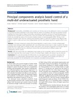
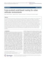
Figure 1a shows a typical SEM image of the as-grown
ZNA prepared through hydrothermal process, which pre-
sents a rodlike morphology with a hexagonal cross-section.
The nanorods are uniform on size with an average diameter
of about 500 ± 10 nm. The cross-section of SEM image
(Fig. 1b) demonstrates the nanorods are aligned along the
perpendicular direction of the Si (100) wafer. The length of
nanorods is about 6 lm.
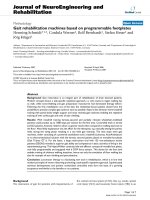
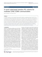
The field emission properties of the pure ZNA and ZNA
film immersed with different concentrations glucose in
PBS buffer solution are illustrated in Figs. 2a, b. As shown
in Fig. 2a, the turn on field (E
to
) of the film of pure ZNA is
5.83 V lm
-1
. The E
to
values of ZNA immersed with 1, 10,
50, 100, and 250 nM glucose solution are 9.82, 9.96, 10.8,
10.97, 11.95 V lm
-1
, respectively. The control experi-
ments show that the E
to
values of ZNA immersing in PBS
buffer solution, the glucose solution in PBS and the GO
x
solution in PBS and are 6.47, 6.5, and 6.83 V lm
-1
,
respectively (Fig. 2b). These values are slightly bigger than
Fig. 1 SEM images of ZNA: a
Top view, b cross-section view
1142 Nanoscale Res Lett (2009) 4:1141–1145
123
that of pure ZNA. The E
to
value of ZNA film immersed in
1 nM glucose solution increases obviously to
9.82 V lm
-1
, which is easily distinguished in comparison
with that of pure ZNA (5.83 V lm
-1
). However, the
measurement of field emission properties on the film of
ZNA indicating no any signal was observed when the
glucose concentration increased to 500 nM. This indicates
that the experimental LOD is 1 nM, which is much lower
compared with the glucose sensors based on GaN/AlGaN
high electron mobility transistors [27].
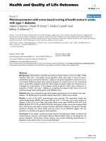
Figure 3a shows the R-M curve of the ZNA field
emitters in different concentrations of 1 nM–50 lM glu-
cose in PBS buffer and GO
x
solutions. It is clearly observed
that the resistance of ZNA field emitter increases promptly
with the increase of the glucose concentration. The resis-
tance of the pure ZNA is about 650 X, which raises 9.3
times in the presence of 1 nM glucose (6,000 X) and about
12 times for 10 nM concentration of glucose (8,000 X).
When the glucose concentration increases to 50 lM, the
resistance is about 420 times that of the pure ZNA. How-
ever, the resistances of the ZNA in control experiments
only increase slightly. Obviously, H
2
O
2
that generated
from the oxidation of glucose by the GO
x
catalyzed (Fig. 4)
has strongly influence on the field emission property and
conductivity of the ZNA. The ratio of R
glucose
/R
ZnO
for the
ZNA field emitter-based glucose sensor presents a good
dependence on the glucose concentrations (Fig. 3b). At this
point, our ZNA field emitter-based glucose sensor sur-
passes previous glucose sensors based on many oxide
semiconductors [22, 25–27, 29].
The properties of ZnO nanostructures are significantly
influenced by surface adsorptions, which have been attra-
cting great attention because the surface absorptions
sometime disturb the fluorescence, field emission, and field
effect transistors [30–32]. In general, the changes of
properties caused by surface energy band bending, induced
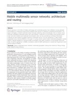
by surface adsorptions. As shown in Fig. 4,GO
x
specifi-
cally catalyzes the oxidation of b-
D-(?)-glucose into glu-
conate and H
2
O
2
.H
2
O
2
and ZnO to form ZnO(OH)
x
on the
surface of ZnO nanorods [25], which depletes the electrons
051015
0
100
200
300
400
500
A
E (Vµµm
-1
)
J(
µµA/cm
2
)
10 nM
50 nM
1 nM
100 nM
250 nM
500 nM
02468101214
0
100
200
300
400
500
600
J(
µ
A/cm
2
)
E (Vµm
-1
)
ZnO+PBS+Glucose
ZnO
ZnO+PBS+GOx
ZnO+PBS
B
Fig. 2 Field emission J–E curves a the ZNA immersing in the
glucose concentration from 1 to 500 nM in 6 mM PBS buffer solution
and 10 U lL
-1
glucose oxidase with a pH value of 5.8. b The control
experiments, the ZNA immersing 6 mM PBS buffer solution, 1 mM
glucose in 6 mM PBS buffer solution, 10 U lL
-1
glucose oxidase in
6 mM PBS buffer solution, respectively, with a pH value of 5.8
10
0
10
1
10
2
10
3
10
4
0.0
5.0x10
4
1.0x10
5
1.5x10
5
2.0x10
5
2.5x10
5
3.0x10
5
A
R
(ΩΩ)
Glucose(nM)
10
0
10
1
10
2
10
3
10
4
0
100
200
300
400
500
B
R
Glucose
/R
ZnO
Glucose(nM)
Fig. 3 a Plot of change resistance (R) as a function of concentrations
(M) from 1 nM to 50 lM in 6 mM PBS buffer solution and
10 U lL
-1
glucose oxidase with a pH value of 5.8. b Dependence
relation between response sensitivity and glucose concentrations
Nanoscale Res Lett (2009) 4:1141–1145 1143
123
on the ZnO nanorods and yields oxygen ions (O
-
,O
2-
,or
O
2
-
)[33]. Leading to the electrons on the ZnO nanorods
are trapped by the adsorbed oxygen molecules, and the
surface depletion region of ZnO nanorods can be formed,
making resistance increase. While the H
2
O
2
is raised with
the increase of glucose, more electrons are captured by the
oxygen molecules at the nanorod surface. Thus, the surface
depletion region is widened and the carrier density in the
ZnO nanorod is decreased even more. At the same time,
gluconolactone can be absorbed strongly on the surface of
ZnO nanorods by hydrogen bonding with ZnO(OH)
x
,
which induces the surface passivation to block the elec-
trons emission of ZnO nanorods under electronic field. The
surface passivation increases to completely deplete the
electrons emission of ZnO nanorods with the increase of
ZnO(OH)
x
on the surface of ZnO nanorods. The result
indicates that field emission properties were not observed
on the ZNA at the glucose concentration C500 nM.
Conclusions
In summary, we demonstrated a new glucose sensor based
on the field emitter of ZNA. The results showed that a wide
range of glucose concentrations from 1 nM to 50 lMis
easily detected, which exhibits ultra-sensitivity for glucose
detection. The experimental LOD of glucose concentration
is 1 nM, which is lower than previous reported glucose
sensor based on EST, FST, and SPR. The new glucose
sensor shows the potential to detect low levels of glucose in
biological system.
Acknowledgments This work was supported by the National Nat-
ure Science Foundation of China (10874187 and 20873155) and the
National Basic Research 973 Program of China.
References
1. N.A. Rakow, K.S. Suslick, Nature 406, 710 (2000)
2. B. Lee, V.H. Perez-Luna, Anal. Chem. 77, 7204 (2005)
3. K. Aslan, J.R. Lakowicz, C.D. Geddes, Anal. Chem. 77, 2007
(2005)
4. X D. Ge, L. Tolosa, G. Rao, Anal. Chem. 76, 1403 (2004)
5. D.L. Meadows, J.S. Schultz, Talanta 35, 145 (1988)
6. G. Blagoi, N. Rosenzweig, Z. Rosenzweig, Anal. Chem. 77, 393
(2005)
7. F. He, Y. Tang, M. Yu, S. Wang, Y. Li, D. Zhu, Adv. Funct.
Mater. 16, 91 (2006)
8. F. He, F. Feng, S. Wang, Y. Li, D. Zhu, J. Mater. Chem. 17, 3702
(2007)
9. W. Chen, H. Yao, C.H. Tzang, J. Zhu, M. Yang, S T. Lee, Appl.
Phys. Lett. 88, 213104 (2006)
10. T. Chen, K.A. Friedman, I. Lei, A. Heller, Anal. Chem. 72, 3757
(2000)
11. A.G. Sapre, A. Bedekar, A.V. Deshpande, A.M. Lali, Biotechnol.
Lett. 22, 569 (2000)
12. J. Wang, M. Musameh, Y.H. Lin, J. Am. Chem. Soc. 125, 2408
(2003)
13. H.D. Duong, J. Il Rhee, Talanta 73, 899 (2007)
14. M. Curreli, C. Li, Y.H. Sun, B. Lei, M.A. Gundersen, M.E.
Thompson, C.W. Zhou, J. Am. Chem. Soc. 127, 6922 (2005)
15. Y. Liu, M. Wang, F. Zhao, Z. Xu, S. Dong, Biosens. Bioelectron.
21, 984 (2005)
16. J.I. Yeh, A. Lazareck, J. Ho Kim, J. Xu, S. Du, Biosens. Bio-
electron. 23, 568 (2007)
17. E. Topoglidis, A.E.G. Cass, B. ORegan, J.R. Durrant, J. Elec-
troanal. Chem. 517, 20 (2001)
18. Y.H. Yang, H.F. Yang, M.H. Yang, Y.L. Liu, G.L. Shen, R.Q.
Yu, Anal. Chim. Acta. 525, 213 (2004)
ZNA
O
CH
2
OH
OH
OH
OH
HO
O
CH
2
OH
OH
OH
HO
O
Glucose
Oxidase
O
2
H
2
O
2
Glucose
Gluconolactone
2ZnO
2ZnO(OH)
Fig. 4 A schematic illustration
of the ZNA field emitter-based
glucose sensor operating
principles
1144 Nanoscale Res Lett (2009) 4:1141–1145
123
19. C.X. Xu, X.W. Sun, Appl. Phys. Lett. 83, 3806 (2003)
20. Z.L. Wang, J.H. Song, Science 312, 242 (2006)
21. H.T. Wang, B.S. Kang, F. Ren, L.C. Tien, P.W. Sadik, D.P.
Norton, S.J. Pearton, J. Lin, J. Appl. Phys. Lett. 86, 243503
(2005)
22. J.X. Wang, X.W. Sun, A. Wei, Y. Lei, X.P. Cai, C.M. Li, Z.L.
Dong, Appl. Phys. Lett. 88, 233106 (2006)
23. Z.Y. Fan, J.G. Lu, Appl. Phys. Lett. 86, 123510 (2005)
24. X.W. Sun, S.F. Yu, C.X. Xu, C. Yuen, B.J. Chen, S. Li, Jpn. J.
Appl. Phys. 2. 42, L1229 (2003)
25. A. Wei, X.W. Sun, J.X. Wang, Y. Lei, X.P. Cai, C.M. Li, Z.L.
Dong, W. Huang, Appl. Phys. Lett. 89, 123902 (2006)
26. J. Zang, C.M. Li, X. Cui, J.X. Wang, X.W. Sun, H. Dong, C.Q.
Sun, Electroanalysis 19, 1008 (2007)
27. B.S. Kang, H.T. Wang, F. Rena, S.J. Pearton, T.E. Morey, D.M.
Dennis, J.W. Johnson, P. Rajagopal, J.C. Roberts, E.L. Piner, K.J.
Linthicum, Appl. Phys. Lett. 91, 252103 (2007)
28. L.E. Greene, M. Law, J. Goldberger, F. Kim, J.C. Johnson, Y.F.
Zhang, R.J. Saykally, P.D. Yang, Angew. Chem. Int. Ed. 42,
3031 (2003)
29. S.J. Bao, C.M. Li, J.F. Zang, X.Q. Cui, Y. Qiao, J. Guo, Adv.
Funct. Mater. 18, 591 (2008)
30. D.S. Bohle, C.J. Spina, J. Am. Chem. Soc. 129, 12380 (2007)
31. Y. Zhang, A. Kolmakov, S. Chretien, H. Metiu, M. Moskovits,
Nano Lett. 4, 403 (2004)
32. S. Song, W.K. Hong, S.S. Kwon, T. Lee, Appl. Phys. Lett. 92,
263109 (2008)
33. Q.H. Li, Y.X. Liang, Q. Wan, T.H. Wang, Appl. Phys. Lett. 85,
6389 (2004)
Nanoscale Res Lett (2009) 4:1141–1145 1145
123