Innovations in Biotechnology Part 16 potx
Bạn đang xem bản rút gọn của tài liệu. Xem và tải ngay bản đầy đủ của tài liệu tại đây (2.31 MB, 24 trang )
Monoclonal Antibody Development and Physicochemical
Characterization by High Performance Ion Exchange Chromatography
441
advanced antibody engineering technologies, almost all antibody products currently in
development are humanized or fully human mAbs and their derivatives.
3. Monoclonal antibody production
Recombinant monoclonal antibodies are typically produced in mammalian cell lines under
defined cell culture conditions. Commercial scale production processes vary depending on
the mAb, but generally, cells are taken from a master cell bank and inoculated into small-
scale bioreactors. The cell culture is transferred to increasingly larger bioreactors until it
reaches the final commercial scale bioreactor. Currently, final scale reactors have volumes
ranging from 12,000 L to 24,000 L (Gottschalk, 2009). The cells are cultured in a controlled
environment for days to weeks, and then the cell culture fluid is harvested by centrifugation
(Shukla & Kandula, 2008). In mammalian cells, the product monoclonal antibodies are
secreted from the cells into the supporting fluid medium. Centrifugation separates the cells
from the fluids and facilitates simpler recovery procedures downstream.
Commercial mAb production requires considerable preproduction effort to ensure that the cell
line is stable and can produce commercially appropriate quantities of antibody. In addition,
the commercial production process must produce a product that meets the quality
expectations of regulatory authorities. In the past few years, improvements have been made in
critical areas, such as cell line generation and large-scale cell culture production, to maximize
specific antibody productivity from a given cell line and improve overall productivity in
bioreactors. These advances include the use of new expression vectors and transfection
technology, high-throughput, robust screening technologies to select the highest producing
clones rapidly and more effectively, improvements in cell culture and optimized bioreactor
processes (Li et al., 2010; Schlatter et al., 2005). As a result, the production of cell lines
expressing multigram quantities of antibody per liter of culture medium is now routine.
The product quality and product heterogeneity of every mAb is highly dependent on its
manufacturing process (Abu-Absi et al., 2010; Horvath et al., 2010). The ideal manufacturing
conditions would have optimal production levels of product in conjunction with the desired
product quality profile. Attributes that are typically deemed critical in selecting stable clones
and cell culture conditions are the product titer and product heterogeneity, including
charged species and aggregates. Production titers directly correlate to the costs of the
process and are desired to be as high as possible with minimal impact to other quality
attributes of the product (Kelley, 2009). Critical quality attributes of the product, such as the
level of aggregation, are carefully monitored, as failure to control critical quality attributes
may pose a safety risk to the patient (Rosenberg, 2006).
4. Monoclonal antibody purification and formulation
Once monoclonal antibodies are produced in cells, the mAbs must be recovered and
purified. Recovery and purification processes vary widely depending on the manufacturing
process and specific mAb characteristics, but generally, the isolation and purification of
mAbs involve a centrifugation step to separate the cells from the cell culture fluid
containing the mAb product, one or more chromatography steps, which can include affinity
chromatography, cation or anion exchange chromatography, hydrophobic interaction
chromatography (HIC) and displacement chromatography (Shukla et al., 2007), and
Innovations in Biotechnology
442
filtration or precipitation steps (Gottschalk, 2009). Many of the purification steps are
designed to remove contaminants and adventitious agents (e.g., bacteria, fungi, viruses, and
mycoplasma).
After elution from the final chromatographic purification step, a unit operation is required
to exchange the components of the chromatography elution buffer with the chosen
formulation components. The predominant technology that has been used in the industry
for buffer exchange and concentration is ultrafiltration/diafiltration using tangential-flow
filtration (Genovesi, 1983; Shiloach, 1988; van Reis, 2001). After this step, the drug substance
is filtered and typically frozen as bulk for storage until filling occurs to produce the final
drug product.
The formulation of the mAb therapeutic is chosen in part to ensure product quality during
shelf life. Formulations are designed to minimize protein aggregation, decrease viscosity,
and increase shelf life through preventing degradation (Shire, 2009). High protein
concentration formulations are being developed to allow for subcutaneous or intramuscular
delivery of mAb products (Shire et al., 2004). Historically, the most conventional route of
delivery for protein drugs has been intravenous administration because of poor
bioavailability by most other routes, greater control during clinical administration, and
faster pharmaceutical development. Subcutaneous delivery allows for home administration
and improved patient compliance. However, development of high protein concentration
formulations involves unique manufacturing challenges compared to low concentration
formulations, such as higher viscosities and necessary changes to unit operation steps.
5. Monoclonal antibody characterization and release testing
Biopharmaceutical manufacturing of monoclonal antibodies produces a heterogeneous
product of structurally related species. Antibody speciation can occur throughout the
manufacturing process at various steps, including cell culture, harvest, purification,
formulation, filling and during shelf life. Full-length monoclonal antibodies are high
molecular weight proteins (around 150,000 Da), and have highly complex secondary and
tertiary structures, subject to post-translational modifications. Therefore, product
characterization and quality control testing are required at critical points throughout clinical
development and manufacturing to control for these species (Harris et al., 2004). Figure 2
depicts the structure of a monoclonal antibody compared to a small molecule drug,
illustrating the increased complexity of a biologic compared to a small molecule therapeutic.
Antibodies can be characterized by many physicochemical properties including hydrated
size (Stokes radius), molecular weight, charge, hydrophobicity, electrophoretic mobility,
isoelectric point (pI), sedimentation velocity, glycosylation, and spectral properties. The
nature of each species can be related to differences in their primary, secondary, tertiary, or
quaternary protein structures. In addition, monoclonal antibodies are susceptible to
chemical or enzymatic modification, particularly at sites that are exposed to the protein-
liquid interface. Product heterogeneity can be caused by a number of modifications, such as
C-terminal processing of lysine residues (Harris, 1995; Santora et al., 1999; Weitzhandler et
al., 1998), deamidation (Di Donato et al., 1993; Hsu et al., 1998), glycation (nonenzymatic
glucose addition) (Quan et al., 2008), amino acid sequence variations (Yang et al., 2010), and
noncovalent complexes (Santora et al., 2001).
Monoclonal Antibody Development and Physicochemical
Characterization by High Performance Ion Exchange Chromatography
443
Antibody products are characterized by physicochemical, immunochemical, and biological
methods. Guidance documents have been issued by regulatory agencies and industry
representatives recommending approaches for protein characterization (International
Conference on Harmonisation of Technical Requirements for the Registration of
Pharmaceuticals for Human Use (ICH), 1999; Schnerman et al., 2004). These orthogonal
assays include potency, identity and purity assays, which evaluate “critical quality
attributes” such as size and charge heterogeneity. These critical quality attributes are part of
the overall target product profile, which is based on the desired clinical performance. The
extent of characterization is linked to the level of risk associated with each phase of drug
development. For example, while there may not be sufficient time or resources for extensive
characterization of an antibody during early stage development, it is expected that the
molecule will be well-characterized before the Biologic License Application (BLA) is
submitted to the regulatory agencies.
Fig. 2. Comparison of the structures of a mAb (Herceptin) and a small molecule therapeutic
(Tarceva).
Many of the recommended protein characterization assays are based on liquid
chromatography methods, such as ion exchange chromatography (IEC) for charge
heterogeneity analysis, size exclusion chromatography (SEC) for size heterogeneity, and
reversed-phase high performance liquid chromatography (RP-HPLC) for peptide mapping
(Chirino & Mire-Sluis, 2004). The remainder of this chapter will primarily focus on ion
exchange chromatography methods for analyzing charge heterogeneity for characterization
and support of formulation and process development, as well as for lot release testing of
drug substance and drug product (Schnerman et al., 2004).
Innovations in Biotechnology
444
5.1 Analyzing mAb charge heterogeneity using IEC
As mentioned previously, monoclonal antibodies are large proteins that are quite complex.
While the light chain and heavy chain sequences of a particular mAb may be known, a
number of modifications can introduce heterogeneity in the product. Thus, it is important to
develop appropriate analytical methods to resolve the minor forms of the product.
Analytical biochemists routinely use IEC for resolving charge variants of the protein. The
scientist must then utilize orthogonal analytical methods to characterize the separated peaks
of the ion exchange chromatogram. The characterization of a mAb is particularly important
if the modifications occur in the complementarity-determining regions (CDR), as
modifications in the CDR can affect the binding activity and potency of the mAb.
A strategy for the assignment of peaks from a weak cation exchange (WCX) mAb
separation using a salt gradient has been published (Harris et al., 2001). Seven forms of a
therapeutic recombinant antibody were resolved by cation-exchange chromatography.
The peak fractions were collected, and structural differences were assigned by peptide
mapping, which involves digesting the mAb with an enzyme and injecting the digest onto
a reverse-phase column coupled to a mass spectrometer, and by hydrophobic interaction
chromatography (HIC) after papain digestion. The peaks in this particular case were
attributed to deamidation, isomerization, and succinimide intermediates. Other
orthogonal analytical methods were used to characterize the IEC peaks; one of these
methods—potency testing—determined that one minor peak demonstrated much lower
potency than the main peak.
In another study, a recombinant humanized monoclonal IgG1 antibody with different
states of glycosylation on the conserved asparagine residue in the CH2 domain was
analyzed by cation exchange chromatography (Gaza-Bulseco et al., 2008). Two major
peaks were observed and were further characterized by enzymatic digestion and mass
spectrometry. It was found that this recombinant monoclonal antibody contained three
glycosylation states—zero, one or two glycosylated heavy chains. The peak that eluted
earlier on the cation exchange column contained antibodies with two glycosylated heavy
chains containing fucosylated biantennary complex oligosaccharides with zero, one or
two terminal galactose residues. The peak that eluted later from the column contained
antibodies with zero, one or two glycosylated heavy chains. The oligosaccharide on the
antibodies that eluted in the later peak was composed of only two GlcNAc residues. These
results indicate that conformational changes, caused by different types of neutral
oligosaccharides as well as the absence of certain oligosaccharides, can be differentiated
by cation exchange column chromatography.
5.2 Lot release testing of mAbs
Once the mAb is purified and formulated, the resulting drug substance must be tested prior
to lot release. A set of tests and acceptance criteria are established based on mAb
characterization and regulatory requirements in order to ensure product quality (Food &
Drug Administration (FDA), 1999). These tests typically include appearance, identity,
purity, protein concentration, potency of the molecule, microbial limits or bioburden, and
bacterial endotoxins (Table 1). IEC is one of the most frequently used lot release methods for
purity for mAbs (Schnerman et al., 2004). Once these tests are performed and the results
Monoclonal Antibody Development and Physicochemical
Characterization by High Performance Ion Exchange Chromatography
445
meet the established acceptance criteria, a Certificate of Analysis (COA) is generated and the
lot is released for use. Finally, adequate stability studies should be performed on the mAb
drug substance (e.g. frozen bulk for storage) and drug product (e.g. final vial) according to
regulatory guidelines (Food & Drug Administration (FDA), 2003).
Attribute Test Name
Appearance Color, Opalescence and Clarity
Identity
Peptide Mapping by RP-HPLC (Reverse-Phase HPLC), or
MALDI (Matrix-Assisted Laser Deionization) Mass Spectrometry, or
UV Spectroscopy (2
nd
Derivative)
Purity
Limulus Amebocyte Lysate (Endotoxin)
Size Exclusion Chromatography (SEC)
CE-SDS (Capillary Electrophoresis-Sodium Dodecyl Sulfate)
IEC (Ion Exchange Chromatography) or icIEF (Imaged Capillary
Isoeletric Focusing)
Glycosylation Profile
Peptide Mapping by RP-HPLC
Potency Potency (ELISA/Cell-Based Assay)
Strength UV Spectroscopy
General Tests
Osmolality
pH
Surfactant Concentration (e.g. Polysorbate 20)
Table 1. Commonly used tests found on a Certificate of Analysis for lot release; a selected
subset is used for stability testing of mAbs.
6. Mechanism of ion exchange chromatography of mAbs
Ion exchange chromatography (IEC) has been a platform for monoclonal antibody
purification and characterization for many years. For the analysis of charged species of
proteins, IEC is a popular method due to the fact that it preserves the native conformation
and maintains bioactivity of the protein, is relatively easy of use, is supported by the
maturity of the equipment and consumables market, and has widespread use in the
biopharmaceutical industry (Rea et al., 2010).
Charge-based methods are an integral component of characterization studies and quality
control strategies because they are sensitive to many types of modifications. Charge
profiling of intact antibodies can resolve species related to protein conformation, size,
sequence species, glycosylation and post-translational modifications (Gaza-Bulseco et al.,
2008; Harris et al., 2001; He et al., 2010). Although IEC can be used to track specific species, it
is common to group all species not associated with the main peak and report them as either
acidic or basic species (Figure 3). In addition, fractions collected from an IEC run can often
be directly injected onto orthogonal columns for further analysis, such as reverse-phase and
size exclusion chromatography columns, or submitted for potency testing.
Innovations in Biotechnology
446
IEC separates proteins based on differences in the surface charge of the molecules, with
separation being dictated by the protein interaction with the stationary phase. The two main
categories of ion exchange chromatography are cation exchange (CEX) and anion exchange
(AEX). Cation exchange chromatography retains biomolecules by the interaction of the
negatively-charged resin with histidine (pK ~ 6.5), lysine (pK ~ 10) and arginine (pK ~ 12) in
the protein. Anion exchange chromatography primarily retains biomolecules by the
interaction of the positively-charged resin with aspartic or glutamic acid side chains, which
have pKa of ~4.4. In addition to the amino acid residues, cation exchange columns can also
separate deamidated, glycated and other charged variants. Anion exchange columns have also
been useful for separating phosphorylated and hydroxyl modified amino acids. When the pH
equals the pI value of the protein, the net charge on the molecule is zero. However, significant
retention can occur for proteins even when the pH of the mobile phase is equal to the pI of the
molecule; despite an overall net charge of zero, only a portion of the mAb molecule will interact
with the stationary phase, and there will be a net charge on that portion of the molecule because
of an uneven distribution of charged groups throughout the molecule (Vlasak & Ionescu, 2008).
Thus, it is possible to separate proteins having very similar charge (Figure 4), or even structural
isomers with identical pI values, by ion exchange chromatography.
Fig. 3. Typical cation exchange chromatogram for analytical characterization of a mAb.
Integration is shown, and main peak, acidic and basic regions are denoted.
There are two ways to elute the protein from the IEC column: 1) increasing salt
concentration with time or 2) by varying the mobile phase pH value as a function of time.
Increasing the salt concentration elutes the protein by increasing the ionic strength of the
mobile phase, thus affecting the charge interaction of the mAb and the stationary phase. A
pH gradient elutes the protein by changing the charge on the molecule, thus affecting the
binding of the molecule to the stationary phase. While conventional salt gradient cation
exchange chromatography is regarded as the gold standard for charge sensitive antibody
analysis (Vlasak & Ionescu, 2008), method parameters such as column type, mobile phase
pH, and salt concentration gradient often need to be optimized for each individual antibody.
A recent publication described a multi-product pH gradient IEC method for the separation
of mAb charge species for a variety of mAbs using a single method (Farnan & Moreno,
2009). The following sections will discuss both salt-gradient and pH-gradient based elution
methods, and the combination of the two modalities (hybrid methods).
Monoclonal Antibody Development and Physicochemical
Characterization by High Performance Ion Exchange Chromatography
447
Fig. 4. Separation of mAbs differing by only one charge, a single amino acid change to
primary structure. The elution buffer (0.5 M NaCl in 20 mM Tris, pH 7.3) was increased
linearly on a ProPac WCX-10 column (4 x 250 mm), which was held at 50 ºC and had a flow
rate of 1 mL min
-1
.
7. Developing a salt-based IEC method
Salt-based IEC separations are developed by choosing a cation or anion exchange column
and varying the buffer system, mobile phase pH value, and ionic strength gradient of the
elution buffer. Figure 5 shows a typical development workflow for salt-based IEC and pH-
based IEC development, and can serve as a guide for initial IEC method development. The
following sections will cover in more detail the outputs to consider when screening various
parameters during development. More general considerations regarding HPLC method
development can be found in various texts (Kastner, 2000; Snyder et al., 1997).
Fig. 5. Sequential salt-gradient IEC and pH-gradient IEC method development and
optimization work flow.
Salt Gradient IEC pH Gradient IEC
Innovations in Biotechnology
448
7.1 Column selection, buffers and operating parameters for salt gradient IEC
Column selection is perhaps the most subjective part of the optimization process; picking
between the different vendor offerings and functionalities can be difficult. Prior experience,
data in the literature or unpublished results within the organization are often the best
starting points.
Analytical ion exchange chromatography of proteins is typically carried out using mobile
phases that are relatively neutral in pH values, 5.5 to 8.5 This general practice is
recommended because at pH extremes, the protein is more likely to degrade. The selection
of whether to use anion or cation exchange chromatography is also driven by the isoelectric
point of the protein (pI) and the species to be resolved, e.g., phosphorylated species, C-
terminal lysine variants, etc.
If the pI value of the mAb is greater than 8, a CEX column is evaluated at pH 6-7 initially.
CEX primarily retains mAbs by the interaction of acid groups on the CEX resin with lysine,
arginine and histidine side chains on the mAb. Since mAbs are positively charged at a
mobile phase pH below their pI, the mAb species would likely be retained and resolved on a
CEX column under the recommended mobile phase pH range.
If the pI value of the mAb is less than 6, an AEX column is evaluated at a pH above 6
initially. AEX primarily retains biomolecules by the interaction of amine groups on the ion
exchange resin with aspartic or glutamic acid side chains. Since mAbs are negatively
charged at a mobile phase pH above their pI, the mAb species would likely be retained and
resolved on an AEX column.
For intermediate pI values of 6-8, both CEX and AEX are evaluated because of the
possibility that the portion of the mAb that interacts with the stationary phase, typically the
side chains that are exposed to the mobile phase, has a different charge than the pI would
suggest, e.g., the surface charge of the mAb is positive despite the entire mAb having an
overall negative charge. Ultimately, the species of interest that are to be resolved determine
whether CEX or AEX is chosen for molecules with intermediate pI’s; the separation mode
that better separates the species of interest is usually the one that is chosen for mAb analysis.
Figure 6 shows CEX and AEX chromatograms of a Fab (mAb fragment) reference sample
and thermally stressed sample. In this case, the Fab molecule has a nominal pI value for the
main species of 7.6. It should be noted that the separations on the AEX and CEX columns
were each optimized independently for column type, pH value and salt gradient. It should
also be noted that the terms “strong” and “weak” (in SAX, strong anion exchange, and
WCX, weak cation exchange) refer to the extent of variation of ionization with pH due to the
functional groups on the resin and not the strength of binding. Strong ion exchangers are
completely ionized over a wide pH range whereas with weak ion exchangers, the degree of
dissociation and thus exchange capacity varies much more markedly with pH. For this
example, SAX results in significantly more peaks and much better resolution of the charge
species in comparison to the WCX chromatogram. Particularly interesting is that the
difference between the WCX and SAX elution profiles are much more vivid for the stressed
samples than for the reference materials. We have seen examples where the converse is true
and the CEX separation is better than that observed on the AEX. This contrast between the
AEX and CEX profiles highlights an important feature of IEC that electrophoretic methods
don’t exhibit, which is the ability to magnify particular aspects of the protein structure and
accentuate the separation of species relating to particular motifs (Vlasak & Ionescu, 2008).
Monoclonal Antibody Development and Physicochemical
Characterization by High Performance Ion Exchange Chromatography
449
Fig. 6. Separation of Fab charge species using a weak cation exchange column (WCX) and a
strong anion exchange column (SAX). Thermally stressed samples are labeled by incubation
time and temperature of incubation.
In general, we have observed that for the separation of mAb variants using ion-exchange
chromatography, the optimized chromatogram has a relatively shallow gradient over a
narrow range of salt concentration. A typical method results in 100 mM NaCl as the center
point of the gradient, with salt concentration increasing over 70 mM NaCl in a linear
gradient. It is recommended to perform iterative gradient optimizations to narrow the NaCl
gradient down to around 2 mM/mL min
-1
. Iterative cycles are quicker and more predictive
than performing a very long shallow gradient.
Chromatograms obtained during the mobile phase pH value optimization for a mAb with a
pI value around 9.5 are shown in Figure 7. Buffer species and buffer concentration for salt-
gradient IEC are generally not significant factors, but should be chosen considering target
pH and buffer pKa.
Although temperature does not significantly affect electrostatic interactions, it often affects
the pH value of the mobile phase. This is particularly of concern for a Good’s buffer system
(group of buffers described in the research of Dr. Norman Good et al. in 1966, often used for
IEC and other biochemistry applications) (Good et al., 1966), which can exhibit a change in
pH value of around 0.02 per °C temperature change. This sensitivity creates a need to
control the column temperature carefully. A column compartment is always used, typically
set at a value greater than 30°C to ensure good temperature stability in compartments that
can only apply heat. Above 30°C, temperature control within +/- 1°C is readily achievable
with commercially available equipment.
Innovations in Biotechnology
450
Fig. 7. Effect of mobile phase pH on mAb separation by WCX. The elution buffer (0.5 M
NaCl) was increased linearly at 1 mM min
-1
at a flow rate of 1 mL min
-1
on a ProPac WCX-10
column (4 x 250 mm), which was held at 30 ºC. Different initial salt concentrations were
optimized for each pH value. Integration is shown, and main peak, acidic and basic regions
are denoted.
Subtle variations in selectivity with temperature may result from temperature-induced
changes in mobile phase pH value (Figure 8). In Figure 8, the elution profile changes in two
distinct regions as a function of temperature. Below 40°C, subtle changes in elution profile
and retention times are observed consistent with minor changes to the mobile phase pH
value as a function of temperature. However, above 40°C, the profiles exhibit much more
radical changes with increasing temperature. This is interpreted to be related to the mAb
having lost higher order structure at those elevated temperatures due to protein denaturing.
For the mAb in Figure 8, it is clear that moderately elevated temperatures are not possible
while maintaining the higher order structure; in general for IgG1 mAbs, chromatography at
temperatures up to 55°C is readily possible. In summary, while mobile phase temperature
does not affect protein charge directly, temperature can affect mobile phase pH and the
structure of the protein, which can affect chromatographic separations. Thus, column
temperature should be optimized considering these temperature effects.
Monoclonal Antibody Development and Physicochemical
Characterization by High Performance Ion Exchange Chromatography
451
Fig. 8. Effect of temperature on mAb separation by CEX. The elution buffer (0.2 M sodium
sulfate) was increased linearly on a ProPacWCX-10 column (4 x 250 mm).
8. Developing a pH gradient-based IEC method
Despite good resolving power and robustness, salt-based ion exchange separations are
usually protein-specific and time-consuming to develop. A novel pH-based separation of
proteins by cation exchange chromatography that was multi-product, high-resolution, and
robust against variations in sample matrix salt concentration and pH was recently reported
(Farnan & Moreno, 2009). A pH gradient-based separation method using cation exchange
chromatography was also evaluated in a mock validation and deemed highly robust (Rea et
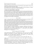
al., 2011). Figure 9 depicts the separation of 16 mAbs by pH gradient IEC (pH-IEC). Each
mAb was injected sequentially, demonstrating that in contrast to salt-based IEC, pH-IEC can
be used to analyze multiple mAbs with a single method.
Fig. 9. Separation of 16 mAbs using a ProPac WCX-10 column by pH gradient IEC. Each
mAb was analyzed using the same pH-IEC method, and each mAb was injected
sequentially. mAb pI values ranged from pI 7.3 to pI 9.4.
Similar to salt-gradient IEC methods, pH-IEC separations are developed by choosing a cation or
anion exchanging column and varying the buffer system, pH of the mobile phases, and other
operating parameters, such as temperature and flow rate. Figure 5 shows a typical development
workflow for pH-IEC, and can serve as a guide for initial pH-IEC method development.
Innovations in Biotechnology
452
8.1 Column selection, buffers and operating parameters for pH gradient IEC
Like conventional IEC, the conditions chosen for pH-IEC separations, such as buffer, pH,
column temperature, and sample load, are dependent on the type of column selected. To
choose a column, the pI of the mAb and the expected charge species should be considered.
Considerations for column selection may differ slightly for pH-IEC compared to conventional
IEC. For example, because the column will be exposed to a pH gradient, the column must be
able to perform adequately over a large pH range, i.e., the charged groups on the
chromatography resin must maintain their charge over the operating pH range. Also, buffer
strength can affect resolution, and pH-IEC mobile phases typically have lower buffer strengths
than conventional salt-gradient IEC. Several pH-IEC buffer systems have been published for
mAb separations; these buffer systems can be used as starting points for formulating buffers
for pH-IEC methods (Farnan and Moreno, 2009; Rea et al., 2011; Rozhkova, 2009).
8.2 High-throughput multi-product separations using pH-IEC
To increase the throughput of the analytical methods, smaller particle sizes and shorter
column lengths are being utilized to reduce run time. In Figure 10, the utilization of a 3 µm
particle size column reduced analysis time 16-fold compared to a 10 µm particle size
column. Analysis times are greatly reduced using smaller particle sizes because as the
particle size decreases, there a significant gain in column efficiency, and the efficiency does
not decrease at increased flow rates or linear velocities (Swartz, 2005). In addition, because
different mAbs can be analyzed using the same pH-IEC method in the same sequence, these
high-throughput methods are capable of analyzing hundreds of mAbs per day, which is not
possible with conventional, product-specific salt-based IEC.
Fig. 10. Separation of a mAb using (A) a WCX column, 10 µm and (B) a SCX column, 3 µm,
by pH gradient IEC. Each mAb was analyzed using the same buffers and gradient volume.
Monoclonal Antibody Development and Physicochemical
Characterization by High Performance Ion Exchange Chromatography
453
9. Hybrid/combination modes of IEC
Salt and pH may be combined to elute proteins from IEC columns. Combination or hybrid
methods can be employed if either salt-based or pH-based methods prove inadequate for
resolving species of interest, especially at extreme pHs. When pH increases above their pKa,
amines, as used exclusively in the pH-IEC piperazine/imidazole/tris buffer system, become
deprotonated and uncharged, resulting in decreased ionic strength. The bound proteins will
also deprotonate and carry less charge. However, adequate amounts of positively charged
ions are required to displace the bound proteins and to elute them off the cation exchange
resin. Since the buffer salts alone can not provide enough positively charged ions at higher
pH, additional salt is added to the pH-IEC elution buffers to maintain ionic strength. Figure
11 depicts measured conductivity as a function of elution time in pH-IEC with and without
salt. In this case, adding salt to the elution buffer will compensate for the loss of ionic
strength (represented by conductivity) due to deprotonation of buffer ions.
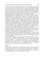
In Figure 12, separation of the charge species of three mAbs using pH-IEC with and without
salt is compared. Without salt, mAb-1 with a pI of 9.4 did not elute, and mAb-3, with a low
pI of 6.2, showed a very broad peak with significant tailing and no resolution of charge
species. With the addition of salt, adequate separation of charge species is obtained for both
high pI and low pI mAbs.
Fig. 11. Measured conductivity as a function of elution time in pH-IEC A) without, and B)
with salt. Buffers are A) 11.6 mM piperazine, 1.5 mM imidazole, 2.4 mM Tris, B) 4 mM
piperazine, 4 mM imidazole, 4 mM Tris, 16 mM NaCl.
10. Equipment configurations to accelerate development
Ionic strength gradient ion-exchange methods are typically product-specific, with each
method requiring a unique pair of mobile phases and experimental conditions. As discussed
above, a significant number of mobile phase pH values and gradient profiles need to be
evaluated. Changing mobile phase pH values normally requires user intervention to supply
new mobile phase pairs; the time needed to manually change the system can slow down
Innovations in Biotechnology
454
development. In order to more efficiently develop analytical IEC methods, alternative
equipment configurations have been utilized to accelerate the selection of operational
parameters, including quaternary buffer systems and customized solvent selection valves on
the pump inlets to allow selection from an array of available solvents. Such modifications to
the equipment and workflows can allow consecutive performance of significantly more
experiments without requiring user intervention.
0
20
40
60
80
100
120
140
0 1020304050
Absorbance (mAu)
Time (min)
mAb-3 pI 6.2
mAb-2 pI 8.2
mAb-1 pI 9.4
blank
Piperazine 11.6 mM
Imidazole: 1.5 mM
Tris: 2.4 mM
0
20
40
60
80
100
120
140
0 102030405060
Absorbance (mAu)
Time (min)
blank
Piperazine 4 mM
Imidazole: 4 mM
Tris: 4 mM
16mM NaCl in Buffer B
mAb-3 pI 6.2
mAb-2 pI 8.2
mAb-1 pI 9.4
A: without salt
B: with salt
0
20
40
60
80
100
120
140
0 1020304050
Absorbance (mAu)
Time (min)
mAb-3 pI 6.2
mAb-2 pI 8.2
mAb-1 pI 9.4
blank
Piperazine 11.6 mM
Imidazole: 1.5 mM
Tris: 2.4 mM
0
20
40
60
80
100
120
140
0 102030405060
Absorbance (mAu)
Time (min)
blank
Piperazine 4 mM
Imidazole: 4 mM
Tris: 4 mM
16mM NaCl in Buffer B
mAb-3 pI 6.2
mAb-2 pI 8.2
mAb-1 pI 9.4
A: without salt
B: with salt
Fig. 12. Separation of the charge species of three mAbs using pH gradient with and without
salt in a ProPac WCX-10 column. A) pH 5 to 9.5 in 45 minutes, gradient 0.1 pH unit/min; B)
pH 5 - 10.8 in 58 minutes, gradient 0.1 pH unit/min.
A quaternary buffer system can be utilized to develop a method and reduce the amount of
user intervention by using a pair of buffer solvents (solvent lines A and B) to allow the pump
to admix to achieve the desired mobile phase pH, and using two other solvents (solvent lines
C and D) to generate the ionic strength gradient for elution. The quaternary system can apply
different combinations of salt and pH by automatically programming the percentages of the
four solvents to be mixed and applied to the column. Thus, programs can be generated to
screen a variety of salt and pH conditions in a single sequence using only four buffers.
Another approach, which is particularly important for binary pump systems, is to add a
multi-port solvent selection valve to the system prior to the pump. Although the customized
Monoclonal Antibody Development and Physicochemical
Characterization by High Performance Ion Exchange Chromatography
455
valve system requires the production of many buffers, the multiple valve configurations can
allow users to further customize buffer components and concentrations (as opposed to only
salt gradient and pH) compared to the quaternary system. Such a system allows up to a
dozen more buffer combinations to be evaluated without intervention.
Because pH-IEC is performed by using a pH gradient and not a salt gradient, simply
reversing the pH gradient allows for the chromatography mode to be switched between
CEX and AEX. This reversal of gradient can be automatically performed through the
chromatography software. Thus, multiple CEX and AEX columns can be screened by using
only two buffers at different pH’s and using a column switching valve to screen different
column types (Figure 13). During development, it is helpful to have online pH and
conductivity meters to ensure that the pH gradient is roughly linear and that the
conductivity does not interfere with the separation efficiency.
Fig. 13. HPLC column compartment equipped with a 6-port column switching valve, for
screening of up to six different columns for pH-IEC without the need to change buffers or
columns.
11. Method robustness and validation
The robustness of an analytical procedure is a measure of its capability to remain unaffected
by small, but deliberate variations in method parameters and provides an indication of its
reliability during normal usage. For IEC, robustness can be evaluated by varying parameters
such as injection volume, buffer pH, flow rate, and column temperature. In addition to
robustness, intermediate precision can be demonstrated by evaluating inter-laboratory
variations, such as different days of analysis and different analysts. Furthermore, the ability
to use different instrument and column manufacturers for a particular method greatly
reduces the business risk of the method; if a column supplier cannot meet demand or if an
instrument manufacturer ceases production of a particular instrument model, method
transfer to other instruments and columns can occur without loss of performance.
11.1 Obtaining robust performance
Obtaining robust performance of an IEC method often goes beyond the design of the
method itself, and involves good equipment hygiene, elimination of metal corrosion (e.g.
formation of iron oxide) and contamination (e.g. presence of metal ions such as Fe
3+
ions),
Innovations in Biotechnology
456
and mitigates the differences between instrument types. Problematic metal contamination
typically results from corrosion of the fluid-contacting metal parts and can be avoided by
using PEEK or titanium materials in the fluid paths. Good practices on obtaining robust
method performance are discussed in the following sections.
11.1.1 Equipment hygiene
Maintaining good equipment hygiene is important in order to achieve robust performance.
The following are good practices to ensure instrument hygiene:
1. Filter mobile phases that are amenable to microbial growth with 0.2 mm filters prior to
use; replace solvent reservoir filters (sinkers) each time mobile phase bottles are
replenished;
2. Flush and store HPLC system in 10% isopropanol in water when not in use, to prevent
growth of microbes;
3. Leave the system running at low flow rates to prevent salt build up and clogging;
4. Keep all lines flushing as opposed to just a single channel;
5. Flush auto-sampler components as needed with 10% isopropanol in water;
6. Follow manufacturer’s instructions regarding proper maintenance of HPLC
instrumentation.
11.1.2 Metal contamination
Metals can negatively affect the ion-exchange chromatography of proteins. Protein chelation
with metals are a secondary retention mechanism to the primary electrostatic interaction of
ion-exchange chromatography. This secondary interaction results in peak tailing. These
interactions can either occur with metal contaminating the column or with corroded
surfaces within the HPLC. In addition to affecting separation, corrosion can result in
physical damage to system, such pump seal failure and compromised performance of the
detector cells. Halide containing eluents readily corrode HPLC systems manufactured from
stainless steel, as stainless steel has the propensity to form rust (Collins et al., 2000a).
Sodium acetate or sodium sulfate can be used as an eluting salt instead of halides; however,
sulfate is divalent, thus concentrations in the eluting mobile phase would be different
compared to using a halide, as halides are monovalent.
Metal contamination may be reversed by flushing with chelating agents such as oxalic acid
dihydrate (Rao & Pohl, 2011). Also, stainless steel systems may require periodic passivation
for reliable usage (Collins et al., 2000b). In light of the drawbacks of using a stainless steel
HPLC system, more manufacturers are including biocompatible equipment (e.g. Titanium
or PEEK) for analyzing mAbs and other protein products.
11.1.3 Transferring methods between instrument types
Transferring methods between instruments from different manufacturers can pose
challenges due to the differences between instruments. As mentioned previously,
equipment composition (e.g. stainless steel vs. titanium) is one of the factors to be
considered when transferring a method between instrument types, in addition to gradient
delay, mixing volumes, pump capabilities, and column compartment temperature ranges.
Gradient delay and mixing volumes can differ between instruments, but they are generally
Monoclonal Antibody Development and Physicochemical
Characterization by High Performance Ion Exchange Chromatography
457
only a significant concern for very fast gradient separations. In addition, shallow IEC
gradients can challenge the performance of an HPLC; however, most gradients are >70 mM
salt and can be proportioned over 30-40% of the pump range, well within the capabilities of
modern pumps. Often a gradient hold for 5 minutes at the initial salt concentration is
included, just in case a method is particularly sensitive when being transferred from one
equipment type to another. In such cases the hold time can be adjusted to compensate for
differences in the gradient delay volume between the instruments.
Temperatures inside the column are dependent on oven design and plumbing
configuration. Having a pre-column heat exchanger in line or out of line could make a
several degree difference in the temperature at which the column chemistry occurs. This is
particularly concerning for buffers with which the pH can change rapidly with temperature.
Figure 14 shows a comparison of column compartment temperature settings for two
different instruments from different manufacturers. To make the correlation, thermocouples
were fitted into T-pieces in the fluid path inside the column oven, but just prior to the
column, and temperatures were measured for a range of column compartment set points
and mobile phase flow rates. These measurements were used to estimate the temperature of
mobile phase going through the column for each set point. By equating the measured fluid
temperatures for each flow rate, the correlation of column compartment temperatures were
plotted. It is noted in this correlation that there was also a significant effect of the mobile
phase flow rate on the correlation.
Different detectors can sometimes yield differences in baseline slope. This can occur when
moving from a single/double wavelength detector with a reference beam to a photodiode
array (PDA) detector. The selection of an appropriate reference wavelength and bandwidth
on the PDA can overcome detector variance.
Fig. 14. Comparison of column compartment temperature settings required to achieve the
same columns compartment temperature for two different HPLC models at different flow
rates. Results are shown for a 4 x 250 mm Dionex ProPac column. The Agilent 1100 HPLC
was configured using only the left hand side heat exchanger. The Waters 2695 HPLC was
configured with the solvent pre-heater in-line.
Innovations in Biotechnology
458
11.2 Method validation
Before an analytical method can be incorporated into a characterization platform or a quality
control system, it must first be demonstrated that the method is suitable for its intended
purpose. Guidelines for validation of analytical methods have been published in the United
States Pharmacopeia, by the International Conference on Harmonization (ICH), US Food
and Drug Administration (FDA), and in published reviews (Bakshi & Singh, 2002). Methods
must be evaluated considering regulatory requirements and validation procedures. In other
words, the “validatability” of these methods must be assessed before implementation.
Validation tests include precision, accuracy, and linearity. Intermediate precision is tested
by using multiple instruments, multiple analysts, and multiple column lots. Methods must
be validated and documented according to regulatory requirements prior to implementation
into a control system for lot release of drug substance and drug product. Robustness studies
can also be performed in conjunction with method validation. It has been our experience
that the most significant effects on method robustness are: mobile phase pH value, column
temperature, metal contamination and column age.
A system suitability range can be obtained from robustness studies. This range is often
based on the standard deviation of the mean for a particular measured component, such as
main peak relative area. The system suitability range indicates the precision of the method.
pH-IEC may demonstrate an improvement in precision over conventional salt-based IEC
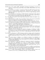
(Rea et al., 2011). The 6σ ranges in Figure 15, which predicts a 99% method success rate,
demonstrate the improved precision of the pH-gradient IEC method over conventional IEC,
which can have a 6σ range of up to 8% main peak relative area (Figure 15).
Fig. 15. Six sigma range (±3SD) for main peak relative area for salt gradient IEC (diamonds)
and pH gradient IEC (circles) for a variety of mAbs.
Monoclonal Antibody Development and Physicochemical
Characterization by High Performance Ion Exchange Chromatography
459
12. Automation in sample preparation and data handling
In addition to high-throughput and multi-product analytical methods, the use of robotics
for sample preparation automation may further reduce sample analysis time and cost.
There are several companies that provide liquid handling automation instruments,
including LEAP Technologies and TECAN. The LEAP Technologies CTC PAL liquid
handling system is capable of on-the-fly sample preparation, such as protein dilution and
digestion. On-the-fly sample preparations are viable if the sample preparation takes less
time than the analytical method. For sample preparations that take longer than the
analytical run time, batch sample preparation can be performed using robotic liquid
handling systems such as the TECAN Freedom EVO, which can handle multi-well plates
for increased sample throughput. Robotic liquid samplers can increase reproducibility,
efficiency and safety compared to manual handling of samples.
The final steps to most characterization workflows include data analysis and report
generation. Several software packages are available that are designed to reduce the time
necessary to complete post-data acquisition tasks. For liquid chromatography applications,
commercially available chromatography data software, such as Dionex’s Chromeleon
Chromatography Management Software and Waters Corporation’s Empower
Chromatography Data Software, include features such as automated peak integration and
one-click report generation. In addition, laboratories are increasingly implementing
electronic laboratory notebooks, which has advantages over traditional laboratory
notebooks, including ease of data sharing and collaboration, streamlined review and
witnessing processes, standardized documentation, and long-term data preservation.
13. Conclusion
Monoclonal antibodies are valuable therapeutic products that are approved for a variety of
indications. In this chapter, mAb development, production, purification, formulation,
characterization and regulatory requirements were discussed, followed by a more detailed
discussion on charge species analysis using IEC. Method development strategies, method
robustness, validation and automation, as well as applications of salt-gradient and pH-
gradient IEC methodologies for the analysis of mAbs were also covered. This chapter is
intended to be a reference text for scientists such that a concise strategy can be implemented
for developing robust IEC methods for the characterization of therapeutic mAbs, resulting
in shorter method development times and enabling faster analysis of mAb products to
support biopharmaceutical pipelines.
14. Acknowledgment
The authors would like to acknowledge Liangyi Zhang at Genentech and Mark van Gils at
Dionex (A Thermo-Fisher Company) for contributions to this work.
15. References
Abu-Absi, S.F., Yang, L., Thompson, P., Jiang, C., Kandula, S., Schilling, B., & Shukla, A.A.
(2010). Defining Process Design Space for Monoclonal Antibody Cell Culture.
Biotechnology and Bioengineering, Vol. 106, No. 6, (August 2010), pp. 894-905, ISSN
1097-0290
Innovations in Biotechnology
460
Bakshi, M., & Singh, S. (2002). Development of Validated Stability-Indicating Assay
Methods—Critical Review. Journal of Pharmaceutical and Biomedical Analysis, Vol. 28,
No. 6, (June 2002), pp. 1011-1040, ISSN 0731-7085
Carter, P.J., & Presta, L.G. (2000). Humanized antibodies and methods for making them. U.S.
Patent No. 6,054,297, Washington, DC, USA
Carter, P.J. (2001). Improving the Efficacy of Antibody-Based Cancer Therapies. Nature
Reviews Cancer, Vol. 1, No. 2, pp. 118-129, (November 2001), ISSN 1474-175X
Carter, P.J., & Presta, L.G. (2002). Method for making humanized antibodies. U.S. Patent No.
6,407,213, Washington, DC, USA
Chirino, A.J., & Mire-Sluis, A. (2004). Characterizing Biological Products and Assessing
Comparability Following Manufacturing Changes. Nature Biotechnology, Vol. 22,
No. 11, (November 2004), pp. 1383-1391, ISSN 1087-0156
Co, M.S., & Queen, C. (1991). Humanized Antibodies for Therapy. Nature, Vol. 351, No.
6326, (June 1991), pp. 501–502, ISSN 0028-0836
Collins, K.E., Collins, C.H., & Bertran, C.A. (2000). Stainless Steel Surfaces in LC systems: I.
Corrossion and Erosion. LC–GC, Vol. 18, No. 6, (June 2000), pp. 600–608, ISSN 0888-
9090
Collins, K.E., Collins, C.H., & Bertran, C.A. (2000). Stainless Steel Surfaces in LC systems: II.
Passivation and Practical Recommendations. LC–GC, Vol. 18, No. 6, (June 2000), pp.
688–692, ISSN 0888-9090
Dickson, M., & Gagnon, J.P. (2004). Key Factors in the Rising Cost of New Drug Discovery
and Development. Nature Reviews Drug Discovery, Vol. 3, No. 5, (May 2004), pp.
417–429, ISSN 1474-1776
Di Donato, A., Ciardiello, M.A., de Nigris, M., Piccoli, R., Mazzarella, L., & D'Alessio, G.
(1993). Selective Deamidation of Ribonuclease A. Isolation and Characterization of
the Resulting Isoaspartyl and Aspartyl Derivatives. Journal of Biological Chemistry,
Vol. 268, No. 7, (March 1993), pp. 4745-4751, ISSN 0021-9258
DiMasi, J.A., Hansen, R.W., & Grabowski, H.G. (2003). The Price of Innovation: New
Estimates of Drug Development Costs. Journal of Health Economics, Vol. 22, No. 2,
(March 2003), pp. 151–185, ISSN 0167-6296
DiMasi, J.A., & Grabowski, H.G. (2007). The Cost of Biopharmaceutical R&D: Is Biotech
Different? Managerial and Decision Economics, Vol. 28, No. 4-5, (August 2007), pp.
469–479, ISSN 0143-6570
Farnan, D., & Moreno, G.T. (2009). Multiproduct High-Resolution Monoclonal Antibody
Charge Variant Separations by pH Gradient Ion-Exchange Chromatography.
Analytical Chemistry, Vol. 81, No. 21, (November 2001), pp. 8846-8857, ISSN 0003-2700
Food & Drug Administration (FDA). (1999). Guidance for Industry: Q6B Test Procedures and
Acceptance Criteria for Biotechnological/Biological Products. FDA, Silver Spring, MD, USA
Food & Drug Administration (FDA). (2003). Guidance for Industry: Q1A(R2) Stability Testing
of New Drug Substances and Products. FDA, Silver Spring, MD, USA
Gaza-Bulseco, G., Bulseco, A., Chumsae, C., & Liu, H. (2008). Characterization of the
Glycosylation State of a Recombinant Monoclonal Antibody Using Weak Cation
Exchange Chromatography and Mass Spectrometry. Journal of Chromatography B,
Vol. 862, No. 2, (February 2008), pp. 155-160, ISSN 1570-0232
Monoclonal Antibody Development and Physicochemical
Characterization by High Performance Ion Exchange Chromatography
461
Genovesi, C.S. (1983). Several Uses for Tangential-Flow Filtration in The Pharmaceutical
Industry. Journal Parenteral Science and Technology, Vol. 37, No. 3, (May-June 1983),
pp. 81-86, ISSN 0279-7976
Good, N.E., Winget, G.D., Winter, W., Connolly, T.N., Izawa, S. & Singh, R.M.M. (1966).
Hydrogen Ion Buffers for Biological Research. Biochemistry, Vol. 5, No. 2, (February
1966), pp. 467–477, ISSN 0001-527X
Gottschalk, U. (2009). Process Scale Purification of Antibodies. John Wiley & Sons, ISBN 978-0-
470-20962-2, Hoboken, New Jersey, U.S.A.
Green, M.C., Murray, J.L., & Hortobagyi, G.N. (2000). Monoclonal Antibody Therapy for
Solid Tumors. Cancer Treatment Reviews, Vol. 26, No. 4, (August 2000), pp. 269-286,
ISSN 0305-7372
Harris, R.J. (1995). Processing of C-terminal Lysine and Arginine Residues of Proteins
Isolated from Mammalian Cell Culture, Journal of Chromatography A, Vol. 705, No. 1,
(June 1995), pp. 129-134, ISSN 0021-9673
Harris, R.J., Kabakoff, B., Macchi, F.D., Shen, F.J., Kwong, M., Andya, J.D., Shire, S.J., Bjork,
N., Totpai, K., & Chen, A.B. (2001). Identification of Multiple Sources of Charge
Heterogeneity in a Recombinant Antibody. Journal of Chromatography B, Vol. 752,
No. 2, (March 2001), pp. 233-245, ISSN 1570-0232
Harris, R.J., Shire, S.J., & Winter, C.W. (2004). Commercial Manufacturing Scale Formulation
and Analytical Characterization of Therapeutic Recombinant Antibodies. Drug
Development Research, Vol. 61, No. 3, (March 2004), pp. 137-154, ISSN 1098- 2299
He, Y., Lacher, N.A., Hu, W., Wang, Q., Isele, C., Starkey, J., & Ruesch, M. (2010). Analysis of
Identity, Charge Variants, and Disulfide Isomers of Monoclonal Antibodies with
Capillary Zone Electrophoresis in an Uncoated Capillary Column. Analytical
Chemistry, Vol. 82, No. 8, (April 2010), pp. 3222-3230, ISSN 0003-2700
Horvath, B., Mun, M., & Laird, M.W. (2010). Characterization of a Monoclonal Antibody
Cell Culture Production Process Using a Quality by Design Approach. Molecular
Biotechnology, Vol. 45, No. 3, (July 2010), pp. 203-206, ISSN 1559-0305
Hsu, Y.R., Chang, W.C., Mendiaz, E.A., Hara, S., Chow, D.T., Mann, M.B., Langley, K.E., &
Lu, H.S. (1998). Selective Deamidation of Recombinant Human Stem Cell Factor
During In Vitro Aging: Isolation and Characterization of the Aspartyl and
Isoaspartyl Homodimers and Heterodimers. Biochemistry. Vol. 37, No. 8, (February
1998), pp. 2251-2262, ISSN 0001-527X
Hudson, P.J. & Souriau, C. (2003). Engineered Antibodies. Nature Medicine, Vol. 9, No. 1,
(January 2003), pp. 129-134, ISSN 1078-8956
International Conference on Harmonisation of Technical Requirements for the Registration
of Pharmaceuticals for Human Use (ICH). (1999). ICH Topic Q6B: Specifications: Test
Procedures and Acceptance Criteria for Biotechnological/Biological Products. ICH,
Geneva, Switzerland, 1999.
Jones, S.D., Castillo, F.J., & Levine, H.L. (2007). Advances inthe Development of Therapeutic
Monoclonal Antibodies. BioPharm International, Vol. 20, No. 10, (October 2007), pp.
96-114, ISSN 1542 -166X
Kastner, M. (2000). Protein Liquid Chromatography (Journal of Chromatography Library). Elsevier
Science, ISBN 0-444-50210-6, Amsterdam, The Netherlands
Innovations in Biotechnology
462
Kelley, B. (2009). Industrialization of mAb Production Technology: The Bioprocessing
Industry at a Crossroads. Mabs, Vol. 1, No. 5, (September 2009), pp. 443-452, ISSN
1942-0870
Kuus-Reichel, K., Grauer, L.S., Karavodin, L.M., Knott, C., Krusemeier, M., & Kay, N.E.
(1994). Will Immunogenicity Limit the Use, Efficacy, and Future Development of
Therapeutic Monoclonal Antibodies? Clinical and Diagnostic Laboratory Immunology,
Vol. 1, No. 4, (July 1994), pp. 365–372, ISSN 1071-412X
Li, F., Vijayasankaran, N., Shen, A.Y., Kiss, R., & Amanullah A. (2010). Cell Culture
Processes for Monoclonal Antibody Production. Mabs, Vol. 2, No. 5, (November
2010), pp. 466-479, ISSN 1942-0870
Lonberg, N. (2005) Human Antibodies from Transgenic Animals. Nature Biotechnology, Vol.
23, No. 9, (September 2005), pp. 1117–1125, ISSN 1087-0156
McCafferty, J., Griffiths, A.D., Winter, G. & Chiswell, D.J. (1990). Phage Antibodies:
Filamentous Phage Displaying Antibody Variable Domains. Nature, Vol. 348, No.
6301, (December 1990), pp. 552–554, ISSN 0028-0836
Morrison, S.L., Johnson, M.J., Herzenberg, L.A. & Oi, V.T. (1984). Chimeric Human
Antibody Molecules: Mouse Antigen-Binding Domains with Human Constant
Domains. Proceedings of the National Academy of Sciences USA, Vol. 81, No. 21,
(November 1984), pp. 6851–6855, ISSN 0027-8424
Quan, C., Alcala, E., Petkovska, I., Matthews, D., Canova-Davis, E., Taticek, R., & Ma, S.
(2008). A Study in Glycation of a Therapeutic Recombinant Humanized
Monoclonal Antibody: Where It Is, How It Got There, and How It Affects Charge-
Based Behavior. Analytical Biochemistry, Vol. 373, No. 2, (February 2008), pp. 179-91,
ISSN 0003-2697
Rao, S., & Pohl, C. (2011). Reversible Interference of Fe3+ with Monoclonal Antibody
Analysis in Cation Exchange Columns. Analytical Biochemistry, Vol. 409, No. 2,
(February 2011), pp. 293-295, ISSN 0003-2697
Rea, J.C., Moreno, G.T., Lou, Y., Parikh, R., & Farnan, D. (2010). High-Throughput Multi-
Product Liquid Chromatography for Characterization of Monoclonal Antibodies.
BioPharm International, Vol. 23, No. 11, (November 2010), pp. 44-51, ISSN 1542 -166X
Rea, J.C., Moreno, G.T., Lou, Y., & Farnan, D. (2011). Validation of a pH Gradient-Based Ion-
Exchange Chromatography Method for High-Resolution Monoclonal Antibody
Charge Variant Separations. Journal of Pharmaceutical and Biomedical Analysis, Vol.
54, No. 2, (January 2011), pp. 317–323, ISSN 0731-7085
Reichert, J.M., Rosensweig, C.J., Faden, L.B., & Dewitz, M.C. (2005). Monoclonal Antibody
Successes in the Clinic. Nature Biotechnology, Vol. 23, No. 9, (September 2005), pp.
1073-1078, ISSN 1087-0156
Reichert, J.M. & Valge-Archer, V.E. (2007). Development Trends for Monoclonal Antibody
Cancer Therapeutics. Nature Reviews Drug Discovery, Vol. 6, No. 5, (May 2007), pp.
349-356, ISSN 1474-1776
Reichert, J.M. (2009). Global Antibody Development Trends. Mabs. Vol. 1, No. 1,
(January/February 2009), pp. 86-87, ISSN 1942-0870
Reichert, J.M. (2011). Antibody-Based Therapeutics to Watch in 2011. MAbs. Vol. 3, No. 1,
(January-February 2011), pp. 76–99, ISSN 1942-0870
Reichmann, L., Clark, M., Waldmann, H. & Winter, G. (1988). Reshaping Human Antibodies
for Therapy. Nature, Vol. 332, No. 6162 (March 1988), pp. 323–327, ISSN 0028-0836
Monoclonal Antibody Development and Physicochemical
Characterization by High Performance Ion Exchange Chromatography
463
Rosenberg, A.S. (2006). Effects of Protein Aggregates: An Immunologic Perspective. AAPS
Journal, Vol. 8, No. 3, (August 2006), pp. E501-E507, ISSN 1550-7416
Rohzkova, A. (2009). Quantitative Analysis of Monoclonal Antibodies by Cation-Exchange
Chromatofocusing. Journal of Chromatography A, Vol. 1216, No. 32, (August 2009),
pp. 5989-5994, ISSN 0021-9673
Santora, L.C., Krull, I.S., & Grant, K. (1999). Characterization of Recombinant Human
Monoclonal Tissue Necrosis Factor-α Antibody Using Cation-Exchange HPLC and
Capillary Isoelectric Focusing, Analytical Biochemistry, Vol. 275, No. 1, (November
1999), pp. 98-108, ISSN 0003-2697
Santora, L.C., Kaymakcalan, Z., Sakorafas, P., Krull, I.S., & Grant, K. (2001).
Characterization of Noncovalent Complexes of Recombinant Human Monoclonal
Antibody and Antigen Using Cation Exchange, Size Exclusion Chromatography,
and BIAcore. Analytical Biochemisty, Vol. 299, No. 2, (December 2001), pp. 119-129,
ISSN 0003-2697
Schlatter, S., Stansfield, S.H., Dinnis, D.M., Racher, A.J., Birch, J.R., & James, D.C. (2005).
On the Optimal Ratio of Heavy to Light Chain Genes for Efficient Recombinant
Antibody Production by CHO cells. Biotechnology Progress, Vol. 21, No. 1,
(January 2005), pp. 122-133, ISSN 8756-7938
Schneider, C.K. (2008). Monoclonal Antibodies - Regulatory Challenges. Current
Pharmaceutical Biotechnology, Vol. 9, No. 6, (December 2008), pp. 431-438, ISSN
1389-2010
Schnerman, M.A., Sunday, B.R., Kozlowski, S., Webber, K., Gazzano-Santoro, H., & Mire-
Sluis, A. (2004). CMC Strategy Forum Report: Analysis and Structure
Characterization of Monoclonal Antibodies. BioProcess International, Vol. 2, No. 2,
(February 2004), pp. 42–52, ISSN 1542-6319
Scolnik, P.A. (2009). MAbs: A Business Perspective. Mabs, Vol. 1, No. 2, (March 2009), pp.
179-184, ISSN 1942-0870
Shawler, D.L., Bartholomew, R.M., Smith, L.M. & Dillman, R.O. (1985). Human Immune
Response to Multiple Injections of Murine Monoclonal IgG. Journal of Immunology,
Vol. 135, No. 2, (August 1985), pp. 1530–1535, ISSN 0022-1767
Shiloach, J., Martin, N., & Moes, H. (1988). Tangential Flow Filtration. Advances in
Biotechnological Processes, Vol. 8, pp. 97-125, ISSN 0736-2293
Shire, S.J., Shahrokh, Z., & Liu, J. (2004). Challenges in the Development of High Protein
Concentration Formulations. Journal of Pharmaceutical Sciences, Vol. 93, No. 6, (June
2004), pp. 1390-1402, ISSN 0022-3549
Shire, S.J. (2009). Formulation and Manufacturability of Biologics. Current Opinion in
Biotechnology, Vol. 20, No. 6, (December 2009), pp. 708-714, ISSN 0958-1669
Shukla, A.A., Hubbard, B., Tressel, T., Guhan, S., & Low, D. (2007). Downstream Processing
of Monoclonal Antibodies—Application of Platform Approaches. Journal of
Chromatography B, Vol. 848, No. 1, (March 2007), pp. 28-39, ISSN 1570-0232
Shukla, A.A. & Kandula, J.R. (2008). Harvest and Recovery of Monoclonal Antibodies from
Large-Scale Mammalian Cell Culture. BioPharm International, Vol. 21, No. 5, (May
2008), pp. 18-25, ISSN 1542 -166X
Snyder, L.R., Kirkland, J.J., & Glajch, J.L. (1997). Practical HPLC Method Development. John
Wiley & Sons, ISBN 0-471-00703-X, Hoboken, New Jersey, U.S.A.
Innovations in Biotechnology
464
Swartz, M.E. (2005). Ultra Performance Liquid Chromatography (UPLC): An Introduction.
LC-GC North America, Vol. 23, No. 5, pp. 8-14, ISSN 1527-5949
van Reis, R., & Zydney, A. (2001). Membrane Separations in Biotechnology. Current Opinion
in Biotechnology, Vol. 12, No. 2, (April 2001), pp. 208-211, ISSN 0958-1669
Vlasak, J., & Ionescu, R. (2008). Heterogeneity of Monoclonal Antibodies Revealed by
Charge-Sensitive Methods. Current Pharmaceutical Biotechnology, Vol. 9, No. 6,
(December 2008), pp. 468–481, ISSN 1389-2010
Waldmann, T.A. (2003). Immunotherapy: Past, Present and Future. Nature Medicine, Vol. 9,
No. 1, (January 2003), pp. 269-277, ISSN 1078-8956
Weitzhandler, M., Farnan, D., Horvath, J., Rohrer, J.S., Slingsby, R.W., Avdalovic, N., &
Pohl, C. (1998). Protein Variant Separations Using Cation Exchange
Chromatography on Grafted, Polymeric Stationary Phases, Journal of
Chromatography A, Vol. 828, No. 1-2, (December 1998), pp. 365-372, ISSN 0021-9673
Yang, Y., Strahan, A., Li, C., Shen, A., Liu, H., Ouyang, J., Katta, V., Francissen, K. & Zhang,
B. (2010). Detecting Low Level Sequence Variants in Recombinant Monoclonal
Antibodies. MAbs, Vol. 2, No. 3, (May/June 2010), pp. 285–298, ISSN 1942-0870
Ziegelbauer, K., & Light, D.R. (2008). Monoclonal Antibody Therapeutics: Leading
Companies to Maximise Sales and Market Share. Journal of Commercial
Biotechnology, Vol. 14, No. 1, (January 2008), pp. 65-72, ISSN 1462-8732