Báo cáo hóa học: " Characterization of Titania Incorporated with Alumina Nanocrystals and Their Impacts on Electrical Hysteresis and Photoluminescence" pot
Bạn đang xem bản rút gọn của tài liệu. Xem và tải ngay bản đầy đủ của tài liệu tại đây (358.64 KB, 5 trang )
NANO EXPRESS
Characterization of Titania Incorporated with Alumina
Nanocrystals and Their Impacts on Electrical Hysteresis
and Photoluminescence
Lei Shi Æ Zhiguo Liu Æ Bo Xu Æ Ligang Gao Æ
Yidong Xia Æ Jiang Yin
Received: 14 March 2009 / Accepted: 15 June 2009 / Published online: 28 June 2009
Ó to the authors 2009
Abstract The structural and optical characterizations of
titania incorporated with alumina nanocrystals have been
presented in this paper and the films exhibit excellent
properties like low current density, small hysteresis as well
as high photoluminescence quantum yields of about
361 nm. These properties are promising for the applica-
tions in future electronic devices.
Keywords Nanocrystal Á Electrical hysteresis Á
Photoluminescence Á Pulsed laser deposition
Introduction
During the past few years, many metal-oxide nanocrystals
have attracted much attention because of their interesting
electronic and optical properties for a wide range of
applications. For example, SnO
2
nanocrystals by doping
with various additives have shown perfect detection of
analytes in ppm concentration and long-term stability as
metal-oxide gas sensors [1–3]. Similarly, ZnO
2
nanocrys-
tals have demonstrated the efficient blue-green emission
for fluorescence-based applications [4, 5]. The research on
new oxide materials with homogeneous nanocrystals is of
key importance in order to achieve optimum performance
in different electronic devices.
The amazing potential for these nano-size materials
arise from the fact that it is possible to fabricate structures
of radius smaller than the electron hole pair (exciton) Bohr
radius [6, 7]. Because of the quantum confinement effect,
the charge carriers can strongly be confined in nanocrys-
tals. Therefore, the band gap will increase obviously as
compared with the bulk material. Furthermore, in the
confinement region, the band gap is conveniently tuned by
virtue of adjusting the nanocrystal diameter to achieve
some special electrical or optical properties. This particular
property of nanocrystals supplies with the prime motiva-
tion to further investigate and optimize the new oxide
materials.
Recently, it has been found that titania-incorporated
alumina pseudobinary films as the next generation gate
dielectrics can enlarge the band gap and restrain the
exceeding leakage current [8]. Although these properties
are very attractive for the alternative gate dielectrics, it has
also been reported that during high temperature (approach
to the crystallization temperature) annealing of the amor-
phous films, the composition may decompose into some
nanocrystals, and this may degrade the electrical charac-
teristics of the gate dielectric, especially, for the pseudob-
inary system [9, 10]. Unfortunately, the thermal treatment is
inevitable for current complementary metal-oxide semi-
conductor (CMOS) technique. In this regard, the electrical
and optical properties of the Ti
x
Al
1-x
O
y
films with thermal
treatment might differ largely from the amorphous films in
the case of the existence of the nanocrystals.
Materials and Methods
Through a large number of experiments of the pseudobi-
nary titania/alumina system, the deposition conditions and
L. Shi Á Z. Liu (&) Á B. Xu Á L. Gao Á Y. Xia Á J. Yin
National Laboratory of Solid State Microstructures, Nanjing
University, Hankou Road 22, 210093 Nanjing,
People’s Republic of China
e-mail:
L. Shi
e-mail:
123
Nanoscale Res Lett (2009) 4:1178–1182
DOI 10.1007/s11671-009-9382-y
the film composition have been optimized. Here, we
describe the characterization of the Ti
0.25
Al
0.75
O
x
thin films
grown on n type silicon (100) substrates by a pulsed laser
deposition procedure. The dense Ti
0.25
Al
0.75
O
x
target used
in the experiment was prepared by a solid-state reaction
process with pure starting materials of Al
2
O
3
and TiO
2
in a
mole ratio of 1.5:1. The mixed powder in this ratio was
ball-milled for 24 h, and then sintered at 1,500 °C for 7 h
to form a dense ceramic target. The Ti
0.25
Al
0.75
O
x
thin
films were deposited on silicon substrates with q = 2–
3 X cm at 400 °C in a chamber of a low oxygen partial
pressure 6.0 9 10
-5
Pa. A KrF excimer laser (COMPex,
Lambda Physik, 248 nm in wavelength, 30 ns in pulse
width) running at 5 Hz with an average energy density of
about 1.6–2.0 J/cm
2
per pulse was employed. The distance
between the substrate and the target was about 8 cm. The
silicon substrates were ultrasonically cleaned by acetone
and de-ionized water. Afterward the silicon substrates were
immersed in the diluted hydrofluoric acid solution to
remove the native silicon dioxide, thus leaving a hydrogen-
terminated silicon surface. After the deposition, the amor-
phous films were in situ annealed at 400 °C in the chamber
for 20 min to reduce the defects in the films. Based on the
earlier research, the crystallization temperature of the film
is a bit higher than 800 °C[11]. Therefore, the deposited
films were then annealed at 800 and 900 °C in the hermetic
quartzose tubes full of argon for 1 h, respectively (named
as S-1 and S-2 below). The samples were character-
ized by high-resolution transmission electron microscopy
(HRTEM), current–voltage (I–V) measurement, and pho-
toluminescence (PL) excitation spectroscopy. The PL
excitation measurement was carried out using excitation
source of 255 nm of xenon lamp at room temperature.
Samples with different thicknesses according to the dif-
ferent measurements were prepared in the same procedure.
Results and Discussion
The 50-nm-thick pseudobinary Ti
0.25
Al
0.75
O
x
films were
post-annealed at 800 and 900 °C after deposition, respec-
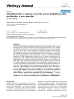
tively. The cross-sectional HRTEM image of the S-1 is
shown in Fig. 1. A representative image displays a fairly
smooth interface layer between the film and the silicon
substrate. Some changes have appeared in the bulk of the
S-1 after post-annealing treatment. There are several
observable bright/dark contrast fluctuations in the film. The
electron diffraction pattern of the S-1 as inset of Fig. 1 has
shown a typical amorphous halo, which indicates the film is
still in amorphous state. Therefore, it could be deduced that
these locations are some composition-rich regions, even a
few small nanocrystals. When the films have been annealed
approaching to the crystallization temperature, the grain
coarsening has occurred, and the increase in grain size has
been observed.
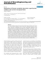
In comparison, several crystal regions have been
observed in the HRTEM image of the S-2 and are shown in
Fig. 2. The fast Fourier transformation (FFT) measurement
has been carried out on these regions to obtain the complex
situations of these nanocrystals, and the relevant image is
shown in the right as inset figure. From the figure one can
observe that it is a mixed nanocrystal region, because the
diffraction pattern is a superposition of the patterns from
two pieces of nanocrystals. Both of interplanes spacings,
whose values are about 0.237 nm and lie at an angle of
near to 60°, are of regular parallelogram with a center and
corresponding to the
"
101ðÞand 1
"
10ðÞplanes of the hex-
agonal Al
2
O
3
, respectively. As for the other dots, the
evaluated two interplanes spacings are equivalent to
0.242 nm. It is presumed that the two spots correspond to
the (004) and 00
"
4ðÞplanes of orthorhombic TiAl
2
O
5
,
respectively.
As indicated above, the HRTEM cross-section and
electron diffraction patterns of the Ti
0.25
Al
0.75
O
x
films
demonstrated the formation of nano-sized crystals. Only
one-dimensional diffraction patterns of orthorhombic
TiAl
2
O
5
phase in the images indicate that the nanocrystals
of the film preferably promote epitaxial c-axis-oriented
growth. This promotion has been demonstrated by the
results derived from the X-ray diffraction [11]. With the
X-ray diffraction results, only a small crystal peak attrib-
uted to the orthorhombic TiAl
2
O
5
phase could be observed.
Fig. 1 HRTEM cross-section image of S-1. Inset electron diffraction
image of S-1
Nanoscale Res Lett (2009) 4:1178–1182 1179
123
From the macroscopical aspect, the preferable orientation
is obvious, and the crystallization of the Ti
0.25
Al
0.75
O
x
film
is anisotropic. Because of the nonstoichiometric composi-
tion, no evidence of the presence of TiO
2
nanocrystals was
detected in this sample.
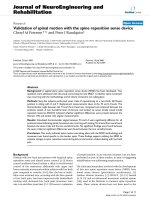
The typical I–V measurements performed on the
respective samples are shown in Fig. 3a, b. The S-1 has
very good insulating properties, as apparent from the sub-
stantial current of about 10
-6
A/cm
2
at an electric field of
-2 MV/cm applied between the silicon substrate and the
metal contact. Comparably, the S-2 exhibits a significantly
increased leakage current of 10
-2
A/cm
2
at the same
electric field, which is almost as much as 4 orders of
magnitude derived from S-1. The large leakage currents of
S-2 possibly originate from the formation of nanocrystals.
Considering the HRTEM results, this confirms the crucial
role of the amorphous Al
2
O
3
in the insulating properties of
the dielectric stack, despite its small amount and thickness.
However, the sweep loop characteristics of the investi-
gated samples disclose the hysteresis. It is ascribed to traps
located within the bulk Ti
0.25
Al
0.75
O
x
film or near the
Ti
0.25
Al
0.75
O
x
film/silicon interface, such as oxygen
vacancies and the other defects, which get filled with
electrons from the applied electrical field upon sweeping to
Fig. 2 HRTEM cross-section image of S-2. The inset on the right shows the magnified image and the FFT image of the selected nanocrystals
Fig. 3 Current density versus
bias electric field for a S-1 and b
S-2 at room temperature
1180 Nanoscale Res Lett (2009) 4:1178–1182
123
more positive gate voltages. At room temperature, the
hysteresis of S-1 is larger than that of S-2. Such a decrease
in hysteresis with annealing temperature reveals the pres-
ence of trap charging upon the temperature factor. More-
over, in the absence of applied electrical field, the negative
shift (*0.2 MV/cm) of S-1 proves the existence of posi-
tive charges in the bulk film as well. By virtue of its
capacitance–voltage curves (not shown here), it is calcu-
lated that the oxide trapped charges density is about as
much as 10
12
/cm
2
.
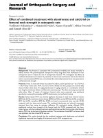
As we all known, Raman spectrum provides a fast and
convenient method to detect the small structural changes.
Typical Raman spectra from S-1 and S-2 are shown in
Fig. 4 that show the same peaks at about 618 cm
-1
and
814 cm
-1
, which are usually detected in amorphous Ti–O
materials, and ascribed to Ti–O stretching and Ti–O–Ti
stretching, respectively [12, 13]. The latter stretching may
also have contributions from a Ti–O stretch assigned to a
short Ti–O bond. Therefore, the large intensity of this peak
indicates the a two dimensional connectivity and provides
the evidence of the presence of –Ti–O–Ti– chainlike
structure with a shortening Ti–O bond distance. The other
peak for S-2 at *1,080 cm
-1
is the signature of TiAl
2
O
5
phase [14]. Its full width at half maximum (FWHM) of
50 cm
-1
is in contrast to the S-1 of FWHM = 40 cm
-1
at
the similar peak region. The Raman linewidth broadening,
primarily caused by phonons confinement in nanocrystals,
is inversely proportional to the size of the nanocrystals.
In order to further understand the nature of the charge
carrier trapping, migration and transfer in Ti
0.25
Al
0.75
O
x
films with small nanocrystals and the PL excitation spec-
troscopy with the emission wavelength fixed at 255 nm
were performed for the sample S-2. In general, it is difficult
to observe the photoluminescence phenomenon at room
temperature for bulk TiO
2
due to its indirect transition
nature. However, some nano-sized TiO
2
particles and
mesoporous-structured powders have been reported to
exhibit room temperature photoluminescence [15]. Fig-
ure 5 shows the PL excitation spectroscopy of a broad
excitation peak centered at *361 nm. The samples exhibit
the very small Stokes shift between the absorption and the
emission, which characterizes the energy relaxation
resulting from interfacial roughness, defects, and other
structural imperfection. Herein, the main probability lies in
the defects of nanoclusters and/or nanocrystals in the bulk
film. Generally, the electrons are trapped by oxygen
vacancies or confined within quantum dots in nanocrystals
region. On the other hand, the excited electrons can transfer
from the valance band to the new levels that exist upper of
the conduction band introduced by the dopant. Thus, the
photoluminescence efficiency will be restrained with the
thermal treatment. Nevertheless, such a meaningful value
has not been previously reported for nanostructural films
comprising titania and alumina, and its realization within
the present films is notable consideration that no attempts
were made to control the size of the nanocrystals.
Conclusions
In conclusion, we have performed a systematical analysis
of titania-incorporated alumina nanocrystals. The present
experiments demonstrate that the nanocrystals exhibit
excellent properties like low current density and small
hysteresis. Moreover, they offer high photoluminescence
quantum yields at room temperature. This approach can be
extended to other conditions such as low temperature,
anion doping, and crystal size controlling.
Fig. 4 Optical Raman spectra for S-1 and S-2
Fig. 5 Photoluminescence emission spectra for S-2, excitation
wavelength 255 nm. Inset the energy band structure of the sample
Nanoscale Res Lett (2009) 4:1178–1182 1181
123
Acknowledgments This work was sponsored by National Natural
Science Foundation of China (Grant number of 60576023 and
60636010), the State Key Program for Basic Research of China
(2004CB619004), the State Key Program for Science and Technology
of China (2009ZX02101-4) and Jiangsu Province Planned Projects for
Postdoctoral Research Funds (0204003426).
References
1. G.S. Pang, S.G. Chen, Y. Koltypin, A. Zaban, S.H. Feng, A.
Gedanken, Nano. Lett. 1(12), 723 (2001)
2. D.F. Zhang, L.D. Sun, J.L. Yin, C.H. Yan, Adv. Mater. 15, 1022
(2003)
3. A. Tricoli, M. Graf, S.E. Pratsinis, Adv. Funct. Mater. 18,1
(2008)
4. S. Rakshit, S. Vasudevan, ACS. Nano. 2(7), 1473 (2008)
5. D.M. Bagnall, Y.F. Chen, Z. Zhu, T. Yao, M.Y. Shen, T. Goto,
Appl. Phys. Lett. 73, 1038 (1998)
6. D.L. Klein, R. Roth, A.K.L. Lim, A.P. Alivisatos, P.L. McEuen,
Nature 389, 699 (1997)
7. A. Sashchiuk, L. Amirav, M. Bashouti, M. Krueger, U. Sivan, E.
Lifshitz, Nano. Lett. 1(4), 159 (2004)
8. L. Shi, J. Yin, K.B. Yin, F. Gao, Y.D. Xia, Z.G. Liu, Appl. Phys.
A: Mater. Sci. Process. 90(2), 379 (2008)
9. H. Kim, P.C. Mclntyre, J. Appl. Phys. 92, 5094 (2002)
10. G. Lucovsky, G.B. Rayner Jr, Appl. Phys. Lett. 77, 2912 (2000)
11. L. Shi, Y.D. Xia, B. Xu, Z.G. Liu, J. Appl. Phys. 101, 034102
(2007)
12. M. Fernadndez, X.Q. Wang, C. Belver, J.C. Hanson, J.A.
Rodriguez, J. Phys. Chem. C. 111, 674 (2007)
13. L.S. Hsu, R. Rujkorakarn, J.R. Sites, Y. She, J. Appl. Phys. 59,
3475 (1986)
14. G. Mestl, N.F.D. Verbruggen, F.C. Lange, B. Tesche, H.
Knoezinger, Langmuir 12, 1817 (1996)
15. D. Li, H. Haneda, S. Hishita, N. Ohashi, Chem. Mater. 17, 2596
(2005)
1182 Nanoscale Res Lett (2009) 4:1178–1182
123