Báo cáo hóa học: " A Novel Route for Preparation of Hollow Carbon Nanospheres Without Introducing Template" ppt
Bạn đang xem bản rút gọn của tài liệu. Xem và tải ngay bản đầy đủ của tài liệu tại đây (338.22 KB, 6 trang )
NANO EXPRESS
A Novel Route for Preparation of Hollow Carbon Nanospheres
Without Introducing Template
Minmin Li Æ Qingsheng Wu Æ Ming Wen Æ
Jianlin Shi
Received: 2 June 2009 / Accepted: 21 July 2009 /Published online: 22 August 2009
Ó to the authors 2009
Abstract A newly developed route for the synthesis of
hollow carbon nanospheres without introducing template
under hydrothermal conditions was reported. Hollow car-
bon nanospheres with the diameter of about 100 nm were
synthesized using alginate as reagent only. Many instru-
ments were applied to characterize the morphologies and
structures of carbon hollow nanospheres, such as XRD,
TEM, and Raman spectroscopy. The possible formation
and growth mechanism of carbon hollow spheres were
discussed on the basis of the investigation of reaction
influence factors, such as temperature, time, and content.
The findings would be useful for the synthesis of more
materials with hollow structure and for the potential use in
many aspects. The loading of SnO
2
on the surface of car-
bon hollow spheres was processed, and its PL property was
also characterized.
Keywords Synthesis Á Nanostructure Á
Carbon hollow nanospheres
Introduction
Inorganic hollow spheres with tailored structural, optical,
and surface properties represent an important class of
materials that may find applications in a wide range of
areas such as delivery vehicle systems, photonic crystals,
fillers, and catalysts [1–4]. Generally, the synthesis of
inorganic hollow spheres can be realized by means of
sacrificial templates, including ‘‘hard templates’’, such as
silica spheres, polystyrene latex spheres, and resin spheres
[5–8], and ‘‘soft templates’’, such as vesicles, liquid drop-
lets, emulsion droplets as well as block copolymer micelles
[9–11]. But synthesis of hollow structures without intro-
ducing templates has scarcely been reported in recent
years.
Researchers have paid great attention to carbon
spheres, as they have significant application in the
preparation of diamond films, lubricating materials, and
special rubber additives, owing to their properties similar
to fullerene and graphite [12–14]. However, harsh envi-
ronment was necessary for the synthesis of these hollow
carbon spheres up to now [15–18]. Hydrothermal method
provide a comparatively mild circumstance and is widely
used in the synthesis of carbon materials. Till now, only
hollow carbon spheres with the diameter of few microns
were obtained through this method [19]. In this report,
hollow carbon nanospheres with the diameter of about
100 nm were reported through hydrothermal treatment
without introducing template, and this process was sel-
dom reported in the synthesis of inorganic hollow
structures, especially in the synthesis of carbon hollow
spheres. SnO
2
nanoparticles loading on the surface of
these hollow spheres were synthesized and the fluores-
cence property of the complicate materials was also be
characterized.
Electronic supplementary material The online version of this
article (doi:10.1007/s11671-009-9406-7) contains supplementary
material, which is available to authorized users.
M. Li Á Q. Wu (&) Á M. Wen
Department of Chemistry, Tongji University, 1239 Siping Road,
200092 Shanghai, People’s Republic of China
e-mail:
Q. Wu
Shanghai Key Laboratory of Molecular Catalysis
and Innovative Materials, Fudan University,
200433 Shanghai, People’s Republic of China
J. Shi
Shanghai Institute of Ceramics, Chinese Academy
of Sciences, 1295 Dingxi Road, 200050 Shanghai,
People’s Republic of China
123
Nanoscale Res Lett (2009) 4:1365–1370
DOI 10.1007/s11671-009-9406-7
Experimental Works
Synthesis of Hollow Carbon Nanospheres
All chemicals were purchased from Sinopharm group
chemical reagent Co. Ltd with analytic-grade purity and used
directly without further treatment. The carbon spheres were
synthesized under hydrothermal conditions. In a typical
procedure, 0.3 g sodium alginate was dissolved in 16 mL
deionized water and ultrasonic processed for 20 min and
sealed in a 20 mL Teflon autoclave and maintained at 180 °C
for 10 h. The autoclave was naturally cooled down to the
room temperature when the reaction was complete. The
black products were collected by using a centrifuge and
washed several times with distilled water and absolute eth-
anol, respectively, and dried under vacuum at 80 °C for 5 h.
Loading of SnO
2
on the Surface of Hollow Carbon
Nanospheres
The loading of SnO
2
on the surface of hollow carbon
nanospheres was performed referring to coating of SnO
2
nanoparticles on the surface of carbon nanotubes in the
Zhou’s report [20]. Using a desired amount of HCl acid
(0.7 ml of 38% HCl in 40 ml H
2
O) is the key to obtaining
uniformly dispersed SnO
2
nanoparticles loading on the
surface of hollow carbon nanospheres.
Characterization
The structures of synthesized products were measured with
X-ray powder diffraction (XRD) and Raman spectroscopy.
XRD measurements were recorded using a Netherlands
1,710 diffractometer with graphite monochromatized Cu
Ka irradiation (k = 1.54056 A
˚
) and Raman spectroscopy
using Renishaw company, equipped with an Ar ? laser at
514.5 nm. Infrared spectrum was characterized by a
Nicolet 5DX FTIR spectrometer equipped with a TGS/PE
detector and a silicon beam splitter with 1 cm
-1
resolution.
The micromorphologies of products were inspected by
transmission electron microscopy (TEM) (JEOL JEM2010,
Japan) at an accelerating voltage of 200 Kv. Emission
spectra were measured on a Perkin-Elmer LS-55 fluores-
cence spectrophotometer. All the measurements were taken
at room temperature.
Results and Discussion
Morphologies and Structure
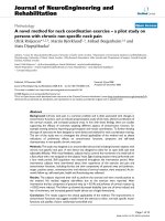
XRD as a kind of important manner can be used to
characterize the phase and structure of samples. The
XRD pattern of products obtained in the hydrothermal
system is shown in Fig. 1a. The broad peak indicates
that the amorphism of product is because of poor
crystallization. As a kind of usual fashion, Raman
spectroscopy is a powerful technique for characterizing
the carbon materials. Figure 1b displays the Raman
spectrum of synthesized materials that verifies carbon
structure of products. A strong peak at 1,588 cm
-1
and
a weak peak at 1,333 cm
-1
corresponding to typical
Raman peaks of graphitized carbon spheres are
observed. The peak at 1,333 cm
-1
could be assigned to
the vibrations of carbon atoms with dangling bonds in
planar terminations of disordered graphite. The peak at
1,588 cm
-1
(G-band) corresponds to an E2 g mode of
graphite and is related to the vibration of sp2-bonded
carbon atoms [21, 22]. The high intensity ratio of D to
G band suggests the poor graphitization of the products,
which is consistent with the XRD pattern. FT-IR is also
used to characterize the function group of the hollow
carbon nanospheres.
In our experiment, FT-IR spectrum (Fig. 1c) was used to
identify the functional groups of the hollow carbon nano-
spheres for the sake of further understanding the structure
Fig. 1 a XRD patterns of synthesized products after hydrothermal process 5 h at initial content = 0.3 g; b Raman spectrum of synthesized
products; c IR spectrum of synthesized products
1366 Nanoscale Res Lett (2009) 4:1365–1370
123
of carbon. As a kind of amylose aggregated from mono-
glucuronide, aromatization is usually regarded as a process
of decreasing the number of functional groups [23]. The
bands at 1,710 and 1,620 cm
-1
can be attributed to C = O
and C = C vibrations, respectively. These results reveal
that aromatization of chitosan has taken place during
hydrothermal treatment. Compared with the aromatization
of glucose under hydrothermal condition [24], the bands in
the range of 1,000 * 1,300 cm
-1
are hardly seen in the
FT-IR spectrum of our products, indicating few C–OH
stretching and OH bending vibrations and implying few
residual hydroxyl groups appear. This is in accordance with
the polymer structure of alginate. The residues of CHO
groups are covalently bonded to the carbon frameworks,
which makes it more potential application as templates for
hybrid complex structures and opens a new way to hollow
core-shell materials.
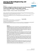
Typical TEM images of hollow carbon nanospheres
obtained in 0.3 g sodium alginate solution after hydro-
thermal process for 5 h are presented in Fig. 2a. The
strong contrast between the dark edge and the pale center
of the spherical particles evidences their hollow structure.
The diameter of the hollow carbon spheres is about 70–
120 nm, with an average diameter of about 100 nm, and
the wall thickness is about 20 nm. The related electron
diffraction pattern (not shown) is circular, indicating the
amorphous structure of carbon, consistent with the XRD
pattern and Raman spectrum. The possible reason might
be that a low temperature process leads to the poor
crystalline.
The Influence Factors of Reaction
The time-dependent experiments were also carried out to
investigate the influence of reaction time on morphologies
of products. Hollow carbon nanospheres were obtained in a
series of experiment times. When the reaction time was
less than 2 h, carbon could not be formed. That is, com-
plete carbonization of alginate is not possible at this
reaction time. This result showed the importance of reac-
tion time on the formation of carbon spheres. Extending the
reaction time to as long as 12 h, the products remained
hollow carbon nanospheres. The hollow nanospheres
obtained changed from single hollow nanospheres (in
Fig. 2a) to a ringlike structure of walled hollow nano-
spheres (in Fig. 2b) and then to a linear structure of walled
hollow nanospheres (in Fig. 2c) when the reaction time is
5, 7, 10 h, respectively. No distinct changes in the thick-
ness of the wall of synthesized hollow nanospheres were
found and the network made of many hollow nanospheres
appeared with the prolonged time. Probably, the reason for
the occurrence of these phenomena lies in the linear
polymer structure.
The content-dependent experiments were carried out to
monitor the influence of the initial content of the product.
The different amounts of alginate were put into autoclaves,
and other parameters were kept constant. Some typical
TEM images are given in Fig. 2. The TEM images showed
that morphologies of obtained products gradually changed
from a few single hollow nanospheres (Fig. 2d) to a great
deal of hollow nanospheres (Fig. 2e) and then to cross-
Fig. 2 TEM images of
prepared hollow carbon
nanospheres at different
reaction time and content. a, b,
c products after 5, 7, 10 h, 0.3 g
sodium alginate d, e, f products
after 7, 10, 18 h, 0.1 g sodium
alginate
Nanoscale Res Lett (2009) 4:1365–1370 1367
123
linked hollow nanospheres (Fig. 2f) when the content
changed from 0.1 to 0.3 g and then to 0.5 g (the reaction
condition is kept at 180 °C for 7 h in all reactions). These
varieties of products revealed that the content is a crucial
factor for preparing carbon nanospheres in a large scale.
Because the carbonization process was actually a defunc-
tionalization process, the content of reagents largely
affected the collision rate among base groups. These results
reveal that carbon spheres could be achieved only the
alginate is up to a certain content. The alginate solution is
up to critical supersaturation and nucleation burst when
these macromolecules dehydrate gradually.
The influence of temperature on products was also
explored. When the reaction temperature is decreased to
160 ° C, even if reaction time is kept at 12 h, carbonization
reaction could not be complete and brown reaction solution
was obtained when the content was reduced to 0.1 g, which
identified the occurrence of aromatization. While a higher
temperature (200 °C) was used, it led to accelerated
dehydration of alginate intermolecules and a burst nucle-
ation around spherical chain, which could result in the
formation of cross-linked hollow spheres. These results
revealed that temperature was a key factor in the prepa-
ration of carbon nanospheres through dehydration, aroma-
tization, and carbonization. At lower temperatures, the
energies of intermolecular collisions and of intramolecular
collisions were not high enough to carbonize, leading to the
failure of formation of carbon nanospheres. Compared with
the carbonization of glucose [24], the carbonization of
alginate was slower and needed higher temperature
although glucose and alginate sodium were a kind of sac-
charide. The possible reason lies in the polymer structure
of alginate. On the one hand, the polymer structure con-
tained fewer –OH group and slowed the dehydration
intermolecular process. More time and higher temperature
were needed to realize polymerization and carbonization of
alginate according to the theory of the rate of chemical
reaction.
The filling ratio as an important parameter of hydro-
thermal systems has a critical influence on the reaction
pressure, solubility of solute, viscosity, density, and
dielectric constant of solution at constant temperature in a
sealed hydrothermal system. To investigate the influence of
filling ratio on the obtained products, a series of parallel
experiments were performed with different filling ratios
from 40 to 80% at 180 °C for 8 h. Obtained products
congregate more easily and become randomly when the
filling ratio of the reagent is low to 40%, compared with the
filling ratio is up to 80%. It is well known that the viscosity
of alginate depends on temperature, density, and the stir-
ring rate. With the decrease in filling ratio, the alginate
solution becomes denser, which makes carbonization
reaction more intense.
Formation Mechanism
The formation mechanism of hollow carbon nanospheres
was also explored. At first, the formation of carbon spheres
was a nucleation and growth process (Fig. 3). At a certain
temperature, the alginate solution can form spherical
micelles and further nucleate by dewatering. Compared
with the dehydration of glucose [24], the dehydration of
glucose became more difficult because less –OH group
made intermolecular dehydration take place only when
reaction system had higher energy. It may be explained
why carbonization of alginate needed higher temperature
than for carbonization of glucose. Then nucleation of
alginate took place when critical supersaturation of alginate
was got to. Finally, the growth of nucleus is controlled by
diffusion or carbonization reaction according to the theory
of Ostwald ripening [25].
The comparative experiment was made without ultra-
sonic processing, and irregular carbon chips were obtained.
That is, ultrasonic process was key to the formation of the
hollow structure. So we speculated that the formation of
hollow structure was as follows: At first, sodium alginate
was wholly dissolved in the water by heating the solution.
Then hollow sodium alginate nanospheres were formed by
cavitation of ultrasonic process. A great number of air
bubbles formed and grew in the zone of negative pressure,
single hollow nanoshperes
cross-linked hollow nanospheres
linear hollow nanospheres
sodium alginate solution
ultrasonic
processing
hydrothermal
processing
Fig. 3 Formation mechanism
of hollow carbon nanosphere
1368 Nanoscale Res Lett (2009) 4:1365–1370
123
and they were occluded in the zone of positive pressure
during the ultrasonic process. This kind of cavitation led to
air bubbles formed in the molecular of alginate. When the
solution was placed in the hydrothermal condition at some
temperature, carbonization took place in situ, and hollow
carbon nanospheres were synthesized. According to the
content of reactant, different structures made of hollow
nanospheres were formed.
The Loading of SnO
2
Nanoparticles
Carbon hollow structures, typically in the form of capsules
converted from their core-shell precursors, exhibited higher
current and power density when used as a catalyst support
in the direct methanol fuel cell [26]. SnO
2
-nanoparticles-
coated carbon spheres are useful functional nanocomposite
in many applications including gas sensors, batteries, and
optics. The special configuration in this nanocomposite is
expected to prevent the SnO
2
nanoparticles from aggre-
gation and to increase its conductivity, hence the perfor-
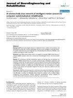
mance. In this article, SnO
2
nanoparticles are loaded onto
the surfaces of hollow carbon nanospheres by room -tem-
perature surface oxidation method. To reveal the compo-
sition and structure of the above sample, XRD was carried
out. Figure 4d shows the XRD pattern, in which all dif-
fraction peaks were in good agreement with tetragonal
rutile SnO
2
(JCPDS No: 41-1445). The morphology of this
kind of complicate material was characterized with TEM.
The TEM image and amplified TEM image are given in
Fig. 4a and b. SnO
2
nanoparticles of several nanometers
were loaded on the surface of hollow carbon nanospheres.
The PL spectrum of the composite material was charac-
terized by two peaks at 376 and 424 nm, and a broad peak
centered at 476 nm in the wavelength of range 450–
516 nm under excitation at 310 nm. The emission in the
wavelength range 450–550 nm may be related to the
intrinsic defect structures, in particular the oxygen vacan-
cies originated from the oxygen deficiency [27] induced
during the growth. The prominent band at 420 nm is
attributed to the recombination of the deep-trapped charged
and photogenerated electron from the conduction band
[28].
Conclusion
To conclude, hollow carbon nanospheres with the diameter
of 100 nm were synthesized without template under
Fig. 4 a, b TEM and amplified
TEM images of synthesized
SnO
2
@C, c its PL properties
and d XRD pattern of
synthesized SnO
2
@C composite
Nanoscale Res Lett (2009) 4:1365–1370 1369
123
hydrothermal condition via ultrasonic pretreatment. And
the wall thickness was about 20 nm. The influence of the
reaction time and the content was also observed. Then a
possible forming mechanism was given. Hollow carbon
nanospheres loading SnO
2
nanoparticles were synthesized
and its photoluminescence peak appeared at 376, 424, and
476 nm. The hollow carbon nanospheres and their loading
structure have potential application in many fields such as
carriers, storage, and catalysts.
Acknowledgments The authors acknowledge the National Natural
Science Foundation (No. 50772074) of China, the State Major
Research Plan (973) of China (No. 2006CB932302), the Nano-
Foundation of Shanghai in China (No. 0852nm01200), and the
Shanghai Key Laboratory of Molecular Catalysis and Innovative
Materials (No. 2009KF04).
References
1. F. Caruso, Adv. Mater. 13, 11 (2001)
2. Z. Zhong, Y. Yin, B. Gates, Y. Xia, Adv. Mater. 12, 206 (2000)
3. C.E. Fowler, D. Khushalani, S. Mann, Chem. Commun. 2028
(2001). doi:10.1039/b104879c
4. D.H. Shin, H.C. Shim, J.W. Song, S. Kim, C. Han, Scr. Mater. 60,
607 (2009). doi:10.1016/j.scriptamat.2008.12.019
5. K.P. Velikov, A. van Blaaderen, Langmuir 17, 4779 (2001). doi:
10.1021/la0101548
6. K.H. Davis, F. Caruso, B. Zhang, S. Mann, Chem. Mater. 12,
2832 (2000)
7. R.A. Caruso, A. Susha, F. Caruso, Chem. Mater. 13, 400 (2001)
8. M.L. Breen, A.D. Donsmore, R.H. Pink, S.Q. Qadri, B.R. Ratna,
Langmuir 17, 903 (2001). doi:10.1021/la0011578
9. H.T. Schmidt, A.E. Ostafin, Adv. Mater. 14, 532 (2002)
10. C.E. Fowler, D. Khushalani, S. Mann, J. Mater. Chem. 11, 1968
(2001)
11. A.M. Collins, C. Spickermann, S. Mann, J. Mater. Chem. 13,
1112 (2003)
12. A.L.M.R.S. Ramaprabhu, Nanoscale Res. Lett. 3, 76 (2008). doi:
10.1007/s11671-008-9116-6
13. Z.P. Dong, B. Yang, J. Jin, J. Li, H.W. Kang, X. Zhong, R. Li,
J.T. Ma, Nanoscale Res. Lett. 4, 335 (2009). doi:10.1007/s11671-
008-9248-8
14. P.M. Ajayan, Chem. Rev. 99, 1787 (1999). doi:10.1021/cr97
0102g
15. X.Y. Liu, B.C. Huang, N.J. Covolle, Carbon 40, 2791 (2002)
16. Ph. Serp, R. Feurer, Ph. Kalck, Y. Kihn, J.L. Faria, J.L. Figuei-
redo, Carbon 39, 621 (2001)
17. J.S. Qiu, Y.F. Li, Y.P. Wang, C.H. Liang, T.H. Wang, D.B.
Wang, Carbon 41, 767 (2003). doi:10.1016/S0008-6223(02)
00392-5
18. Y.J. Xiong, Y. Xie, Z.Q. Li, C.Z. Wu, R. Zhang, Chem. Com-
mun. 904 (2003). doi: 10.1039/b211996j
19. X.M. Sun, Y.D. Li, J. Colloid Interface Sci. 291, 7 (2005). doi:
10.1016/j.jcis.2005.04.101
20. J.G. Zhou, H.T. Fang, J.M. Maley, J.Y.P. Ko, M. Murphy, Y.
Chu, R. Sammynaiken, T.K. Sham, J. Phys. Chem. C 113(15),
6114–6117 (2009). doi:10.1021/jp810639y
21. Y. Huang, R.J. Young, Carbon 33, 97 (1995)
22. R.J. Nemanich, S.A. Solin, Phys. Rev. B 20, 392 (1979)
23. T. Sakaki, M. Shibata, T. Miki, H. Hirosue, N. Hayashi, Biore-
sour. Technol. 58, 197 (1996)
24. X.M. Sun, Y.D. Li, Angew. Chem. Int. Ed. 43, 597 (2004). doi:
10.1002/anie.200352386
25. T. Sugimoto, AIChE J. 24
, 1125 (1978). doi:10.1002/aic.6902
40629
26. S.J. Han, Y.K. Yun, K.W. Park, Y.E. Sung, T. Hyeon, Adv.
Mater. 15, 1922 (2003). doi:10.1002/adma.200305697
27. Q. Kuang, Z.Y. Jiang, Z.X. Xie, S.C. Lin, Z.W. Lin, S.Y. Xie,
R.B. Huang, L.S. Zheng, J. Am. Chem. Soc. 127, 11777 (2005).
doi:10.1021/ja052259t
28. F. Gu, S.F. Wang, M.K. Lu, G.J. Zhou, D. Xu, D.R. Yuan, J.
Phys. Chem. B 108, 8119 (2004). doi:10.1021/jp036741e
1370 Nanoscale Res Lett (2009) 4:1365–1370
123