Báo cáo hóa học: " A Novel Docetaxel-Loaded Poly (e-Caprolactone)/Pluronic F68 Nanoparticle Overcoming Multidrug Resistance for Breast Cancer Treatment" pot
Bạn đang xem bản rút gọn của tài liệu. Xem và tải ngay bản đầy đủ của tài liệu tại đây (416.11 KB, 10 trang )
NANO EXPRESS
A Novel Docetaxel-Loaded Poly (e-Caprolactone)/Pluronic F68
Nanoparticle Overcoming Multidrug Resistance for Breast
Cancer Treatment
Lin Mei Æ Yangqing Zhang Æ Yi Zheng Æ Ge Tian Æ Cunxian Song Æ
Dongye Yang Æ Hongli Chen Æ Hongfan Sun Æ Yan Tian Æ Kexin Liu Æ
Zhen Li Æ Laiqiang Huang
Received: 12 June 2009 / Accepted: 1 September 2009 /Published online: 16 September 2009
Ó to the authors 2009
Abstract Multidrug resistance (MDR) in tumor cells is a
significant obstacle to the success of chemotherapy in
many cancers. The purpose of this research is to test the
possibility of docetaxel-loaded poly (e-caprolactone)/Plu-
ronic F68 (PCL/Pluronic F68) nanoparticles to overcome
MDR in docetaxel-resistance human breast cancer cell line.
Docetaxel-loaded nanoparticles were prepared by modified
solvent displacement method using commercial PCL and
self-synthesized PCL/Pluronic F68, respectively. PCL/
Pluronic F68 nanoparticles were found to be of spherical
shape with a rough and porous surface. The nanoparticles
had an average size of around 200 nm with a narrow size
distribution. The in vitro drug release profile of both
nanoparticle formulations showed a biphasic release pat-
tern. There was an increased level of uptake of PCL/Plu-
ronic F68 nanoparticles in docetaxel-resistance human
breast cancer cell line, MCF-7 TAX30, when compared
with PCL nanoparticles. The cytotoxicity of PCL nano-
particles was higher than commercial Taxotere
Ò
in the
MCF-7 TAX30 cell culture, but the differences were not
significant (p [ 0.05). However, the PCL/Pluronic F68
nanoparticles achieved significantly higher level of cyto-
toxicity than both of PCL nanoparticles and Taxotere
Ò
(p \ 0.05), indicating docetaxel-loaded PCL/Pluronic F68
nanoparticles could overcome multidrug resistance in
human breast cancer cells and therefore have considerable
potential for treatment of breast cancer.
Keywords Nanoparticles Á MDR Á Pluronic F68 Á
Poly (e-caprolactone) Á Docetaxel Á Breast cancer
Introduction
Cancer remains the leading cause of death worldwide. The
global incidence and mortality of breast cancer remains
high despite extraordinary progress in understanding the
molecular mechanisms underlying carcinogenesis, tumor
promotion, and the establishment of molecular targeted
therapies [1]. Although early detection and screening of
breast cancer is associated with less invasive surgical
procedures and may increase survival, the 5-year survival
rate of metastatic breast cancer (stage IV) is still below
15%. Multidrug resistance (MDR) to anticancer agents
remains a major barrier to successful cancer treatment.
L. Mei (&) Á Y. Zhang Á Y. Zheng Á D. Yang Á L. Huang (&)
The Shenzhen Key Lab of Gene and Antibody Therapy,
Center for Biotech and Bio-Medicine and Division of Life
Sciences, Graduate School at Shenzhen, Tsinghua University,
L308, Tsinghua Campus, Xili University Town,
518055 Shenzhen, Guangdong, China
e-mail:
L. Huang
e-mail:
L. Mei Á G. Tian Á Y. Tian Á K. Liu Á Z. Li
College of Pharmacy, Dalian Medical University,
116027 Dalian Liaoning, China
C. Song Á H. Sun
Institute of Biomedical Engineering,
Peking Union Medical College & Chinese Academy of Medical
Sciences, The Tianjin Key Laboratory of Biomaterial Research,
300192 Tianjin, China
D. Yang
Department of Gastroenterology,
Xiangya Second Hospital, Central South University,
410011 Changsha, China
H. Chen
Department of Life Science and Technology,
Xinxiang Medical University, 453003 Xinxiang, China
123
Nanoscale Res Lett (2009) 4:1530–1539
DOI 10.1007/s11671-009-9431-6
Thus, the development of effective therapies overcoming
MDR against invasive breast cancer and particularly highly
metastatic disease still remains a significant priority.
Nanoparticulate delivery systems in cancer therapies pro-
vide better penetration of therapeutic and diagnostic sub-
stances within the body at a reduced risk in comparison
with conventional cancer therapies. Nanoparticles could
reduce the multidrug resistance (MDR) that characterizes
many anticancer drugs, including docetaxel, by a mecha-
nism of internalization of the drug [2], reducing its efflux
from cells mediated by the P-glycoprotein [3]. Nanoparti-
cle distribution within the body is based on various
parameters such as their relatively small size resulting in
longer circulation times and their ability to take advantage
of tumor characteristics. In comparison to conventional
cancer treatments, the nanoscale of these particulate sys-
tems also minimizes the irritant reactions at the injection
site. Nanoparticles and their use in drug delivery is a far
more effective cancer treatment method than conventional
chemotherapy, which is typically limited by the toxicity of
drugs to normal tissues, short circulation half-life in
plasma, limited aqueous solubility, and nonselectivity
restricting therapeutic efficacy [4].
Docetaxel is a poorly water-soluble, semi-synthetic
taxane analog commonly used in the treatment of breast
cancer, oval cancer, small and nonsmall cell lung cancer,
prostate cancer, etc. Its commercial formulation Taxotere
Ò
is formulated in high concentration of Tween 80, which has
been found associated with severe side effects including
hypersensitivity reactions, cumulative fluid retention, nau-
sea, mouth sores, hair loss, peripheral neuropathy, fatigue,
and anemia [5, 6] and has shown incompatibility with the
common PVC intravenous administration sets [7]. In order
to eliminate the Tween 80-based adjuvant and in the
attempt to increase the drug solubility, alternative formu-
lations have been attempted, such as liposomes [5], nano-
particles [8–10], docetaxel-fibrinogen-coated olive oil
droplets [6]. Among them, the nanoparticle formulation
holds greatest promise for this purpose. The nanoparticles
showed advantages such as more stable during storage over
others. Moreover, such a colloidal system is able to
extravasate solid tumors into the inflamed or infected site,
where the capillary endothelium is defective [3, 4].
Nanoparticles serving in anticancer therapies may be
comprised, in whole or in part, of various lipids and natural
and synthetic polymers. Most commonly used synthetic
polymers to prepare nanoparticles for drug delivery are
biodegradable. Among the various biodegradable polymers
approved by the US Food and Drug Administration (FDA),
poly(lactide) (PLA), poly(
D,L-lactide-co-glycolide) (PLGA),
and poly (caprolactone) (PCL) are used most often in the
literature. In the family of polyesters, PCL occupies a unique
position: it is at the same time biodegradable and miscible
with a variety of polymers, and it crystallizes very readily
[11]. A lack of toxicity and great permeability has already
found wide use for PCL in medical applications [11]. Plu-
ronic F68 is a difunctional block copolymer surfactant ter-
minating in primary hydroxyl groups. It is both water and
organic solvent soluble. Poloxamers and poloxamine non-
ionic surfactants have diverse applications in various bio-
medical fields ranging from drug delivery and medical
imaging to management of vascular diseases and disorders
[12]. In the present study, Pluronic F68 was incorporated
into PCL as a pore-forming agent and drug-releasing
enhancer. Previous studies by our group have demonstrated
the amount of Pluronic F68 blended into PCL affected the
microspheres morphology and controlled paclitaxel release
[13]. In addition, it has been demonstrated that Pluronic
block copolymers interact with multidrug-resistant (MDR)
tumors resulting in drastic sensitization of these tumors with
respect to various anticancer agents [13, 14]. The key attri-
bute for the biological activity of Pluronics is their ability to
incorporate into membranes followed by subsequent trans-
location into the cells and affecting various cellular func-
tions, such as mitochondrial respiration, ATP synthesis,
activity of drug efflux transporters, apoptotic signal trans-
duction, and gene expression. As a result, Pluronics cause
drastic sensitization of MDR tumors to various anticancer
agents including docetaxel, enhance drug transport across
the blood–brain barriers (BBB) and intestinal barriers and
cause transcriptional activation of gene expression both in
vitro and in vivo [14, 15]. Furthermore, recent studies
indicated that Pluronic F68 is a potent in vitro inhibitor of
both P-gp and CYP3A4 [16]. Thus, in this research we
investigate the hypothesis that a novel docetaxel-loaded
PCL/Pluronic F68 nanopaticles overcoming multidrug
resistance (MDR) will achieve better therapeutic effects in
docetaxel-resistance human breast adenocarcinoma MCF-7
cell line.
Materials and Methods
Materials
In brief, docetaxel of purity 99% was purchased from
Shanghai Jinhe Bio-Technology Co. Ltd, Shanghai, China.
Polycaprolactone (Mn * 42,500) was obtained from
Sigma–Aldrich (St. Louis, MO, USA). Cell Counting Kit-8
(CCK-8) was from Dojindo Molecular Technologies Inc.,
Kumamoto, Japan. e–Caprolactone monomer with 99.9%
purity was from Aldrich Chemical Co., USA. The mono-
mer was further purified by vacuum distillation over CaH
2
.
Pluronic F68 with molecular weight (Mw) around 8,300
containing about 80% poly (ethyl oxide) (PEO) segment
and 20% of poly (propyl oxide) (PPO) segment was
Nanoscale Res Lett (2009) 4:1530–1539 1531
123
purchased from BASF, Germany. The Pluronic F68 was
incorporated into PCL matrix in 10% of weight ratio as a
molecular distribution, which would leach out in aqueous
medium to leave microporous structure in the PCL matrix
(Sun et al., 2006). Polyvinyl alcohol (PVA) (MW 30 000–
70 000) was obtained from Sigma, Chemical Co (St Louis,
MO). Acetonitrile and methanol used as mobile phase in
high performance liquid chromatography (HPLC) were
purchased from EM Science (ChromAR, HPLC grade,
Mallinckrodt Baker, USA). All other chemicals were
HPLC grade and were used without further purification.
Millipore water was prepared by a Milli-Q Plus System
(Millipore Corporation, Breford, USA).
Synthesis of PCL/Pluronic F68 Compound
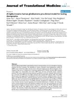
PCL/Pluronic F68 compound was synthesized by ring-
opening polymerization as shown in Fig. 1;[17]. Briefly,
the terminal hydroxyl groups in Pluronic F68 molecules
were capped with acetyl so that it became inactive and
would not participate in the polymerization reaction of
e-caprolactone. The acetyl-capped Pluronic F68 was dis-
solved in e-caprolactone monomer before polymerization
so that Pluronic F68 was incorporated in PCL matrixes as a
molecular dispersion instead of forming a copolymer. The
polymerization was carried out at 140
Æ
C under high vac-
uum for 24 h with 0.04% stannous octoate as catalyst.
Preparation of Nanoparticles
The nanoparticles were prepared by modified solvent dis-
placement method as described previously [18]. Briefly,
100 mg of PCL/Pluronic F68 compound and 17.65 mg of
docetaxel were dissolved in 100 mL of acetone by mild
heating and sonication. The mixed solution was gently
poured into 50 mL of deionized water containing 1,000 mg
of PVA under magnetic stirring. The emulsion was then
evaporated overnight under reduced pressure to remove the
organic solvent. The resulting suspension of nanoparticles
was centrifuged at 23,000 rpm for 30 min. The pellet was
washed twice with distilled water to remove free drug and
PVA. The resulted particles were freeze-dried for 2 days.
Docetaxel-loaded PCL nanoparticles and empty PCL/Plu-
ronic F68 nanoparticles were prepared by the same method.
In addition, the fluorescent coumarin-6-loaded nanoparti-
cles were prepared in the same way except 0.05% (w/v)
coumarin-6 was encapsulated instead of docetaxel.
Characterization of Nanoparticles
Surface Morphology
The nanoparticles were imaged by a field emission scanning
electron microscopy (FESEM) system at an accelerating
voltage of 5 kV. To prepare samples for FESEM, the par-
ticles were fixed on the stub by a double-sided sticky tape
and then coated with platinum layer by JFC-1300 automatic
fine platinum coater (JEOL, Tokyo, Japan) for 80 s.
Size Analysis and Zeta Potential
The particle size and size distribution were measured by
laser light scattering (Brookhaven Instruments. Corpora-
tion, Holtsville, NY 90-PLUS analyzer). Before measure-
ment, the freshly prepared particles were appropriately
diluted. Zeta potential of the docetaxel-loaded nanoparti-
cles was detected by laser Doppler anemometry (Zeta Plus
zeta potential analyzer, Brookhaven Corporation, Holts-
ville, NY). The particles (about 2 mg) were suspended in
deionized water before measurement. The data were
obtained with the average of three measurements.
Drug Loading and Encapsulation Efficiency
Drug content in the nanoparticles was assayed by HPLC
(Agilent LC 1100, Santa Clara, CA, USA). A reverse-phase
Fig. 1 The end-capping
reaction of Pluronic F68 and the
polymerization of PCL
1532 Nanoscale Res Lett (2009) 4:1530–1539
123
Inertsils ODS-3 column (150 lm 9 4.6 lm, pore size
5 lm, GL science Inc, Tokyo, Japan) was used. Briefly,
5 mg particles were dissolved in 1 mL DCM under vig-
orous vortexing. This solution was transferred to 5 mL of
mobile phase consisting of deionized water, methanol, and
acetonitrile (50:45:5, v/v). DCM was evaporated in nitro-
gen atmosphere and the clear solution was obtained for
HPLC analysis. The solution was transferred into HPLC
vial after filtered through 0.22 mm syringe filter. The flow
rate of mobile phase was 1 mL/min. The column effluent
was detected at 230 nm with a UV/VIS detector. The
measurement was performed triplicate. The encapsulation
efficiency (EE) was expressed as the percentage of the drug
loaded in the final product.
Differential Scanning Calorimetry (DSC)
The physical status of docetaxel inside the nanoparticles
was investigated by differential scanning calorimetry (DSC
822e, Mettler Toledo, Switzerland). The samples were
purged with dry nitrogen at a flow rate of 20 mL/min. The
temperature was raised at 10 °C/min.
In Vitro Drug Release
Dialysis method was selected to examine the drug release
in vitro. Briefly, 15 mg nanoparticles were dispersed in
5 mL release medium (phosphate buffer solution (PBS) of
pH 7.4 containing 0.1% w/v Tween 80) to form a sus-
pension. Tween 80 was used to increase the solubility of
docetaxel in the buffer solution and avoid the binding of
docetaxel to the tube wall. The suspension was put into a
standard grade regenerated cellulose dialysis membrane
(Spectra/Por
Ò
6, MWCO = 1,000, Spectrum, Houston,
TX, USA). Then, the closed bag was put into a centrifuge
tube and immersed in 15 mL release medium. The tube
was put in an orbital water bath shaking at 120 rpm at
37.0 ° C. At given time intervals, 10 mL samples was
sucked out for analysis and replaced with fresh medium. In
this research, the sink condition was maintained by the
addition of Tween 80 and frequent replacement of fresh
buffer during the in vitro release experiment. The newly
collected samples were extracted with 2 mL DCM and
reconstituted in 5 mL mobile phase. The DCM was evap-
orated by nitrogen stream. The analysis procedure was
similar as for the measurement of EE.
Cell Culture
In this research, human breast cancer cell lines MCF-7
cells of passages between 26 and 31 (American Type
Culture Collection, VA) were cultured in Dubelco’s mod-
ified essential medium (DMEM) supplemented with 10%
FBS, 100 mM sodium pyruvate, 1.5 g/L of sodium bicar-
bonate, and 1% penicillin–streptomycin and incubated in
SANYO CO
2
incubator at 37 °C in a humidified-environ-
ment of 5% carbon dioxide. Then, docetaxel-resistance
human breast cancer cells (MCF-7 TAX30) were created as
described previously [19]. Briefly, the cells were made
resistant to docetaxel by short-term in vitro exposure to
docetaxel for 1 h, which was immediately followed by
washing of the cells several times with culture media,
trypsinization, and splitting the cells for subsequent cell
growth recovery. The cells were initially exposed to
10 nmol/L docetaxel increasing to 500 nmol/L for 1 h.
After this point, the cells were exposed to 1 lmol/L
docetaxel increasing to 30 lmol/L docetaxel for 24 h.
Cellular Uptake of Nanoparticles
For quantitative study, docetaxel-resistance human breast
cancer cells (MCF-7 TAX30) were seeded into 96-well black
plates (Costar, IL, USA) of 1.3 9 10
4
cells/well, and after
the cells reached confluence, the cells were equilibrated with
HBSS at 37 °C for 1 h and then incubated with coumarin-6-
loaded PCL/Pluronic F68 nanoparticle suspension. The
nanoparticles were dispersed in the medium at a concentra-
tion of 100, 250, and 500 lg/mL. The wells with nanopar-
ticles were incubated at 37 °C for 2 h. After incubation, the
suspension was removed, and the wells were washed three
times with 50 lL cold PBS to eliminate traces of nanopar-
ticles left in the wells. Afterthat, 50 lL of 0.5% Triton X-100
in 0.2N NaOH was introduced into each sample wells to lyse
the cells. The fluorescence intensity of each sample well was
measured by microplate reader (GENios, Tecan, Switzer-
land) with excitation wave length at 430 nm and emission
wavelength at 485 nm. Cell uptake efficiency was expressed
as the percentage of cells-associated fluorescence versus the
fluorescence present in the feed solution.
For the qualitative study, cells were reseeded in the
chambered-cover glass system (LABTEK
Ò
, Nagle Nunc,
IL). After the cells were incubated with 250 lg/mL cou-
marin-6-loaded nanoparticles at 37 °C for 2 h, they were
rinsed with cold PBS for three times and then fixed by eth-
anol for 20 min. The cells were further washed twice with
PBS, and the nuclei were counterstained with propidium
iodide (PI) for 30 min. The cell monolayer was washed
twice with PBS and mounted in Dako
Ò
fluorescent mounting
medium (Glostrup, Denmark) to be observed by confocal
laser scanning microscope (CLSM) (LSM 410, Zeiss, Jena,
Germany) with an imaging software, Fluoview FV500.
In Vitro Cytotoxicity
Cancer cell viability of the drug-loaded PCL/Pluronic F68
nanoparticles was evaluated by CCK-8 assay. CCK-8 is a
Nanoscale Res Lett (2009) 4:1530–1539 1533
123
kind of cell viability assay reagent with a higher sensitivity
and a better reproducibility than MTT. Hundred lLof
MCF-7 TAX30 cells were seeded in 96-well plates (Costar,
IL, USA) at the density of 5 9 10
3
viable cells/well and
incubated at 24 h to allow cell attachment. The cells were
incubated with docetaxel-loaded PCL/Pluronic F68 nano-
particle suspension, docetaxel-loaded PCL nanoparticle
suspension, Taxotere
Ò
at 0.025, 0.25, 2.5, 10, and 25 lg/
mL equivalent docetaxel concentrations and empty PCL/
Pluronic F68 (PCL/F68) nanoparticles with the same
nanoparticle concentrations of 0.25, 2.5, 25, 100, and
250 lg/mL for 24, 48, and 72 h, respectively. At desig-
nated time intervals, the medium was removed, and the
wells were washed with PBS for two times. Ten lLof
CCK-8 solution was added to each well of the plate and
incubated for 1–4 h in the incubator. The absorbance was
measured at 450 nm using a microplate reader. Cell via-
bility was calculated by the following equation.
Cell viability %ðÞ¼ðAbs
s
=Abs
control
ÞÂ100
where Abs
s
is the fluorescence absorbance of the cells
incubated with the nanoparticle suspension, and Abs
control
is the fluorescence absorbance of the cells incubated with
the culture medium only (positive control). IC50, the drug
concentration at which inhibition of 50% cell growth was
observed, in comparison with that of the control sample,
was calculated by curve fitting of the cell viability data.
Statistical Methodology
The results are expressed as mean ± SD. The significance
of differences was assessed using Student’s t test and was
termed significance when p = 0.05.
Results and Discussion
Characterization of Nanoparticles
PCL/Pluronic F68 compound with viscosity average
molecular weight of 44,000 was successfully synthesized.
Previous studies by our group have demonstrated that drug
release rate was greatly enhanced by increasing content of
Pluronic F68 in PCL matrix from 0 to 10%, but there was no
further increase in release rate when the content of Pluronic
F68 increased to 15% [13]. Therefore, we decided to use the
PCL/Pluronic F68 (90/10, wt/wt) matrix as the final drug
carrier to fabricate PCL/Pluronic F68 nanoparticles. The
nanoparticles were characterized in terms of mean size and
size distribution, morphology, surface charge, and physical
state of encapsulated drug. As shown in Table 1, the aver-
age size of PCL/Pluronic F68 nanoparticles was much
smaller, and the particle size distribution was much nar-
rower than those of PCL nanoparticles. Nonionic emulsifier,
especially Pluronic F68, offered additional steric stabiliza-
tion effect avoiding aggregation of the fine particles in the
colloidal system [20]. In this sense, Pluronic F68 may act as
a coemulsifier in the fabrication process, resulting in
smaller particle size and narrow size distribution. The drug
loading level of docetaxel encapsulated in the PCL/Pluronic
F68 and PCL nanoparticles was 10.02% and 9.76%,
respectively. In addition, the results revealed that the drug
encapsulation efficiency (EE%) of both nanoparticle for-
mulations was almost the same and more than 65%.
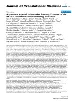
As shown by Fig. 2, the docetaxel-loaded nanoparticles
(PFNP) observed by FESEM were spherical in shape, and
their size was around 200 nm. The surface of nanoparticles
appears rough and porous. As mentioned earlier, Pluronic
F68 is both organic and water-soluble. So the pores in the
surface of PCL/Pluronic F68 nanoparticles could be
attributed to the hydrophilicity of Pluronic F68. Pluronic
F-68 leached out due to the water phase during fabrication
process, therefore creating porous structure in the surface
of the PCL/F68 nanoparticles [17]. In addition, Pluronics
adsorb strongly onto the surface of hydrophobic nano-
spheres [e.g. polystyrene, poly(lactide-co-glycolide),
poly(phosphazene), poly(methyl methacrylate), and poly
(butyl 2-cyanoacrylate) nanospheres] via their hydrophobic
POP center block [21]. This mode of adsorption leaves the
hydrophilic POE side-arms in a mobile state because they
extend outward from the particle surface. These side-arms
provide stability to the particle suspension by a repulsion
effect through a steric mechanism of stabilization, involv-
ing both enthalpic and entropic contribution [22, 23].
Table 1 Characterization of nanoparticles
Group Size (nm)(n = 3) Polydispersion
(n = 3)
Drug loading (%) Encapsulation
efficiency (%)
Zeta potential
(mV)(n = 3)
Polymer
PCNP 293.2 ± 3.6 0.172 9.76 65.08 -48.70 ± 3.11 PCL
PFNP 201.7 ± 10.1 0.096 10.02 69.10 -12.50 ± 0.86 PCL/F68
CCNP 281.2 ± 5.5 0.145 -35.70 ± 2.99 PCL
CFNP 222.7 ± 5.4 0.133 -20.50 ± 1.34 PCL/F68
Note: Group CCNP and CFNP represent coumarin-6-loaded PCL nanoparticles and PCL/Pluronic F68 nanoparticles, respectively
1534 Nanoscale Res Lett (2009) 4:1530–1539
123
Zeta potential, i.e., surface charge can greatly influence
the particles stability in suspension through the electro-
static repulsion between the particles. It is also an impor-
tant factor to determine their interaction in vivo with the
cell membrane, which is usually negatively charged. In
addition, from the zeta potential measurement, we can
roughly know the dominated component on the particles
surface. The detection of laser Doppler anemometry
showed that zeta potential of docetaxel-loaded PCL/Plu-
ronic F68 nanoparticles was -12.5 mV, a great increase
compared with that of PCL nanoparticles, with zeta
potential around -48.7 mV. Since Pluronic F68 is non-
ionic, this surface charge increase demonstrated the pres-
ence of Pluronic F68 layer on the surface, which shifted the
shear plane of the diffusive layer to a larger distance [24].
However, high absolute value of zeta potential is necessary
to ensure stability and avoid aggregation of particles. It
thus could be concluded that PCL/Pluronic F68 nanopar-
ticles were electrically less stable than PCL nanoparticles.
DSC studies were performed to investigate the physical
state of the drug in the nanoparticles, because this aspect
could influence the in vitro and in vivo release of the drug
from the systems. Figure 3 shows the DSC thermograms of
pure docetaxel, PCL/Pluronic F68 nanoparticles, and PCL
nanoparticles. The melting endothermic peak of pure
docetaxel appeared at 173 °C. However, no melting peak
was detected for both nanoparticle formulations, evidenc-
ing the absence of crystalline drug in the nanoparticles, at
least at the particle surface level. It might be hypothesized
that the polymer inhibited the crystallization of docetaxel
during nanoparticles formation. Therefore, it could be
concluded that docetaxel in the nanoparticles was in an
amorphous or disordered crystalline phase of a molecular
dispersion or a solid solution state in the PCL/Pluronic F68
matrix after the production.
In Vitro Drug Release
Maintaining sink condition for poorly water-soluble drugs
has been one of the difficulties in designing in vitro release
experiments. In this research, the sink condition was
maintained by the addition of Tween 80 and frequent
replacement of fresh buffer during the in vitro release
experiment. The in vitro drug release profiles of the
docetaxel-loaded nanoparticles in the first 32 days are
shown in Fig. 4. The initial burst of 35.57 and 47.01% in
the first 5 days can be observed for PCL nanoparticles and
Fig. 3 DSC thermograms of the pure docetaxel and docetaxel-loaded
nanoparticles
Fig. 2 FESEM images of
docetaxel-loaded PCL/Pluronic
F68 nanoparticles
Fig. 4 The in vitro release profile of docetaxel-loaded nanoparticles
Nanoscale Res Lett (2009) 4:1530–1539 1535
123
PCL/Pluronic F68 nanoparticles, respectively, which is
followed by an approximately first–order release afterward.
After 32 days, the accumulative drug release from PCL/
Pluronic F68 nanoparticles was found to be 67.91%, which
was significantly faster than PCL nanoparticles, which is
57.60%. The present studies confirmed our previous results
that the amount of Pluronic F68 blended into PCL could
facilitate drug release and affect the microspheres mor-
phology [13]. Thus, Pluronic F68 blended into PCL could
also be used as a pore-forming agent and drug-releasing
enhancer in nanoparticle formulation.
Uptake of Coumarin-6-Loaded Nanoparticles
by MCF-7 TAX30 Cells
It is clear that the therapeutic effects of the drug-loaded
nanoparticles would depend on internalization and sus-
tained retention of the nanoparticles by the diseased cells
[25]. Although in vitro and in vivo experiment could pro-
duce different results, an in vitro investigation can provide
some preliminary evidence to show advantages of nano-
particle formulation versus free drug. Coumarin-6, a fluo-
rescence marker, has been widely used as a probe for
marking nanoparticles in cellular uptake experiment,
because of its biocompatibility, high fluorescence activity,
low dye loading (\0.5%, w/w), and low leaking rate, which
is used to replace the drug in the nanoparticle formulation to
visualize and measure cellular uptake of polymeric nano-
particles [26]. The cellular uptake efficiency of the fluo-
rescent coumarin 6-loaded-nanoparticles by MCF-7 TAX30
cells was assayed upon 2 h incubation, and the results are
shown in Fig. 5. It can be clearly observed that for both
formulations, the cellular uptake efficiency of nanoparticles
by MCF-7 TAX30 cells (Fig. 4) was found decreased with
increase of the incubated particle concentration from 100 to
500 lg/ml, indicating the saturated and limited capability of
cellular uptake of the nanoparticles. Such saturated and
limited characteristic of cellular uptake of particles was also
observed by others [27, 28]. The cellular uptake efficiency
of PCL/Pluronic F68 nanoparticles was 1.47-, 1.36-, and
1.67-fold higher than that of PCL nanoparticles at the
incubated particle concentration of 100, 250, and 500 lg/
ml, respectively. As shown in Table 1, the coumarin-6-
loaded nanoparticles were highly relevant to the docetaxel-
loaded nanoparticles in terms of size and zeta potential.
Harush-Frenkel et al. [29] found that both cationic and
anionic nanoparticles are targeted mainly to the clathrin
endocytic machinery. A fraction of both nanoparticle for-
mulations is suspected to internalize through a macro-
pinocytosis-dependent pathway. A significant amount of
nanoparticles transcytose accumulate at the basolateral
membrane. Some anionic but not cationic nanoparticles
transited through the degradative lysosomal pathway. Plu-
ronic block copolymers could enhance cellular uptake of
drugs, proteins or polynucleotides [15, 30]. In addition, it
was demonstrated that the mechanism of cellular uptake of
biodegradable microparticles or nanoparticles is size
dependent [2, 31]. Thus, it is reasonable that PCL/Pluronic
F68 nanoparticles with incorporation of Pluronics and
smaller particle size would have higher cellular uptake.
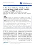
Figure 6 shows the confocal laser scanning microscopy
(CLSM) images of MCF-7 TAX30 cells after 2 h incuba-
tion with coumarin-6-loaded PCL/Pluronic F68 nanoparti-
cles at 250 lg/mL nanoparticle concentration, in which the
upper left image was obtained from FITC channel (green),
the lower left one was from propidium iodide (PI) channel
(red), the upper right image was from transmitted light
channel (black and white), and the lower right image was
the combination of all the three images. We can see from
this figure that the fluorescence of the coumarin-6-loaded
PCL/Pluronic F68 nanoparticles (green) is closely located
around the nuclei (red, stained by PI), which indicates that
the nanoparticles have been internalized by the cells.
In Vitro Cell Viability of Nanoparticles
Figure 7 shows the in vitro viability of MCF TAX30 cells
cultured with the drug formulated in Taxotere
Ò
, PCL/Plu-
ronic F68 nanoparticles, and PCL nanoparticles at the same
equivalent docetaxel concentration of 0.025, 0.25, 2.5, 10,
and 25 lg/mL and empty PCL/Pluronic F68 (PCL/F68)
nanoparticles with the same nanoparticle concentrations of
0.25, 2.5, 25, 100, and 250 lg/ml, respectively (n = 6). It
can be seen from this figure that in general (1) the drug
formulated in PCL nanoparticles showed equivalent or
better effects against the cancer cells than Taxotere
Ò
and
(2) PCL/Pluronic F68 nanoparticles achieved even better
therapeutic effect than PCL nanoparticles and Taxotere
Ò
.
Fig. 5 Cellular uptake of coumarin-6-loaded nanoparticles. Data
represent mean ± SD (n = 6)
1536 Nanoscale Res Lett (2009) 4:1530–1539
123
For example, the MCF-7 TAX30 cell viability after 1 day
incubation at the 10 lg/mL drug concentration was
decreased from 54.37% for Taxotere
Ò
to 49.16% (i.e. a
11.42% increase in cytotoxicity, p [ 0.05) for PCL NP
formulation and 36.63% (i.e. a 38.88% increase in cyto-
toxicity, p \0.05) for PCL/Pluronic F68 NP formulation.
Similarly, it can be evaluated from Fig. 7 that compared
with commercial Taxotere
Ò
, the cytotoxicity of MCF-7
TAX30 cells was increased 5.07% (p [ 0.05) and 9.31%
(p [ 0.05) for the PCL NP formulation, and 42.37%
(p \ 0.05) and 38.32% (p \ 0.05) for PCL/Pluronic F68
NP formulation after 2 and 3 day incubation at the 10 lg/
mL drug concentration, respectively. However, the empty
PCL/Pluronic F68 nanoparticles had no significant influ-
ence on cell viability of MCF TAX30 cells. The higher
cytotoxicity of the drug formulated in the two nanoparticle
formulations can be attributed to the higher cellular uptake
as well as the sustained drug release manner in comparison
with Taxotere
Ò
. In addition, nanoparticles could reduce the
multidrug resistance (MDR) that characterizes many anti-
cancer drugs, including docetaxel, by a mechanism of
internalization of the drug [2], reducing its efflux from cells
mediated by the P-glycoprotein [3]. The reason of the
advantage of PCL/Pluronic F68 nanoparticles over PCL
nanoparticles may be attributed to the higher cellular
uptake of the nanoparticles as well as the faster drug
release from the nanoparticles, which was shown before in
Fig. 4. More importantly, Pluronics could cause drastic
sensitization of MDR tumors to various anticancer agents
including docetaxel [12, 14]. The mechanisms of Pluronic
effects in MDR cells were thoroughly investigated. It was
demonstrated that Pluronic block copolymers could (1)
incorporate into membranes changing its microviscosity;
(2) induce a dramatic reduction in ATP levels in cancer and
barrier cells; (3) inhibit drug efflux transporters, such as
P-gp [14, 32], breast cancer resistance protein [33] and
multidrug resistance proteins [34]; (4) induce release of
cytochrome C and increase of reactive oxygen species
levels in the cytoplasm [15]; (5) enhance proapoptotic
signaling and decreasing antiapoptotic defense in MDR
cells [35]; (6) inhibit the glutathione/glutathione S-trans-
ferase detoxification system [33]; and (7) abolish drug
sequestration within cytoplasmic vesicles [36]. The key
attribute for the biological activity of Pluronics is their
ability to incorporate into membranes followed by sub-
sequent translocation into the cells and affecting various
cellular functions, such as mitochondrial respiration, ATP
synthesis, activity of drug efflux transporters, apoptotic
Fig. 7 Viability of MCF-7 TAX30 cells cultured with docetaxel-
loaded PCL nanoparticles and PCL/Pluronic F68 (PCL/F68) nano-
particles in comparison with that of Taxotere
Ò
at the same docetaxel
dose and empty PCL/Pluronic F68 (PCL/F68) nanoparticles with the
same amount of nanoparticles (n = 6)
Fig. 6 Confocal laser scanning microscopy (CLSM) image of MCF-
7 TAX30 cells after 2 h incubation with coumarin-6-loaded PCL/
Pluronic F68 nanoparticles at 37.0 °C. The cells were stained by
propidium iodide (red) and the coumarin-6-loaded nanoparticles are
green. The cellular uptake is visualized by overlaying images
obtained by white light, FITC filter, and PI filter: upper left image
from FITC channel; upper right image from transmitted light channel;
lower left image from PI channel; lower-right image from combined
transmitted light channel, PI channel, and FITC channel
Nanoscale Res Lett (2009) 4:1530–1539 1537
123
signal transduction, and gene expression [15]. Moreover,
recent studies indicated that Pluronic F68 is a potent in
vitro inhibitor of both P-gp and CYP3A4 [16]. Other
similar drug carrier such as mPEG-PCL copolymer [37]
and n-(2-hydroxypropyl)methacrylamide (HPMA) copoly-
mer [38] could also overcome the multidrug resistance of
cancer cells.
The in vitro therapeutic effects of a dosage form can be
quantitatively evaluated by its IC
50
, which is defined as the
drug concentration at which 50% of the cells in culture
have been killed in a designated time period. Table 2 gives
the IC
50
value of MCF-7 TAX 30 cells after 24-, 48-, and
72-h incubation with docetaxel formulated in Taxotere
Ò
,
PCL, and PCL/Pluronic F68 nanoparticles at various drug
concentrations, respectively. The data are impressive to
show the advantage of the nanoparticle formulation versus
the pristine drug as well as that of PCL/Pluronic F68
nanoparticles versus the PCL nanoparticles in docetaxel
formulation. It can be found from Table 2 that the IC
50
value for MCF-7 TAX30 cells was decreased from 10.380,
8.726, and 5.945 lg/mL for Taxotere
Ò
to 7.388, 3.643, and
1.244 lg/mL for PCL NP formulation and to 1.019, 0.384,
and 0.196 lg/mL for PCL/Pluronic F68 NP formulation
after 24, 48, and 72 h incubation, respectively. Such
advantages of the NP formulations in achieving higher
cytotoxicity would become even more significant if the
controlled release manner of the drug from the nanoparti-
cles is further considered. It can be seen from Fig. 4 that
the accumulative drug release was only 13.57, 20.61, and
27.94% for PCL nanoparticles and 22.40, 31.09, and
37.44% for PCL/Pluronic F68 nanoparticles after 1, 2,
and 3 days, respectively, and the release started from 0%
while the Taxotere
Ò
immediately became 100% available
for the MCF-7 TAX30 cells in culture.
Conclusions
For the first time, a novel docetaxel-loaded PCL/Pluronic
F68 nanoparticle formulation was prepared to overcome
multidrug resistance in human breast cancer cells. The
results revealed that there was an increased level of uptake
of PCL/Pluronic F68 nanoparticles in docetaxel-resistance
human breast cancer cell line, MCF-7 TAX30, when
compared with PCL nanoparticles. The cytotoxicity of PCL
nanoparticles was higher than commercial Taxotere
Ò
in the
MCF-7 TAX30 cell culture, but the differences were not
significant (p [ 0.05). However, the PCL/Pluronic F68
nanoparticles achieved significantly higher level of cyto-
toxicity than both of PCL nanoparticles and Taxotere
Ò
(p \ 0.05), indicating docetaxel-loaded PCL/Pluronic F68
nanoparticles could overcome multidrug resistance in
human breast cancer cells and therefore have considerable
potential for treatment of breast cancer.
Acknowledgments The authors are grateful for financial support
from the National Natural Science Foundation of China (NSFC) under
Grant No 30500239 and the Shenzhen Municipal Government and
Bureau of Science, Technology & Information for providing funding
supports (to LQH) through the programs of Shenzhen National Key
Lab of Health Science and Technology and the Key Lab of Gene and
Antibody Therapy.
References
1. T. Tanaka, P. Decuzzi, M. Cristofanilli, J.H. Sakamoto, E. Tas-
ciotti, F.M. Robertson, M. Ferrari, Nanotechnology for breast
cancer therapy. Biomed. Microdevices. 11(1), 49–63 (2009)
2. J. Davda, V. Labhasetwar, Characterization of nanoparticle
uptake by endothelial cells. Int. J. Pharm. 233, 51–59 (2002)
3. I. Brigger, C. Dubernet, P. Couvreur, Nanoparticles in cancer
therapy and diagnosis. Adv. Drug Deliv. Rev. 54, 631–651 (2002)
4. K.Y. Kim, Nanotechnology platforms and physiological chal-
lenges for cancer therapeutics. Nanomedicine 3(2), 103–110
(2007)
5. M.L. Immordino, P. Brusa, S. Arpicco, B. Stella, F. Dosio, L.
Cattel, Preparation, characterization, cytotoxicity and pharma-
cokinetics of liposomes containing docetaxel. J. Control. Release
91, 417–429 (2003)
6. F.K. Engels, R.A. Mathot, J. Verweij, Alternative drug formu-
lations of docetaxel: a review. Anticancer Drugs 8(2), 95–103
(2007)
7. S.D. Baker, M. Zhao, P. He, M.A. Carducci, J. Verweij, A.
Sparreboom, Simultaneous analysis of docetaxel and the formu-
lation vehicle polysorbate 80 in human plasma by liquid chro-
matography/tandem mass spectrometry. Anal. Biochem. 324,
276–284 (2004)
8. J. Cheng, B.A. Teply, I. Sherifi, J. Sung, G. Luther, F.X. Gu, E.
Levy-Nissenbaum, A.F. Radovic-Moreno, R. Langer, O.C. Far-
okhzad, Formulation of functionalized PLGA-PEG nanoparticles
for in vivo targeted drug delivery. Biomaterials 28(5), 869–876
(2007)
9. F. Quaglia, L. Ostacolo, A. Mazzaglia, V. Villari, D. Zaccaria,
M.T. Sciortino, The intracellular effects of non-ionic amphiphilic
cyclodextrin nanoparticles in the delivery of anticancer drugs.
Biomaterials 30(3), 374–382 (2009)
10. M.V. Lozano, D. Torrecilla, D. Torres, A. Vidal, F. Dominguez,
M.J. Alonso, Highly efficient system to deliver taxanes into
tumor cells: docetaxel-loaded chitosan oligomer colloidal carri-
ers. Biomacromolecules 9(8), 2186–2193 (2008)
11. V.M. Hiljanen, T. Karjalainen, J. Seppa
¨
la
¨
, Biodegradable lactone
copolymers. I. Characterization and mechanical behavior of
Table 2 IC
50
of MCF-7 TAX30 cells after 24-, 48-, and 72-h incu-
bation with docetaxel formulated in Taxotere
Ò
, PCL, and PCL/Plu-
ronic F68 nanoparticles at various drug concentrations
Incubation
time (h)
IC
50
(lg/ml)
PCL/Pluronic
F68 NPs
PCL NPs Taxotere
Ò
24 1.019 7.388 10.380
48 0.384 3.643 8.726
72 0.196 1.244 5.945
1538 Nanoscale Res Lett (2009) 4:1530–1539
123
e-caprolactone and lactide copolymers. J. Appl. Polym. Sci. 59,
1281–1288 (1996)
12. S.M. Moghimi, A.C. Hunter, Poloxamers and poloxamines in
nanoparticle engineering and experimental medicine. Trends
Biotechnol. 18, 412–420 (2000)
13. G. Ma, C. Song, PCL/poloxamer 188 blend microsphere for
paclitaxel delivery: Influence of poloxamer 188 on morphology
and drug release. J. Appl. Polym. Sci. 104(3), 1895–1899 (2007)
14. A.V. Kabanov, E.V. Batrakova, V.Y. Alakhov, Pluronic block
copolymers for overcoming drug resistance in cancer. Adv. Drug
Deliv. Rev. 54(5), 759–779 (2002)
15. E.V. Batrakova, A.V. Kabanov, Pluronic block copolymers:
evolution of drug delivery concept from inert nanocarriers to
biological response modifiers. J. Control. Release 130(2), 98–106
(2008)
16. J. Huang, L. Si, L. Jiang, Z. Fan, J. Qiu, G. Li, Effect of pluronic
F68 block copolymer on P-glycoprotein transport and CYP3A4
metabolism. Int. J. Pharm. 356(1–2), 351–353 (2008)
17. H. Sun, L. Mei, C. Song, X. Cui, P. Wang, The in vivo degra-
dation, absorption and excretion of PCL-based implant. Bioma-
terials 27, 1735–1740 (2006)
18. L. Mei, H. Sun, C. Song, Local delivery of modified paclitaxel-
loaded poly(epsilon-caprolactone)/pluronic F68 nanoparticles for
long-term inhibition of hyperplasia. J. Pharm. Sci. 98, 2040–2050
(2009)
19. I. Brown, K. Shalli, S.L. McDonald, S.E. Moir, A.W. Hutcheon,
S.D. Heys, A.C. Schofield, Reduced expression of p27 is a novel
mechanism of docetaxel resistance in breast cancer cells. Breast
Cancer Res. 6(5), R601–R607 (2004)
20. G. Reich, In vitro stability of poly (D, L-lactide) and poly (D,
L-lactide)/poloxamer nanoparticles in gastrointestinal fluids.
Drug Dev Ind. Pharm. 23(6), 1191–1198 (1997)
21. G. Storm, S.O. Belliot, T. Daemen, D.D. Lasic, Surface modifi-
cation of nanoparticles to oppose uptake by the mononuclear
phagocyte system. Adv. Drug Deliv. Rev. 16, 31–48 (1995)
22. S.M. Moghimi, I.S. Muir, L. Illum, S.S. Davis, V. Kolb-Bacho-
fen, Coating particles with block co-polymer (poloxamine-908)
suppresses opsonization but permits the activity of dysopsonins in
the serum. Biochim. Biophys. Acta 1179, 157–165 (1993)
23. J.T. Li, K.D. Caldwell, Plasma protein interactions with Pluronic
treated colloids. Colloids Surfaces B-Biointerfaces 7, 9–22
(1996)
24. R. Gref, A. Domb, P. Quellec, T. Blunk, R.H. Muller, J.M.
Verbavatz, R. Langer, The controlled intravenous delivery of
drugs using PEG-coated sterically stabilized nanospheres. Adv.
Drug Deliv. Rev. 16, 215–233 (1995)
25. C. Jin, L. Bai, H. Wu, F. Tian, G. Guo, Radiosensitization of
paclitaxel, etanidazole and paclitaxel ? etanidazole nanoparti-
cles on hypoxic human tumor cells in vitro. Biomaterials 28(25),
3724–3730 (2007)
26. K.Y. Win, S.S. Feng, Effects of particle size and surface coating
on cellular uptake of polymeric nanoparticles for oral delivery of
anticancer drugs. Biomaterials 26, 2713–2722 (2005)
27. M.P. Desai, V. Labhasetwar, E. Walter, R.J. Levy, G.L. Amidon,
The mechanism of uptake of biodegradable microparticles in
Caco-2 cells is size dependent. Pharm. Res. 14(11), 1568–1573
(1997)
28. M.G. Qaddoumi, H. Ueda, J. Yang, J. Davda, V. Labhasetwar,
V.H.L. Lee, The characteristic and mechanisms of PLGA nano-
particles in rabbit conjunctival epithelial cell layers. Pharm. Res.
21, 641–648 (2004)
29. O. Harush-Frenkel, E. Rozentur, S. Benita, Y. Altschuler, Surface
charge of nanoparticles determines their endocytic and transcy-
totic pathway in polarized MDCK cells. Biomacromolecules 9,
435–443 (2008)
30. Z. Yang, G. Sahay, S. Sriadibhatla, A.V. Kabanov, Amphiphilic
block copolymers enhance cellular uptake and nuclear entry of
polyplex-delivered DNA. Bioconjug Chem 19(10), 1987–1994
(2008)
31. J. Rejman, V. Oberle, I.S. Zuhorn, D. Hoekstra, Size-dependent
internalization of particle via the pathways of clathrin- and cav-
eolae-mediated endocytosis. Biochem. J. 377(Pt 1), 159–169
(2004)
32. G. Szakacs, J.K. Paterson, J.A. Ludwig, C. Booth-Genthe, M.M.
Gottesman, Targeting multidrug resistance in cancer. Nat Rev
Drug Discov. 5, 219–234 (2006)
33. T. Yamagata, H. Kusuhara, M. Morishita, K. Takayama,
H. Benameur, Y. Sugiyama, Improvement of the oral drug
absorption of topotecan through the inhibition of intestinal
xenobiotic efflux transporter, breast cancer resistance protein, by
excipients. Drug Metab. Dispos. 35, 1142–1148 (2007)
34. E.V. Batrakova, S. Li, V.Y. Alakhov, W.F. Elmquist, D.W.
Miller, A.V. Kabanov, Sensitization of cells overexpressing
multidrug-resistant proteins by pluronic P85. Pharm. Res. 20,
1581–1590 (2003)
35. T. Minko, E.V. Batrakova, S. Li, Y. Li, R.I. Pakunlu, V.Y.
Alakhov, A.V. Kabanov, Pluronic block copolymers alter apop-
totic signal transduction of doxorubicin in drug-resistant cancer
cells. J Control Release. 105, 269–278 (2005)
36. A. Venne, S. Li, R. Mandeville, A. Kabanov, V. Alakhov,
Hypersensitizing effect of Pluronic L61 on cytotoxic activity,
transport, and subcellular distribution of doxorubicin in multiple
drug-resistant cells. Cancer Res. 56, 3626–3629 (1996)
37. P. Elamanchili, C. McEachern, H. Burt, Reversal of multidrug
resistance by methoxypolyethylene glycol-block-poly-
caprolactone diblock copolymers through the inhibition of
P-glycoprotein function. J. Pharm. Sci. 98, 945–958 (2009)
38. T. Minko, P. Kopeckova, V. Pozharov, J. Kopecek, HPMA
copolymer bound adriamycin overcomes MDR1 gene encoded
resistance in a human ovarian carcinoma cell line. J Control
Release. 54, 223–233 (1998)
Nanoscale Res Lett (2009) 4:1530–1539 1539
123