Báo cáo hóa học: " Synthesis of Aqueous CdTe/CdS/ZnS Core/shell/shell Quantum Dots by a Chemical Aerosol Flow Method" pot
Bạn đang xem bản rút gọn của tài liệu. Xem và tải ngay bản đầy đủ của tài liệu tại đây (391.94 KB, 6 trang )
NANO EXPRESS
Synthesis of Aqueous CdTe/CdS/ZnS Core/shell/shell Quantum
Dots by a Chemical Aerosol Flow Method
Chuanmiao Yan
•
Fangqiong Tang
•
Linlin Li
•
Hongbo Li
•
Xinglu Huang
•
Dong Chen
•
Xianwei Meng
•
Jun Ren
Received: 6 September 2009 / Accepted: 5 October 2009 / Published online: 23 October 2009
Ó to the authors 2009
Abstract This work described a continuous method to
synthesize CdTe/CdS/ZnS core/shell/shell quantum dots.
In an integrated system by flawlessly combining the
chemical aerosol flow system working at high temperature
(200–300°C) to generate CdTe/CdS intermediate products
and an additional heat-up setup at relatively low tempera-
ture to overcoat the ZnS shells, the CdTe/CdS/ZnS multi-
shell structures were realized. The as-synthesized CdTe/
CdS/ZnS core/shell/shell quantum dots are characterized
by photoluminescence spectra, X-ray diffraction (XRD),
energy-dispersive X-ray spectra (EDS), transmission elec-
tron microscopy (TEM), and high-resolution transmission
electron microscopy (HRTEM). Fluorescence and XRD
results confirm that the obtained quantum dots have a core/
shell/shell structure. It shows the highest quantum yield
above 45% when compared to the rhodamine 6G. The core/
shell/shell QDs were more stable via the oxidation exper-
iment by H
2
O
2
.
Keywords Chemical aerosol flow Á CdTe/CdS/ZnS Á
Aqueous Á Core–shell Á Quantum dot
Introduction
In recent years, colloidal semiconductor nanocrystals or
quantum dots (QDs) have attracted great scientific and
technological interest due to their unique size-dependent
properties [1–3]. By changing the size or composition of
the QDs, their optical properties can be easily tuned to suit
for various requirement [4, 5]. In the biological labeling
especially [6, 7], numerous successful efforts have been
made by using the QDs as the fluorescence agent. The
organometallic synthesis of CdSe and CdTe QDs provides
a good method to synthesize high quality QDs, which were
synthesized using high boiling point solvents such as tri-
octylphosphine (TOP), trioctylphosphine oxide (TOPO)
and so on. However, they were hydrophobic and need time
consuming operation to convert into water soluble disper-
sions. The high toxicity of precursors and high price also
limit their wide application. Aqueous colloid approaches
become more attractive because they are much cheaper and
suitable for biological studies.
Aqueous CdTe [CdTe (aq)] QDs with high fluorescence
can be synthesized using a metal salt and NaHTe with
mercaptan acid as the stabilize agent [8, 9]. It provides a
simple way to generate aqueous QDs. But several hours to
several days’ reaction time is needed. Chemical aerosol
flow (CAF) synthesis of QDs [10] was first published by
Suslick et al. and has been widely used to generate many
kinds of nanoparticles, including mesoporous silica [11,
12], mesoporous carbon [13, 14], semiconductor nano-
crystals [10, 15, 16], and other nanomaterials [17–19]. It
provides a simple and fast way to continuously synthesize
C. Yan Á F. Tang (&) Á L. Li Á H. Li Á X. Huang Á
X. Meng Á J. Ren
Laboratory of Controllable Preparation and Application
of Nanomaterials, Technical Institute of Physics and Chemistry,
Chinese Academy of Sciences, 100190 Beijing,
People’s Republic of China
e-mail:
J. Ren
e-mail:
D. Chen
Beijing Creative Nanophase Hi-Tech Co., Ltd. China,
100086 Beijing, People’s Republic of China
C. Yan Á H. Li Á X. Huang
Graduate University of Chinese Academy of Sciences,
100039 Beijing, People’s Republic of China
123
Nanoscale Res Lett (2010) 5:189–194
DOI 10.1007/s11671-009-9464-x
nanoparticles. In our previous work, we used the modified
CAF method to generate CdTe/CdS core/shell quantum dots
in several seconds [20]. The QDs showed high stability, high
quantum yield and have large scale. But for bioapplication,
the cadmium-based QDs would release toxic Cd
2?
when
used in the cell or tissues [21]. Capping a shell of ZnS not
only can decrease the toxicity of cadmium but also can
increase the quantum yield [21], forming a core/shell or
core/shell/shell structure with CdTe cores inside and ZnS
shells outside is a good method to solve this problem.
With successive ionic layer adsorption and reaction
(SILAR) [22] method, high quality of core/shell and mul-
tishell quantum dots can be synthesized [23–27] in organic
media. Unfortunately, this method was complex, and it is
hard to be carried out in aqueous solution. Synthesis of
core/shell QDs especially core/shell/shell QDs in aqueous
solution is a hard work as the surface stabilizers were
fragile and sensitive to the environment. Many efforts have
been made to obtain high quality core/shell in water [28];
however, only a few works pay attention to the synthesis of
the CdTe/CdS/ZnS core/shell/shell quantum dots. Because
of the large lattice mismatch between CdTe and ZnS
(16.4%), it is hard to epitaxially grow ZnS shells on CdTe
cores. A shell of CdS between CdTe and ZnS can work as a
transition shell because the band gap and lattice contact of
CdS is just between that of CdTe and ZnS. Using micro-
wave irradiation method, high quantum yield CdTe/CdS/
ZnS quantum dots can be obtained [29], but multistep was
needed, the process was extremely troublesome, and the
yield was low. So developing a low cost, simple, contin-
uous way for preparing hydrophilic QDs with a shell of
ZnS capped is an urgent need.
Here, we investigated a facial way to directly and con-
tinuously synthesize the CdTe/CdS/ZnS core/shell/shell
QDs using a modified chemical aerosol flow method. In
this integrated synthesis system, the Cd, Te precursors were
first carried into the chemical aerosol flow system to result
CdTe/CdS core/shell quantum dots. Then, they were
brought out into a vessel containing Zn, S precursor solu-
tion for coating a shell of ZnS. The obtained core/shell/
shell QDs were characterized by X-ray powder diffraction
(XRD), transmission electron microscopy (TEM), energy-
dispersive X-ray spectrometer (EDS), and PL spectra to
confirm the core/shell/shell structure.
Experimental Section
Chemicals
Cd(NO
3
)
2
Á2H
2
O, Zn(NO
3
)
2
Á2H
2
O, NaOH, NaBH
4
(99%),
tellurium powder (99.8%), thiourea (97%), and 3-mercapto-
propionic acid (MPA, 99%) were purchased from Beijing
Chemical Reagent Co., Ltd. All chemicals were used without
additional purification. Distilled water was used for preparation
of all aqueous solutions.
Precursor Preparation
Briefly, 0.5 mmol Te powder and 2 mmol NaBH
4
were
mixed in a tube, and then 2 mL water was added. The
reaction mixture was heated at 80°C for 30 min to get a
pink NaHTe solution. The NaHTe solution was stored at
4°C for further use. One millimole Cd(NO
3
)
2
Á2H
2
O,
2.4 mmol MPA, and 100 mL water were mixed, and the
pH of the solution was adjusted to 11.5. Subsequently, the
NaHTe solution was injected into the mixture, and a clear
deep red solution was obtained.
One millimole Zn(NO
3
)
2
Á2H
2
O, 2.4 mmol MPA, and
100 mL water were mixed, and the pH was adjusted to
11.5. After stirring for 30 min, 2 mmol thiourea was added.
Synthesis of CdTe/CdS/ZnS
In a typical synthetic procedure, the stock solution con-
taining Cd(MPA) complex and NaHTe was carefully
transferred to the equipment and nebulized into microdro-
plets by a 1.7-MHz ultrasonic generator (Yuyue 402AI,
Shanghai Yuyue Co. Ltd). The mist was carried to the
furnace by a N
2
flow at designed rate through a quartz tube
located in a tube furnace kept at appointed temperature,
and in the furnace, the solvent evaporated, then the reaction
took place, and subsequently CdTe/CdS core/shell quan-
tum dots were obtained; in the following collection and
further reaction stage, the synthesized CdTe/CdS QDs were
pumped into a three-neck flask containing the Zn and
S precursor stock solution with continuous stirring and kept
at about 80°C, capping a shell of ZnS was realized in this
stage.
Characterization Techniques
Fluorescence spectra were measured at room temperature
using a FL-4600 spectrofluorimeter (HITACHI), Powder
X-ray diffraction (XRD) measurements were performed on
a D8 Focus XRD system (Bruker), and samples for XRD
were prepared by dropping a colloidal solution of QDs in
water on a glass sheet. EDS data were obtained on a
scanning electron microscope S-4300 (HITACHI) system.
Cellular Imaging
Chinese hamster ovary (CHO) cells were grown as a
monolayer in a humidified incubator in a 95% air/5% CO
2
atmosphere at 37°C in a dish containing DMEM
190 Nanoscale Res Lett (2010) 5:189–194
123
supplemented with 10% (v/v) heat-inactivated fetal bovine
serum, 100 IU/mL penicillin, and 100 IU/mL streptomy-
cin. The CHO cells were detached mechanically and
adjusted to the required concentration of viable cells as
determined by counting in a hemocytometer. The CHO
cells were plated 24 h before the start of the experiment in
chamber slides at a density of 5 9 10
3
cell/cm
2
. One
milliliter QDs was added and incubated with the CHO cells
for 30 min. The slides were washed twice with PBS and
then examined with a LEICA-Sp5 confocal microscope.
Results and Discussion
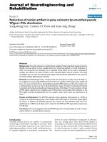
As schemed in Fig. 1, CdTe/CdS/ZnS core/shell/shell
quantum dots were fabricated in this integrated chemical
aerosol flow apparatus through the epitaxial growth stage. In
the nebulizer apparatus, the stock solution containing
Cd(MPA) complex and NaHTe was misted to microdroplets,
carried into the furnace by the 1.5 L/min N
2
flow. CdTe/CdS
core/shell quantum dots were directly formed in the furnace.
Then, at the end of the furnace, the CdTe/CdS QDs flowed
into in a three-neck flask that was kept at 80°C, capping a
shell of ZnS was carried out. About 45 min later, CdTe/CdS/
ZnS core/shell/shell quantum dots were harvested.
In our experiment, the intermediate CdTe/CdS QDs
synthesized show great stability due to the good crystalli-
zation at high reaction temperature, and the in situ
synthesized CdS shells on CdTe cores can play as a buffer
layer to further epitaxially grow ZnS shells. So with this
facile method, high quality of CdTe/CdS/ZnS core/shell/
shell QDs can be synthesized.
As known, in the synthesis of QDs, especially that with
core/shell/shell structure, large-scale production is difficult
for many methods mainly due to its difficulty to ensure the
same temperature and homogeneous mixing in the large
volume of solution, which have a great influence on the
monodispersity of the nanocrystals. Up to now, large-scale
synthesis is still a challenge [23]. Here, in our modified
integrated apparatus, the continuous synthesis method
makes it possible to realize large-scale synthesis of QDs
with core/shell/shell structure. In our experiment, the rate
of production can reach as high as 0.1 g/h. As the currently
used quartz tube in the furnace only has a diameter of
30 mm, the flow rate is 1.5 L/min. It is easy to improve the
production rate using a larger diameter tube or increasing
the flow rate. Owing to its continuity, as much core/shell/
shell QDs can be synthesized.
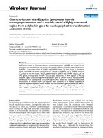
CdTe/CdS/ZnS core/shell/shell QDs with different peak
position of photoluminescence (PL) can be obtained by
changing flow rate and temperature, consistent with our
previous description for synthesizing CdTe/CdS QDs [20].
As shown in Fig. 2a, with a flow rate of 1.5 L/min and
temperature of 200, 225, and 250°C, CdTe/CdS/ZnS core/
shell/shell QDs with PL peak of 525, 554, and 567 nm
were synthesized. The flow rates also had an influence
on the peak position of PL. With increased flow rate, the
PL would be blue-shifted (Data not shown), attributed to
shortened reaction time. All the synthesized CdTe/CdS/
ZnS core/shell/shell QDs showed high fluorescence
(Fig. 2b), whose quantum yields were as high as 45%.
In order to confirm the core/shell/shell structure of the
synthesized QDs, several control experiments were carried
out, as shown in Table 1 and Fig. 3. For control experi-
ment, the Zn and/or S precursors was absent in the part for
ZnS coating. As can be seen, when the Zn precursor and
S precursor were added in the three-neck flask simulta-
neously, the obtained QDs showed a fluorescence peak at
Fig. 1 Apparatus to synthesize the CdTe/CdS/ZnS core/shell/shell
quantum dots
Fig. 2 PL spectra of QDs synthesized at different temperature (a). Image of CdTe/CdS/ZnS QDs solution under ultraviolet light
Nanoscale Res Lett (2010) 5:189–194 191
123
541 nm with high fluorescence intensity. QDs with a
weaker fluorescent intensity were obtained when Zn and S
precursors were both absent. The results reveal that ov-
ercoating a shell of ZnS on the CdTe/CdS greatly enhances
the fluorescence intensity. Similarly, when only S precursor
was added, the resultant QDs showed a red-shifted PL,
because a thicker CdS shell was formed on the outer shell
of the QDs. As we know, the CdTe/CdS core/shell QDs
have more Cd atoms on its surface. However, when only
Zn precursor was added, an aggregated agglomeration with
a very weak red fluorescence was obtained. It is deduced
that due to the coordination between the Zn
2?
and QDs, the
Zn (MPA) complex acts as a flocculant to aggregate the
synthesized QDs in water at the elevated temperature.
From the results, we can clearly see that the coexisted Zn
and S precursor in the second part can form a shell of ZnS
on the outside of CdTe/CdS QDs.
The core/shell/shell structure was further confirmed by
XRD (Fig. 4a). In the XRD pattern, three peak including
(111), (220), and (311) can be clearly seen, which corre-
sponds to the cubic CdTe/CdS and CdTe/CdS/ZnS quan-
tum dots. Figure 4a shows that the scattering peaks of
CdTe/CdS quantum dots were just between the bulk cubic
CdTe and bulk CdS. This is because of the existence of
CdS shell on the edge of CdTe. For the CdTe/CdS/ZnS
QDs, the diffraction patterns shift to higher angles due to
the growth of ZnS shells. The composition of CdTe/CdS/
ZnS was also investigated by energy-dispersive X-ray
spectroscopy (EDS) (Fig. 4b). In the EDS pattern, the
presence of Zn and S was clearly confirmed, and the atomic
ratio of S:Zn:Cd:Te was calculated to be 2.84:1.64:1.38:1.
The carbon and oxygen peak showed in the EDS pattern
were corresponding to the capping agent of MPA. The
TEM and HRTEM of CdTe/CdS/ZnS QDs synthesized
with temperature of 200°C and flow rate of 1.5 L/min were
shown in Fig. 5. We can see that the CdTe/CdS/ZnS QDs
have a narrow size distribution. The average diameter of
the QDs is about 2.5 nm. The existence of lattice planes on
Table 1 Control experiments to confirm the core/shell/shell structure
of synthesized QDs
Precursor A B C D
Zn(MPA) complex HH––
Thiourea H – H –
Fluorescence wavelength (nm) 541 573 563 541
Right character ‘‘H’’ means they have the precursor in the second part
for ZnS coating. While ‘‘–’’ means no precursor was added
Fig. 3 Fluorescence spectra of different kinds of core/shell QDs
Fig. 4 a XRD patterns of CdTe/CdS and CdTe/CdS/ZnS and b EDS of CdTe/CdS/ZnS core/shell/shell QDs
Fig. 5 TEM image for CdTe/CdS/ZnS core/shell/shell quantum dots
prepared at 200°C. Inset is the HRTEM image
192 Nanoscale Res Lett (2010) 5:189–194
123
the HRTEM confirms the good crystallinity of the CdTe/
CdS/ZnS QDs.
It was expected that after coating with a ZnS shell, the
CdTe/CdS/ZnS core/shell/shell QDs would be more stable,
less toxic and have higher quantum efficiency compared to
CdTe/CdS quantum dots and CdTe quantum dots. In order
to investigate the difference between the three kinds of
QDs, CdTe/CdS/ZnS, CdTe/CdS, and CdTe (aq), H
2
O
2
was used as an oxidizing agent to examine their anti-oxide
ability via detecting the change of fluorescence spectra
(Fig. 6). It is known that when reacted with an oxidizer
such as H
2
O
2
, a blue shift would be observed due to the
oxidization of surface atoms [30, 31]. But if the QDs were
core/shell structure, the blue shift would not occur because
the thick shell can prevent the oxidation of cores, which
determines the PL peak position. Different volumes of
0.03% H
2
O
2
were added to equal amount of three kinds of
QDs (4 mL). From the results (Fig. 6), we can clearly see
that when H
2
O
2
solution was added, the fluorescence
intensities were all decreased and gave an approximate
liner change. The difference is that the PL peak position
was almost not changed when ZnS shells existed for CdTe/
CdS/ZnS, while there is obviously a blue shift of about
6 nm for CdTe (aq) QDs. Similarly, for the CdTe/CdS
QDs, a smaller blue shift of about 2 nm can be seen. This
result clearly shows that the CdTe/CdS via CAF has
enhanced anti-oxide ability compared with CdTe (aq).
While overcoating ZnS shells on the CdTe/CdS QDs can
further enhance this effect. When reacted with H
2
O
2
, the
surface atoms of CdTe can be oxided to CdTeO
3
or TeO
2
[31, 32], which causes the decrease in CdTe QDs size and
further leads to the blue shift of fluorescence peak. The
Fig. 6 The fluorescence intensity change of different QDs when added various volumes of 0.03% H
2
O
2
solution. a CdTe/CdS/ZnS, b CdTe/CdS
c CdTe(aq)
Fig. 7 Labeled CHO (Chinese hamster ovary) with CdTe/CdS/ZnS quantum dots, a–c are the fluorescent images of cells. d–f are the
corresponding co-situated picture of cells and fluorescence
Nanoscale Res Lett (2010) 5:189–194 193
123
existence of CdS can weaken this effect, while ZnS shells
can prevent the blue shift. These results reflect that the
overcoating of ZnS shells can greatly enhance its anti-
oxide ability and stability.
With ZnS shells on the CdTe/CdS QDs, its toxicity can
be greatly decreased. It is more suitable to use it in bio-
logical applications such as cellular imaging. To confirm
that the QDs can label the cells, we chose three kinds of
CdTe/CdS/ZnS QDs (brown, yellow, and green) and added
them into the Chinese hamster ovary (CHO) cells. After
30 min incubation, the intracellular distribution of CdTe/
CdS/ZnS QDs was observed by confocal microscopy. It
can be clearly seen that the QDs penetrate into the living
cells and exhibit bright fluorescence (Fig. 7). The distri-
bution of all three kinds of QDs is in the cytoplasm and the
nucleus. This observation demonstrates that QDs are
gradually transported inside the cytoplasm and eventually
to the nucleus. On the basis of these fluorescence images,
we consider that the CHO cells are efficiently labeled with
QDs and can display multicolor images.
Conclusion
In summary, CdTe/CdS/ZnS core/shell/shell quantum dots
were synthesized by chemical aerosol flow method in a
continuous system. This method can provide a simple,
ultrafast, and continuous way to prepare core/shell/shell
quantum dots. Importantly, compared with CdTe QDs
prepared directly in aqueous solution and CdTe/CdS core/
shell QDs synthesized by chemical aerosol flow method,
the CdTe/CdS/ZnS core/shell/shell QDs have enhanced
anti-oxide ability and stability. This is significant for fur-
ther application of aqueous QDs. We also prove that the
QDs can achieve multicolor label in living cells. Benefiting
from their reduced toxicity, enhanced stability, and
increased PLQY, this kind of core/shell/shell QDs has
potential for future in vivo fluorescent imaging.
Acknowledgments The current investigations were financially
supported by the Hi-Tech Research and Development Program of
China (863 program 2007AA021803 and 2009AA03Z302) and the
National Natural Science Foundation of China (NSFC No. 60736001)
and Natural Science Foundation of Beijing (2093044).
References
1. X. Peng, L. Manna, W. Yang, J. Wickham, E. Scher, A.
Kadavanich, A.P. Alivisatos, Nature 404, 59 (2000)
2. S. Kim, B. Fisher, H. Eisler, M. Bawendi, J. Am. Chem. Soc. 125,
11466 (2003)
3. L. Manna, D.J. Milliron, A. Meisel, E.C. Scher, A.P. Alivisatos,
Nat. Mater 2, 382 (2003)
4. N. Al-Salim, A.G. Young, R.D. Tilley, A.J. McQuillan, J. Xia,
Chem. Mater 19, 5185 (2007)
5. J. Ouyang, M. Vincent, D. Kingston, P. Descours, T. Boivineau,
M.B. Zaman, X. Wu, K. Yu, J. Phys. Chem. C 113, 5193 (2009)
6. M. Bruchez, M. Moronne, P. Gin, S. Weiss, A.P. Alivisatos,
Science 281, 5385 (1998)
7. W.C.W. Chan, S. Nie, Science 281, 2016 (1998)
8. A.M. Kapitonov, A.P. Stupak, S.V. Gaponenko, E.P. Petrov,
A.L. Rogach, A. Eychmuller, J. Phys. Chem. B 103, 8259 (1999)
9. A.L. Rogach, A. Kornowski, D. Su, A. Eychmuller, H. Weller,
J. Phys. Chem. B 103, 3065 (1999)
10. Y.T. Didenko, K.S. Suslick, J. Am. Chem. Soc. 127, 12196
(2005)
11. X.M. Jiang, C.J. Brinker, J. Am. Chem. Soc. 128, 4512 (2006)
12. Y.F. Lu, H.Y. Fan, A. Stump, T.L. Ward, T. Rieker, C.J. Brinker,
Nature 398, 223 (1999)
13. S.E. Skrabalak, K.S. Suslick, J. Phys. Chem. C 111, 17807 (2007)
14. S.E. Skrabalak, K.S. Suslick, J. Am. Chem. Soc. 128, 12642
(2006)
15. J.H. Bang, W.H. Suh, K.S. Suslick, Chem. Mater. 20, 4033
(2008)
16. D.J. Kim, K.K. Koo, Crys. Grow. & Des. 9, 1153 (2009)
17. C.R. Stoldt, M.A. Haag, B.A. Larsen, Appl. Phys. Lett. 93,
193107 (2008)
18. Y. Huang, Z. Zheng, Z.H. Ai, L.Z. Zhang, X.X. Fan, Z.G. Zou,
J. Phys. Chem. B 110, 19323 (2006)
19. S.S. Dunkle, R.J. Helmich, K.S. Suslick, J. Phys. Chem. C 113,
11980 (2009)
20. H.B. Li, F.Q. Tang, L.L. Li, C.M. Yan, X.L. Huang, D. Chen,
Crystengcomm 11, 1231 (2009)
21. Y.Y. Su, Y. He, H.T. Lu, L.M. Sai, Q.N. Li, W.X. Li, L.H. Wang,
P.P. Shen, Q. Huang, C.H. Fan, Biomaterials 30, 19 (2009)
22. J.J. Li, Y.A. Wang, W. Guo, J.C. Keay, T.D. Mishima,
M.B. Johnson, X. Peng, J. Am. Chem. Soc. 125, 12567 (2003)
23. S. Deka, A. Quarta, M.G. Lupo, A. Falqui, S. Boninelli, C.
Giannini, G. Morello, M. De Giorgi, G. Lanzani, C. Spinella, R.
Cingolani, T. Pellegrino, L. Manna, J. Am. Chem. Soc. 131, 2948
(2009)
24. H.V. Demir, S. Nizamoglu, E. Mutlugun, T. Ozel, S. Sapra, N.
Gaponik, A. Eychmuller, Nanotechnology 19, 335203 (2008)
25. R.G. Xie, X.G. Peng, Angew. Chem. Int. Ed. 47, 7677 (2008)
26. P. Reiss, M. Protiere, L. Li, Small 5, 154 (2009)
27. P.K. Santra, R. Viswanatha, S.M. Daniels, N.L. Pickett, J.M.
Smith, P. O’Brien, D.D. Sarma, J. Am. Chem. Soc.
131, 470
(2009)
28. Y. He, H.T. Lu, L.M. Sai, W.Y. Lai, Q.L. Fan, L.H. Wang,
W. Huang, 110, 13370 (2006)
29. H. Yao, H.T. Lu, L.M. Sai, Y.Y. Su, M. Hu, C.H. Fan, W. Huang,
L.H. Wang, Adv. Mater. 20, 3416 (2008)
30. R. Beaulac, P.I. Archer, X.Y. Liu, S. Lee, G.M. Salley, M.
Dobrowolska, J.K. Furdyna, D.R. Gamelin, Nano Lett. 8,
1197 (2008)
31. Y J. Chen, X P. Yan, Small 5, 2012 (2009)
32. L.P. Liu, Q. Peng, Y.D. Li, Inorg. Chem. 47, 3182 (2008)
194 Nanoscale Res Lett (2010) 5:189–194
123